Kinematic and dynamic analysis of an anatomically based knee joint Kok-Meng Lee n , Jiajie Guo The George W. Woodruff School of Mechanical Engineering, Georgia Institute of Technology, Atlanta, GA 30332-0405, USA article info Article history: Accepted 2 February 2010 Keywords: Bio joint Sliding velocity Exoskeleton Dynamic model abstract This paper presents a knee-joint model to provide a better understanding on the interaction between natural joints and artificial mechanisms for design and control of rehabilitation exoskeletons. The anatomically based knee model relaxes several commonly made assumptions that approximate a human knee as engineering pin-joint in exoskeleton design. Based on published MRI data, we formulate the kinematics of a knee-joint and compare three mathematical approximations; one model bases on two sequential circles rolling a flat plane; and the other two are mathematically differentiable ellipses- based models with and without sliding at the contact. The ellipses-based model taking sliding contact into accounts shows that the rolling-sliding ratio of a knee-joint is not a constant but has an average value consistent with published measurements. This knee-joint kinematics leads to a physically more accurate contact-point trajectory than methods based on multiple circles or lines, and provides a basis to derive a knee-joint kinetic model upon which the effects of a planar exoskeleton mechanism on the internal joint forces and torque during flexion can be numerically investigated. Two different knee-joint kinetic models (pin-joint approximation and anatomically based model) are compared against a condition with no exoskeleton. The leg and exoskeleton form a closed kinematic chain that has a significant effect on the joint forces in the knee. Human knee is more tolerant than pin-joint in negotiating around a singularity but its internal forces increase with the exoskeleton mass-to-length ratio. An oversimplifying pin-joint approximation cannot capture the finite change in the knee forces due to the singularity effect. & 2010 Elsevier Ltd. All rights reserved. 1. Introduction With rapid advances in robotics and mechatronics, exoskeletons have been widely developed to assist in or rehabilitate human body motions; for examples, the commercialized Lokomat (Colombo et al., 2000) for treadmill training, assistant exoskeleton (Kiguchi et al., 2003) for elderly persons, hybrid assistive limb (Kawamoto and Sankai, 2005) to assist in walking and climbing, the ankle–foot orthosis powered by artificial pneumatic muscles (Ferris et al., 2005), and the Berkeley lower extremity exoskeleton (Zoss et al., 2006) to assist in carrying heavy loads over rough terrain. Experiments (Gordon and Ferris, 2007) on a robotic exoskeleton controlled by muscle activity could be useful tools for testing neural mechanism of human locomotor adaptation. While mechanical exoskeletons have the advantages of adding energy to human motions and adjusting motion patterns, it could perturb normal motions and potentially cause pains or even damages to human joints. To minimize negative effects of an artificial exoskeleton on human joints, the design and control of rehabilitation exoskeletons require a good understanding of the natural bio-joint kinematics and kinetics. Natural bio-joints (that exoskeletons are designed to rehabi- litate) are commonly modeled in kinematics as non-slip revolute joints (such as a pin or ball joint). Unlike an engineering joint which typically has a fixed rotational center, bio-joints often consist of non-uniform shaped contact parts that roll and slide. Human bodies also vary widely in shapes and sizes and change over years; engineering joints are usually not compliant to accommodate these variations. Although exoskeletons help users adapt their motion after repetitive training, there are potentials for damages to bio-joints in long term usage if natural joint variations are not accounted for. Compliant mechanisms that reduce assembled joints (and thus friction) can be employed in exoskeleton to accommodate (shape, size and motion) variations in human joints. ‘‘Soft’’ pneumatic muscle actuators (pMA) are used as power sources (Tsagarakis and Caldwell, 2003), and flexible cables are used for transmission in LOPES (Veneman et al., 2006). However, most of the works on compliance have been motivated by power efficiency rather than the interaction between a human body and an exoskeleton. Recent works also concern about shock absorption; for example, flexible geared joint and rubber footpad (Low, 2005). Besides gaining energy, an exoskeleton should not interfere negatively ARTICLE IN PRESS Contents lists available at ScienceDirect journal homepage: www.elsevier.com/locate/jbiomech www.JBiomech.com Journal of Biomechanics 0021-9290/$ - see front matter & 2010 Elsevier Ltd. All rights reserved. doi:10.1016/j.jbiomech.2010.02.001 n Corresponding author. Tel.: + 1 404 8947402. E-mail address: [email protected] (K.-M. Lee). Journal of Biomechanics 43 (2010) 1231–1236
anatomically based knee model relaxes several commonly made assumptions that approximate a
human knee as engineering pin-joint in exoskeleton design. Based on published MRI data, we formulate
the kinematics of a knee-joint and compare three mathematical approximations; one model bases on
two sequential circles rolling a flat plane; and the other two are mathematically differentiable ellipses-
based models with and without sliding at the contact. The ellipses-based model taking sliding contact
into accounts shows that the rolling-sliding ratio of a knee-joint is not a constant but has an average
value consistent with published measurements. This knee-joint kinematics leads to a physically more
accurate contact-point trajectory than methods based on multiple circles or lines, and provides a basis
to derive a knee-joint kinetic model upon which the effects of a planar exoskeleton mechanism on the
internal joint forces and torque during flexion can be numerically investigated. Two different knee-joint
kinetic models (pin-joint approximation and anatomically based model) are compared against a
condition with no exoskeleton. The leg and exoskeleton form a closed kinematic chain that has a
significant effect on the joint forces in the knee. Human knee is more tolerant than pin-joint in
negotiating around a singularity but its internal forces increase with the exoskeleton mass-to-length
ratio. An oversimplifying pin-joint approximation cannot capture the finite change in the knee forces
due to the singularity effect.
& 2010 Elsevier Ltd. All rights reserved.
1. Introduction
With rapid advances in robotics and mechatronics, exoskeletonshave been widely developed to assist in or rehabilitate human bodymotions; for examples, the commercialized Lokomat (Colombo et al.,2000) for treadmill training, assistant exoskeleton (Kiguchi et al.,2003) for elderly persons, hybrid assistive limb (Kawamoto andSankai, 2005) to assist in walking and climbing, the ankle–footorthosis powered by artificial pneumatic muscles (Ferris et al.,2005), and the Berkeley lower extremity exoskeleton (Zoss et al.,2006) to assist in carrying heavy loads over rough terrain.Experiments (Gordon and Ferris, 2007) on a robotic exoskeletoncontrolled by muscle activity could be useful tools for testing neuralmechanism of human locomotor adaptation. While mechanicalexoskeletons have the advantages of adding energy to humanmotions and adjusting motion patterns, it could perturb normalmotions and potentially cause pains or even damages to humanjoints. To minimize negative effects of an artificial exoskeleton onhuman joints, the design and control of rehabilitation exoskeletons
ll rights reserved.
. Lee).
require a good understanding of the natural bio-joint kinematics andkinetics.
Natural bio-joints (that exoskeletons are designed to rehabi-litate) are commonly modeled in kinematics as non-slip revolutejoints (such as a pin or ball joint). Unlike an engineering jointwhich typically has a fixed rotational center, bio-joints oftenconsist of non-uniform shaped contact parts that roll and slide.Human bodies also vary widely in shapes and sizes and changeover years; engineering joints are usually not compliant toaccommodate these variations. Although exoskeletons help usersadapt their motion after repetitive training, there are potentialsfor damages to bio-joints in long term usage if natural jointvariations are not accounted for.
Compliant mechanisms that reduce assembled joints (and thusfriction) can be employed in exoskeleton to accommodate (shape,size and motion) variations in human joints. ‘‘Soft’’ pneumaticmuscle actuators (pMA) are used as power sources (Tsagarakisand Caldwell, 2003), and flexible cables are used for transmissionin LOPES (Veneman et al., 2006). However, most of the works oncompliance have been motivated by power efficiency rather thanthe interaction between a human body and an exoskeleton.Recent works also concern about shock absorption; for example,flexible geared joint and rubber footpad (Low, 2005). Besidesgaining energy, an exoskeleton should not interfere negatively
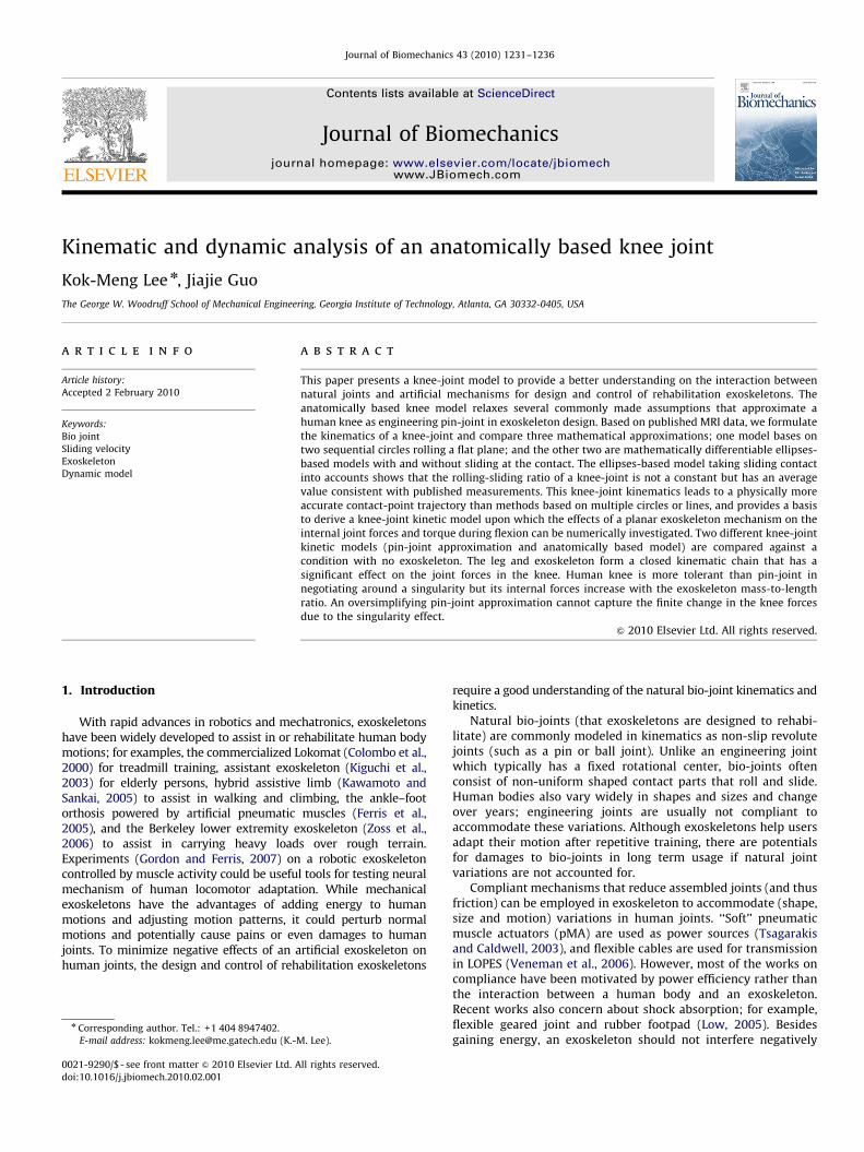
Fig. 1. Bio-joint illustration: (a) MRI of a cadaver knee (Iwaki et al., 2000) where
the two ellipses approximate the contact regions of the knee. (b) Bio-joint model
(Lee and Guo, 2008) for analyzing the contact kinematics based on elliptical
geometries.
K.-M. Lee, J. Guo / Journal of Biomechanics 43 (2010) 1231–12361232
with normal human-joint motions; otherwise, it could result indiscomfort and potential damages. For these reasons, problems oninteraction between bio-joints and compliant mechanism areworthy of exploration.
Among challenges in the modeling of human body motion arethe characterization of the joint mechanics and musculoskeletalgeometry (Pandy, 2001). Different experimental methods havebeen developed to detect joint structures, and measure the kneejoint motions (Bull and Amis, 1998). These include the mostcommonly used skin-marker systems (Lafortune et al., 1992; Luand O’ Connor, 1999) where soft tissues often make them difficultto apply, and the radiation radiographic/fluoroscopic techniques(Peterfy et al., 2003; Li et al., 2008). More recently, theelectromagnetic techniques (MRI) (Iwaki et al., 2000) provide away to shed light on the internal geometries of joints. Withgeometrical details of a joint available from experiments,kinematic models can be built for analyses. A computer modelwith surface friction (Sathasivam and Walker, 1997) wassuggested showing that the condylar geometries could haveimportant differences in kinematics, function and wear. Accuratesolutions of tibiofemoral contact behavior were obtained from a3D finite-element model (Donahue et al., 2002). Recently, acomputational model allowing for anatomical details was builtusing CAD/CAE methods to predict the lower leg kinematics(Liacouras and Waynel, 2007).
Motivated by the need to provide a good understanding on theinteraction between natural joints and artificial mechanisms fordesign and control of rehabilitation exoskeletons, this paperintroduces a mathematical bio-joint model originally proposed(Lee and Guo, 2008) for mechanical deboning of chicken breastmeat (Guo and Lee, 2008). The remainder of this paper offers thefollowings:
�
We formulate an anatomically based model for characterizinga human knee joint so that its internal joint forces/torquesduring flexion can be accurately analyzed. The bio-joint modelrelaxes several assumptions commonly made on a knee joint inexoskeleton design. � Two examples are given. The first investigates the effects of
different bio-joint geometrical approximations (with/withoutsliding) on the contact locations. The second numericallyinvestigates the effects of a planar exoskeleton on a humanknee joint by comparing two different models for predictingthe forces/torque acting on the knee; pin-joint approximationand bio-joint model.
2. Methods
With MRI data, a model can be built to provide a good understanding of the
kinematics and kinetics of a bio-joint (consisting of non-uniform shaped contact
parts), and estimate its contact locations, rolling/sliding velocities and forces/
torques involved.
Fig. 1(a) shows a lateral sagittal MRI of an unloaded cadaver knee (Iwaki et al.,
2000), where the two white circles are approximated geometries for the femoral
articular surfaces. Data are presented as positions of the extension facet center
(EFC) and flexion facet center (FFC) in Fig. 1(a), where the contact is modeled as a
point between a circle and a plane. To provide a continuous differentiable function,
a more general bio-joint representation based on elliptical geometries is proposed
in Fig. 1(b) to characterize the observed data for analyzing the contact kinematics
and kinetics, where OA and OB are two bodies with surfaces GA and GB,
respectively; and the angular velocity o describes the motion of OA rolling on
OB at the instantaneous contact point C.
2.1. Knee joint kinematics
Given a contact point on GA, there always exists a tangential plane with a
normal vector n such that the angular velocity x describes the motion of OA at the
contact point. An instantaneous osculating plane perpendicular to this tangent
plane can then be defined as shown in Fig. 1(b) to characterize the 3D motion of a
biological joint, which depends on the location of the contact point (and hence is a
function of time). The contact points on GA and GB move incrementally from CA
and CB to C along the respective osculating circles, where (OA, rA) and (OB, rB) are
the centers and radii of the osculating circles intersecting at GA and GB,
respectively. The rotation axis changes with the contact point as OA rolls and
slides on OB.
For the knee joint in Fig. 1(a), the normal component (along n) of the angular
velocity is generally nonzero but very small as compared to the component on the
tangent plane, and thus neglected to simplify the bio-joint kinematic analysis.
The 3D kinematics of a biological joint is reduced to finding the contact location in
the motion of the osculating circle and the position and orientation of OA. The
rolling velocity vroll due to the angular velocity about the rotation center OA is
defined in Eq. (1) as follows:
vrollðyÞ ¼orA where o¼ dy=dt ð1Þ
For a specified ellipse, rA is a function of the contact position (Lee and Guo, 2008;
Davis and Snider, 1987). As observed in published data (Iwaki et al., 2000), the
sliding velocity vslide also depends on the flexion angle y as
vslideðyÞ ¼dsslide
dt¼o dsslide
dyð2Þ
where sslide is the difference between the rolling displacement sroll (or integrated
vroll over y) and the path length of the contact point from the MRI data.
Using a lumped-parameter approach in polar coordinates (r, y) where the
reference (y=0) is along the longitudinal axis of the femur (Fig. 2), the lower limb
(leg and foot) is modeled as a mass centered at O. As illustrated in Fig. 2(b) where
Ci is the initial contact point on the femur, the distance r0 from the center of the
lateral tibial condyle to O is a constant; and C is measured from the ipsilateral
posterior tibial cortices (IPTC). The bio-joint kinematics can be computed from the
following:
_r ¼o dr
dy; and €r ¼ _o dr
dyþo2 d2r
dy2ð3a;bÞ
where r provides a means to account for the change in the rotational center.
Engineering ball joints are special cases where both spherical surfaces are
concentric and hence, _r ¼ €r ¼ 0. The (mass center O) trajectory can be computed in
terms of y from the following:
r¼ r er ; v¼ _r erþr _y ey ð4a;bÞ
a¼ ð€r�r _y2Þ erþð2_r _yþr €yÞey ð4cÞ
2.2. Knee joint dynamics
The calf dynamics (relative to the upper leg) are given by
ma¼ fgþfyþfrþfe ð5aÞ
ð J €yþ2mr _r _yÞk¼ sgþsaþse where k¼ er � ey ð5bÞ
where m is the calf mass; fg is the gravity force; and fr and fy are the resultant
forces exerted by the surrounding bones and tissues (muscle and ligament) in er
ARTICLE IN PRESS
Fig. 2. Coordinates illustrating the knee joint rotation: (a) the tibia rotation in
polar coordinates centered at the initial contact point Ci. (b) Schematics
illustrating the relationship between the distance r and flexion angle y.
Table 1Geometry approximation.
Circles (Iwaki et al., 2000) Ellipse (green dash)
r1=21 mm r2=32 mm rmaj=25.3 mm rmin=21.1 mm
Ellipse (blue) Ellipse (red)
rmaj=33.6 mm rmin=23 mm rmaj=28.8 mm rmin=18.8 mm
Initial contact position=31 mm;
Angular velocity o=1.57 rad/s
Fig. 3. Comparison of current contact point C positions between different models
showing the sliding effect in the knee rotation.
K.-M. Lee, J. Guo / Journal of Biomechanics 43 (2010) 1231–1236 1233
and ey directions, respectively. Within a bio-joint, bones primarily support
compressive forces; and soft tissues can only exert tensile forces. For example, fr
represents the force from the tissues if tensile force dominates, or otherwise from
the bones. With rehabilitation applications in mind, we include fe to account for
the force exerted by an external device (such as an exoskeleton) and reaction from
the ground. On the left hand side of Eq. (5b), the 1st term accounts for the
moment-of-inertia J (about Ci) due to the leg rotation while the 2nd term describes
the interaction between _yand_rdue to the variation in r. In Eq. (5b), all the torques
are computed about Ci: sg and se denote the torques due to the gravity and
external device, respectively; and sa is a net torque accounting for fr, fy, and tissue
contraction within the knee.
The vector Eqs. (5a, b) can be recast into three scalar equations (6a, b, c) from
which fr, fy and ta can be solved as
mð€r�r _y2Þ ¼ fg sinyþ frþ ferðyÞ ð6aÞ
mð2_r _yþr €yÞ ¼ fg cosyþ fyþ feyðyÞ ð6bÞ
J €yþ2mr _r _y ¼ tgþtaþteðyÞ ð6cÞ
3. Results and discussion
Two sets of results are presented here. The 1st set investigatesthe effect of the geometrical approximations on the contact(between the femoral and tibial articular surfaces) with publisheddata (Iwaki et al., 2000) as a basis for comparison. The 2nd setillustrates the effects of an exoskeleton on the human knee joint.
3.1. Effect of bio-joint models on contact point trajectory
In (Iwaki et al., 2000), two circles, each of which rolls on adifferent flat facet, were used for the sagittal section of the medialtibiofemoral compartment but for the lateral tibiofemoral com-partment, two circles roll on the same flat facet. In this paper, thesimulations focus on the lateral part as it has a larger displacementthan the medial part. The following three models are compared:
Model 1: Two sequential circles roll on a flat plane (Iwakiet al., 2000).Model 2: One ellipse rolls on a flat plane.Model 3: One ellipse rolls on another ellipse.
The dimensions of the approximated circles and ellipses(Fig. 1b) are listed in Table 1. With the contact location definedas a horizontal distance of C measured from the IPTC in Fig. 1(a),results are given as a function of the flexion angle y in Fig. 3 forcomparing three models against published data. Fig. 4 simulates
(on the basis of Model 3) the snap-shot trajectory of the lower legas it rotates from its initially full extension, and its corresponding(rolling/sliding) displacements, velocities as well as the sroll/sslide
ratio. Observations in Figs. 3 and 4 are discussed as follows:
a)
For Model 1, the sroll/sslide ratio is given as 1.7. As the slidingvelocity of each rolling circle is assumed constant, the contactpoint is a linear function of y. The overall result, however, is not asmooth curve (Fig. 3) due to the transition from circles r2 to r1.The difference between the 2-circle model and experimentalresults can be observed when y4901. This is because the rota-tional axis of the circle is tilted by a small angle; when projectedon the camera plane, the tilted circle is essentially as an ellipse.
b)
Based on the above observation, we model bio-joints usingelliptical surfaces as they offer a more realistic characterizationthan a multi-circle model, and are mathematically differenti-able. Fig. 3 compares Models 2 and 3 against published data.With only rolling, Model 2 (that simplifies the tibial condyle asa planar surface) results in some negative contact positions;this is intuitively incorrect as the knee joint does not losecontact. Given the close match between Model 3 (whenconsidering both sliding and rolling in the joint kinematics)and the experiment data, Model 3 with sliding is used for thesubsequent analysis.
c)
The displacements, sroll(y) and sslide(y), normalized to the majorradius of the femoral condyle, are given by Eqs. (7a) and (b),respectively, and their ratio is plotted in Fig. 4(b):
srollðyÞ=rmaj ¼ 0:093y5�0:409y4
þ0:57y3�0:448y2
�0:926y ð7aÞ
sslideðyÞ=rmaj ¼ 0:334y5�1:518y4
þ2:12y3�0:996y2
þ0:513y ð7bÞ
ARTICLE IN PRESS
Fig. 4. Rolling and sliding velocities of the current contact point (Model 3): (a) snapshots of the femoral condyle and position changes of initial/current contact points.
(b) Plots of normalized rolling and sliding displacements showing their ratio is not a constant. (c) Plots of normalized rolling and sliding velocities as the knee rotates at a
constant speed (901/s).
Table 2Physical parameters of human’s lower leg.
Human Exoskeleton
length (m)
Length (m) Mass (kg)
Upper leg 0.40 7.02 0.40
Lower leg/foot 0.37/0.27 2.44/1.18 0.37
r0 (m) 0.2453
K.-M. Lee, J. Guo / Journal of Biomechanics 43 (2010) 1231–12361234
The sroll/sslide ratio is not a constant, but its average valueof 1.69 closely agrees with the experimental observation(Iwaki et al., 2000) of 1.7. Fig. 4(c) graphs vslide calculated fromEq. (2); negative vslide means sliding forward instead of backward.
Fig. 5. Distance, velocity and acceleration of the tibia mass-center during knee
rotation.
3.2. Effect of exoskeleton design on joint forces/torque
Due to the kinematic constraint imposed by the contact, thehuman knee joint embodies two degrees of freedom (DOF),rotation and translation for its planar motion. To investigate the
ARTICLE IN PRESS
K.-M. Lee, J. Guo / Journal of Biomechanics 43 (2010) 1231–1236 1235
effects of a planar exoskeleton on human knee joints, we comparetwo different models in predicting the forces and moments actingon the knee; namely,
�
Figmo
Fig�5
Pin-joint (PJ) engineering approximation and
� Bio-joint knee (BJK) Model 3.
The exoskeleton consists of a revolute (pin) joint between tworigid links attached to the lower and upper legs with pin joints.This design has three-DOF from its three pin-joints and thus hasone redundancy. For a nonzero flexion angle, there are twopossible solutions. However, only one solution is physicallyfeasible.
For clarity and ease of illustration, the following assumptionsare made: (1) the human subject sits with the upper leg heldstatic and horizontal and the lower link rotates with the tibia fromits initial state (full extension); and (2) the lower link is attachedat O with the revolute joint centered at the initial contact point Ci
. 6. Comparing snapshots of an exoskeleton between two different knee joint
dels.
. 7. Force and torque comparison between two different knee joint models with/wi
1oyo901 and underestimated in y4901 as compared to the bio-joint knee model
(Fig. 2). Numerical values used in this study are given in Table 2(Nomiyama et al., 2007) and from Figs. 2(b) and 4(a),
rðyÞ ¼ 1:078y4�11:184y3
þ26:542y2�0:825yþ263:59 ð8Þ
Eq. (8) and its derivatives are graphed in Fig. 5. Fig. 6 shows thelink kinematics (solid lines) as the tibia rotates, where dash linessimulate the knee as a pin joint (commonly assumed inexoskeleton designs) for comparison.
To account for the exoskeleton mass in the kinetic study, thetwo links are assumed to have the same mass-to-length ratio Z of0.5 kg/m. Unlike the condition with no exoskeleton where thehuman leg is an open-chain mechanism, the leg and exoskeletonform a closed kinematic chain that has a significant effect on theinternal joint forces and torque of the knee. Fig. 7(a–c) arecalculated results from Eq. (6) showing the internal forces andtorque in the knee as the tibia accelerates from the initial staticstate (y=�51) to y=201 for 0.5 s, then maintains at an angularvelocity for 1 s to y=951, and finally decelerates to the final staticstate y=1151 in another 0.5 s. Throughout the trajectory, the foot isoff the ground and thus, there is no ground reaction. In Fig. 7 wherethe solid and dashed lines are results of the BJK and PJ models,respectively, the internal forces and torque for a condition with noexoskeleton are plotted as a basis for comparison.
Several observations can be made from Figs. 5–7:
1.
thou
. (b)
The sign of the force fr in Fig. 7(a) can be explained as follows.During the initial flexion (yo0), fr is positive since the force isprimarily supported by the femur, but becomes negative as theretraction force from the soft tissues gradually plays a moredominant role as the knee rotates downward.
2.
The distance r increases as much as 30 mm (Fig. 5). For the samework done, this increase in r tends to reduce fr in the knee. As thePJ approximation assumes a constant r and neglects the jointgeometry, the effect of the r(y) variation on the attaching point(Fig. 6) and on the forces/torque (Fig. 7) cannot be accounted for.As compared to the BJK model in Fig. 7(a), the PJ approximationoverestimates 9fr9 in the range (01oyo901) and underestimatesas y approaches its rotation limit.
t exoskeleton: (a) pin-joint knee approximation overestimates fr in the range
Tangential force fy. (c) Moment ta.
ARTICLE IN PRESS
K.-M. Lee, J. Guo / Journal of Biomechanics 43 (2010) 1231–12361236
3.
Near y=01, the exoskeleton loses one DOF along the er
direction causing a finite change in fr as well as fy and ta asshown in Fig. 7. Human knee (that can roll and slide) is moretolerant than a pin-joint to a singularity along er as illustratedin Fig. 7(a). However, these internal forces and torque increasewith the mass-to-length ratio Z of the exoskeleton. An increasein Z from 0.5 to 1 kg/m implies that fr (at y=0� , y=0+) wouldincrease from (39N, �24N) to (73N, �51N). The PJ approx-imation, which neglects the r(y) variation, cannot capture thefinite change in fr and also grossly underestimates thesingularity effect on fy and ta.
4.
The trapezoidal-velocity y trajectory (commonly used inrobotics) has an effect on the tangential force fy and momentta. As seen in Fig. 7(b) and (c), the two sudden changes aty=201 and y=951 (on the simulated fy and ta) are reactionsfrom the soft tissues in order to meet the acceleration changesspecified in the y trajectory.
4. Conclusions
A general method for mathematical modeling a bio-joint hasbeen introduced, which provides a better understanding on theinteraction between natural joints and artificial mechanisms fordesign and control of rehabilitation exoskeletons. With the aid ofMRI data (Iwaki et al., 2000), the ellipsoid-based bio-joint modelhas been shown to offer a physically more accurate account ofboth rolling and sliding motion within bio-joint than a geome-trically simple pin-joint approximation or methods based onmultiple circles and lines. The bio-joint model shows that thesliding-rolling displacement ratio is not a constant but has anaverage value consistent with published measurements and itsmathematically differentiable property facilitates the analysis ofrolling/sliding velocity. Finally, the effects of a planar exoskeletonon a human knee joint have been numerically illustrated bycomparing results of two different knee models (pin-jointapproximation and bio-joint model derived from published MRIdata). A single-DOF pin-joint approximation (that oversimplifiesthe knee joint geometry) cannot account for the effect of thetranslational variation on the attaching point of the exoskeleton,and on the internal forces and torque in the knee. While a detailedexoskeleton design to accommodate joint flexibility of a knee isbeyond the scope of this paper, some intuitive insights presentedhere are potentially useful considerations for future design ofrehabilitation exoskeletons.
Conflict of interest statement
The authors declare that they have no conflict of interest.
Acknowledgment
The project is partially funded by the Georgia AgriculturalTechnology Research Program.
References
Bull, A.M.J., Amis, A.A., 1998. Knee joint motion: description and measurement. In:Proceedings of the Institution of Mechanical Engineers, Part H, Journal ofEngineering in Medicine 212(5), 357–372.
Colombo, G., Joerg, M., Schreier, R., Dietz, V., 2000. Treadmill training of paraplegicpatients using a robotic orthosis. Journal of Rehabilitation Research andDevelopment 37 (6), 693–700.
Donahue, T.L.H., Hull, M.L., Rashid, M.M., 2002. A finite element model of thehuman knee joint for the study of tibio-femoral contact. ASME Journal ofBiomechanical Engineering 124 (3), 273–280.
Ferris, D.P., Czerniecki, J.M., Hannaford, B., 2005. An ankle–foot orthosis poweredby artificial pneumatic muscles. Journal of Applied Biomechanics 21 (2),189–197.
Gordon, K.E., Ferris, D.P., 2007. Learning to walk with a robotic ankle exoskeleton.Journal of Biomechanics 40 (12), 2636–2644.
Guo, J., Lee, K.-M. , 2008. Effects of musculoskeleton model on flexible deboningautomation. In: International Symposium on Flexible Automation, 23–26 June2008, Atlanta GA, USA.
Iwaki, H., Pinskerova, V., Freeman, M.A.R., 2000. Tibiofemoral movement 1: theshapes and relative movements of the femur and tibia in the unloaded cadaverknee. Journal of Bone and Joint Surgery 82-B, 1189–1195.
Kawamoto, H., Sankai, Y., 2005. Power assist method based on phase sequenceand muscle force condition for HAL. Advanced Robotics 19 (7),717–734.
Kiguchi, K., Iwami, K., Yasuda, M., Watanabe, K., Fukuda, T., 2003. An exoskeletalrobot for human shoulder joint motion assist. IEEE/ASME Transactions onMechatronics 8 (1), 125–135.
Lafortune, M.A., Lambert, C.E., Lake, M.J., 1992. Skin marker displacement at theknee joint. In: Proceedings of the 2nd North American Congress onBiomechanics, Chicago, IL, August 24–28.
Lee, K.-M., Guo, J., 2008. Biological joint kinematic model for flexible deboningautomation. In: International Symposium on Flexible Automation, 23–26 June2008, Atlanta GA, USA.
Li, G.A., de Velde, S.K.V., Bingham, J.T., 2008. Validation of a non-invasivefluoroscopic imaging technique for the measurement of dynamic knee jointmotion. Journal of Biomechanics 41 (7), 1616–1622.
Liacouras, P.C., Waynel, J.S., 2007. Computational modeling to predict mechanicalfunction of joints: application to the lower leg with simulation of twocadaver studies. ASME Journal of Biomechanical Engineering 129 (6),811–817.
Low, K.H., 2005. Initial experiments on a leg mechanism with a flexible gearedjoint and footpad. Advanced Robotics 19 (4), 373–399.
Lu, T.W., O’ Connor, J.J., 1999. Bone position estimation from skin marker co-ordinates using global optimisation with joint constraints. Journal ofBiomechanics 32 (2), 129–134.
Nomiyama, T., Lawi, A., Katsuhara, T., Hirokawa, S., 2007. Model Analysis of LowerLimb at Deep Knee Flexion. In: Proceedings of the International Conference onElectrical Engineering and Informatics, 17–19 June 2007, Institut TeknologiBandung, Indonesia.
Pandy, M.G., 2001. Computer modeling and simulation of human movement.Annual Review of Biomedical Engineering 3, 245–273.
Peterfy, C., Li, J., Zaim, S., Duryea, J., Lynch, J., Miaux, Y., Yu, W., Genant, H.K., 2003.Comparison of fixed-flexion positioning with fluoroscopic semi-flexedpositioning for quantifying radiographic joint-space width in the knee:test–retest reproducibility. Skeletal Radiology 32 (3), 128–132.
Sathasivam, S., Walker, P.S., 1997. A computer model with surface friction for theprediction of total knee kinematics. Journal of Biomechanics 30 (2),177–184.
Tsagarakis, N.G., Caldwell, D.G., 2003. Development and control of a ‘‘soft-actuated’’ exoskeleton for use in physiotherapy and training. AutonomousRobots 15 (1), 21–33.
Veneman, J.F., Ekkelenkamp, R., Kruidhof, R., van der Helm, F.C.T., 2006. A serieselastic- and Bowden-cable-based actuation system for use as torque actuatorin exoskeleton-type robots. International Journal of Robotics Research 25 (3),261–281.
Zoss, A.B., Kazerooni, H., Chu, A., 2006. Biomechanical design of the Berkeley lowerextremity exoskeleton (BLEEX). IEEE/ASME Transactions on Mechatronics 11(2), 128–138.