Kinetic phases of distribution and tumor targeting by T cell receptor engineered lymphocytes inducing robust antitumor responses Richard C. Koya a,1 , Stephen Mok a , Begoña Comin-Anduix a , Thinle Chodon b , Caius G. Radu c , Michael I. Nishimura d , Owen N. Witte c,e,f,g,1 , and Antoni Ribas a,b,g,h,1 a Department of Surgery, Division of Surgical Oncology, b Department of Medicine, Division of Hematology/Oncology, c Department of Molecular and Medical Pharmacology, e Department of Microbiology, Immunology and Molecular Genetics, f Howard Hughes Medical Institute, g Broad Stem Cell Research Center, and h Jonsson Comprehensive Cancer Center, University of California, Los Angeles, CA 90095; and d Department of Surgery, Medical University of South Carolina, Charleston, SC 29403 Contributed by Owen N. Witte, June 18, 2010 (sent for review March 3, 2010) A key issue in advancing the use of adoptive cell transfer (ACT) of T cell receptor (TCR) engineered lymphocytes for cancer therapy is demon- strating how TCR transgenic cells repopulate lymphopenic hosts and target tumors in an antigen-specific fashion. ACT of splenocytes from fully immunocompetent HLA-A2.1/K b mice transduced with a chimeric murine/human TCR specific for tyrosinase, together with lymphode- pletion conditioning, dendritic cell (DC)-based vaccination, and high- dose interleukin-2 (IL-2), had profound antitumor activity against large established MHC- and antigen-matched tumors. Genetic labeling with bioluminescence imaging (BLI) and positron emitting tomogra- phy (PET) reporter genes allowed visualization of the distribution and antigen-specific tumor homing of TCR transgenic T cells, with traffick- ing correlated with antitumor efficacy. After an initial brief stage of systemic distribution, TCR-redirected and genetically labeled T cells demonstrated an early pattern of specific distribution to antigen- matched tumors and locoregional lymph nodes, followed by a more promiscuous distribution 1 wk later with additional accumulation in antigen-mismatched tumors. This approach of TCR engineering and molecular imaging reporter gene labeling is directly translatable to humans and provides useful information on how to clinically develop this mode of therapy. adoptive cell transfer therapy | molecular imaging | tumor immunotherapy A doptive cell transfer (ACT) of antigen-specific T cells involves the administration of large pools of autologous an- tigen-specific T cells generated by ex vivo expansion of cytotoxic T lymphocytes (CTLs) from peripheral blood mononuclear cells (PBMC) (1) or by expanding tumor antigen-reactive tumor-in- filtrating lymphocytes (TIL) (2). These approaches have resulted in significant antitumor activity in patients with metastatic mela- noma, but they are primarily limited by the need for lengthy ex vivo cell expansion time (several weeks) followed by the selection of antigen-specific cells for ACT. T cell receptor (TCR) engi- neering represents an alternative approach that attempts to shorten this process because the transfer of the alpha and beta TCR genes is necessary and sufficient to endow recipient T cells with the specificity of donor T cells (3, 4). The pioneering work by investigators at the Surgery Branch, National Cancer Institute, provided proof of principle that the ACT of TCR-engineered lymphocytes in humans is feasible and leads to objective tumor responses in patients with metastatic melanoma (5, 6). However, early clinical studies with ACT of TCR-engineered cells suggest that their antitumor activity lags behind the response rates achieved with ACT of TILs (2). Using two different TCRs, the response rate of ACT of TCR transgenic cells to patients with metastatic melanoma was in the range of 25%, whereas the same full ACT protocol but using TILs generated response rates in the 50–70% range in patients with metastatic melanoma (6, 7). There are several possible explanations for this discrepancy, one of them being a differential trafficking of peripheral blood lymphocytes genetically modified to express transgenic TCRs compared with the ability of TILs to traffic back to peripherally located tumors. The study of the in vivo dynamics of the infused cells and how they specifically target tumors would provide information about po- tential problems, such as lack of specific tumor homing following ex vivo expansion, inappropriate sequestration in nonantigen positive sites, or rapid cell death and inability for the adoptively transferred TCR transgenic cells to persist in vivo. The study of these possibilities can be achieved with modern molecular imaging techniques with reporter gene labeling of cells to allow non- invasive detection of adoptively transferred TCR transgenic cell populations in recipients (8). In the current work we have taken the molecular imaging gene marking approach of antigen-specific T cells one step closer to the clinic by simultaneously redirecting the TCR specificity of T cells and providing genetic labeling for molecular imaging demonstrating robust antitumor activity cor- related with specific tumor targeting. Results Efficient TCR and Reporter Transgene Expression and Function with 2A-Linked Viral Constructs. We used a TCR obtained from a TIL clone (TIL 1383I) that specifically recognizes the MHC class I-restricted tyrosinase 368–376 peptide presented by HLA-A2.1 (9, 10). A chimeric murine/human modification of this tyrosinase- specific TCR, with proximal constant TCR subunits being murine and the distal variable subunits being human and restricted to HLA-A2.1 (Fig. 1A), allowed its testing in a fully immunocom- petent mouse model, recognizing tyrosinase antigen-expressing tumors that had been engineered to express a corresponding chi- meric murine/human MHC molecule derived from HLA-A2.1/K b mice (11). This modification turned the tumors syngeneic to the HLA-A2.1/K b mice, which express the HLA-A2.1 α1 and α2 domains that allow their cells to present the same epitopes as HLA-A2.1 subjects and maintain the murine α3 domain, permit- ting murine CD8 coreceptor engagement (11). We generated lentiviral and retroviral vectors coexpressing the alpha and beta TCR chains of this TCR and molecular imaging reporter genes linked by picornavirus-derived “self-cleaving” 2A-like sequences (Fig. 1B). The 2A sequences allow the stoichiometric expression of Author contributions: R.C.K., C.G.R., M.I.N., O.N.W., and A.R. designed research; R.C.K., S.M., B.C.-A., and T.C. performed research; R.C.K., B.C.-A., C.G.R., M.I.N., O.N.W., and A.R. con- tributed new reagents/analytic tools; R.C.K., C.G.R., M.I.N., O.N.W., and A.R. analyzed data; and R.C.K., O.N.W., and A.R. wrote the paper. The authors declare no conflict of interest. Freely available online through the PNAS open access option. See Commentary on page 13977. 1 To whom correspondence may be addressed. E-mail: [email protected], aribas@ mednet.ucla.edu or [email protected]. This article contains supporting information online at www.pnas.org/lookup/suppl/doi:10. 1073/pnas.1008300107/-/DCSupplemental. 14286–14291 | PNAS | August 10, 2010 | vol. 107 | no. 32 www.pnas.org/cgi/doi/10.1073/pnas.1008300107
Transcript
Kinetic phases of distribution and tumor targeting byT cell receptor engineered lymphocytes inducingrobust antitumor responsesRichard C. Koyaa,1, Stephen Moka, Begoña Comin-Anduixa, Thinle Chodonb, Caius G. Raduc, Michael I. Nishimurad,Owen N. Wittec,e,f,g,1, and Antoni Ribasa,b,g,h,1
aDepartment of Surgery, Division of Surgical Oncology, bDepartment of Medicine, Division of Hematology/Oncology, cDepartment of Molecular and MedicalPharmacology, eDepartment of Microbiology, Immunology and Molecular Genetics, fHoward Hughes Medical Institute, gBroad Stem Cell Research Center, andhJonsson Comprehensive Cancer Center, University of California, Los Angeles, CA 90095; and dDepartment of Surgery, Medical University of South Carolina,Charleston, SC 29403
Contributed by Owen N. Witte, June 18, 2010 (sent for review March 3, 2010)
Akey issue inadvancing theuseof adoptive cell transfer (ACT)ofT cellreceptor (TCR) engineered lymphocytes for cancer therapy is demon-strating how TCR transgenic cells repopulate lymphopenic hosts andtarget tumors in an antigen-specific fashion. ACT of splenocytes fromfully immunocompetentHLA-A2.1/Kbmice transducedwithachimericmurine/human TCR specific for tyrosinase, together with lymphode-pletion conditioning, dendritic cell (DC)-based vaccination, and high-dose interleukin-2 (IL-2), had profound antitumor activity againstlargeestablishedMHC-andantigen-matchedtumors.Genetic labelingwith bioluminescence imaging (BLI) and positron emitting tomogra-phy (PET) reporter genes allowed visualization of the distribution andantigen-specific tumor homingof TCR transgenic T cells, with traffick-ing correlated with antitumor efficacy. After an initial brief stage ofsystemic distribution, TCR-redirected and genetically labeled T cellsdemonstrated an early pattern of specific distribution to antigen-matched tumors and locoregional lymph nodes, followed by a morepromiscuous distribution 1 wk later with additional accumulation inantigen-mismatched tumors. This approach of TCR engineering andmolecular imaging reporter gene labeling is directly translatable tohumans and provides useful information on how to clinically developthis mode of therapy.
adoptive cell transfer therapy | molecular imaging | tumor immunotherapy
Adoptive cell transfer (ACT) of antigen-specific T cellsinvolves the administration of large pools of autologous an-
tigen-specific T cells generated by ex vivo expansion of cytotoxicT lymphocytes (CTLs) from peripheral blood mononuclear cells(PBMC) (1) or by expanding tumor antigen-reactive tumor-in-filtrating lymphocytes (TIL) (2). These approaches have resultedin significant antitumor activity in patients with metastatic mela-noma, but they are primarily limited by the need for lengthy exvivo cell expansion time (several weeks) followed by the selectionof antigen-specific cells for ACT. T cell receptor (TCR) engi-neering represents an alternative approach that attempts toshorten this process because the transfer of the alpha and betaTCR genes is necessary and sufficient to endow recipient T cellswith the specificity of donor T cells (3, 4). The pioneering work byinvestigators at the Surgery Branch, National Cancer Institute,provided proof of principle that the ACT of TCR-engineeredlymphocytes in humans is feasible and leads to objective tumorresponses in patients with metastatic melanoma (5, 6).However, early clinical studies with ACT of TCR-engineered
cells suggest that their antitumor activity lags behind the responserates achieved with ACT of TILs (2). Using two different TCRs,the response rate of ACT of TCR transgenic cells to patients withmetastatic melanoma was in the range of 25%, whereas the samefull ACT protocol but using TILs generated response rates in the50–70% range in patients with metastatic melanoma (6, 7). Thereare several possible explanations for this discrepancy, one of thembeing a differential trafficking of peripheral blood lymphocytes
genetically modified to express transgenic TCRs compared withthe ability of TILs to traffic back to peripherally located tumors.The study of the in vivo dynamics of the infused cells and how theyspecifically target tumors would provide information about po-tential problems, such as lack of specific tumor homing followingex vivo expansion, inappropriate sequestration in nonantigenpositive sites, or rapid cell death and inability for the adoptivelytransferred TCR transgenic cells to persist in vivo. The study ofthese possibilities can be achieved with modern molecular imagingtechniques with reporter gene labeling of cells to allow non-invasive detection of adoptively transferred TCR transgenic cellpopulations in recipients (8). In the current work we have takenthe molecular imaging gene marking approach of antigen-specificT cells one step closer to the clinic by simultaneously redirectingthe TCR specificity of T cells and providing genetic labeling formolecular imaging demonstrating robust antitumor activity cor-related with specific tumor targeting.
ResultsEfficient TCR and Reporter Transgene Expression and Function with2A-Linked Viral Constructs. We used a TCR obtained from a TILclone (TIL 1383I) that specifically recognizes the MHC classI-restricted tyrosinase368–376 peptide presented by HLA-A2.1 (9,10). A chimeric murine/human modification of this tyrosinase-specific TCR, with proximal constant TCR subunits being murineand the distal variable subunits being human and restricted toHLA-A2.1 (Fig. 1A), allowed its testing in a fully immunocom-petent mouse model, recognizing tyrosinase antigen-expressingtumors that had been engineered to express a corresponding chi-meric murine/human MHC molecule derived from HLA-A2.1/Kb
mice (11). This modification turned the tumors syngeneic to theHLA-A2.1/Kb mice, which express the HLA-A2.1 α1 and α2domains that allow their cells to present the same epitopes asHLA-A2.1 subjects and maintain the murine α3 domain, permit-ting murine CD8 coreceptor engagement (11). We generatedlentiviral and retroviral vectors coexpressing the alpha and betaTCR chains of this TCR and molecular imaging reporter geneslinked by picornavirus-derived “self-cleaving” 2A-like sequences(Fig. 1B). The 2A sequences allow the stoichiometric expression of
Author contributions: R.C.K., C.G.R., M.I.N., O.N.W., and A.R. designed research; R.C.K., S.M.,B.C.-A., and T.C. performed research; R.C.K., B.C.-A., C.G.R., M.I.N., O.N.W., and A.R. con-tributed new reagents/analytic tools; R.C.K., C.G.R., M.I.N., O.N.W., and A.R. analyzeddata; and R.C.K., O.N.W., and A.R. wrote the paper.
The authors declare no conflict of interest.
Freely available online through the PNAS open access option.
several transgenes under a single promoter in a reasonably sizedgene transfer vector (12, 13), which are key features for the successof this approach. The reporter genes were luciferase for bio-luminescence imaging (BLI) and an optimized herpes simplexvirus 1 thymidine kinase (HSV1-tk) termed sr39tk for microPETimaging (14, 15). Transduction of cells with viral supernatantsefficiently induced the expression of both alpha and beta TCRchains with more than 90% cleavage efficiency mediated by the2A sequences as assessed by immunoblotting (Fig. 1C). A highlevel of cell surface expression of the TCRs was confirmedby flow cytometry with a clonotypic antibody to the beta chain(Fig. 1D) and by a specific HLA-A2.1 tetramer loaded with ty-rosinase368–376 peptide (Fig. 1E). The vector constructs alsoallowed expression of high levels of the fluorescent marker GFP(Fig. 1 D and E) concomitant with the PET reporter gene sr39tk.The functionality of sr39tk in transduced cells was analyzed byuptake of radiolabeled penciclovir (Fig. 1F) and by its use asa suicide gene when treated with ganciclovir (Fig. S1).
Transduced Primary T Cells Specifically Recognize Antigen-MatchedCell Targets. We then generated tyrosinase TCR retargeted pri-mary murine T cells from HLA-A2/Kb transgenic mice by ret-
roviral transduction with a limited expansion protocol of a total of4 d (including the 2 d of viral vector transduction), in an attemptto limit the alteration of the functional phenotype of T cells usedfor ACT. These cells displayed specific functional activity dem-onstrated by high tyrosinase-restricted polyfunctional cytokineproduction (Fig. 2 and Fig. S2).
Potent Melanoma Tumor Eradication in Vivo with TCR-TransducedSyngeneic Primary T Cells. Fully immunocompetent HLA-A2/Kb
transgenic mice with flank B16-A2/Kb murine melanoma tumorsof 4 mm average diameters underwent whole body myelodepletingirradiation followed by i.v. ACT of tyrosinase TCR-transducedsyngeneic splenocytes, tyrosinase368–376 peptide-pulsed DC vacci-nation, and high-dose IL-2 (Fig. 3A). The melanoma tumors grewrapidly in the control group where mice received the completetherapy including myelodepletion, DC vaccination, and IL-2 butthe splenocytes were transduced with a control retrovirus. Micereceiving adoptive transfer of tyrosinase TCR T cells had robustantitumor activity (Fig. 3B) and improved survival (P = 0.0006).
In Vivo T Cell Tracking with Bioluminescence Imaging Shows DiscretePatterns of TCR Transgenic Distribution and Specific Tumor Targeting.ACT of TCR transgenic cells need to repopulate a lymphopenichost, expand in vivo, target antigen-matched tumors, and thenexert their specific cytotoxic activity. This process can be se-quentially studied using noninvasive molecular imaging. HLA-A2/Kb transgenic mice had isogenic tumors implanted that stablyexpressed tyrosinase (EL4-A2/Kb-Tyr) or did not express thistumor antigen (EL4-A2/Kb) in contralateral lower abdominalflanks. When tumors reached average diameters of 6 mm, micewere conditioned with whole body irradiation and then re-ceived i.v. ACT of tyrosinase TCR/firefly luciferase retroviralvector-transduced syngeneic T cells, tyrosinase368–376 peptide
A B
C D
E F
Fig. 1. Model system, vector schematic, and transgene expression. (A)Schematic of the chimeric murine/human interaction between the trans-genic TCR and MHC molecules in the A2.1/Kb mouse model. In gray are theproximal murine sequences, and in white the distal human sequencesallowing the presentation and recognition of peptide antigen with humanrestriction. (B) Tyrosinase (Tyr)-TCR/sr39TK-GFP and firefly luciferase vectorschematic representation. (C) Immunoblotting of 293T cells transduced withcontrol lentiviral vector expressing GFP (LV-GFP) or the tyrosinase TCR andsr39tk (LV-TCR/sr39TK-GFP) vectors, incubation with rabbit anti-2A primaryantibody. Arrows indicate cleaved products with sizes corresponding tothe TCR α and β chains. (D) Flow-cytometric analysis of 293T cells expressingCD3 transduced with LV-TCR/sr39TK-GFP vector, stained with specific clono-typic anti-Vβ12 antibody. (E) Tyrosinase368–376 specific and negative controlpeptide HLA-A2.1 tetramer assay of Jurkat cells transduced with LV-TCR/sr39TK-GFP vector. (F) Penciclovir uptake assay of Jurkat cells transduced withnegative control LV-GFP, positive control LV-L/GFP/TK, or LV-TCR/sr39TK-GFPvectors.
pg/ m
l 106
cel
ls/2
4h
A
B
Fig. 2. In vitro functional analysis of murine primary T cells transduced withtyrosinase TCR retroviral supernatants. (A) ELISPOT assay for cellular IFN-γsecretion of control T cells and tyrosinase TCR transduced T cells incubatedwith control scrambled or tyrosinase368–376 peptides. (B) ELISA for total IFN-γsecretion of 24 h-collection supernatants of EL4-A2/Kb pulsed with control ortyrosinase peptides coincubated with control T cells or tyrosinase TCRtransduced T cells.
Koya et al. PNAS | August 10, 2010 | vol. 107 | no. 32 | 14287
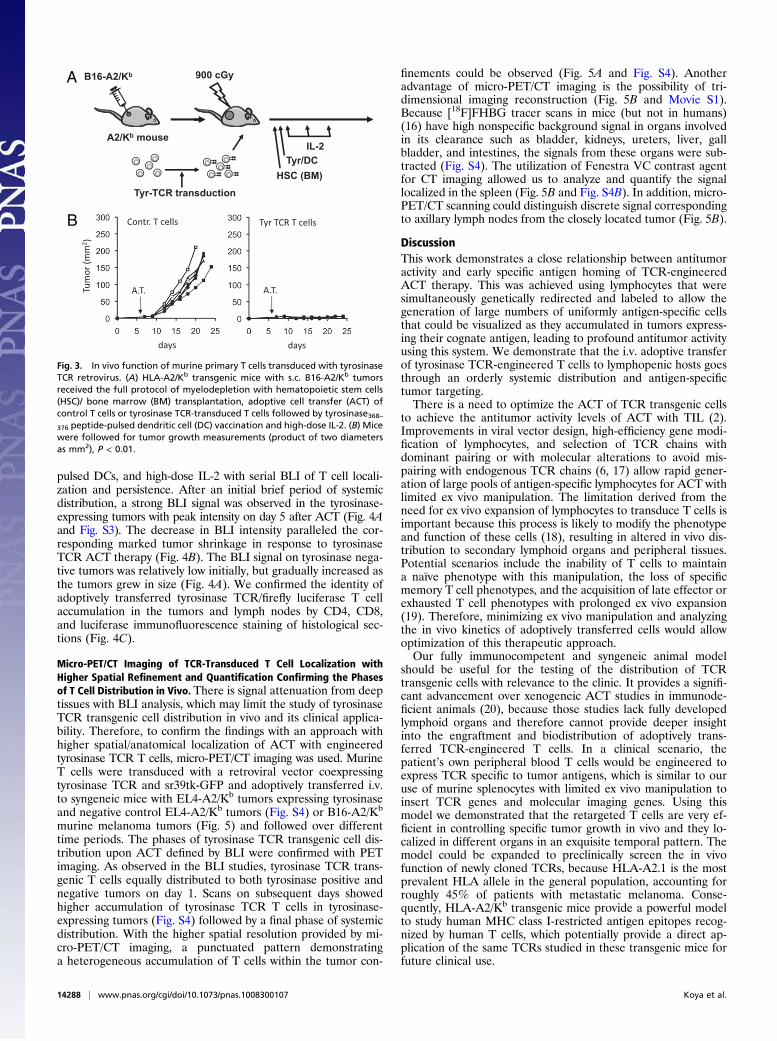
pulsed DCs, and high-dose IL-2 with serial BLI of T cell locali-zation and persistence. After an initial brief period of systemicdistribution, a strong BLI signal was observed in the tyrosinase-expressing tumors with peak intensity on day 5 after ACT (Fig. 4Aand Fig. S3). The decrease in BLI intensity paralleled the cor-responding marked tumor shrinkage in response to tyrosinaseTCR ACT therapy (Fig. 4B). The BLI signal on tyrosinase nega-tive tumors was relatively low initially, but gradually increased asthe tumors grew in size (Fig. 4A). We confirmed the identity ofadoptively transferred tyrosinase TCR/firefly luciferase T cellaccumulation in the tumors and lymph nodes by CD4, CD8,and luciferase immunofluorescence staining of histological sec-tions (Fig. 4C).
Micro-PET/CT Imaging of TCR-Transduced T Cell Localization withHigher Spatial Refinement and Quantification Confirming the Phasesof T Cell Distribution in Vivo. There is signal attenuation from deeptissues with BLI analysis, which may limit the study of tyrosinaseTCR transgenic cell distribution in vivo and its clinical applica-bility. Therefore, to confirm the findings with an approach withhigher spatial/anatomical localization of ACT with engineeredtyrosinase TCR T cells, micro-PET/CT imaging was used. MurineT cells were transduced with a retroviral vector coexpressingtyrosinase TCR and sr39tk-GFP and adoptively transferred i.v.to syngeneic mice with EL4-A2/Kb tumors expressing tyrosinaseand negative control EL4-A2/Kb tumors (Fig. S4) or B16-A2/Kb
murine melanoma tumors (Fig. 5) and followed over differenttime periods. The phases of tyrosinase TCR transgenic cell dis-tribution upon ACT defined by BLI were confirmed with PETimaging. As observed in the BLI studies, tyrosinase TCR trans-genic T cells equally distributed to both tyrosinase positive andnegative tumors on day 1. Scans on subsequent days showedhigher accumulation of tyrosinase TCR T cells in tyrosinase-expressing tumors (Fig. S4) followed by a final phase of systemicdistribution. With the higher spatial resolution provided by mi-cro-PET/CT imaging, a punctuated pattern demonstratinga heterogeneous accumulation of T cells within the tumor con-
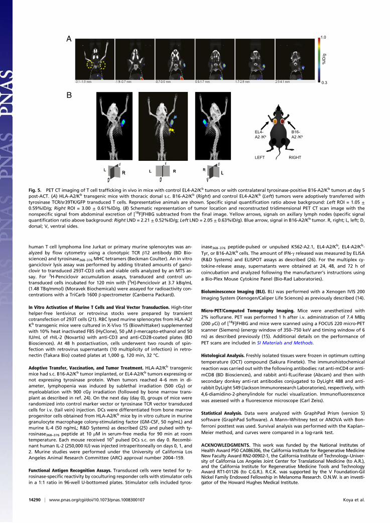
finements could be observed (Fig. 5A and Fig. S4). Anotheradvantage of micro-PET/CT imaging is the possibility of tri-dimensional imaging reconstruction (Fig. 5B and Movie S1).Because [18F]FHBG tracer scans in mice (but not in humans)(16) have high nonspecific background signal in organs involvedin its clearance such as bladder, kidneys, ureters, liver, gallbladder, and intestines, the signals from these organs were sub-tracted (Fig. S4). The utilization of Fenestra VC contrast agentfor CT imaging allowed us to analyze and quantify the signallocalized in the spleen (Fig. 5B and Fig. S4B). In addition, micro-PET/CT scanning could distinguish discrete signal correspondingto axillary lymph nodes from the closely located tumor (Fig. 5B).
DiscussionThis work demonstrates a close relationship between antitumoractivity and early specific antigen homing of TCR-engineeredACT therapy. This was achieved using lymphocytes that weresimultaneously genetically redirected and labeled to allow thegeneration of large numbers of uniformly antigen-specific cellsthat could be visualized as they accumulated in tumors express-ing their cognate antigen, leading to profound antitumor activityusing this system. We demonstrate that the i.v. adoptive transferof tyrosinase TCR-engineered T cells to lymphopenic hosts goesthrough an orderly systemic distribution and antigen-specifictumor targeting.There is a need to optimize the ACT of TCR transgenic cells
to achieve the antitumor activity levels of ACT with TIL (2).Improvements in viral vector design, high-efficiency gene modi-fication of lymphocytes, and selection of TCR chains withdominant pairing or with molecular alterations to avoid mis-pairing with endogenous TCR chains (6, 17) allow rapid gener-ation of large pools of antigen-specific lymphocytes for ACT withlimited ex vivo manipulation. The limitation derived from theneed for ex vivo expansion of lymphocytes to transduce T cells isimportant because this process is likely to modify the phenotypeand function of these cells (18), resulting in altered in vivo dis-tribution to secondary lymphoid organs and peripheral tissues.Potential scenarios include the inability of T cells to maintaina naïve phenotype with this manipulation, the loss of specificmemory T cell phenotypes, and the acquisition of late effector orexhausted T cell phenotypes with prolonged ex vivo expansion(19). Therefore, minimizing ex vivo manipulation and analyzingthe in vivo kinetics of adoptively transferred cells would allowoptimization of this therapeutic approach.Our fully immunocompetent and syngeneic animal model
should be useful for the testing of the distribution of TCRtransgenic cells with relevance to the clinic. It provides a signifi-cant advancement over xenogeneic ACT studies in immunode-ficient animals (20), because those studies lack fully developedlymphoid organs and therefore cannot provide deeper insightinto the engraftment and biodistribution of adoptively trans-ferred TCR-engineered T cells. In a clinical scenario, thepatient’s own peripheral blood T cells would be engineered toexpress TCR specific to tumor antigens, which is similar to ouruse of murine splenocytes with limited ex vivo manipulation toinsert TCR genes and molecular imaging genes. Using thismodel we demonstrated that the retargeted T cells are very ef-ficient in controlling specific tumor growth in vivo and they lo-calized in different organs in an exquisite temporal pattern. Themodel could be expanded to preclinically screen the in vivofunction of newly cloned TCRs, because HLA-A2.1 is the mostprevalent HLA allele in the general population, accounting forroughly 45% of patients with metastatic melanoma. Conse-quently, HLA-A2/Kb transgenic mice provide a powerful modelto study human MHC class I-restricted antigen epitopes recog-nized by human T cells, which potentially provide a direct ap-plication of the same TCRs studied in these transgenic mice forfuture clinical use.
A
B
Fig. 3. In vivo function of murine primary T cells transduced with tyrosinaseTCR retrovirus. (A) HLA-A2/Kb transgenic mice with s.c. B16-A2/Kb tumorsreceived the full protocol of myelodepletion with hematopoietic stem cells(HSC)/ bone marrow (BM) transplantation, adoptive cell transfer (ACT) ofcontrol T cells or tyrosinase TCR-transduced T cells followed by tyrosinase368–376 peptide-pulsed dendritic cell (DC) vaccination and high-dose IL-2. (B) Micewere followed for tumor growth measurements (product of two diametersas mm2), P < 0.01.
14288 | www.pnas.org/cgi/doi/10.1073/pnas.1008300107 Koya et al.
The definition of the pattern of distribution of adoptivelytransferred cells eliciting robust antitumor activity can be used tolater analyze individual components of this combinatorial ap-proach before going into clinical trials. In addition to screeningdifferent TCRs with human MHC class I restriction elements,variables like the surface functional phenotype of lymphocytes andtheir skewing while being activated ex vivo for TCR transductionaffecting their in vivo distribution upon ACT, the maximization ofcell persistence in vivo and the role of supporting therapies like theconditioning regimen, the use of high-dose IL-2, and antigen-specific vaccines can be systematically analyzed. Therefore, mo-lecular imaging with reporter genes enables monitoring of adop-tively transferred TCR-engineered retargeted cells to study theirbiodistribution, expansion/contraction, and persistence, allowingpattern-based prediction of T cell-based immunotherapeutic ef-ficiency and clinical outcome.
Materials and MethodsSubcloning and Vector Construction. The α and β chains of the TIL 1383I TCR (9,10) had their corresponding constant regions entirely substitutedwithmurinecounterparts by standard PCR techniques to generate a hybrid human/murine
TCR construct. The 2A self-cleaving sequences were inserted between thetransgenes by overlap PCR (12, 13). Thewhole constructwas then inserted intoa self-inactivating third generation lentiviral vector (21) or a retroviral vectorderived from a murine stem cell virus (pMSCV) backbone (22) containing a 5′long terminal repeat-driven truncated version of the sr39tk (15) fused withenhancedgreenfluorescent protein (eGFP), or afirefly luciferase gene (14). Toobtain the HLA-A2/Kb transgene to engineer syngeneic tumor targets, totalmRNA was obtained from hepatocytes of HLA-A2/Kb transgenic mice (fromLinda Sherman, The Scripps Research Institute, La Jolla, California) (11). ThisHLA-A2/Kb transgene was inserted into a lentiviral vector with a MND-drivenpromoter (23) andused to transduceB16andEL4 cells togenerate the cell linesB16-A2/Kb and EL4-A2/Kb, respectively. Similarly, the full-length human ty-rosinase cDNA was obtained by PCR cloning and used to generate a lentiviralvector with MND promoter for the transduction of EL4-A2/Kb cells to expressthe tyrosinase gene (EL4-A2/Kb-Tyr).
Analysis of Transgene Expression by TCR Engineering Viral Vectors. Lentivirusvectors were produced using a transient transfection protocol (21). Testing oftransgene expression was performed by transducing 293T-CD3 cells (fromDavid Baltimore, California Institute of Technology, Pasadena, California).Cells were analyzed by Western blot using an anti-2A antibody (from DarioVignali, St. Jude Children’s Research Hospital, Memphis, Tennessee) as pre-viously described (13). TCR expression upon transduction of 293T-CD3, the
A
B
C
Fig. 4. Bioluminescence imaging (BLI) of T cell trafficking in vivo. (A) HLA-A2/Kb transgenic mice with inguinal s.c. EL4-A2/Kb-expressing tyrosinase (Left)and control EL4-A2/Kb (Right) tumors received the full protocol of adoptive cell transfer (ACT) with tyrosinase TCR/fLuciferase-transduced T cells. Micewere followed from day 1 to 10 post-ACT and bioluminescence signal of ventral views were recorded and quantified on region of interest (ROI) drawn ontumor sites. Representative animals are shown. Pink, EL4-A2/Kb-tyrosinase+; yellow, control EL4-A2/Kb. (B) These mice were followed for tumor growthmeasurements (product of two diameters as mm2). (C ) EL4-A2/Kb-tyrosinase+ tumors were costained with DAPI (nucleus localization), anti-CD8, and anti-fLuciferase (40×).
Koya et al. PNAS | August 10, 2010 | vol. 107 | no. 32 | 14289
IMMUNOLO
GY
SEECO
MMEN
TARY
human T cell lymphoma line Jurkat or primary murine splenocytes was an-alyzed by flow cytometry using a clonotypic TCR β12 antibody (BD Bio-sciences) and tyrosinase368–376 MHC tetramers (Beckman Coulter). An in vitroganciclovir lysis assay was performed by adding titrated amounts of ganci-clovir to transduced 293T-CD3 cells and viable cells analyzed by an MTS as-say. For 3H-Penciclovir accumulation assays, transduced and control un-transduced cells incubated for 120 min with [3H]-Penciclovir at 3.7 kBq/mL(1.48 TBq/mmol) (Moravek Biochemicals) were assayed for radioactivity con-centrations with a TriCarb 1600 β-spectrometer (Canberra Packard).
In Vitro Activation of Murine T Cells and Viral Vector Transduction. High-titerhelper-free lentivirus or retrovirus stocks were prepared by transientcotransfection of 293T cells (21). RBC lysed murine splenocytes from HLA-A2/Kb transgenic mice were cultured in X-Vivo 15 (Biowhittaker) supplementedwith 10% heat inactivated FBS (HyClone), 50 μM β-mercapto-ethanol and 50IU/mL of rhIL-2 (Novartis) with anti-CD3 and anti-CD28-coated plates (BDBiosciences). At 48 h postactivation, cells underwent two rounds of spin-fection with retrovirus supernatants (10 multiplicity of infection) in retro-nectin (Takara Bio) coated plates at 1,000 g, 120 min, 32 °C.
Adoptive Transfer, Vaccination, and Tumor Treatment. HLA-A2/Kb transgenicmice had s.c. B16-A2/Kb tumor implanted, or EL4-A2/Kb tumors expressing ornot expressing tyrosinase protein. When tumors reached 4–6 mm in di-ameter, lymphopenia was induced by sublethal irradiation (500 cGy) ormyeloablation with 900 cGy irradiation (followed by bone marrow trans-plant as described in ref. 24). On the next day (day 0), groups of mice wererandomized into control marker vector or tyrosinase TCR vector transducedcells for i.v. (tail vein) injection. DCs were differentiated from bone marrowprogenitor cells obtained from HLA-A2/Kb mice by in vitro culture in murinegranulocyte macrophage colony-stimulating factor (GM-CSF, 50 ng/mL) andmurine IL-4 (50 ng/mL; R&D Systems) as described (25) and pulsed with ty-rosinase368–376 peptide at 10 μM in serum-free media for 90 min at roomtemperature. Each mouse received 105 pulsed DCs s.c. on day 0. Recombi-nant human IL-2 (250,000 IU) was injected intraperitoneally on days 0, 1, and2. Murine studies were performed under the University of California LosAngeles Animal Research Committee (ARC) approval number 2004–159.
Functional Antigen Recognition Assays. Transduced cells were tested for ty-rosinase-specific reactivity by coculturing responder cells with stimulator cellsin a 1:1 ratio in 96-well U-bottomed plates. Stimulator cells included tyros-
inase368–376 peptide-pulsed or unpulsed K562-A2.1, EL4-A2/Kb, EL4-A2/Kb-Tyr, or B16-A2/Kb cells. The amount of IFN-γ released was measured by ELISA(R&D Systems) and ELISPOT assays as described (26). For the multiplex cy-tokine-release assay, supernatants were obtained at 24, 48, and 72 h ofcoincubation and analyzed following the manufacturer’s instructions usinga Bio-Plex Mouse Cytokine Panel (Bio-Rad Laboratories).
Bioluminescence Imaging (BLI). BLI was performed with a Xenogen IVIS 200Imaging System (Xenogen/Caliper Life Sciences) as previously described (14).
Micro-PET/Computed Tomography Imaging. Mice were anesthetized with2% isoflurane. PET was performed 1 h after i.v. administration of 7.4 MBq(200 μCi) of [18F]FHBG and mice were scanned using a FOCUS 220 micro-PETscanner (Siemens) (energy window of 350–750 keV and timing window of 6ns) as described previously (15). Additional details on the performance ofPET scans are included in SI Materials and Methods.
Histological Analysis. Freshly isolated tissues were frozen in optimum cuttingtemperature (OCT) compound (Sakura Finetek). The immunohistochemicalreaction was carried out with the following antibodies: rat anti-mCD4 or anti-mCD8 (BD Biosciences), and rabbit anti-fLuciferase (Abcam) and then withsecondary donkey anti-rat antibodies conjugated to DyLight 488 and anti-rabbit DyLight 549 (Jackson Immunoresearch Laboratories), respectively, with4,6-diamidino-2-phenylindole for nuclei visualization. Immunofluorescencewas assessed with a fluorescence microscope (Carl Zeiss).
Statistical Analysis. Data were analyzed with GraphPad Prism (version 5)software (GraphPad Software). A Mann–Whitney test or ANOVA with Bon-ferroni posttest was used. Survival analysis was performed with the Kaplan–Meier method, and curves were compared in a log-rank test.
ACKNOWLEDGMENTS. This work was funded by the National Institutes ofHealth Award P50 CA086306, the California Institute for Regenerative MedicineNew Faculty Award RN2-00902-1, the California Institute of Technology–Univer-sity of California Los Angeles Joint Center for Translational Medicine (to A.R.),and the California Institute for Regenerative Medicine Tools and TechnologyAward RT1-01126 (to C.G.R.). R.C.K. was supported by the V Foundation-GilNickel Family Endowed Fellowship in Melanoma Research. O.N.W. is an investi-gator of the Howard Hughes Medical Institute.
EL4-A2 /K b
LEFT RIGHT
L R
%ID
/g
1.0
0.3
B16-A2 /Kb
A
B
Fig. 5. PET CT imaging of T cell trafficking in vivo in mice with control EL4-A2/Kb tumors or with contralateral tyrosinase-positive B16-A2/Kb tumors at day 5post-ACT. (A) HLA-A2/Kb transgenic mice with thoracic dorsal s.c. B16-A2/Kb (Right) and control EL4-A2/Kb (Left) tumors were adoptively transferred withtyrosinase TCR/sr39TK/GFP transduced T cells. Representative animals are shown. Specific signal quantification ratio above background: Left ROI = 1.05 ±0.59%ID/g; Right ROI = 3.00 ± 0.61%ID/g. (B) Schematic representation of tumor location and reconstructed tridimensional PET CT scan image with thenonspecific signal from abdominal excretion of [18F]FHBG subtracted from the final image. Yellow arrows, signals on axillary lymph nodes (specific signalquantification ratio above background: Right LND = 2.21 ± 0.52%ID/g; Left LND = 2.05 ± 0.63%ID/g). Blue arrow, signal in B16-A2/Kb tumor. R, right; L, left; D,dorsal; V, ventral sides.
14290 | www.pnas.org/cgi/doi/10.1073/pnas.1008300107 Koya et al.
1. Yee C, et al. (2002) Adoptive T cell therapy using antigen-specific CD8+ T cell clones forthe treatment of patients with metastatic melanoma: In vivo persistence, migration, andantitumor effect of transferred T cells. Proc Natl Acad Sci USA 99:16168–16173.
2. Rosenberg SA, Restifo NP, Yang JC, Morgan RA, DudleyME (2008) Adoptive cell transfer:A clinical path to effective cancer immunotherapy. Nat Rev Cancer 8:299–308.
3. Dembić Z, et al. (1987) Transfection of the CD8 gene enhances T-cell recognition.Nature 326:510–511.
4. Schumacher TN (2002) T-cell-receptor gene therapy. Nat Rev Immunol 2:512–519.5. Morgan RA, et al. (2006) Cancer regression in patients after transfer of genetically
engineered lymphocytes. Science 314:126–129.6. Johnson LA, et al. (2009) Gene therapy with human and mouse T-cell receptors
mediates cancer regression and targets normal tissues expressing cognate antigen.Blood 114:535–546.
7. Dudley ME, et al. (2008) Adoptive cell therapy for patients with metastatic melanoma:Wvaluation of intensive myeloablative chemoradiation preparative regimens. J ClinOncol 26:5233–5239.
8. Dubey P, et al. (2003) Quantitative imaging of the T cell antitumor response bypositron-emission tomography. Proc Natl Acad Sci USA 100:1232–1237.
9. Nishimura MI, et al. (1999) MHC class I-restricted recognition of a melanoma antigenby a human CD4+ tumor infiltrating lymphocyte. Cancer Res 59:6230–6238.
10. Roszkowski JJ, et al. (2003) CD8-independent tumor cell recognition is a property ofthe T cell receptor and not the T cell. J Immunol 170:2582–2589.
11. Vitiello A, Marchesini D, Furze J, Sherman LA, Chesnut RW (1991) Analysis of the HLA-restricted influenza-specific cytotoxic T lymphocyte response in transgenic micecarrying a chimeric human-mouse class I major histocompatibility complex. J Exp Med173:1007–1015.
12. de Felipe P, Martín V, Cortés ML, Ryan M, Izquierdo M (1999) Use of the 2A sequencefrom foot-and-mouth disease virus in the generation of retroviral vectors for genetherapy. Gene Ther 6:198–208.
13. Szymczak AL, et al. (2004) Correction of multi-gene deficiency in vivo using a single‘self-cleaving’ 2A peptide-based retroviral vector. Nat Biotechnol 22:589–594.
14. Prins RM, et al. (2008) Anti-tumor activity and trafficking of self, tumor-specific T cellsagainst tumors located in the brain. Cancer Immunol Immunother 57:1279–1289.
15. Shu CJ, et al. (2009) Quantitative PET reporter gene imaging of CD8+ T cells specificfor a melanoma-expressed self-antigen. Int Immunol 21:155–165.
16. Yaghoubi SS, et al. (2005) Imaging progress of herpes simplex virus type 1 thymidinekinase suicide gene therapy in living subjects with positron emission tomography.Cancer Gene Ther 12:329–339.
17. Kuball J, et al. (2007) Facilitating matched pairing and expression of TCR chainsintroduced into human T cells. Blood 109:2331–2338.
18. Hinrichs CS, et al. (2009) Adoptively transferred effector cells derived from naiverather than central memory CD8+ T cells mediate superior antitumor immunity. ProcNatl Acad Sci USA 106:17469–17474.
19. Klebanoff CA, Gattinoni L, Restifo NP (2006) CD8+ T-cell memory in tumorimmunology and immunotherapy. Immunol Rev 211:214–224.
20. Bobisse S, et al. (2009) Reprogramming T lymphocytes for melanoma adoptiveimmunotherapy by T-cell receptor gene transfer with lentiviral vectors. Cancer Res 69:9385–9394.
21. Koya RC, Kasahara N, Pullarkat V, Levine AM, Stripecke R (2002) Transduction ofacute myeloid leukemia cells with third generation self-inactivating lentiviral vectorsexpressing CD80 and GM-CSF: Effects on proliferation, differentiation, and stimulationof allogeneic and autologous anti-leukemia immune responses. Leukemia 16:1645–1654.
22. Hawley RG, Lieu FH, Fong AZ, Hawley TS (1994) Versatile retroviral vectors forpotential use in gene therapy. Gene Ther 1:136–138.
23. Robbins PB, et al. (1997) Increased probability of expression from modified retroviralvectors in embryonal stem cells and embryonal carcinoma cells. J Virol 71:9466–9474.
24. Wrzesinski C, et al. (2007) Hematopoietic stem cells promote the expansion andfunction of adoptively transferred antitumor CD8 T cells. J Clin Invest 117:492–501.
25. Ribas A, et al. (1997) Genetic immunization for the melanoma antigen MART-1/Melan-A using recombinant adenovirus-transduced murine dendritic cells. Cancer Res57:2865–2869.
26. Comin-Anduix B, et al. (2008) Detailed analysis of immunologic effects of the cytotoxicT lymphocyte-associated antigen 4-blocking monoclonal antibody tremelimumab inperipheral blood of patients with melanoma. J Transl Med 6:22.
Koya et al. PNAS | August 10, 2010 | vol. 107 | no. 32 | 14291