90
FETAL AND NEONATAL PHYSIOLOGY
| Date post: | 03-Jun-2018 |
| Category: |
Documents |
| Upload: | sheilla-elfira |
| View: | 238 times |
| Download: | 0 times |
8/12/2019 Kul. Fisiologi
http://slidepdf.com/reader/full/kul-fisiologi 1/90
8/12/2019 Kul. Fisiologi
http://slidepdf.com/reader/full/kul-fisiologi 2/90
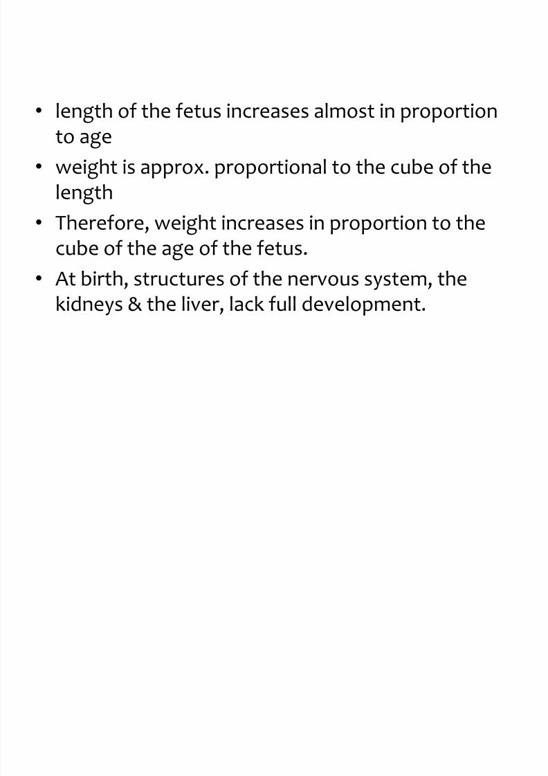
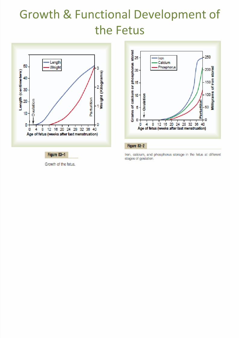
• length of the fetus increases almost in proportion
to age
• weight is approx. proportional to the cube of the
length• Therefore, weight increases in proportion to the
cube of the age of the fetus.
•
At birth, structures of the nervous system, thekidneys & the liver, lack full development.
8/12/2019 Kul. Fisiologi
http://slidepdf.com/reader/full/kul-fisiologi 3/90
8/12/2019 Kul. Fisiologi
http://slidepdf.com/reader/full/kul-fisiologi 4/90
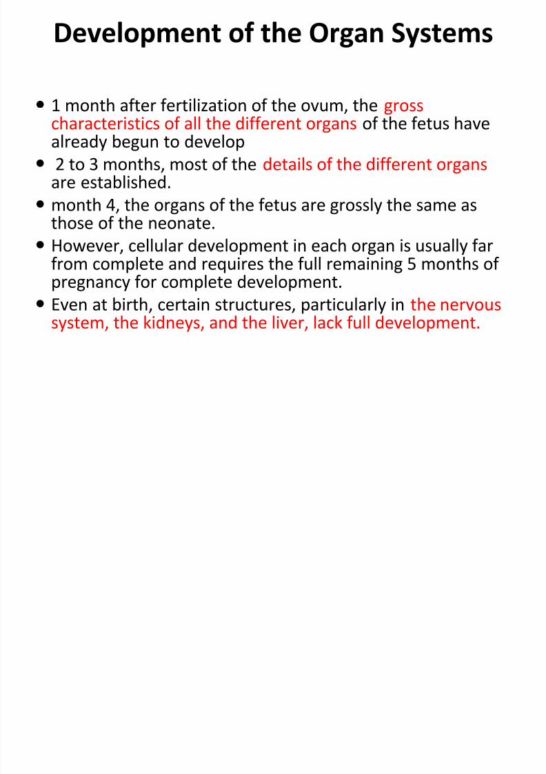
Development of the Organ Systems
1 month after fertilization of the ovum, the grosscharacteristics of all the different organs of the fetus havealready begun to develop
2 to 3 months, most of the details of the different organs
are established. month 4, the organs of the fetus are grossly the same as
those of the neonate.
However, cellular development in each organ is usually farfrom complete and requires the full remaining 5 months of
pregnancy for complete development. Even at birth, certain structures, particularly in the nervous
system, the kidneys, and the liver, lack full development.

8/12/2019 Kul. Fisiologi
http://slidepdf.com/reader/full/kul-fisiologi 5/90
Circulatory System
• The human heart begins beating during the
fourth week after fertilization.
• contracting at a rate of about 65 beats/min.
• This increases steadily to about 140 beats/min
immediately before birth until the neonatal
period
8/12/2019 Kul. Fisiologi
http://slidepdf.com/reader/full/kul-fisiologi 6/90
Fetal Circulation
• By the third month of development, all major
blood vessels are present and functioning.
• Fetus must have blood flow to placenta.
• Resistance to blood flow is high in lungs.
8/12/2019 Kul. Fisiologi
http://slidepdf.com/reader/full/kul-fisiologi 7/90
Formation of Blood Cells
3rd week – nRBC begin form’n in the yolk sac &mesothelial layers of the placenta
4th-5th week – form’n of nRBC by fetal mesenchyme &endothelium of the fetal blood vessels
6th week – liver begins to form blood cells
3rd month – spleen & other lymphoid tissues formblood cells
> 3
rd
month – bone marrow becomes the principalsource of RBC & most WBC, except continuedlymphocytic & plasma cell production in lymphoidtissues
8/12/2019 Kul. Fisiologi
http://slidepdf.com/reader/full/kul-fisiologi 8/90
Respiratory System
• Respiration cannot occur during fetal lifebecause there is no air to breathe in theamniotic cavity.
• However, attempted respiratory movementsdo take place beginning at the end of the firsttrimester of pregnancy.
•
Tactile stimuli and fetal asphyxia especiallycause these attempted respiratorymovements.
8/12/2019 Kul. Fisiologi
http://slidepdf.com/reader/full/kul-fisiologi 9/90
Respiratory System the last 3 to 4 months of pregnancy, the
respiratory movements of the fetus are mainlyinhibited, for reasons unknown, and the lungsremain almost completely deflated.
The inhibition of respiration during the later
months of fetal life prevents filling of the lungswith fluid and debris from the meconium excretedby the fetus’s gastrointestinal tract into theamniotic fluid.
small amounts of fluid are secreted into the lungsby the alveolar epithelium until the moment ofbirth, thus keeping only clean fluid in the lungs.
8/12/2019 Kul. Fisiologi
http://slidepdf.com/reader/full/kul-fisiologi 10/90
Nervous System
• Most reflexes of the fetus that involve the spinal
cord and even the brain stem are present by the
third to fourth months of pregnancy.
• Nervous system functions that involve thecerebral cortex are still only in the early stages of
development even at birth.
• Myelinization of some major tracts of the brainbecomes complete only after about 1 year of
postnatal life

8/12/2019 Kul. Fisiologi
http://slidepdf.com/reader/full/kul-fisiologi 11/90
Gastrointestinal.
• By mid-pregnancy – fetus begins to ingest &
absorb large quantities of amniotic fluid
• The last 2 to 3 months, gastrointestinal
function approaches that of the normal
neonate.

8/12/2019 Kul. Fisiologi
http://slidepdf.com/reader/full/kul-fisiologi 12/90
Meconium
• By that time, small quantities of meconiumare continually formed in the gastrointestinaltract and excreted from the anus into the
amniotic fluid.• Meconium is composed partly of residue
from swallowed amniotic fluid and partly ofmucus and other residues of excretory products from the gastrointestinal mucosa andglands.
8/12/2019 Kul. Fisiologi
http://slidepdf.com/reader/full/kul-fisiologi 13/90
Kidneys.
The fetal kidneys begin to excrete urine during thesecond trimester pregnancy, and fetal urine accountsfor about 70 to 80 per cent of the amniotic fluid.
Abnormal kidney development or severe impairment
of kidney function in the fetus greatly reduce theformation of amniotic fluid (oligohydramnios) and canlead to fetal death.
The renal control systems for regulating fetalextracellular fluid volume and electrolyte balances, and
especially acid base balance, are almost nonexistentuntil late fetal life and do not reach full developmentuntil a few months after birth.

8/12/2019 Kul. Fisiologi
http://slidepdf.com/reader/full/kul-fisiologi 14/90
Fetal Metabolism
• The fetus uses mainly glucose for energy
• and it has a high capability to store fat and
protein
8/12/2019 Kul. Fisiologi
http://slidepdf.com/reader/full/kul-fisiologi 15/90
Metabolism of Calcium and
Phosphate
22.5 grams of calcium and 13.5 grams of phosphorusare accumulated in the average fetus during gestation.
One half of these accumulate during the last 4 weeksof gestation, which is coincident with the period ofrapid ossification of the fetal bones and with the periodof rapid weight gain of the fetus.
During the earlier part of fetal life, the bones arerelatively unossified and have mainly a cartilaginous
matrix. until after the fourth month of pregnancy.
the total amounts of calcium and phosphate needed bythe fetus during gestation only 2 per cent of themother’s bones.
8/12/2019 Kul. Fisiologi
http://slidepdf.com/reader/full/kul-fisiologi 16/90
Accumulation of Iron
iron accumulates in the fetus more rapidly thancalcium and phosphate.
Most of the iron is in the form of hemoglobin -- beginsto be formed at third week after fertilization .
Small amounts of iron are concentrated in the mother’suterine progestational endometrium even beforeimplantation -- ingested into the embryo by thetrophoblastic cells and is used to form the very earlyred blood cells.
One third of the iron in fetus is normally stored in theliver -- can be used for several months after birth -- forformation of additional hemoglobin.

8/12/2019 Kul. Fisiologi
http://slidepdf.com/reader/full/kul-fisiologi 17/90

8/12/2019 Kul. Fisiologi
http://slidepdf.com/reader/full/kul-fisiologi 18/90

8/12/2019 Kul. Fisiologi
http://slidepdf.com/reader/full/kul-fisiologi 19/90
Utilization & Storage of Vit.
• Vit. K
– used by the fetal liver for form’n of Factor VII,prothrombin, & several other blood coag’n factors
– formed by bacterial action in the mother’s colon
– neonate has no adequate source during the 1st week of postnatal life until normal colonicbacterial flora become established
8/12/2019 Kul. Fisiologi
http://slidepdf.com/reader/full/kul-fisiologi 20/90
Utilization & Storage of Vit.
• Vit. K
– thus, prenatal storage is essential to prevent fetal
hemorrhage hemorrhage in the brain when the
head is traumatized by squeezing through thebirth canal
– also the reason for vit. K intramuscular injection
after birth as part of routine newborn care
8/12/2019 Kul. Fisiologi
http://slidepdf.com/reader/full/kul-fisiologi 21/90
ADJUSTMENTS OF THE INFANTTO EXTRAUTERINE LIFE
8/12/2019 Kul. Fisiologi
http://slidepdf.com/reader/full/kul-fisiologi 22/90
Onset of Breathing
• The most obvious effect of birth on the baby is
loss of the placental connection with the
mother
• This means loss of metabolic support.
• One of the most important immediate
adjustments required of the infant is to begin
breathing.
8/12/2019 Kul. Fisiologi
http://slidepdf.com/reader/full/kul-fisiologi 23/90
Cause of Breathing at Birth
The child begins to breathe within seconds
N ormal respiratory rhythm within less than 1 minuteafter birth.
The breathing is initiated by sudden exposure to theexterior world, resulting from (1) a slightly asphyxiatedstate incident to the birth process (2) sensory impulsesthat originate in the suddenly cooled skin.
Infant who does not breathe immediately -- the bodybecomes more hypoxic and hypercapnic -- provideadditional stimulus to the respiratory center and usuallycauses breathing within an additional minute afterbirth.
8/12/2019 Kul. Fisiologi
http://slidepdf.com/reader/full/kul-fisiologi 24/90
Delayed or Abnormal Breathing at
Birth mother has been depressed by a general anesthetic during
delivery --- the fetus as well -- the onset of respiration islikely to be delayed for several minutes -- importance ofusing as little anesthesia as feasible.
Many infants have head trauma during delivery or whoundergo prolonged delivery are slow to breathe orsometimes do not breathe at all. This can result from:
1. First, in a few infants, intracranial hemorrhage or braincontusion causes a concussion syndrome with a greatlydepressed respiratory center.
2. Second, and probably much more important, prolongedfetal hypoxia during delivery can cause serious depressionof the respiratory center
8/12/2019 Kul. Fisiologi
http://slidepdf.com/reader/full/kul-fisiologi 25/90
Causes of Prolonged Fetal Hypoxia
during Delivery
1. compression of the umbilical cord – “cord coil”
2. premature separation of the placenta – abruptio
placenta
3. excessive uterine contraction – cut-off blood flowto the placenta
4. excessive maternal anesthesia- depressed
oxygenation to her own blood
5. head trauma – ICH or brain contusion
8/12/2019 Kul. Fisiologi
http://slidepdf.com/reader/full/kul-fisiologi 26/90
Degree of Hypoxia That an Infant Can
Tolerate
• Adult, failure to breathe for only 4 minutes oftencauses death
• A neonate often survives as long as 10 minutes of
failure to breathe after birth.• Permanent and very serious brain impairment
often ensues If breathing is delayed more than 8to 10 minutes -- actual lesions develop mainly in
the thalamus, inferior colliculi, and in other brainstem areas -- permanently affecting many of themotor functions -- cerebral palsy
8/12/2019 Kul. Fisiologi
http://slidepdf.com/reader/full/kul-fisiologi 27/90
Expansion of the Lungs at Birth
At birth, the walls of the alveoli are at first collapsedbecause of the surface tension of the viscid fluid that fillsthem.
More than 25 mm Hg of negative inspiratory pressure in thelungs is usually required to oppose the effects of thissurface tension and to open the alveoli for the first time.
But once the alveoli open, further respiration can beeffected with relatively weak respiratory movements.
The first inspirations of the normal neonate are extremely
powerful, usually capable of creating as much as 60 mm Hgnegative pressure in the intrapleural space. the tremendousnegative intrapleural pressures required to open the lungsat the onset of breathing.

8/12/2019 Kul. Fisiologi
http://slidepdf.com/reader/full/kul-fisiologi 28/90
• Note that the second breath is much easier,
with far less negative and positive pressures
required. Breathing
• does not become completely normal until
about 40 minutes after birth.
8/12/2019 Kul. Fisiologi
http://slidepdf.com/reader/full/kul-fisiologi 29/90
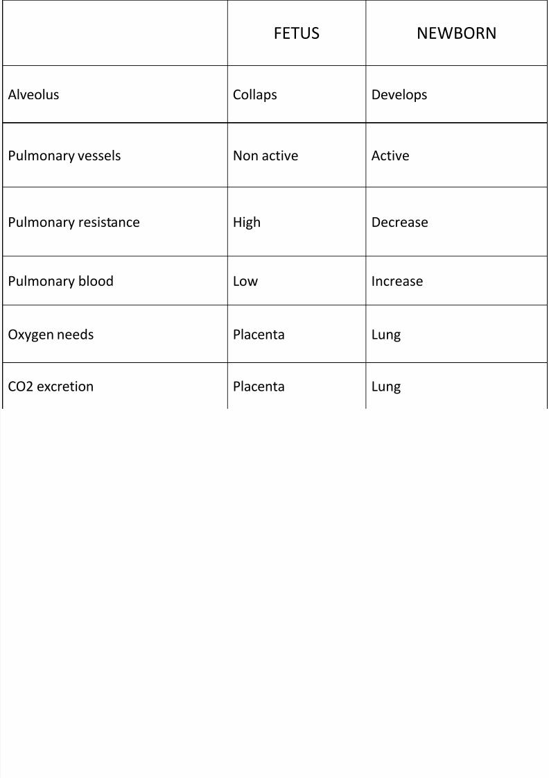
FETUS NEWBORN
Alveolus Collaps Develops Pulmonary vessels Non active Active
Pulmonary resistance High Decrease
Pulmonary blood Low Increase
Oxygen needs Placenta Lung
CO2 excretion Placenta Lung
8/12/2019 Kul. Fisiologi
http://slidepdf.com/reader/full/kul-fisiologi 30/90
Respiratory Distress Syndrome
Caused When Surfactant Secretion Is Deficient.
A small number of infants, especially premature infantsand infants born of diabetic mothers, develop severerespiratory distress in the early hours to the first several
days after birth, and some die within the next day or so. The alveoli of these infants at death contain large
quantities of proteinaceous fluid(pure plasma hadleaked out of the capillaries into the alveoli) -- The fluidalso contains desquamated alveolar epithelial cells --
called hyaline membrane disease because microscopicslides of the lung show the material filling the alveoli tolook like a hyaline membrane.

8/12/2019 Kul. Fisiologi
http://slidepdf.com/reader/full/kul-fisiologi 31/90
Respiratory Distress Syndrome
One of the most characteristic findings in respiratorydistress syndrome is failure of the respiratory epithelium tosecrete adequate quantities of surfactant, a substancenormally secreted into the alveoli that decreases thesurface tension of the alveolar fluid, therefore allowing the
alveoli to open easily during inspiration. The surfactant- secreting cells (type II alveolar epithelial
cells) do not begin to secrete surfactant until the last 1 to 3months of gestation.
Therefore, many premature babies and a few full-termbabies are born without the capability to secrete sufficientsurfactant, which causes both a collapse tendency of thealveoli and development of pulmonary edema. The role ofsurfactant in preventing these effects
8/12/2019 Kul. Fisiologi
http://slidepdf.com/reader/full/kul-fisiologi 32/90
Specific Anatomical Structure of the
Fetal Circulation
• Because the lungs are mainly nonfunctionalduring fetal life
• and because the liver is only partially functional,
it is not necessary for the fetal heart to pumpmuch blood through either the lungs or the liver.
• However, the fetal heart must pump largequantities of blood through the placenta.
• Therefore, special anatomical arrangementscause the fetal circulatory system to operatemuch differently from that of the newborn baby.
8/12/2019 Kul. Fisiologi
http://slidepdf.com/reader/full/kul-fisiologi 33/90
blood returning from the placenta through theumbilical vein passes through the ductus venosus,mainly bypassing the liver.
Then most of the blood entering the right atrium from
the inferior vena cava is directed in a straight pathwayacross the posterior aspect of the right atrium andthrough the foramen ovale directly into the left atrium.
Thus, the well-oxygenated blood from the placenta
enters mainly the left side of the heart, rather than theright side, and is pumped by the left ventricle mainlyinto the arteries of the head and forelimbs.
8/12/2019 Kul. Fisiologi
http://slidepdf.com/reader/full/kul-fisiologi 34/90
• The blood entering the right atrium from thesuperior vena cava is directed downward throughthe tricuspid valve into the right ventricle.
•
This blood is mainly deoxygenated blood fromthe head region of the fetus, and it is pumped bythe right ventricle into the pulmonary artery andthen mainly through the ductus arteriosus into
the descending aorta, then through the twoumbilical arteries into the placenta, where thedeoxygenated blood becomes oxygenated.

8/12/2019 Kul. Fisiologi
http://slidepdf.com/reader/full/kul-fisiologi 35/90
• gives the relative percentages of the total bloodpumped by the heart that pass through thedifferent vascular circuits of the fetus.
•
that 55 per cent of all the blood goes through theplacenta, leaving only 45 per cent to pass throughall the tissues of the fetus. Furthermore, duringfetal life, only 12 per cent of the blood flows
through the lungs;• Immediately after birth, virtually all the blood
flows through the lungs.
8/12/2019 Kul. Fisiologi
http://slidepdf.com/reader/full/kul-fisiologi 36/90
8/12/2019 Kul. Fisiologi
http://slidepdf.com/reader/full/kul-fisiologi 37/90
Primary Changes in Pulmonary and Systemic Vascular
Resistances at Birth
In the unexpanded fetal lungs,-- the blood vessels arecompressed -- Immediately on expansion, thesevessels are no longer compressed -- the resistance toblood flow decreases several fold.
in fetal life -- hypoxia of the lungs --- tonicvasoconstriction of the lung blood vessels, -- whenaeration of the lungs eliminates the hypoxia --vasodilatation.
All these changes together reduce the resistance to
blood flow through the lungs as much as fivefold,which reduces the pulmonary arterial pressure, rightventricular pressure, and right atrial pressure
8/12/2019 Kul. Fisiologi
http://slidepdf.com/reader/full/kul-fisiologi 38/90
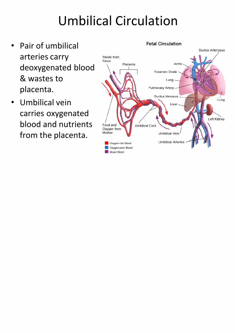
Umbilical Circulation
• Pair of umbilical
arteries carry
deoxygenated blood
& wastes to
placenta.
• Umbilical vein
carries oxygenated
blood and nutrientsfrom the placenta.
8/12/2019 Kul. Fisiologi
http://slidepdf.com/reader/full/kul-fisiologi 39/90
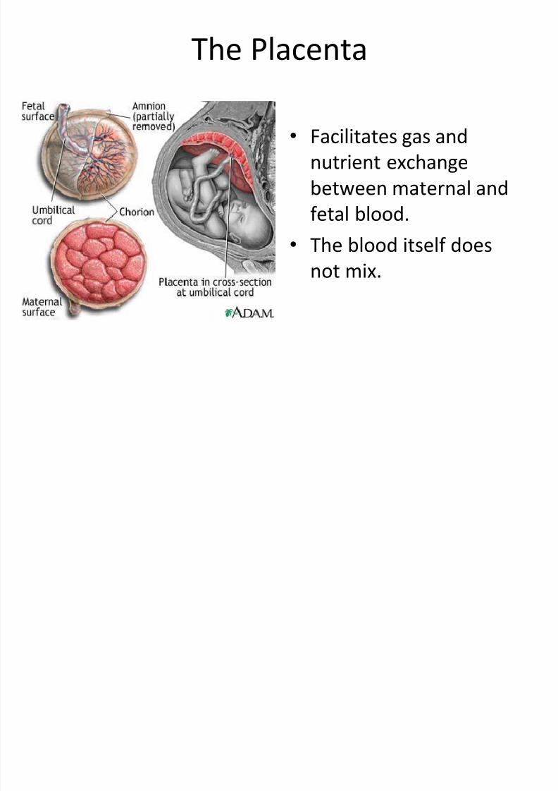
The Placenta
• Facilitates gas and
nutrient exchange
between maternal and
fetal blood.
• The blood itself does
not mix.

8/12/2019 Kul. Fisiologi
http://slidepdf.com/reader/full/kul-fisiologi 40/90
8/12/2019 Kul. Fisiologi
http://slidepdf.com/reader/full/kul-fisiologi 41/90
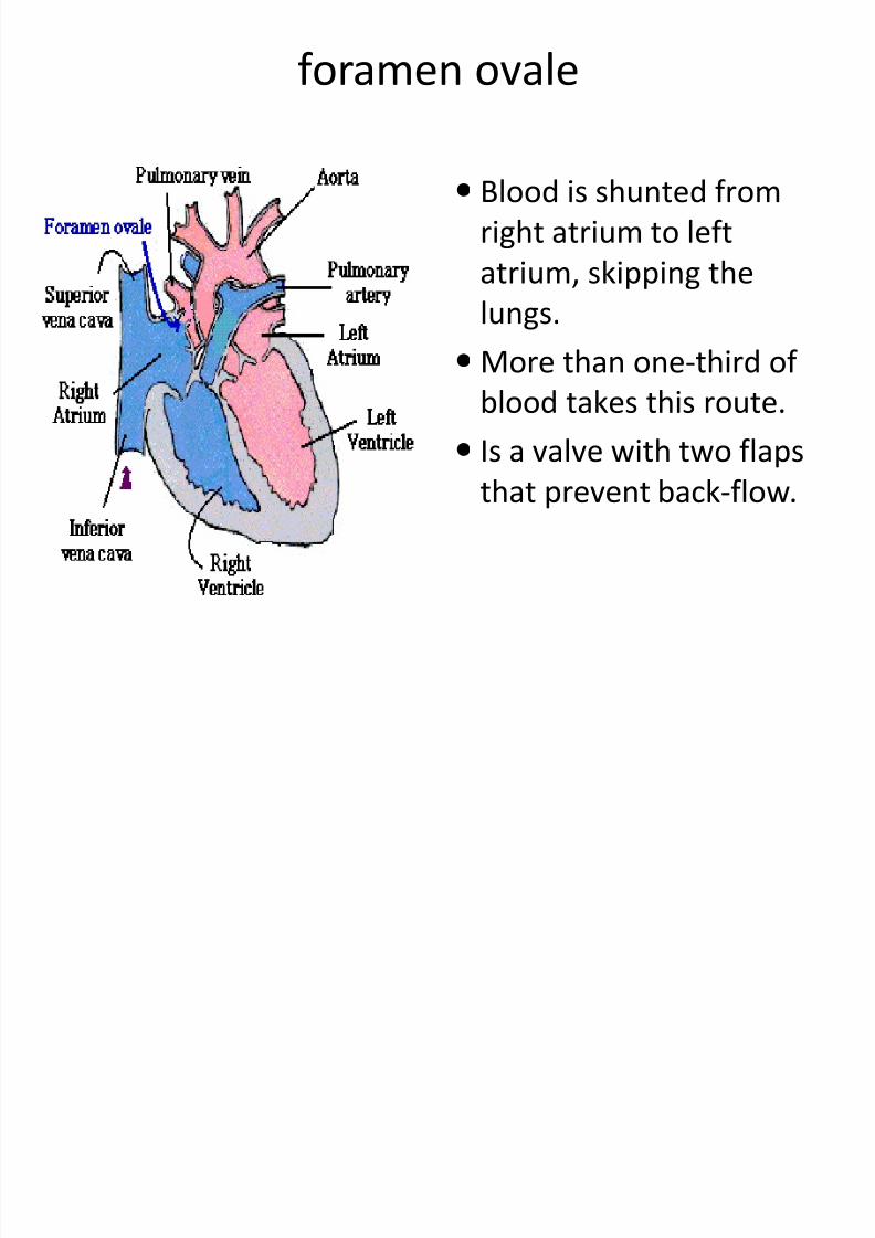
foramen ovale
Blood is shunted from
right atrium to left
atrium, skipping the
lungs. More than one-third of
blood takes this route.
Is a valve with two flapsthat prevent back-flow.
8/12/2019 Kul. Fisiologi
http://slidepdf.com/reader/full/kul-fisiologi 42/90
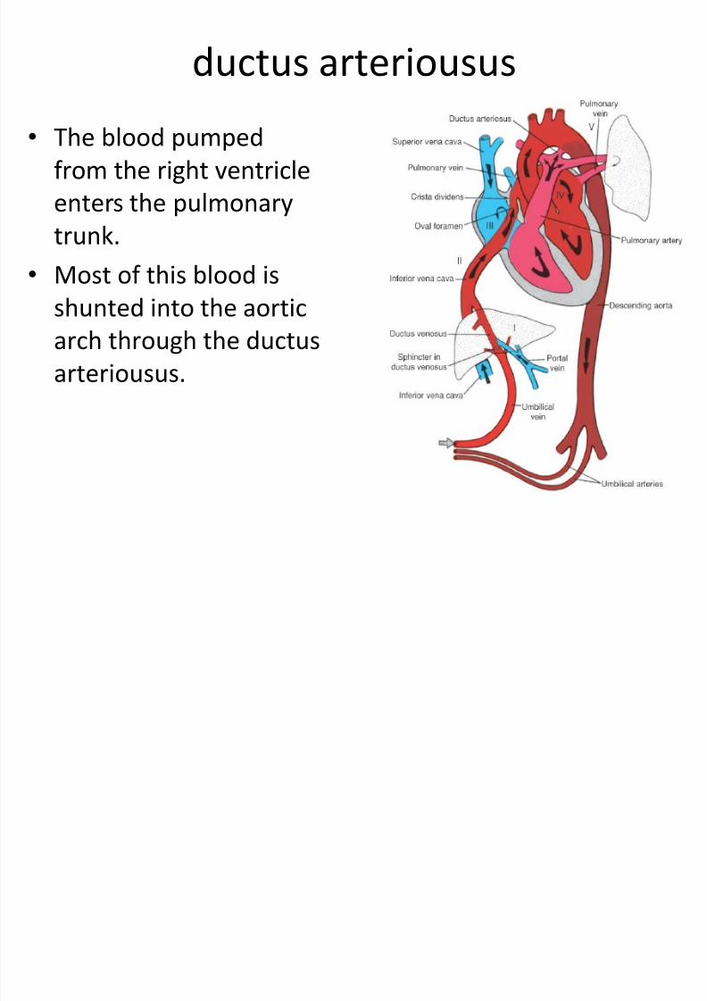
ductus arteriousus
• The blood pumpedfrom the right ventricle
enters the pulmonary
trunk.
• Most of this blood is
shunted into the aortic
arch through the ductus
arteriousus.
Wh h bi h?
8/12/2019 Kul. Fisiologi
http://slidepdf.com/reader/full/kul-fisiologi 43/90
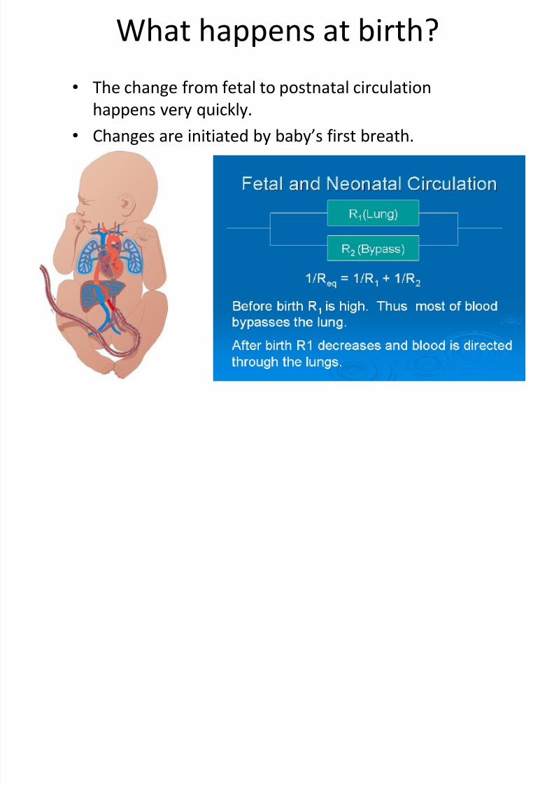
What happens at birth?
• The change from fetal to postnatal circulation
happens very quickly.
• Changes are initiated by baby’s first breath.
f
8/12/2019 Kul. Fisiologi
http://slidepdf.com/reader/full/kul-fisiologi 44/90
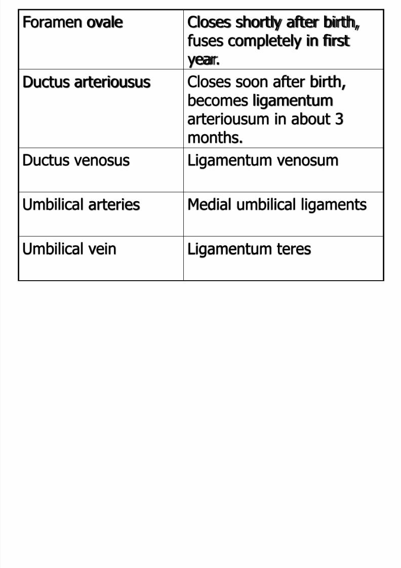
Foramen ovale Closes shortly after birth,fuses completely in firstyear.
Ductus arteriousus Closes soon after birth,becomes ligamentumarteriousum in about 3
months.Ductus venosus Ligamentum venosum
Umbilical arteries Medial umbilical ligaments
Umbilical vein Ligamentum teres
8/12/2019 Kul. Fisiologi
http://slidepdf.com/reader/full/kul-fisiologi 45/90
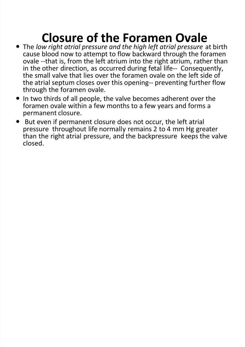
Closure of the Foramen Ovale The low right atrial pressure and the high left atrial pressure at birth
cause blood now to attempt to flow backward through the foramenovale --that is, from the left atrium into the right atrium, rather thanin the other direction, as occurred during fetal life-- Consequently,the small valve that lies over the foramen ovale on the left side ofthe atrial septum closes over this opening-- preventing further flowthrough the foramen ovale.
In two thirds of all people, the valve becomes adherent over theforamen ovale within a few months to a few years and forms apermanent closure.
But even if permanent closure does not occur, the left atrial
pressure throughout life normally remains 2 to 4 mm Hg greaterthan the right atrial pressure, and the backpressure keeps the valveclosed.
P bl i h i
8/12/2019 Kul. Fisiologi
http://slidepdf.com/reader/full/kul-fisiologi 46/90
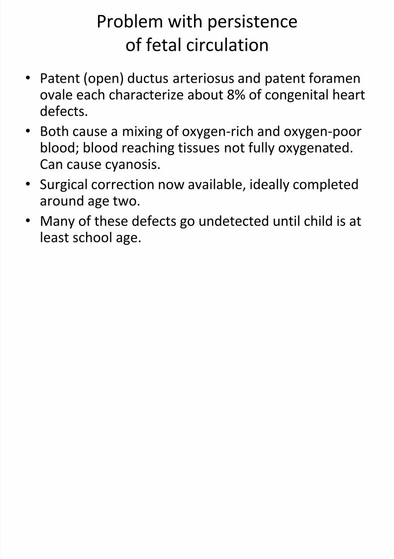
Problem with persistence
of fetal circulation
• Patent (open) ductus arteriosus and patent foramenovale each characterize about 8% of congenital heartdefects.
• Both cause a mixing of oxygen-rich and oxygen-poorblood; blood reaching tissues not fully oxygenated.Can cause cyanosis.
• Surgical correction now available, ideally completedaround age two.
• Many of these defects go undetected until child is atleast school age.
8/12/2019 Kul. Fisiologi
http://slidepdf.com/reader/full/kul-fisiologi 47/90
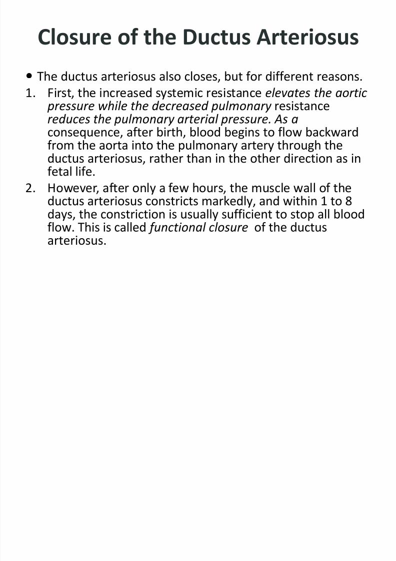
Closure of the Ductus Arteriosus
The ductus arteriosus also closes, but for different reasons.1. First, the increased systemic resistance elevates the aortic
pressure while the decreased pulmonary resistancereduces the pulmonary arterial pressure. As aconsequence, after birth, blood begins to flow backward
from the aorta into the pulmonary artery through theductus arteriosus, rather than in the other direction as infetal life.
2. However, after only a few hours, the muscle wall of theductus arteriosus constricts markedly, and within 1 to 8
days, the constriction is usually sufficient to stop all bloodflow. This is called functional closure of the ductusarteriosus.
8/12/2019 Kul. Fisiologi
http://slidepdf.com/reader/full/kul-fisiologi 48/90
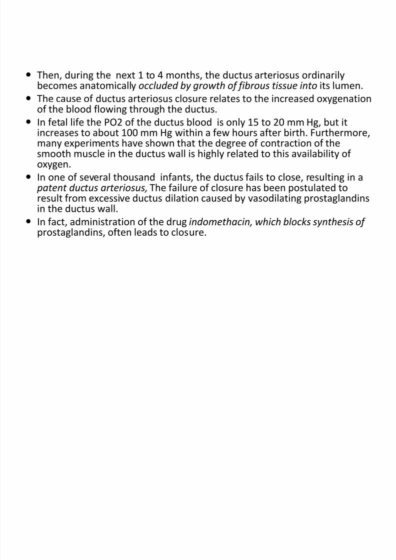
Then, during the next 1 to 4 months, the ductus arteriosus ordinarilybecomes anatomically occluded by growth of fibrous tissue into its lumen.
The cause of ductus arteriosus closure relates to the increased oxygenationof the blood flowing through the ductus.
In fetal life the PO2 of the ductus blood is only 15 to 20 mm Hg, but itincreases to about 100 mm Hg within a few hours after birth. Furthermore,
many experiments have shown that the degree of contraction of thesmooth muscle in the ductus wall is highly related to this availability ofoxygen.
In one of several thousand infants, the ductus fails to close, resulting in a patent ductus arteriosus, The failure of closure has been postulated toresult from excessive ductus dilation caused by vasodilating prostaglandins
in the ductus wall. In fact, administration of the drug indomethacin, which blocks synthesis of
prostaglandins, often leads to closure.

8/12/2019 Kul. Fisiologi
http://slidepdf.com/reader/full/kul-fisiologi 49/90
Closure of the Ductus Venosus
In fetal life, the portal blood from the fetus’s abdomen joins theblood from the umbilical vein, and these together pass by way ofthe ductus venosus directly into the vena cava immediately belowthe heart but above the liver, thus bypassing the liver.
Immediately after birth, blood flow through the umbilical vein
ceases, but most of the portal blood still flows through the ductusvenosus, with only a small amount passing through the channels ofthe liver.
However, within 1 to 3 hours the muscle wall of the ductus venosuscontracts strongly and closes this avenue of flow. As a consequence,the portal venous pressure rises from near 0 to 6 to 10 mm Hg,
which is enough to force portal venous blood flow through the liversinuses.
Although the ductus venosus rarely fails to close, we know almostnothing about what causes the closure.
CIRCULATORY ADAPTATION
8/12/2019 Kul. Fisiologi
http://slidepdf.com/reader/full/kul-fisiologi 50/90
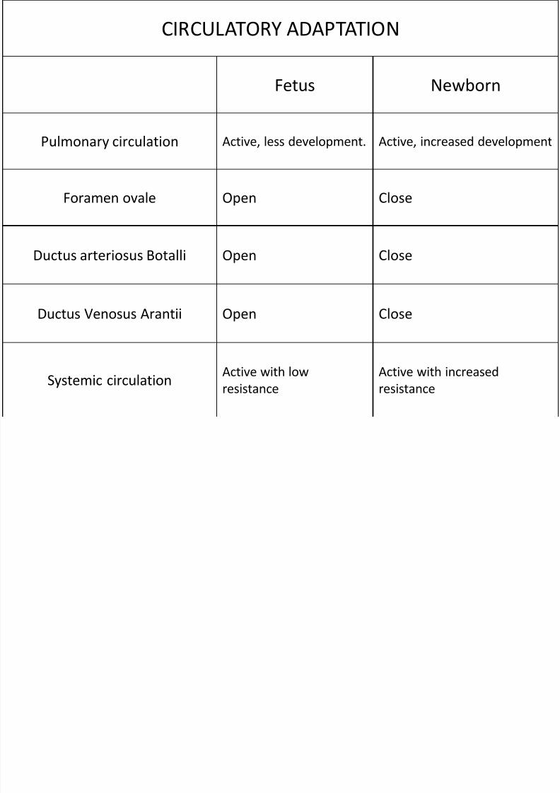
CIRCULATORY ADAPTATION Fetus Newborn
Pulmonary circulation Active, less development. Active, increased development
Foramen ovale Open Close Ductus arteriosus Botalli Open Close
Ductus Venosus Arantii Open Close
Systemic circulation Active with low
resistance Active with increased
resistance
8/12/2019 Kul. Fisiologi
http://slidepdf.com/reader/full/kul-fisiologi 51/90
Nutrition of the Neonate
Before birth – all energy from glucose obtained from themother’s blood.
After birth -- glucose stored in the infant’s body in the formof liver and muscle glycogen is sufficient to supply theinfant’s needs for only a few hours.
The liver of the neonate is still far from functionallyadequate at birth -- prevents significant gluconeogenesis. --the infant’s blood glucose concentration frequently falls thefirst day 30 to 40 mg/dl of plasma, ( <1/2 Normal value) --appropriate mechanisms are available for the infant to use
its stored fats and proteins for metabolism until mother’smilk can be provided 2 to 3 days later.
8/12/2019 Kul. Fisiologi
http://slidepdf.com/reader/full/kul-fisiologi 52/90
• Special problems are associated with gettingan adequate fluid supply to the neonate --because the infant’s rate of body fluid
turnover averages seven times that of anadult, and the mother’s milk supply requiresseveral days to develop -- Ordinarily, theinfant’s weight decreases 5 to 10 per cent and
sometimes as much as 20 per cent within thefirst 2 to 3 days of life. Most of this weight lossis loss of fluid rather than of body solids.
8/12/2019 Kul. Fisiologi
http://slidepdf.com/reader/full/kul-fisiologi 53/90
Special Functional Problems in the Neonate
(Adjustments of the Neonate)
Neonate - characterized by instability of the
various hormonal and neurogenic control
systems
• Due to
1. Immature development of the different organ
system
2. Control system have not become fully adjustedto the new way of life.
Respiratory System
8/12/2019 Kul. Fisiologi
http://slidepdf.com/reader/full/kul-fisiologi 54/90
Respiratory System
The normal rate of respiration in a neonate is about 40breaths per minute
Tidal air with each breath averages 16 milliliters.
Total minute respiratory volume of 640 ml/min, which isabout twice as great in relation to the body weight as thatof an adult.
The functional residual capacity of the infant’s lungs is onlyone half that of an adult in relation to body weight. Thisdifference causes excessive cyclical increases and decreasesin the newborn baby’s blood gas concentrations if therespiratory rate becomes slowed because it is the residualair in the lungs that smooths out the blood gas variations.

8/12/2019 Kul. Fisiologi
http://slidepdf.com/reader/full/kul-fisiologi 55/90
Circulation
The blood volume of a neonate immediately after birthaverages about 300 milliliters
Placenta left attached or umbilical cord was stripped maycause additional blood volume = + 75 ml -- 375 milliliters.
Then, during the ensuing few hours, fluid is lost into theneonate’s tissue spaces from this blood, which increases thehematocrit but returns the blood volume once again to thenormal value of about 300 milliliters.
Some pediatricians believe that this extra blood volumecaused by stripping the umbilical cord can lead to mildpulmonary edema with some degree of respiratory distress,but the extra red blood cells are often very valuable to theinfant.
8/12/2019 Kul. Fisiologi
http://slidepdf.com/reader/full/kul-fisiologi 56/90
Cardiac Output and Arterial Pressure
The cardiac output of the neonate averages 500ml/min, which, like respiration and body metabolism, isabout twice as much in relation to body weight as inthe adult.
The arterial pressure during the first day after birthaverages about 70 mm Hg systolic and 50 mm Hgdiastolic
this increases slowly during the next several months toabout 90/60.
Then there is a much slower rise during the subsequentyears until the adult pressure of 115/70 is attained atadolescence.
8/12/2019 Kul. Fisiologi
http://slidepdf.com/reader/full/kul-fisiologi 57/90
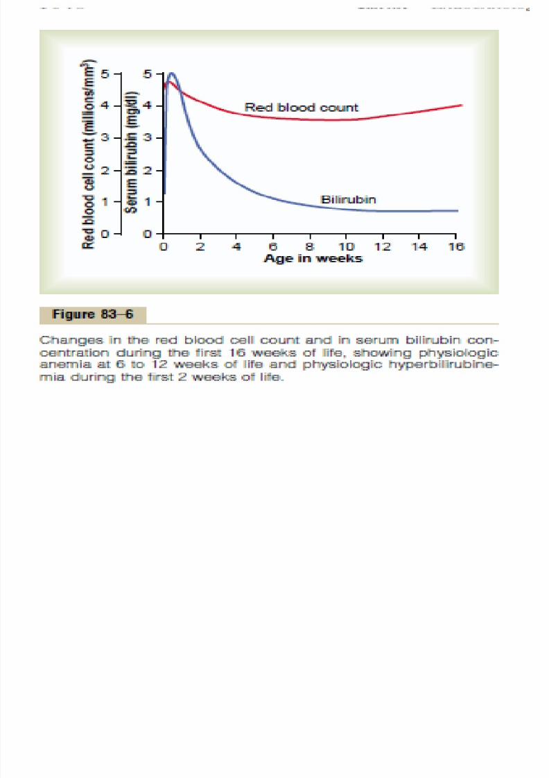
Blood Characteristics
The red blood cell count in the neonate averages about 4million per cubic millimeter.
If blood is stripped from the cord into the infant, the redblood cell count rises an additional 0.5 to 0.75 millionduring the first few hours of life, giving a red blood cell
count of about 4.75 million per cubic millimeter Few new red blood cells are formed in the infant during the
first few weeks of life because the hypoxic stimulus of fetallife is no longer present to stimulate red cell production. ---The average red blood cell count falls to less than 4 million
per cubic millimeter by about 6 to 8 weeks of age. From that time on, increasing activity by the baby provides
the appropriate stimulus for returning the red blood cell
8/12/2019 Kul. Fisiologi
http://slidepdf.com/reader/full/kul-fisiologi 58/90
Fluid BalanceAcid-Base Balance,
and Renal Function
The rate of fluid intake and fluid excretion in the newborn infant isseven times as great in relation to weight as in the adult -- slightpercentage alteration of fluid intake or fluid output can cause rapidlydeveloping abnormalities.
The rate of metabolism in the infant is also twice as great in relationto body mass as in the adult, twice as much acid is normally formed,
-- a tendency toward acidosis in the infant. Functional development of the kidneys is not complete until the end
of about the first month of life. For instance, the kidneys of theneonate can concentrate urine to only 1.5 times the osmolality ofthe plasma instead of the adult three to four times.
Therefore, considering the immaturity of the kidneys, together with
the marked fluid turnover in the infant and rapid formation of acid,one can readily understand that among the most importantproblems of infancy are acidosis, dehydration, and, more rarely,overhydration.
8/12/2019 Kul. Fisiologi
http://slidepdf.com/reader/full/kul-fisiologi 59/90
Liver Function
1. The liver of the neonate conjugates bilirubin with glucuronic acidpoorly and therefore excretes bilirubin only slightly during the firstfew days of life -- physiologic hyperbilirubinemia (jaundice)
2. The liver of the neonate is deficient in forming plasma proteins --the plasma protein concentration falls during the first weeks of lifeto 15 to 20 per cent less than that for older children. Occasionallythe protein concentration falls so low that the infant developshypoproteinemic edema.
3. The gluconeogenesis function of the liver is particularly deficient. Asa result, the blood glucose level of the unfed neonate falls to about30 to 40 mg/dl (about 40 per cent of normal), and the infant must
depend mainly on its stored fats for energy until sufficient feedingcan occur.
4. The liver of the neonate usually also forms too little of the bloodfactors needed for normal blood coagulation
8/12/2019 Kul. Fisiologi
http://slidepdf.com/reader/full/kul-fisiologi 60/90
Digestion, Absorption, and Metabolism of Energy Foods; and Nutrition
In general, the ability of the neonate to digest, absorb, and metabolizefoods is no different from that of the older child, with the following threeexceptions.
1. First, secretion of pancreatic amylase in the neonate is deficient, so thatthe neonate uses starches less adequately than do older children.
2. Second, absorption of fats from the gastrointestinal tract is somewhat
less than that in the older child. Consequently, milk with a high fatcontent, such as cow’s milk, is frequently inadequately absorbed.
3. Third, because the liver functions imperfectly during at least the firstweek of life, the glucose concentration in the blood is unstable and low.
The neonate is especially capable of synthesizing and storing proteins.Indeed, with an adequate diet, as much as 90 per cent of the ingested
amino acids are used for formation of body proteins. This is a muchhigher percentage than in adults.
Metabolic Rate and Body
8/12/2019 Kul. Fisiologi
http://slidepdf.com/reader/full/kul-fisiologi 61/90
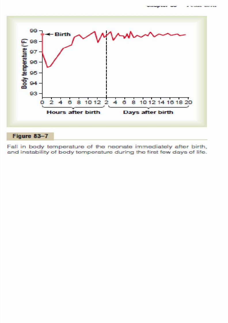
Metabolic Rate and Body
Temperature
• The normal metabolic rate of the neonate inrelation to body weight is about twice that of theadult, which accounts also for the twice as greatcardiac output and twice as great minuterespiratory volume in relation to body weight in theinfant.
• Because the body surface area is large in relation to
body mass, heat is readily lost from the body. As aresult, the body temperature of the neonate,particularly of premature infants, falls easily.
Nutritional Needs During the Early Weeks of Life

8/12/2019 Kul. Fisiologi
http://slidepdf.com/reader/full/kul-fisiologi 62/90
Nutritional Needs During the Early Weeks of Life.
• At birth, a neonate is usually in complete
nutritional balance, provided the mother has had
an adequate diet.
• function of the gastrointestinal system is usuallymore than adequate to digest and assimilate all
the nutritional needs of the infant if appropriate
nutrients are provided in the diet.
• However, three specific problems do occur in the
early nutrition of the infant.
d f l i d i i
8/12/2019 Kul. Fisiologi
http://slidepdf.com/reader/full/kul-fisiologi 63/90
Need for Calcium and Vitamin D
• The neonate is in a stage of rapid ossification ofits bones at birth -- supply of calcium is neededfrom milk.
•
Absorption of calcium by the gastrointestinaltract is poor in the absence of vitamin D --deficient vit Dcan develop severe rickets in only afew weeks this is particularly true in premature
babies because their gastrointestinal tractsabsorb calcium even less effectively than those ofnormal infants.
N i f I i h Di

8/12/2019 Kul. Fisiologi
http://slidepdf.com/reader/full/kul-fisiologi 64/90
Necessity for Iron in the Diet
1. Stored Fe in the infant’s liver depends on the adequacy ofthe Fe content in the mother’s diet RBC formed duringthe next 4-6 mons after birth.
(Fe diet of mons severe anemia in 3mons; may be
prevented by early feeding of the infant with egg yolk, & Fesupplement by the 2nd or 3rd month of life).
Vi i C D fi i i I f

8/12/2019 Kul. Fisiologi
http://slidepdf.com/reader/full/kul-fisiologi 65/90
Vitamin C Deficiency in Infants
• Vitamin C is not stored in significant, quantities in
the fetal tissue - required for proper formation of
cartilage, bone and other intercellular structures
• milk has poor supplies of ascorbic acid, especiallycow’s milk, which has only one fourth as much as
human milk. For this reason, orange juice or
other sources of ascorbic acid are often
prescribed by the third week of life.
Immunity

8/12/2019 Kul. Fisiologi
http://slidepdf.com/reader/full/kul-fisiologi 66/90
Immunity
The neonate inherits much immunity from themother because many protein antibodies diffusefrom the mother’s blood through the placentainto the fetus..
By the end of the first month, the baby’s gammaglobulins, which contain the antibodies, havedecreased to less than one half the original level,with a corresponding decrease in immunity
Thereafter, the baby’s own immunity systembegins to form antibodies, and the gammaglobulin concentration returns essentially tonormal by the age of 12 to 20 months
Immunity

8/12/2019 Kul. Fisiologi
http://slidepdf.com/reader/full/kul-fisiologi 67/90
Immunity
Despite the decrease in gamma globulins soon afterbirth, the antibodies inherited from the mother protectthe infant for about 6 months against most majorchildhood infectious diseases, including diphtheria,
measles, and polio. Therefore, immunization againstthese diseases before 6 months is usually unnecessary.
Conversely, the inherited antibodies against whoopingcough are normally insufficient to protect the neonate;
therefore, for full safety, the infant requiresimmunization against this disease within the firstmonth of life.

8/12/2019 Kul. Fisiologi
http://slidepdf.com/reader/full/kul-fisiologi 68/90
Allergy
• The newborn infant is seldom subject toallergy. Several months later, however, whenthe infant’s own antibodies first begin to form,
extreme allergic states can develop, oftenresulting in serious eczema, gastrointestinalabnormalities, and even anaphylaxis.
• As the child grows older and still higher
degrees of immunity develop, these allergicmanifestations usually disappear.
Endocrine Problems

8/12/2019 Kul. Fisiologi
http://slidepdf.com/reader/full/kul-fisiologi 69/90
Endocrine Problems
• The endocrine system of the infant is highlydeveloped at birth, and the infant seldom exhibitsany immediate endocrine abnormalities. isimportant:
• 1. If a pregnant mother bearing a female child istreated with an androgenic hormone or if anandrogenic tumor develops during pregnancy --
the child will be born with a high degree ofmasculinization of her sexual organs --hermaphroditism.
Endocrine Problems
8/12/2019 Kul. Fisiologi
http://slidepdf.com/reader/full/kul-fisiologi 70/90
Endocrine Problems
2. The sex hormones secreted by the placenta and by themother’s glands during pregnancy occasionally cause theneonate’s breasts to form milk during the first days of life.Sometimes the breasts then become inflamed, or infectiousmastitis develops.
3. An infant born of an untreated diabetic mother will haveconsiderable hypertrophy and hyperfunction of the islets ofLangerhans in the pancreas --- the infant’s blood glucoseconcentration may fall to lower than 20 mg/dl shortly afterbirth.
in the neonate, unlike in the adult, insulin shock or comafrom this low level of blood glucose concentration onlyrarely develops
Endocrine Problems
8/12/2019 Kul. Fisiologi
http://slidepdf.com/reader/full/kul-fisiologi 71/90
Endocrine Problems
Maternal type II diabetes is the most common cause of large babies. Type II diabetes in the mother is associated with resistance to the
metabolic effects of insulin and compensatory increases in plasma insulinconcentration. The high levels of insulin are believed to stimulate fetalgrowth factor and contribute to increased birth weight. Increased supply ofglucose and other nutrients to the fetus may also contribute to increased
fetal growth. However, most of the increased fetal weight is due toincreased body fat; there is usually little increase in body length althoughthe size of some organs may be increased (organomegaly).
In the mother with uncontrolled type I diabetes (caused by lack of insulinsecretion), fetal growth may be stunted because of metabolic deficits inthe mother, and growth and tissue maturation of the neonate are oftenimpaired. Also, there is a high rate of intrauterine mortality, and amongthose fetuses that do come to term, there is still a high mortality rate. Twothirds of the infants who die succumb to respiratory distress syndrome,described earlier in the chapter
Endocrine Problems
8/12/2019 Kul. Fisiologi
http://slidepdf.com/reader/full/kul-fisiologi 72/90
Endocrine Problems
Occasionally a child is born with hypofunctional adrenalcortices, often resulting from agenesis of the adrenal glandsor exhaustion atrophy, which can occur when the adrenalglands have been vastly overstimulated.
5. If a pregnant woman has hyperthyroidism or is treated with
excess thyroid hormone, the infant is likely to be born witha temporarily hyposecreting thyroid gland. Conversely, ifbefore pregnancy a woman had had her thyroid glandremoved, her pituitary gland may secrete great quantities ofthyrotropin during gestation, and the child might be born
with temporary hyperthyroidism.6. In a fetus lacking thyroid hormone secretion, the bones
grow poorly and there is mental retardation. This causes thecondition called cretin dwarfism,

8/12/2019 Kul. Fisiologi
http://slidepdf.com/reader/full/kul-fisiologi 73/90
Special Problem of Prematurity
All the problems in neonatal life just noted areseverely exacerbated in prematurity. They canbe categorized under the following two
headings:(1) immaturity of certain organ systems and
(2) instability of the different homeostaticcontrol systems. Because of these effects, apremature baby seldom lives if it is bornmore than 3 months before term.
8/12/2019 Kul. Fisiologi
http://slidepdf.com/reader/full/kul-fisiologi 74/90
Immature Development of the Premature Infant
Almost all the organ systems of the body are immature in
the premature infant, but some require particularattention if the life of the premature baby is to be saved.
Respiration. The respiratory system is especially likely tobe underdeveloped in the premature infant. The vital
capacity and the functional residual capacity of the lungsare especially small in relation to the size of the infant.Also, surfactant secretion is depressed or absent. As aconsequence, respiratory distress syndrome is a common
cause of death. Also, the low functional residual capacityin the premature infant is often associated with periodicbreathing of the Cheyne-Stokes type.
8/12/2019 Kul. Fisiologi
http://slidepdf.com/reader/full/kul-fisiologi 75/90
Gastrointestinal Function
Another major problem of the premature infant is toingest and absorb adequate food.
If the infant is more than 2 months premature, thedigestive and absorptive systems are almost alwaysinadequate.
The absorption of fat is so poor that the prematureinfant must have a low-fat diet.
The premature infant has unusual difficulty inabsorbing calcium and, therefore, can develop severe
rickets before the difficulty is recognized. For thisreason, special attention must be paid to adequatecalcium and vitamin D intake.
8/12/2019 Kul. Fisiologi
http://slidepdf.com/reader/full/kul-fisiologi 76/90
Function of Other Organs
1. immaturity of the liver, which results in poorintermediary metabolism and often a bleeding tendencyas a result of poor formation of coagulation factors
2. immaturity of the kidneys, which are particularly deficientin their ability to rid the body of acids, thereby
predisposing to acidosis as well as to serious fluid balanceabnormalities
3. immaturity of the blood-forming mechanism of the bonemarrow, which allows rapid development of anemia
4. depressed formation of gamma globulin by the lymphoidsystem, which often leads to serious infection.
8/12/2019 Kul. Fisiologi
http://slidepdf.com/reader/full/kul-fisiologi 77/90
Instability of the Homeostatic Control
Syst Immaturity of the different organ systems in the premature -- creates a
high degree of instability in the homeostatic mechanisms of the body.
1. the acid-base balance can vary tremendously, particularly when the rateof food intake varies from time to time.
2. The blood protein concentration is usually low because of immature liver
development, often leading to hypoproteinemic edema.3. Inability of the infant to regulate its calcium ion concentration frequently
brings on hypocalcemic tetany.
4. The blood glucose concentration can vary between the extremely widelimits of 20 to more than 100 mg/dl, depending principally on theregularity of feeding.
These extreme variations in the internal environment of the prematureinfant --- mortality is high.
8/12/2019 Kul. Fisiologi
http://slidepdf.com/reader/full/kul-fisiologi 78/90
Instability of Body Temperature
the premature infant is inability to maintainnormal body temperature.
Its temperature tends to approach that of itssurroundings.
At normal room temperature, the infant’stemperature may stabilize in the low 90°s or evenin the 80°s F. Statistical studies show that a bodytemperature maintained below 96°F (35.5°C) is
associated with a particularly high incidence ofdeath, which explains the almost mandatory useof the incubator in treatment of prematurity.
Danger of Blindness Caused by Excess Oxygen Therapy in the Premature Infant

8/12/2019 Kul. Fisiologi
http://slidepdf.com/reader/full/kul-fisiologi 79/90
Danger of Blindness Caused by Excess Oxygen Therapy in the Premature Infant
Because premature infants frequently develop respiratory distress,oxygen therapy has often been used in treating prematurity.
It has been discovered that use of excess oxygen in treatingpremature infants, especially in early prematurity, can lead toblindness.
The reason is that too much oxygen stops the growth of new bloodvessels in the retina.
Then when oxygen therapy is stopped, the blood vessels try to makeup for lost time and burst forth with a great mass of vessels growingall through the vitreous humor, blocking light from the pupil to theretina.
And still later, the vessels are replaced with a mass of fibrous tissuewhere the eye’s clear vitreous humor should be.
h O
8/12/2019 Kul. Fisiologi
http://slidepdf.com/reader/full/kul-fisiologi 80/90
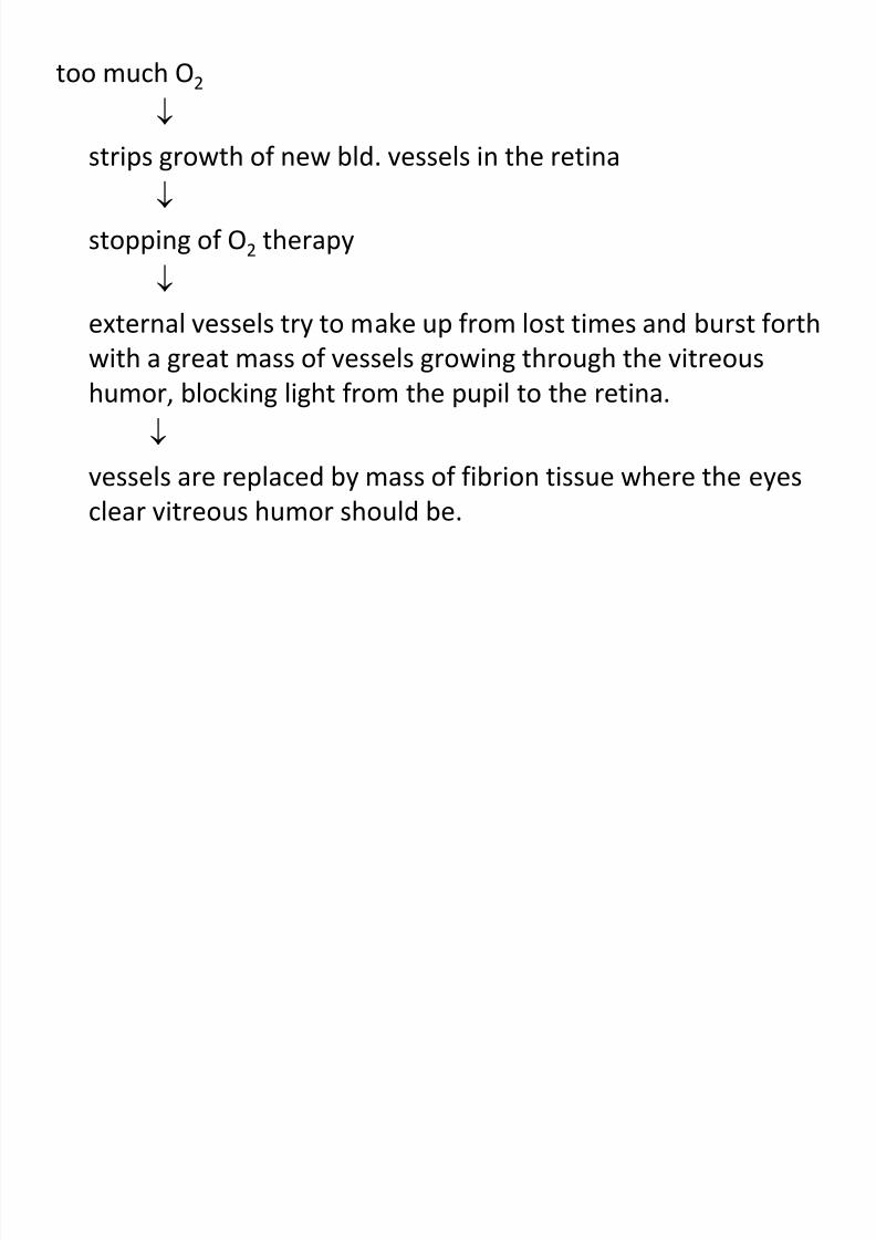
too much O2
strips growth of new bld. vessels in the retina
stopping of O2 therapy
external vessels try to make up from lost times and burst forthwith a great mass of vessels growing through the vitreous
humor, blocking light from the pupil to the retina.
vessels are replaced by mass of fibrion tissue where the eyesclear vitreous humor should be.
8/12/2019 Kul. Fisiologi
http://slidepdf.com/reader/full/kul-fisiologi 81/90
• This condition, known as retrolental fibroplasia,causes permanent blindness. For this reason, it isparticularly important to avoid treatment ofpremature infants with high concentrations of
respiratory oxygen.• Physiologic studies indicate that the premature
infant is usually safe with up to 40 per centoxygen in the air breathed, but some child
physiologists believe that complete safety can beachieved only at normal oxygen concentration inthe air that is breathed.
h d l f h hild

8/12/2019 Kul. Fisiologi
http://slidepdf.com/reader/full/kul-fisiologi 82/90
Growth and Development of the Child
The major physiologic problems of the child
beyond the neonatal pd. are related to special
metabolic needs from growth.
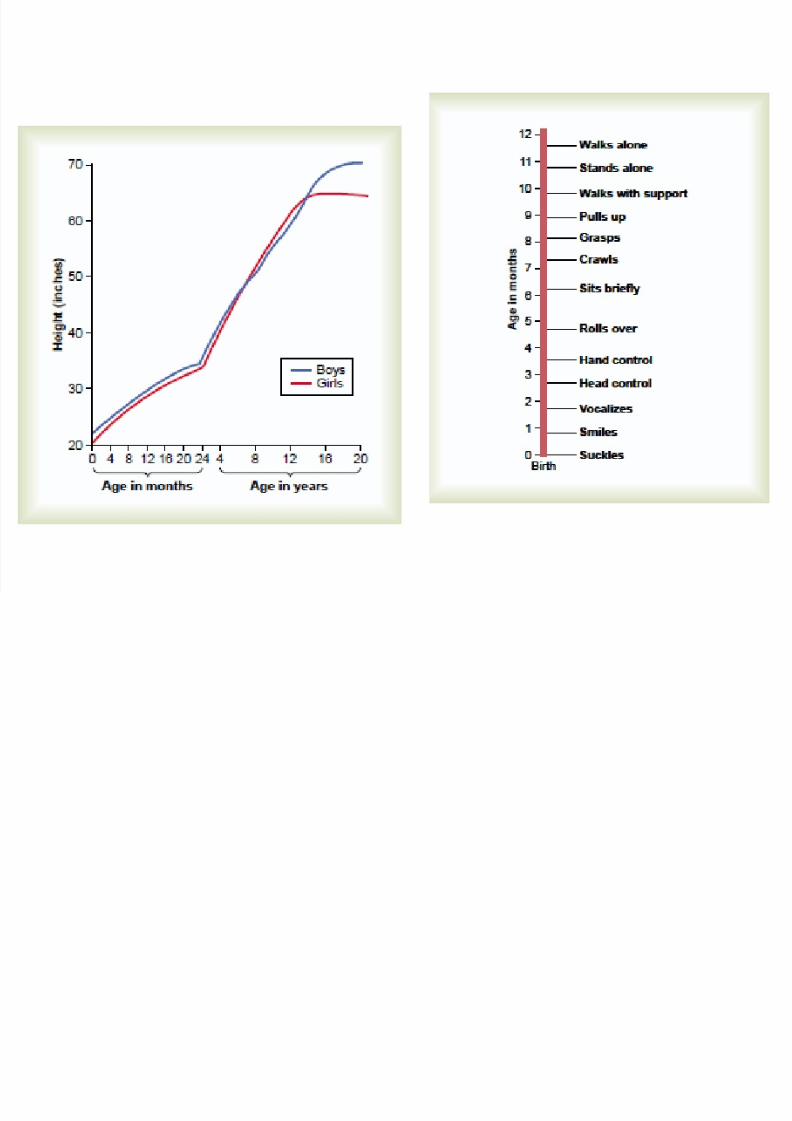
• Height of girls and boys parallel each otheralmost exactly until the end of the 1st decade
of life
G h d D l f h Child

8/12/2019 Kul. Fisiologi
http://slidepdf.com/reader/full/kul-fisiologi 83/90
Growth and Development of the Child
1. Estrogen in females begin to be formed and causesrapid growth in height but early unity of the
epiphyses of the long bones (14-16 y/o)
2. testosterone in males provides extra growthbetween 13-17 y/o - there is much delay in unity of
the epiphysis.
3. male Height > female
B h i l G h
8/12/2019 Kul. Fisiologi
http://slidepdf.com/reader/full/kul-fisiologi 84/90
Behavioral Growth
• Principally a problem of maturity of the nervoussystem
• Complete myelination achieved only until end of the1st yr of life
• nervous system not fully functional. at birth
• Brain growth continuous from birth until the 2nd yr.Of life which is associate with closure of thefontanelles and suture of the skull

8/12/2019 Kul. Fisiologi
http://slidepdf.com/reader/full/kul-fisiologi 85/90
Thank You!!!
Behavioral Growth
8/12/2019 Kul. Fisiologi
http://slidepdf.com/reader/full/kul-fisiologi 86/90
Behavioral growth is principally a problem ofmaturity of the nervous system. It is extremelydifficult to dissociate maturity of the anatomicalstructures of the nervous system from maturity
caused by training. Anatomical studies show thatcertain major tracts in the central nervous systemare not completely myelinated until the end ofthe first year of life. For this reason, it isfrequently stated that the nervous system is notfully functional at birth. The brain cortex and itsassociated functions, such as vision, seem torequire several months after birth for finalfunctional development to occur.
8/12/2019 Kul. Fisiologi
http://slidepdf.com/reader/full/kul-fisiologi 87/90
At birth, the infant brain mass is only 26 per cent of the adult brain mass and 55 per cent at 1 year, but it
reaches almost adult proportions by the end of the
second year. This is also associated with closure of the
fontanels and sutures of the skull, which allows only 20
per cent additional growth of the brain beyond the first
2 years of life. Figure 83 –9 shows a normal progress
chart for the infant during the first year of life. Comparison
of this chart with the baby’s actual development
is used for clinical assessment of mental and behavioral
growth.
8/12/2019 Kul. Fisiologi
http://slidepdf.com/reader/full/kul-fisiologi 88/90
8/12/2019 Kul. Fisiologi
http://slidepdf.com/reader/full/kul-fisiologi 89/90
8/12/2019 Kul. Fisiologi
http://slidepdf.com/reader/full/kul-fisiologi 90/90


