Cardiovascular Medicine Kardiovaskuläre Medizin – Médecine cardiovasculaire Official journal of the Swiss Society of Cardiology, the Swiss Society of Hypertension, the Swiss Society of Angiology and the Swiss Society of Paediatric Cardiology www.cardiovascmed.ch Abstracts P e e r r e v i e w e d j o u r n a l Annual meeting SSC/SSCS Basel (Switzerland), June 5–6, 2018 5 23. 5. 2018
124 Rapid fire abstract session: Pacemaker, defibrillator and electrophysiology
128 SSCS Abstract session
130 Abstract session: Preclinical study
136 Abstract session: clinical cases you don’t want to miss
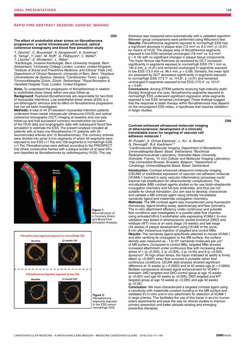
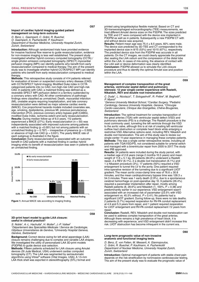
138 Rapid fire abstract session: Cardiac imaging
140 Rapid fire abstract session: Heart failure, vavulopathy and heart replacement therapy
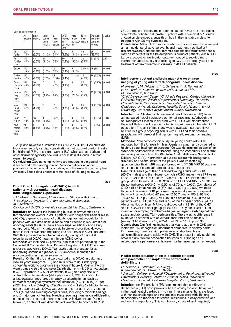
144 Abstract session: Congenital friday
Editorial Board
PD Dr. Andreas J. Flammer, Zürich (ed. in chief); Prof. Dr. François Mach, Genève (ed. in chief); PD Dr. Philippe Meyer, Genève (deputy editor); PD Dr. Jan Steffel, Zürich (deputy editor); Prof. Dr. Thomas F. Lüscher, Zürich (Senior editor); Dr. Katharina Blatter (Managing editor); Dr. Natalie Marty (Managing editor)
Editors
Prof. Dr. Paul Erne, Luzern; Prof. Dr. Augusto Franco Gallino, Bellinzona; Prof. Dr. Bernhard Meier, Bern; Prof. Dr. Matthias Pfisterer, Basel; Prof. Dr. Hans Rickli, St. Gallen; Prof. Dr. Christian Seiler, Bern; Prof. Dr. Bernard Waeber, Lausanne
Section editors
The interesting ECG: Dr. Michael Kühne, Basel; Dr. Jürg Schläpfer, Lausanne. Images in cardiovascular medicine: Dr. Alain Delabays, Morges; Prof. Dr. Michel Zuber, Othmarsingen. The new device: PD Dr. Haran Burri, Genève; Prof. Dr. Stephan Windecker, Bern. The new compound: PD Dr. Georg Ehret, Genève. Evidence-based cardiology: Prof. Dr. Heiner Bucher, Basel; PD Dr. Jens Hellermann, Altstätten; PD Dr. Jörg Muntwyler, Kloten
Advisory Board
The members of the Advisory Board are listed on www.cardiovascmed.ch.
Poster Walks (published in the online version of this issue)
A 1 Poster Walk I. Pacemaker, defibrillator and electrophysiology
A 5 Poster Walk I. Thromboembolic disease, epidemiology, risk factors, rehabilitation
A 9 Poster Walk I. Heart failure, vavulopathy and heart replacement therapy
A 11 Poster Walk II. Heart failure, vavulopathy and heart replacement therapy
A 14 Poster Walk II. Cardiac imaging
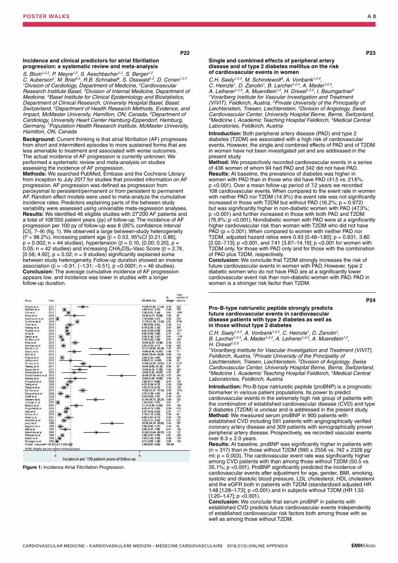
A 16 Poster Walk II. Clinical cases
A 21 Poster Walk II. Congenital and pediatric cardiology
A 24 Poster Walk II. ACS, PCI & CABG
TABLE OF CONTENTS 112
ImpressumCardiovascular MedicineOfficial publication of the Swiss Society of Cardiology, the Swiss Society for Angiology, the Swiss Society of Hypertension and the Swiss Paediatric Cardiology Society.Contact: Gisela Wagner, CVM Editorial office, EMH Medical Publishers Ltd., Farnsburgerstrasse 8, 4132 Muttenz, Phone +41 (0)61 467 85 52, Fax +41 (0)61 467 85 56, [email protected], www.cardiovascmed.chOnline manuscript submission: http://www.edmgr.com/cvm
Marketing / Advertising: Dr. phil. II Karin Würz, Head of Marketing and Communication, Phone +41 (0)61 467 85 49, Fax +41 (0)61 467 85 56, [email protected]
Subscription: EMH Medical Publishers Ltd., Subscriptions, Farnsburgerstrasse 8, 4132 Muttenz, Tel. +41 (0)61 467 85 75, Fax +41 (0)61 467 85 76, [email protected] price (excl. postage): CHF 125.–, students CHF 63.–. Postage prices and single issues see www.sanp.ch
Hinweis: Alle in dieser Zeitschrift publizierten Angaben wurden mit der grössten Sorgfalt überprüft. Die mit Verfassernamen gezeichneten Veröffentlichungen geben in erster Linie die Auffassung der Autoren und nicht zwangsläufig die Meinung der Redaktion von «Cardiovascular Medicine» wieder. Die angegebenen Dosierungen, Indikationen und Applikationsformen, vor allem von Neuzulassungen, sollten in jedem Fall mit den Fachinformationen der verwendeten Medikamente verglichen werden.
Production: Schwabe AG, Muttenz, www.schwabe.ch
CARDIOVASCULAR MEDICINE – KARDIOVASKULÄRE MEDIZIN – MÉDECINE CARDIOVASCULAIRE 2018;21(5):111–146
AUTHOR INDEX 113
AAaron L. P44Adams A. P21Adamson P. P70Aeschbacher S. O23, O32, P18,
P22Aggoun Y. O16, P43Aghlmandi S. O05Ahmadov K. O16, O41, P43, P53,
P57Ajalbert G. O49Akdis C.A. O62Akdis D. O25, O62Akhmedov A. O42, O43, O44,
O45Alonso-Gonzalez R. P62Altmann D. O53, P08, P13, P37Ammann P. O26, O30, O54, P08,
P13, P37Amstalden H. O43, O44Anagnostopoulos A. P34Arnold M. O36, O48, O50Arslani K. O31, O73Asatryan B. O26, P14Aschwanden M. P74Attenhofer-Jost C. O67, O70, O73,
O74, P59, P60, P64Auberson C. P22Auer R. O20, O21Auricchio A. O24, O26, O30, P06Avellan F. O71
BBadertscher P. O01, O02, O27,
O33, O61, P69, P70Baier P. O53, P47Balas B. P30Baldinger S.H. O26, O34, P09,
P14, P20, P51Balmer C. O72, O76Baltensperger N. P38Banz Y. P50Barras N. P31Bastiaansen J.A. P33, P46Baumgartner C. O23Baumgartner I. P23Beale A. O68Beer J.H. O42, O43, O44, O46,
O47Beghetti M. O59, P43, P62Behr-Graves R. O08Benussi S. O65, P01Benvenuti C. P06Benz D. O57, O60Berdat P. O67, O70Bereuter L. P09, P51Berg J. P38Berger S. P18, P22Berger W. P04Berte B. P11Bianchi V. P46Biasco L. P40Bigler M.R. O10Billinger M. P68Bisch L. P12, P49Blanche C. P62Blankenberg S. O02Blum S. P18, P22Bochud M. P31Bode P. O25, O62, P34Boeddinghaus J. O01, O02, O27,
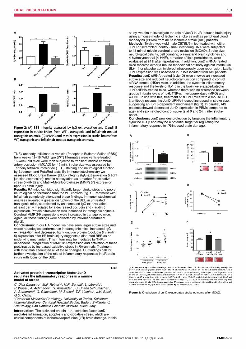
O47Bossard M. O03, O12, P73Bouchardy J. O73Bourantas C. O55Bourhis J. P49
Brandtner E.-M. O17, O18, P15, P16
Brauchlin A. O51Breidthardt T. O61Breitenstein A. O30, O51, P02,
P03, P10, P34Brenner R. P13, P37Briand Schumacher S. O43Briel M. P18, P22Brinkert M. O52, P11Broisat A. O56Brugger N. O19Brugger P. O75Brugnetti D. O38, P71, P75Brunckhorst C. O62Brunec M. O67Brüngger B. O23Buchholz N. P01, P52Buechel R. O57, O60, P34Buhmann R. O13, O52Burkart R. P06Burkhardt T. P65Burri H. O26, O30Buser M. O53, P37Buttu A. P12
CCairns J.A. O12Calado F. P30Calanchini C. P77Camici G.G. O42, O43, O44,
O45, O46, O47Cantor W. O12Caputo M.L. O24, P06Carballo D. O05, O20, O21, O68Carballo S. O68Carrel T.P. O36, O48, O50, P55Cassani D. O64Cattaneo M. P50, P77, P77Cesarovic N. O11, P32Chapman A. P70Cheema A.N. O12Chevailler S. O71Chiodini A. O51Christ J. O13Christ M. O27Chronis I. O03Chua H.C. P14Cikirikcioglu M. O41, P57Colombier S. O40, P54Conen D. O23, P18, P22Conte G. O24, O26Cook S. O49Corti R. P50Costantino S. O45Crea F. P50, P77Cuculi F. O03, O52Cuevas O.A. P27, P34Cullen L. O27Czopak A. O65, P29
DDave H. O72, P28De Boeck B. O52De Pasquale G. O70Degrauwe S. P33, P46Delacrétaz E. O30Delay D. O40, P54Delfine V. O14Deluigi C.C. P55Demertzis S. P19Denegri A. O07Derungs R.S. O44Devoogdt N. O56Dhinoja M.B. P03Di Bernardo S. P41, P42Di Somma S. O27Diaz-Cañestro C. O42, O43, O47,
O44Dickstein K. O30Dietrich P. P44
Dillier R. O26Dimopoulos K. P62Djafarzadeh S. O48Doebele T. O39Dogar A. O49Donati T.G. O66Dressel A. O18Drexel H. O17, O18, P15, P16,
P17, P23, P24Du Fay de Lavallaz J. O01, O27,
O61, P69Duclos F. P49Duda C. O59Duru F. O26, O62, P01, P04Dushaj S. O08Dworak M. P30Džavík V. O12Dzemali O. O08, O38, P56, P66,
P71, P75, P78
EEarley M.J. P03Eberli F. P72, P76Eberli F.R. P44Ebner J. O17, P15, P16Ebrahimi R. O33Eckstein F.S. O39Eeckhout E. O40, O69, P54Ehl N.F. P45, P48Ehret G. P31Elchinova E. O34, P09, P20, P51Engel R. O73Engelhard J. P30Erbay A. O04Erhart L. O66, P26Erne P. O06, O22, P76Eser P. O19 FFabienne W. P76Faeh-Gunz A. O70Faletra F. P40Falk V. O37Fay B. P30Feldmann M. O75Ferrari E. O69Flammer A.J. P34Fontaine G. O25Frank M. P34Fraunberger P. O17, O18, P15,
P16Frey S. P13Frobert A. O49Fröhlich G.M. P67Fuhrer J. O26, O34, P09, P14,
P20, P51Fung A. O12
GGabriel H. O73Gaemperli L. O57Gaemperli O. O57Gahl B. O39, P74Gallino A. P50, P77Garin N. O68Garzoli G. P40Gass M. O72Gaul D.S. O44Geiger K O17, O18, P15, P16Geigy N. O27Gencer B. O05, O20, O21, O66,
P26Genoni M. O08, O38, P56, P66,
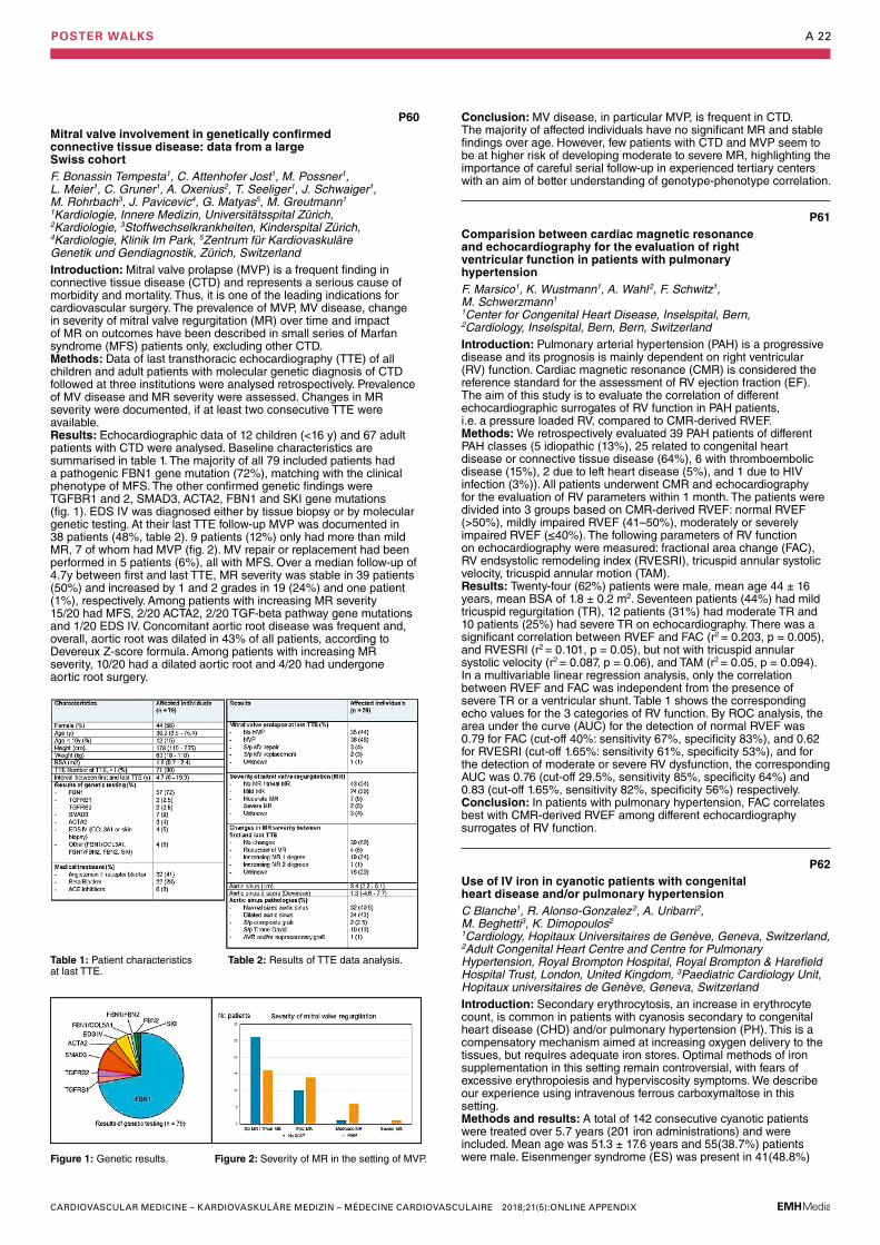
P71, P75, P78Giacalone G. O43, O44Giannitsis E. O02Giannopoulos A. O60Ginami G. P33, P46Girardet A. O35Giraud M.-N. O49Girod G. O40, P54
Gisler F. P55Glutz von Blotzheim L. O74, P02Gobbato S. O47Göldi T. O32Goulouti E. P20Gräni C. O57, O60Graves K. O38Grego S. P19Greutmann M. O73, O74, O75,
P28, P34, P59, P60, P63, P64Grossenbacher R. O10Grossmann J. O62Gruner C. O64, P34, P60Grunow S.S. P30Gruszczynski M. P66, P71, P75,
P78Gugala M. P70Guglielmi G. P57Guidetti F. O51Guidotti A. O11, P32Gulac P. O50 HHaager P.K. O66, P26, P36, P37,
P47Hachulla A.-L. O15, O58, P57Haeberlin A. O34, P09, P14, P20,
P51Hagenbuch N. P26Halbe M. O65Hasse B. P34Häussler A. O08, O38, P56, P71,
P75, P78Heg D. O05, O20, O21, P68Heidecker B. P38Heinzle C. P17, P23, P24Held L. P26Held U. O65Hellermann J. O66Hermann M. O22Hesselink T. O29Hilfiker G. O03Hilfiker S. O76Hofmann M. P28Huber C. O41, P53, P57Hübler M. P28, P59Hullin R. P25, P31, P33, P39,
P46Hunter R.J. P03
IIglesias J.F. P33, P46, P76Inderbitzin D. P01Inderbitzin D.T. O65, P74Isenrich R. O27
JJakob P. P67Joerg L. P36, P45Johannes J. P63Johner N. O28, O35Jolly S.S. O12, P73Joseph S. O71Jumeau R. P49Jüni P. O05 KKaelin A. P77Kamel M. P54Karagiannis A. O55Kaufmann B.A. O56, O66, P26Kaufmann P. O57, O60Kawecki D. O27Kaye D.M. O68Kedev S. O12Keller D. O27Kern I. O20Kessler N. O75Kirsch M. O69, O71, P25, P31,
P33, P35, P39, P58Kissel C.K. P38
CARDIOVASCULAR MEDICINE – KARDIOVASKULÄRE MEDIZIN – MÉDECINE CARDIOVASCULAIRE 2018;21(5):111–146
AUTHOR INDEX 114
Kleber M. O18Klebs S. P30Klersy C. O24, P40Klingenberg R. O05, O20, O21Knecht S. O32, O33, O54, P07,
P08Knirsch W. O75Kobza R. O03, O23, O52, P11Koechlin L. O39Koller M. P05Koskinas K. O55, P68Kottke R. O75Kottwitz J. P38Kovacs B. P04Kozhuharov N. O01, O33, O61Kretschmar O. P28Kuen H.-P. O73Kühne M. O23, O27, O31, O32,
O33, O54, P05, P07, P08, P13Kulic L. O44Kullak-Ublick G.A. O44Kurtal D. P27Kurz D. P72Kuwata S. O11, P29, P32
LLam A. O26, O34, P09, P14, P20,
P51Landmesser U. O04, O05, P67Lanz J. P68Larcher B. O17, O18, P15, P16,
P17, P23, P24Latal B. O75
Lauriers N. P33, P46Lauten A. O04, P67Lavi S. O12Le Bloa M. P12, P49Lehmann P. O76Leiherer A. O17, O18, P15, P16,
P17, P23, P24Leistner D.M. O04, P67Leo A.L. P40Leschka S. P45Leung R.C.M. O12Li G. O25Liaudet L. P25Liberale L. O42, O43, O44Lijovic G. P27Lindahl B. O02Linde C. O30Lipiski M. O11, P32Löblein H. O08, O38, P66, P71,
P75, P78Longnus S. O36, O48, O50Lovrinovic M. P38Luca A. P12Luciani M. O11, P32Luscher T.F. O45Lüscher T. O55Lüscher T.F. O05, O07, O20, O21,
O42, O43, O44, O47, P38
MMach F. O05, O07, O20, O21Mader A. O17, O18, P15, P16,
P17, P23, P24Maeder M. P48Maeder M.T. O53, P36, P37, P45,
P47Maertens A. O71Maerz W. O18Magyar I. P04Mair M. P55Maisano F. O11, O37, O65, P01,
P27, P29, P32, P34, P36, P52, P74
Manka R. P38Marcucci C. P25, P35Mariotti S. P71, P75Marsico F. P61Martinelli M. P50Martin-Sanchez J. O27
Masci P.G. O14Matt P. O13Matter C. O55, O62Matter C.M. O05, O07, O20, O21,
O44Matter-Ensner S. P75Matyas G. P60Maurer D. O70Medeiros-Domingo A. O26, O34,
P09, P14, P20, P51Meeks B. O12Mehta S. O12Meier L. P59, P60, P64Meier L.M. O74Meister T. O46, O49Méndez Carmona N. O36, O48,
O50Merlini M. O42, O44Messerli M. O60Mestres C. P52Mestres C.A. O37, P27, P34, P47Meyer M. P72Meyer P. O68Meyer S. P04Meyre P. O23, P18, P22Mills N. P70Milting H. O62Mira A. P06Miro O. O27Mivelaz Y. P41, P42Moarof I. O03Moccetti M. P40, P50, P77Moccetti T. O24, P06, P19Mochmann H.-C. P67Moeckli R. P49Mohacsi P. P50Mohammed S.A. O45Molitor N. P10Monney P. P31, P35Montecucco F. O44Moreno R. O12Moschovitis A. P68Moschovitis G. O23Moutzouri E. O21, O23Mueller A. O30Mueller C. O01, O02, P69, P70Muendlein A. O17, O18, P15, P16,
P17, P23, P24Muller H. O58Muller O. O07, O69Müller A. O26Müller C. O27, O61Müller H. O66, P26Münch C. P67Murith N. P57Myers P. O59, P43
NNaegeli B. O70Namasivayam J. O69Namdar M. O28, O35Nanchen D. O05, O20, O21Nanni P. O62Natarajan M.K. O12Nestelberger T. O01, O02, O27,
P69, P70Neumann J.T. O02Neurauter E. P45Niclauss L. P54Nicolazzi L. O59Niemann M. O64Niemelä K. O12Niessner A. P18Nietlispach F. P29Niklas F E. P37Nitsch R.M. O44Noble S. O15, O58, P76Noirez L. O14Noll G. P50Noti F. O26, O34, P09, P14, P20,
P51Novak J. P08
Nowacka A. O40, P41, P42, P58Nowak A. O61Ntinopoulos V. P71, P75, P78
OObeid S. O07Ochoa-Espinosa A. O56Odavic D. P66, P71, P75, P78Osswald S. O01, O23, O31, O32,
O33, O54, P05, P07, P13, P18, P22, P70
OSullivan C. P44Ouda A. O65, P27Oxenius A. O74, O75, P60, P64,
P65Özkartal T. O24Ozsahin M. P49
PPaneni F. O45Pascale P. P12, P49Pasotti E. P40Pasterk L. O47Patriki D. P38Paulin L. P31Pavicevic J. O67, O70, P60Pavlovic N. P08Pazhenkottil A. O57, O60, O64Peacock F. O27Pedrazzini G. O06, O55, P19,
P40, P50, P72, P76Pelouze A. O41, P53, P57Perrin N. O15Pfister M. P29Pfister R. P39, P41, P42, P58Pilgrim T. P68Pithon A. P12Pless S.A. P14Poepping I. O27Pop C.M. P48Possner M. O74, P59, P60, P64Pozzoli A. P01, P52Pravatà E. P77Praz F. P68Pretre R. O16, P39, P41, P42,
P43, P58Prêtre R. O59, O69Proenca C.C. P30Provenzi M. P77Pruvot E. O14, P12, P49Puelacher C. O01, O27, O61, P69,
P70Punjabi M. O56
QQanadli S.D. P54
RRaber L. P55Räber L. O05, O07, O20, O21,
O55, P68Radovanovic D. O06, O22, P72,
P76Rauch-Kröhnert U. P67Rechsteiner S. P44Regamey J. P25, P31, P33Regar E. O11, P32Regoli F. O24Reichlin T. O01, O26, O27, O31,
O32, O33, O53, O54, P05, P07, P08, P13, P70
Reiner M. O47Reiner M.F. O43, O44Reinhold J. P06Reinthaler M. P67Reser D. O37, O65Reuthebuch O. O39, P74Rexhaj E. O46, O49Rickli H. O06, O22, P36, P37,
P45, P48, P76Rieubland C. P14Rigamonti F. O20, O21
Rimoldi S.F. O49Rings L. O08, O38, P56, P71,
P75Rivero-Ayerza M. O29Rodondi N. O05, O20, O21, O23Rodriguez-Adrada E. O27Roffi M. O21, O55, P72, P76Roffler N. O73Rogler S. O64Roguelov C. O69Rohrbach M. P60Romanens M. P21Rometsch S. O75Roost E. P50Rosemann T. P72Rossner G.-A. O70Roten L. O26, O34, P09, P12,
P14, P20, P51Rotman S. P33Roumy A. P35Rrahmani B. O39Rubini Gimenez M. O02, P69Rubini Giménez M. O01Rüegg A. O76Ruggiero J. P25Ruschitzka F. O65, P01, P34Ryffel C. O19
SSabti Z. O61Saccocci M. O11, P32Saely C.H. O17, O18, P15, P16,
P17, P23, P24Saguner A. O25, O26, P04Saguner A.M. O62, P02, P10Salgado E. O27Sanz M.N. O48Savarese G. O44Savic V. P47Schaer B. O32, O54Schaller A. O26, P14Schär B. O29, O31, O33, P05,
P13Scharf C. O67Schefer T. P11Scherff F. P38Scherrer U. O46, O49Schilling R.J. P03Schindewolf M. P23Schindler M. P30Schlosser L. O75Schmiady M. P28Schmid A. P34Schmidli J. P50Schmied C. P38Schnabel R.B. P22Schneiders C. P50Schoepfer H. P18Schulte M. O29Schwaiger J. O74, P59, P60, P64Schwalm J.D. P73Schwarz J. O01Schwarz R. P47Schwarz U. P34Schweiger M. P28Schwenkglenks M. O23Schwerzmann M. O73, P61, P63Schwitter J. P49Schwitz F. P61, P63Sebastian C. O20Seeliger T. O74, P60, P64Seifert B. O37Seiler C. O10Seiler J. O26, P09, P14, P20, P51Seilier J. O34Sekarski N. P41, P42Semerano A. O43, O44Servatius H. O26, O34, P09, P14,
P20, P51Sessa M. O43, O44Shah A. P70Shah D. O23
CARDIOVASCULAR MEDICINE – KARDIOVASKULÄRE MEDIZIN – MÉDECINE CARDIOVASCULAIRE 2018;21(5):111–146
AUTHOR INDEX 115
Shah D.C. O28, O35Shahin M. O07Skurk C. P67Sologashvili T. O16, O59, P43,
P53Sörensen N.A. O02Soria R. O46, O49Spescha R.D. O44Spies F. O32, O33, O54, P07Sporton S.S. P03Springer A. O23Stähli B. O04, P67Stähli B.E. P26Stambach D. P28, P65Stämpfli S. O66, P26Stämpfli S.F. O64Stankovic G. O12Staub D. P74Steffel J. O51, P01Steiner J. O04Steinmann K. O76Sticherling C. O29, O30, O31,
O32, O33, O54, P05, P07, P08, P12, P13
Stirnemann J. O68Stivala S. O47Stoller M. O10Stortecky S. P68Strebel I. O27, O33Stuber M. P33, P46Sudano I. P21, P77Suerder D. P50Sürder D. P40Suter T. P50Sweda R. O34, P09, P20, P51Szekessy H. P74
Szucs T. O23, P21
TTanner F. P34, P52Tanner F.C. O64, O66, P26Tanner H. O26, O34, P09, P14,
P20, P51Taramasso M. O63, P29, P52Tevaearai H. O48Tevaearai Stahel H. O50Tevaearai Stahel H.T. O36Than M. O27Theuns D. O29, P05Thöni N. P27Tierney N.J. P06Tobler D. O73, P63Toggweiler S. O03, O13, O52,
P11Tozzi P. O69, O71, P25, P35, P39Trachsel L. O19Tschannen C. O10Twerenbold R. O01, O02, O27,
O61, P69, P70
UUeki Y. O55, P68Umans V. O29Urban P. P76Uribarri A. P62
VValgimigli M. P68Vallee J.-P. O58Vallet V. P49Valsangiacomo E. P28, P65Van Boven N. O29
Van Dam P. O33Van Heeswijk R. B. P33, P46 Van Hemelrijck M. O37, P27, P34Van Tilburg K. O65, P27Verbrugge F. O29Vesin J.-M. P12Vincenti G. O14Viriato D. P30Voegele J. O67, O70Vogt P. O67, O70Von Eckardstein A. O05Von Felten E. O60Vonbank A. P17, P23, P24Vuilleumier N. O20
WWachter R. P30Wahl A. P55, P61Warmuth W. P21Weber A. O37, P27, P34Weber D. P05Weber L. P36, P37Weber R. O72, P65Weberndoerfer V. P11Weidmann L. O07Weilenmann D. O03, P36Weiser M. P06Werner H. O76Westermann D. O02Whithlock R. O12Wichmann C. P66Wijnen W. O45Wildbolz M. O72Wildermuth S. P45Wildi K. O02, P69Wilhelm M. O19, O26
Wilhelm M.J. O65Windecker S. O05, O20, O21,
O55, P68Winter J.L. P73Wirta S.B. P30Wisser J. P65Witassek F. O06, O22, O23Wüest P. O43Wussler D. O01, O27, O61, P69Wustmann K. P55, P61, P63Wyss R.K. O36, O48, O50
XXu L. O56
YYamaji K. O55Yerly J. P33, P46Yerly P. P25, P39Yoon S.-I. O13Yousif N. O07
ZZanchin C. P68Zanchin T. O55, P68Zanolin D. P17, P23, P24Zbinden R. P34Zeljkovic I. O30, O32, O33, P07Zenklusen U. O39Zeverino M. P49Zientara A. P56, P71, P75, P78Zijlstra F. O29Zinkernagel A. P34Zuber M. O11, O64, P29, P32,
P52
CARDIOVASCULAR MEDICINE – KARDIOVASKULÄRE MEDIZIN – MÉDECINE CARDIOVASCULAIRE 2018;21(5):111–146
ORAL PRESENTATIONS 116
O01Direct comparison of the 0/1h- and 0/3h-algorithm for early rule-out of acute myocardial infarctionP. Badertscher, J. Boeddinghaus, R. Twerenbold, T. Nestelberger, M. Rubini Giménez, C. Puelacher, D. Wussler, J. Du Fay de Lavallaz, N. Kozhuharov, J. Schwarz, S. Osswald, T. Reichlin, C. Mueller Cardiovascular Research Institute Basel (CRIB), Universitätsspital Basel | Kardiologie, Basel, SwitzerlandBackground: The 0/1h-algorithm and the 0/3h-algorithm are both recommended by the European Society of Cardiology (ESC) with a class I recommendation for the early rule-out of acute myocardial infarction (AMI). We aimed to directly assess and compare their safety and efficacy. Methods: Among patients presenting with acute chest discomfort to the emergency department, classification towards rule-out by the 0/1h-algorithm or the 0/3h-algorithm were compared against the final adjudication performed by two independent cardiologists using all information including cardiac imaging and serial hs-cTnT measurements. Analyses were performed using high-sensitivity cardiac troponin (hs-cTn) T and hs-cTnI. Safety, as quantified by the negative predictive value (NPV), and efficacy of rule-out were the co-primary endpoints. Results: Among 2547 patients eligible for analysis using hs-cTnT, AMI was the final adjudicated diagnosis in 387 patients (15%). The 0/1h-algorithm provided similar safety (NPV 99.8% (95%CI 99.4–99.9%) versus 99.7% (95%CI 99.2–99.9%), p = 0.645) and higher efficacy as compared to the 0/3h-algorithm (60% (95%CI 58–62%) versus 44% (95%CI 42–46%), p <0.001). Among 2197 patients eligible for analysis using hs-cTnI, AMI was the final diagnosis in 327 patients (15%). The 0/1h-algorithm provided higher safety (NPV 99.6% (95%CI 99.1–99.9%) versus 97.8 (95%CI 96.7–98.5%), p <0.001) and similar efficacy compared to the 0/3h-algorithm (52% (95%CI 50–54%) versus 51% (95%CI 49-53%), p = 0.507, fig. 1). These findings were confirmed in the subgroup of early presenters, and in a second adjudication using serial hs-cTnI measurements.Conclusions: The 0/1h-algorithm is superior to the 0/3h-algorithm using hs-cTnT as well as hs-cTnI, as it more favorably combines safety with efficacy.
O02Impact of age on the performance of the ESC 0/1h-algorithms for early diagnosis of myocardial infarctionJ. Boeddinghaus1, T. Nestelberger1, R. Twerenbold1,2, J.T. Neumann2, B. Lindahl3, E. Giannitsis4, N.A. Sörensen2, P. Badertscher1, M. Rubini Gimenez1, K. Wildi1, D. Westermann2, S. Blankenberg2, C. Mueller1 1Cardiology and Cardiovascular Research Institute Basel (CRIB), University Hospital Basel, Basel, Switzerland, 2Department of General and Interventional Cardiology, Hamburg University Heart Center, Hamburg, Germany, 3Department of Medical Sciences, Uppsala University and Uppsala Clinical Research Centre, Uppsala, Sweden, 4Medizinische Klinik III, University Heidelberg, Heidelberg, GermanyAims: Beyond the presence or absence of myocardial infarction (MI), age seems to be the most important confounder of high-sensitivity cardiac troponin (hs-cTn) T and I blood concentrations. Mildly elevated
hs-cTnT and hs-cTnI blood concentrations are common in elderly individuals without apparent ischemic symptoms. Unfortunately, the impact of age on the diagnostic performance of the European Society of Cardiology (ESC) 0/1h-algorithms is incompletely understood. We aimed to evaluate the impact of age on the performance of the ESC 0/1h-algorithms and to derive and externally validate alternative cut-offs specific to older patients. Methods: We prospectively enrolled patients presenting to the emergency department with symptoms suggestive of acute myocardial infarction (AMI) in three large diagnostic studies. Final diagnoses were adjudicated by two independent cardiologists. High-sensitivity cardiac troponin (hs-cTn) T and I concentrations were measured at presentation and after 1h. Patients were stratified according to age (<55 years [young], ≥55 to <70 years [middle-age], ≥70 years [old]). Results: Among 3123 patients in the main cohort, prevalence of AMI increased with increasing age (young 6.4%, middle-aged 15%, old 27%, p <0.001). The ESC hs-cTnT 0/1h-algorithm ruled-out 956 (85%) young patients (sensitivity 100% [95%CI, 94.9–100]), 606 (65%) middle-aged patients (sensitivity 99.3% [95%CI, 96.0–99.9]), and 317 (30%) old patients (sensitivity 99.3% [95%CI, 97.5–99.8]). Likewise, 92 (8%) young patients (specificity 97.0% [95%CI, 95.8–97.9]), 141 (15%) middle-aged patients (specificity 96.1% [95%CI, 94.5–97.2]), and 272 (25%) old patients (specificity 92.7% [95%CI, 90.7–94.3]) were ruled-in. The proportion of patients triaged within one hour decreased with increasing age (young 93%, middle-aged 80%, old 55%, p <0.001). Similar results were found for the ESC hs-cTnI 0/1h-algorithm. Alternative, slightly higher cut-off concentrations optimized for older patients did not improve the overall diagnostic performance (p = ns). Findings were confirmed in two validation cohorts (n = 2767).Conclusion: While the safety of the ESC 0/1h-algorithms remained very high, increasing age significantly reduced overall efficacy and the accuracy of rule-in. Alternative, slightly higher cut-offs optimized for use in older patients did not result in a relevant improvement of rule-out nor rule-in.
RAPID FIRE ABSTRACT SESSION: ACUTE CORONARY SYNDROME (ACS), PERCUTANEOUS CORONARY INTERVENTION (PCI), CORONARY ARTERY BYPASS GRAFT SURGERY (CABG)
Figure 1
Figure 2: Outcome of patients according to age triaged by the ESC hs-cTnT 0/1h-algorithm.
Figure 1: Performance of the ESC hs-cTnT 0/1h-algorithm according to age.
ORAL PRESENTATIONS 117
CARDIOVASCULAR MEDICINE – KARDIOVASKULÄRE MEDIZIN – MÉDECINE CARDIOVASCULAIRE 2018;21(5):111–146
O05Cysteine-rich angiogenic inducer 61 (Cyr61) in combination with established biomarkers improves GRACE risk score to predict all-cause mortality in ACS patientsR. Klingenberg1, S. Aghlmandi2, L. Räber3, B. Gencer4, D. Carballo4, D. Nanchen5, D. Heg6, P. Jüni7, N. Rodondi8, F. Mach9, S. Windecker3, U. Landmesser10, A. von Eckardstein11, C.M. Matter12, T.F. Lüscher13 1Klinik für Kardiologie, Kerckhoff-Klinik, Bad Nauheim, Germany, 2Clinical Epidemiology and Biostatistics, Unispital Basel, Basel, 3Cardiology, Inselspital Bern University Hospital, Bern, 4Cardiology, University Hospital Geneva, Geneva, 5Ambulatory Care and Community Medicine, University of Lausanne, Lausanne, 6Clinical Trials Unit, Institute of Social and Preventive Medicine, University of Bern, Bern, Switzerland, 7Applied Health Research Centre (AHRC), Li Ka Shing Knowledge Institute of St. Michael’s Hospital, University of Toronto, Toronto, ON, Canada, 8General Internal Medicine, Inselspital Bern University Hospital, Bern, 9Cardiology, University Hospital of Geneva, Geneva, Switzerland, 10Cardiology, Campus Benjamin Franklin, Charité University Medicine, Berlin, Germany, 11Institute of Clinical Chemistry, University Hospital Zurich, 12Cardiology, University Heart Center Zurich, Zurich, 13Center for Molecular Cardiology, University of Zurich, Schlieren, SwitzerlandIntroduction: We have identified cysteine-rich angiogenic inducer 61 (Cyr61) as a novel biomarker of acute myocardial injury improving risk stratification in patients with acute coronary syndromes (ACS). However, the value of Cyr61 for predicting all-cause mortality in ACS patients compared with established biomarkers such as hsTnT, NT-proBNP and hsCRP against the GRACE risk score model remains unclear.Method: Consecutive ACS patients were enrolled in the SPUM-ACS biomarker study at one of four Swiss university hospitals. Patients had blood drawn at coronary angiography. Concentrations of Cyr61 in serum were measured in duplicates of single serum aliquots using a semi-automated solid phase enzyme-linked immunosorbent assay. hsTnT and hsCRP were measured in serum aliquots in addition to NT-proBNP. All-cause mortality within 30 days and at 1 year was the primary outcome as defined in the GRACE risk score. Associations between biomarkers and outcome were assessed using continuous, log-transformed biomarker values and continuous GRACE risk scores. The incremental predictive value of the new marker over and above a reference model was assessed by Harrell’s C-statistics calculated from a Cox proportional-hazard regression model. Results: Among 2168 patients enrolled, 1732 had available biomarker data constituting the study population with a mean age of 63.8 ± 12.3 (SD). STEMI was more prevalent (n = 916, 52.9%) than NSTEMI (n = 747, 43.1%) and unstable angina (n = 69, 4.0%); the majority of patients were treated by PCI (n = 1564, 90.3%). Cyr61 showed good prognostic accuracy compared with the other biomarkers for all-cause mortality at 30 days (hazard ratio 1.77 (1.31, 2.40), p <0.001) and 1 year (hazard ratio 1.81 (1.47, 2.22), p <0.001), similar to hsTnT. Adding Cyr61 to the GRACE risk score as a reference model improved prognostic accuracy for 30 days all-cause mortality (c-statistic 0.87 to 0.88, p = 0.001) and 1 year all-cause mortality (c-statistic 0.77 to 0.80, p <0.001). The best prediction was achieved when combining all biomarkers with the GRACE risk score achieving a significant improvement against the reference model for 30 days all-cause mortality (c-statistic 0.87 to 0.90, p <0.001) and 1 year all-cause mortality (c-statistic 0.77 to 0.84, p <0.001). Conclusion: Cyr61 is a strong predictor of adverse outcome in ACS patients adding independent and incremental information to the GRACE risk score and established cardiovascular biomarkers.
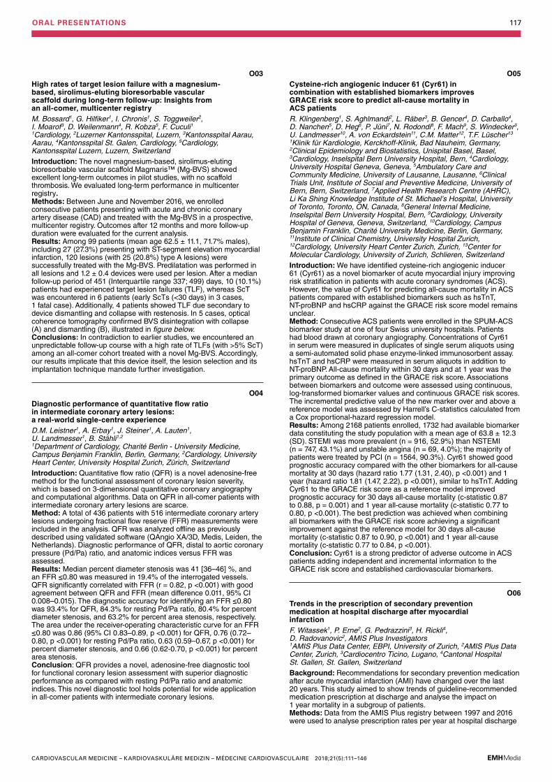
O06Trends in the prescription of secondary prevention medication at hospital discharge after myocardial infarctionF. Witassek1, P. Erne2, G. Pedrazzini3, H. Rickli4, D. Radovanovic2, AMIS Plus Investigators 1AMIS Plus Data Center, EBPI, University of Zurich, 2AMIS Plus Data Center, Zurich, 3Cardiocentro Ticino, Lugano, 4Cantonal Hospital St. Gallen, St. Gallen, SwitzerlandBackground: Recommendations for secondary prevention medication after acute myocardial infarction (AMI) have changed over the last 20 years. This study aimed to show trends of guideline-recommended medication prescription at discharge and analyse the impact on 1 year mortality in a subgroup of patients. Methods: Data from the AMIS Plus registry between 1997 and 2016 were used to analyse prescription rates per year at hospital discharge
O03High rates of target lesion failure with a magnesium- based, sirolimus-eluting bioresorbable vascular scaffold during long-term follow-up: Insights from an all-comer, multicenter registryM. Bossard1, G. Hilfiker1, I. Chronis1, S. Toggweiler2, I. Moarof3, D. Weilenmann4, R. Kobza5, F. Cuculi1 1Cardiology, 2Luzerner Kantonsspital, Luzern, 3Kantonsspital Aarau, Aarau, 4Kantonsspital St. Galen, Cardiology, 5Cardiology, Kantonsspital Luzern, Luzern, SwitzerlandIntroduction: The novel magnesium-based, sirolimus-eluting bioresorbable vascular scaffold Magmaris™ (Mg-BVS) showed excellent long-term outcomes in pilot studies, with no scaffold thrombosis. We evaluated long-term performance in multicenter registry.Methods: Between June and November 2016, we enrolled consecutive patients presenting with acute and chronic coronary artery disease (CAD) and treated with the Mg-BVS in a prospective, multicenter registry. Outcomes after 12 months and more follow-up duration were evaluated for the current analysis.Results: Among 99 patients (mean age 62.5 ± 11.1, 71.7% males), including 27 (27.3%) presenting with ST-segment elevation myocardial infarction, 120 lesions (with 25 (20.8%) type A lesions) were successfully treated with the Mg-BVS. Predilatation was performed in all lesions and 1.2 ± 0.4 devices were used per lesion. After a median follow-up period of 451 (Interquartile range 337; 499) days, 10 (10.1%) patients had experienced target lesion failures (TLF), whereas ScT was encountered in 6 patients (early ScTs (<30 days) in 3 cases, 1 fatal case). Additionally, 4 patients showed TLF due secondary to device dismantling and collapse with restenosis. In 5 cases, optical coherence tomography confirmed BVS disintegration with collapse (A) and dismantling (B), illustrated in figure below.Conclusions: In contradiction to earlier studies, we encountered an unpredictable follow-up course with a high rate of TLFs (with >5% ScT) among an all-comer cohort treated with a novel Mg-BVS. Accordingly, our results implicate that this device itself, the lesion selection and its implantation technique mandate further investigation.
O04Diagnostic performance of quantitative flow ratio in intermediate coronary artery lesions: a real-world single-centre experienceD.M. Leistner1, A. Erbay1, J. Steiner1, A. Lauten1, U. Landmesser1, B. Stähli1,2 1Department of Cardiology, Charité Berlin - University Medicine, Campus Benjamin Franklin, Berlin, Germany, 2Cardiology, University Heart Center, University Hospital Zurich, Zürich, SwitzerlandIntroduction: Quantitative flow ratio (QFR) is a novel adenosine-free method for the functional assessment of coronary lesion severity, which is based on 3-dimensional quantitative coronary angiography and computational algorithms. Data on QFR in all-comer patients with intermediate coronary artery lesions are scarce. Method: A total of 436 patients with 516 intermediate coronary artery lesions undergoing fractional flow reserve (FFR) measurements were included in the analysis. QFR was analyzed offline as previously described using validated software (QAngio XA/3D, Medis, Leiden, the Netherlands). Diagnostic performance of QFR, distal to aortic coronary pressure (Pd/Pa) ratio, and anatomic indices versus FFR was assessed. Results: Median percent diameter stenosis was 41 [36–46] %, and an FFR ≤0.80 was measured in 19.4% of the interrogated vessels. QFR significantly correlated with FFR (r = 0.82, p <0.001) with good agreement between QFR and FFR (mean difference 0.011, 95% CI 0.008–0.015). The diagnostic accuracy for identifying an FFR ≤0.80 was 93.4% for QFR, 84.3% for resting Pd/Pa ratio, 80.4% for percent diameter stenosis, and 63.2% for percent area stenosis, respectively. The area under the receiver-operating characteristic curve for an FFR ≤0.80 was 0.86 (95% CI 0.83–0.89, p <0.001) for QFR, 0.76 (0.72–0.80, p <0.001) for resting Pd/Pa ratio, 0.63 (0.59–0.67, p <0.001) for percent diameter stenosis, and 0.66 (0.62-0.70, p <0.001) for percent area stenosis.Conclusion: QFR provides a novel, adenosine-free diagnostic tool for functional coronary lesion assessment with superior diagnostic performance as compared with resting Pd/Pa ratio and anatomic indices. This novel diagnostic tool holds potential for wide application in all-comer patients with intermediate coronary lesions.
ORAL PRESENTATIONS 118
CARDIOVASCULAR MEDICINE – KARDIOVASKULÄRE MEDIZIN – MÉDECINE CARDIOVASCULAIRE 2018;21(5):111–146
for aspirin, P2Y12, lipid lowering drugs, beta blocker and ACEI/ARB. The proportion of patients who received 1,2,3,4 or 5 of these medications was graphically illustrated over the years. In a subgroup of patients with 1year follow up (FU) (since 2005), we analysed the impact of these medications on 1 year mortality correcting for age, gender and comorbidities using logistic regression. Data on individual treatment decisions, contraindications and medication adherence were, however, not available. Results: Between 1997 and 2016, 39,036 patients with AMI and known discharge medication were included. Prescription rates significantly increased over the last 20 years for all 5 medications (fig. 1). Since 2007, the rate of the patients who received 3 or more of the considered medications rose from 94.9% to 97.7% (p <0.001) (fig. 2). Since 2005, 8117 patients were followed up 1 year after discharge. Of these, 7880 (97.1%) had 3 or more of the considered discharge medication and 237 (2.9%) received <3. Patients with less medication were significantly older, more likely to be female, had significantly more comorbidities such as cardiac insufficiency (CI), cerebrovascular disease (CVD) and cancer and underwent less frequently PCI. Crude 1 year mortality differed significantly between these two groups (>=3: 3.3% vs. <3:14.8%, p <0.001). In a multivariable regression analysis, FU mortality was significantly lower for patients with >=3 medications (OR: 0.62, CI 0.39–0.97) even after correction for age, gender, STEMI, PCI, diabetes, renal disease, CI, CVD and cancer. Conclusions: In Switzerland, prescription rates of secondary prevention medications are currently high and relatively stable during the last years. Between 2005 and 2016, FU mortality was lower if patients received at least three of the guideline-recommended medications even after correction for confounders. However, patients with <3 were also treated less intensively during hospitalisation which could possibly be explained by individual treatment decisions.
O07Influence of pretreatment with aspirin or statins or both on clinical presentation as well as infarct size and inflammation in patients with de novo acute coronary syndromesL. Weidmann1, S. Obeid1, F. Mach2, M. Shahin1, N. Yousif1, A. Denegri1, O. Muller3, L. Räber4, C.M. Matter1, T.F. Lüscher5 1Unispital Zürich, Zürich, 2University Hospital of Geneva, Genf, 3CHUV, Lausanne, 4Inselspital Bern, Bern, 5Zürich Heart House, Zürich, SwitzerlandObjective: To investigate whether pre-treatment with aspirin and/or statins prior to a first acute coronary syndrome (ACS) will affect the clinical presenta-tion, degree of inflammation and infarct size. To that end, we analyzed pa-tients prospectively enrolled in the Swiss Program University Medicine ACS cohort (SPUM-ACS; ClinicalTrials.gov number: NCT01075867). Methods: 1’639 eligible patients were categorized into 4 groups: (1) Those without antiplatelet drugs nor statins prior to their first ACS (n = 1’181); (2) those only on aspirin, but not on statins (n = 157); (3) those only on statins, but not on antiplatelet drugs (n = 133) and (4) those on aspirin and statins (n = 168). Clinical features, ECG, creatinine kinase (CK, U/l), troponin T (TNT, µg/l), brain natriuretic peptide (NT-proBNP, ng/l), leucocytes (Lc, G/l), neutrophils (Nc, G/l), C-reactive protein (CRP, mg/l) and angiographic features were documented and analysed. Results: The incidence of ST-elevation myocardial infarction (STEMI) was 64% in those without either drug, 45% in those on aspirin only, 52% in those with statins only and 40% in those on aspirin and a statin (p <0.0001). At pre-sentation, those pre-treated with aspirin and statin had the lowest CK (145 U/l, interquartile range (IQR) 89–297; p <0.0001) and TNT plasma levels (0.13 µg/l, IQR 0.03–0.52; p = 0.001) and the highest left ventricular ejection fraction (LVEF; 55 ± 12%; p = 0.028) compared to the other groups. Co-medicated subgroups matched for high risk factors showed a significantly smaller infarct size as assessed by CK (p <0.0001) and TNT (p <0.0001) as well as lower plasma levels of CRP (p = 0.01) and presented less frequently as STEMIs compared to those without aspirin or statins (p <0.0001).Conclusion: Pretreatment with either aspirin and statins and particularly with their combination markedly changes the clinical presentation (i.e. STEMI vs. NSTEMI), the degree of inflammation and infarct size in patients suffering their first acute coronary syndrome. Keywords: Aspirin, statins, ECG, infarct size, inflammation, de novo ACS. Abbreviations: SPUM-ACS cohort, Swiss Program University Medicine ACS cohort. De novo, first time.
O08Intraoperative endoluminal quality control of saphenous vein grafts with optical coherence tomography in coronary artery bypass graftingA. Häussler, L. Rings, S. Dushaj, H. Löblein, R. Behr-Graves, O. Dzemali, M. Genoni Allianz Herzchirurgie Zürich, Departement of Cardiac Surgery Stadtspital Triemli Zürich, Zürich, SwitzerlandIntroduction: During bypass surgery saphenous vein grafts (SVGs) remain a widely used conduit. From earlier studies we know that graft harvesting can lead to endothelial lesions which result in graft failure or poor quality of anastomosis. There is no intraoperative endothelial quality control durable, which enables assessment of the inner layer or diamter of the vein and morphologic disorders endoluminal including venous valves. We need fast, dynamic and direct control for conduit and anastomosis intraoperative. Optical Coherence Tomography (OCT) with its very high resolution in time and space offers a new possibility to evaluate luminal features and vessel wall. In this pilot trial intraoperative intraluminal assessment of the graft was done after endoscopic vein harvesting during off-pump coronary bypass grafting (OPCAB). Methods: We included 9 patients undergoing elective OPCAB surgery. After endoscopic vein harvesting we clipped all sidebranches and cannulated with a vessel cannula. After, administration of 0.9% physiologic saline solution since the region of interest has to be blood free for assessment. When OCT catheter is introduced inside the SVG it was pulled back at 10 mm/s while images were acquired at rate of 100 frames/s in real time. Maximal length of 5 cm. SVG of 30 cm is recorded 6 times within 30 seconds at least. Additionally, we operated on 5 pigs on-pump-beating heart. After harvesting of the internal mammary artery we were able to assess the vessel with OCT to get visualization of graft and anastomosis.
Figure 1
Figure 2
ORAL PRESENTATIONS 119
CARDIOVASCULAR MEDICINE – KARDIOVASKULÄRE MEDIZIN – MÉDECINE CARDIOVASCULAIRE 2018;21(5):111–146
Results: 60 video sequences with length of 5 cm each were recorded. Total assessment length 3 meters. In two sequences we found blow outs with aneurysmatic dilatation. In three sections dysmorphic venous valves were indicated. We found no endothelial lesions except at the end of the SVGs at the resection area. Direct visualization of anastomosis in pigs showed good quality of suture and in one pig microthrombus on anastomosis. Conclusion: OCT assessment is the only method for dynamic and direct visualization for conduits and anastomosis. Penetration of soft tissue and vessel wall is excellent. The whole procedure is safe, fast and easy to handle. For cardiac surgery no contrast age is demanded which makes it feasible in all patients. First animal studies were done with intraoperative assessment of graft and anastomosis. Intraoperative findings can lead to graft adjustment or revision of anastomosis to assure best quality for the patient.
O10Functional assessment of myocardial ischemia by intracoronary electrocardiogramM.R. Bigler, R. Grossenbacher, M. Stoller, C. Tschannen, C. Seiler Cardiology, Inselspital Bern University Hospital, Bern, SwitzerlandIntroduction: In patients with chronic stable coronary artery disease (CAD), percutaneous coronary intervention targets hemodynamically significant coronary lesions, i.e., those thought to cause inducible ischemia. The goal of this study is to test the accuracy of intracoronary (ic) ECG during pharmacologic inotropic stress to determine significant coronary lesions in comparison with established physiologic indices (fractional flow reserve (FFR), instantaneous wave-free ratio (iFR)) as well as with quantitatively determined percent diameter stenosis (%S) using biplane coronary angiography. Methods: This is an ongoing prospective, open-label study in patients with chronic stable CAD. The primary study end point is the change in intracoronary ST-segment shift during pharmacologic inotropic stress induced by dobutamine plus atropine measured at the point of maximal heart rate (estimated by the formula 220 – age). IcECG is easily acquired by attaching an alligator clamp to the angioplasty guidewire, positioned downstream of a stenosis. For the pressure-derived ratios, i.e. FFR and iFR, the coronary perfusion pressure downstream of a lesion as well as the aortic pressure are continuously recorded. Results: Using the FFR threshold of 0.80 as reference to determine the hemodynamic significance of coronary lesions, the ROC-analysis of the absolute ST-segment shift showed an area under the curve of 0.732 ± 0.197 (p = 0.037, n = 30, FFR <0.80 n = 11, meanFFR = 0.81). The area under the ROC curve for iFR was 0.995 ± 0.015 (p <0.0001), for percent diameter stenosis it was 0.864 ± 0.134 (p = 0.001). The DeLong-Test of the ROC-curves showed a significant difference for iFR compared to %S and icECG (p = 0.04 respectively p = 0.009). No significant difference in the AUC was shown between %S and the icECG (p = 0.18). Regarding the optimum cut-off point for the icECG, an absolute ST-segment shift of 1mV distinguished best between hemodynamically relevant and irrelevant stenotic lesions; sensitivity 55%, specificity 90%. Conclusions: Intracoronary ECG ST-segment shift during pharmacologic inotropic stress appears to be similarly accurate as structural stenosis assessment in detecting hemodynamically relevant coronary stenotic lesions. Disclosures: None. Keywords: intracoronary ECG - pharmacologic inotropic stress - FFR - iFR - percent diameter stenosis - coronary circulation - percutaneous coronary intervention - myocardial ischemia
O11Diagnostic changes in heart sounds and acoustic cardiography parameters during acute myocardial ischemia in a porcine modelS. Kuwata, M. Luciani, M. Saccocci, N. Cesarovic, M. Lipiski, A. Guidotti, E. Regar, M. Zuber, F. Maisano, Heart Center University Heart Center Zurich, Zurich, SwitzerlandIntroduction: Myocardial ischemia (MI) is commonly associated with ECG changes. The timing and relationship of ECG changes to mechanical alterations in left ventricular (LV) contractility and stiffness has not been extensively explored. We examined heart sound and acoustic cardiography parameters reflecting LV electrical and mechanical function before and after distal left circumflex coronary artery (LCX) occlusion. Methods: Domestic pigs (60 ± 2 kg) were anesthetized using isoflurane and ventilated. MI was introduced by complete occlusion of
the distal LCX. Continuous cardiac output, blood pressure, LV pressure-volume (PV) loops and right heart pressures were acquired. Transvalvular and annular mitral systolic (velocity time integral, VTI) and diastolic functional (E deceleration time, E/A ratio) and intravascular VTI of ascending flow measurements were collected. ECG and heart sounds were simultaneously recorded. Automated acoustic cardiography analysis of the ECG and heart sound signals resulted in measurement of the intensities of the first (S1) and second (S2) heart sounds, diastolic third (S3) and fourth (S4) heart sounds, LV systolic time (from S1 to S2), perfusion time (S2 to Q onset), and electromechanical activation time (EMAT, time from Q onset to S1). Results: Balloon occlusion was successfully induced, maintained for ≥22 minutes and produced ischemic changes in ECG ST segment morphology. Hemodynamics were altered from a baseline state after occlusion as reflected in the PV loops and cardiac output. As ischemia progressed with time, LV stiffness increased as seen in a rise of S4 intensity and LV diastolic dysfunction worsened as reflected by increased S3 intensity. EMAT lengthened indicating reduced contractility and electro-mechanical dysfunction. Example from one animal at baseline, 3 minutes post balloon occlusion and 13 minutes post balloon occlusion. Conclusions: Acute MI results in changes in the ECG along with heart sound alterations reflecting increased ventricular stiffness and impaired contractility. The mechanical impairment increased as the ischemic event proceeded. Diagnostic ability to detect MI may be improved with the combined use of ECG and acoustic cardiography parameters, particularly the fourth heart sound. Thus, non-invasive monitoring with acoustic cardiography might be translated to patient care, potentially improving the clinician’s ability to monitor episodes of ischemia in real-time noninvasively.
O12Coronary artery bypass grafting in patients with ST-segment elevation myocardial infarction – characteristics, timing and outcomes: insights from the TOTAL trialM. Bossard1, V. Džavík2, J.A. Cairns3, S. Mehta4, M.K. Natarajan4, S. Lavi5, R.C.M. Leung6, A. Fung7, R. Whithlock8, A.N. Cheema9, K. Niemelä10, B. Meeks11, W. Cantor12, S. Kedev13, G. Stankovic14, R. Moreno15, S.S. Jolly4
1Luzerner Kantonsspital, Luzern, Switzerland, 2Peter Munk Cardiac Center, University of Toronto, Toronto, ON, 3Cardiology, University of Britisch Columbia, Vancouver, BC, 4Hamilton General Hospital, Cardiology, McMaster University, Hamilton, 5Cardiology, London Health Sciences, Western University, London, ON, 6Cardiology, CK Hui Heart Center, Royal Alexandra Hospital, Edmonton, AB, 7Cardiology, University of Britisch Columbia, Vancouver General Hospital, Vancouver, BC, 8Hamilton General Hospital, Cardiac Surgery, McMaster University, Hamilton, 9Cardiology, University of Toronto, St. Michael’s Hospital, Toronto, ON, Canada, 10Cardiology, Heart Center, Tampere University Hospital, Tampere, Finland, 11McMaster University, Population Health Research Institute, Hamilton, 12Cardiology, University of Toronto, Southlake Regional Health Center, Newmarket, 13University Clinic of Cardiology, Medical Faculty, Skopje, ON, Canada, 14Cardiology, Medical Faculty, University of Belgrade, Belgrad, Serbia, 15University Hospital La Paz, Madrid, SpainIntroduction: Contemporary data about the timing and outcomes of patients requiring coronary artery bypass grafting (CABG) after ST-segment elevation myocardial infarction (STEMI) are scarce. Methods: Data from a large contemporary STEMI trial (N = 10,732) were analyzed. Patients undergoing CABG surgery during the study period were evaluated. Timing of CABG surgery and clinical outcomes were evaluated by using adjusted Cox-regression models. Patients with mechanical complications (N = 28) were excluded. Results: During 1 year follow-up, 422 patients underwent CABG surgery (4.3% of all patients), whereas 330 had a primary PCI and
Figure 1: Results of study.
CARDIOVASCULAR MEDICINE – KARDIOVASKULÄRE MEDIZIN – MÉDECINE CARDIOVASCULAIRE 2018;21(5):111–146
ORAL PRESENTATIONS 120
in the Table. Cardiogenic shock occurred in 16 (11.0%) versus 10 (3.6%) patients from the early versus late CABG group, respectively (p = 0.003). Early versus late surgery was associated with a more than 3-fold higher risk for major bleeds (table). Conclusions: During 1 year follow-up, STEMI patients undergoing CABG surgery have a high risk for cardiovascular events representing a higher risk subgroup. Major hemorrhagic events are a concern in those patients, particularly among those requiring early surgery (≤7 days since index STEMI). New approaches are needed to reduce bleeding in these patients requiring early CABG in STEMI.
146 underwent surgery within 7 days after index STEMI event. Patients undergoing surgery were older and had more frequently diabetes, hypertension, dyslipidemia, previous PCIs, peripheral vascular disease and left main disease. Patients, who underwent CABG surgery, were a higher risk cohort, than those that did not, with a higher risk of repeat MI (7.4% vs. 2.2%, adjusted hazard ratio (HR) 3.17 (95% confidence interval (CI) 2.16–4.64)), stroke/ TIA (2.6% vs. 1.1%, adjusted HR 2.27 (1.21–4.25)) and major bleed (14.0% vs. 1.3%, HR 10.48 (95%CI 7.66–14.33)). The comparison of STEMI patients undergoing early (within 7 days after MI) versus late CABG (>7 days after MI) is shown
O13Chest pain in a young runnerJ. Christ1, S. Toggweiler2, R. Buhmann3, S.-I. Yoon4, P. Matt1 1Herzchirurgie, 2Kardiologie, 3Radiologie, Luzerner Kantonsspital, 4Herzpraxis Sursee, Luzern, SwitzerlandIntroduction: Non invasive imaging studies (e.g. coronary computed tomography angiography (CCTA), coronary magnetic resonance angiography (CMRA)) are increasingly performed in several clinical contexts. Incidental finding of coronary anomalies is challenging. While coronary anomalies are the second most leading cause of sudden cardiac death in young athletes clinical relevance in asymptomatic patients is less clear. Cardiologists have to deal with the questions of clinical significance, prognosis, risk assessment and therapeutical options for patients with coronary anomalies. Methods: A 42year old woman presented with typical chest pain after finishing a training run for a marathon at her family physician. As ECG showed negative T wave in lead III accompanied by increased cardiac troponin T- level (value = 351.6 ng/l; reference value <14 ng/l) she was referred for coronary angiography to our hospital. Results: Coronary angiogram was presumptive for an anomolous origin of the right coronary artery (RCA) from the left coronary sinus. CCTA was performed showing an acute-angeled origin of the RCA from the opposite (= left) sinus with a proximal intramural course within the aortic wall. As cardiac ischemia due to exercise was present we estimated a high risk for cardiac death due to the coronary anomaly. Open heart surgery with reinsertion of the RCA at the right coronary sinus was perfomed. Fully recovery and restarting sportive activity was rapidly achieved after surgery. Conclusions: The management of patients with incidental findings of coronary anomalies is challenging. Depending on the risk assessment for sudden cardiac death therapeutical options range from conservative treatment to more invasive procedures like angioplasty or open heart surgery. Surgical repair is the treatment of choice in symptomatic patients. In asymptomatic patients, especially in young and athletic adults, a positive stress test result should prompt to determine the most appropriate treatment option in a multidisciplinary heart team.
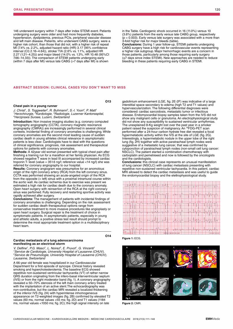
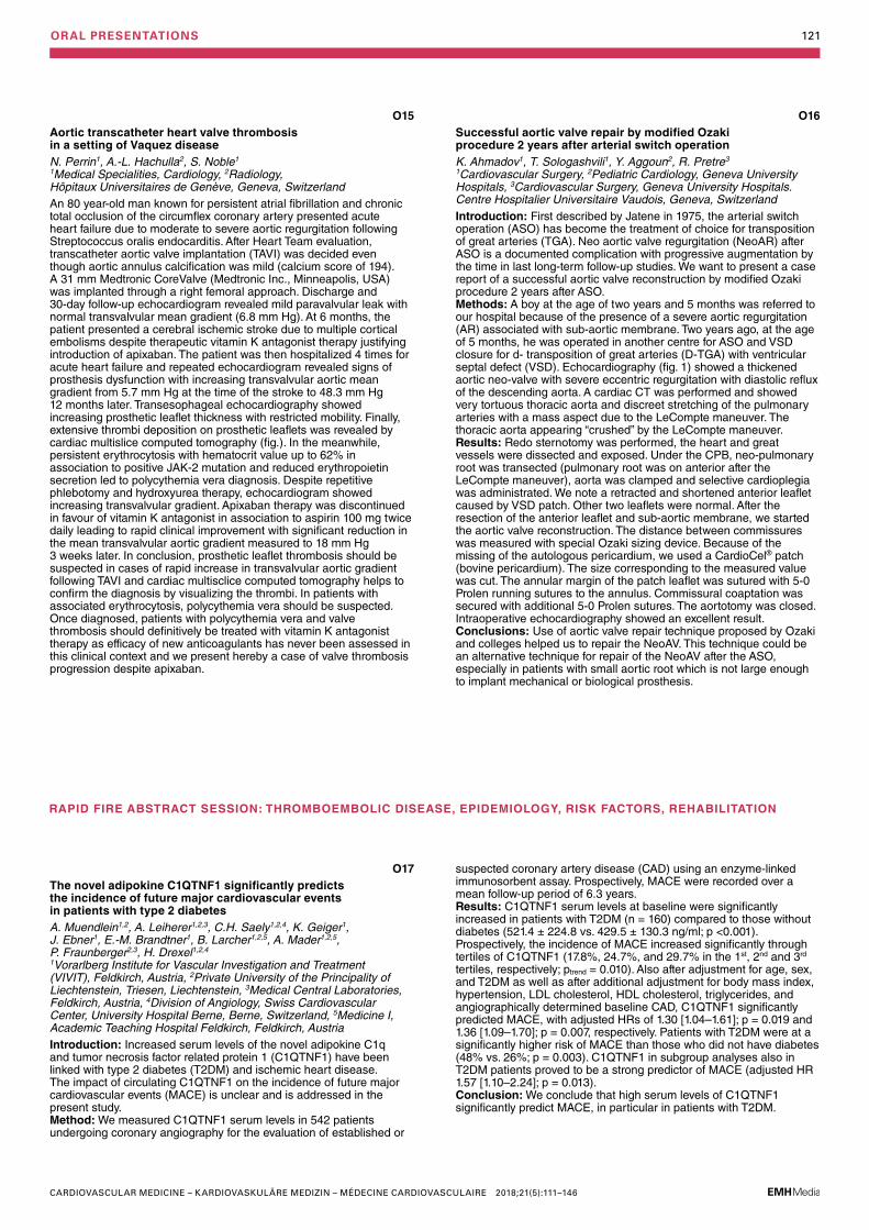
O14Cardiac metastasis of a lung adenocarcinoma manifesting as an electrical stormV. Delfine1, P.G. Masci1, L. Noirez2, E. Pruvot1, G. Vincenti1 1Service de Cardiologie, University Hospital of Lausanne (CHUV), 2Service de Pneumologie, University Hospital of Lausanne (CHUV), Lausanne, SwitzerlandA 66-year old female was hospitalized in our Cardiovascular Department for a first episode of syncope. Clinical history revealed smoking and hypercholesterolemia. The baseline ECG showed repetitive non-sustained ventricular tachycardia (VT) of rather narrow QRS duration originating from the infero-basal interventricular septum (IVS) or from the right moderator band (fig. 1). A coronary angiography revealed a 50–70% stenosis of the left main coronary artery treated with the implantation of an active stent.The echocardiography was non-contributive, but the cardiac MRI revealed a localized thickening of the inferior IVS (fig. 2A) with hyperintense inhomogeneous appearance on T2-weighted images (fig. 2B) confirmed by elevated T2 values (60 ms, normal values <55 ms; fig. 2D) and T1 values (1184 ms, normal values <1050 ms; fig. 2C); the high signal intensity on late
gadolinium enhancement (LGE, fig. 2E-2F) was indicative of a large interstitial space secondary to edema (high T2 and T1 values) and hypervascularization. The following differential diagnoses were considered: cardiac sarcoidosis, metastatic tumor or infectious disease. Endomyocardial biopsy samples taken from the IVS did not show any malignant cells or granuloma. An electrophysiological study did not show any susceptibility to sustained ventricular arrhythmias. The unexplained 8-Kg weight lost over the past year in a smoker patient raised the suspicion of malignancy. An 18FDG-PET scan performed after a 24-hour carbon hydrate free diet revealed a focal hypermetabolic activity within the IVS at the site of LGE (fig. 2G). Interestingly, a hypermetabolic nodule in the upper lobe of the right lung (fig. 2H) together with active paratracheal lymph nodes were suggestive of a metastatic lung cancer, that was confirmed by cytoponction of paratracheal lymph nodes (non-small cell lung cancer: NSCLC). The patient started a combination chemotherapy with carboplatin and pemetrexed and now is followed by the oncologists and the cardiologists. Conclusions: this clinical case represents an unusual manifestation of lung cancer (NSCLC) with cardiac metastasis presenting with repetitive non-sustained ventricular tachycardia. In this patient, cardiac MRI allowed to detect the cardiac metastasis and was useful to guide the endomyocardial biopsy and the elettrophysiological study.
ABSTRACT SESSION: CLINICAL CASES YOU DON’T WANT TO MISS
Figure 1: ECG.
Figure 2: CMR.
ORAL PRESENTATIONS 121
CARDIOVASCULAR MEDICINE – KARDIOVASKULÄRE MEDIZIN – MÉDECINE CARDIOVASCULAIRE 2018;21(5):111–146
O15Aortic transcatheter heart valve thrombosis in a setting of Vaquez diseaseN. Perrin1, A.-L. Hachulla2, S. Noble1 1Medical Specialities, Cardiology, 2Radiology, Hôpitaux Universitaires de Genève, Geneva, SwitzerlandAn 80 year-old man known for persistent atrial fibrillation and chronic total occlusion of the circumflex coronary artery presented acute heart failure due to moderate to severe aortic regurgitation following Streptococcus oralis endocarditis. After Heart Team evaluation, transcatheter aortic valve implantation (TAVI) was decided even though aortic annulus calcification was mild (calcium score of 194). A 31 mm Medtronic CoreValve (Medtronic Inc., Minneapolis, USA) was implanted through a right femoral approach. Discharge and 30-day follow-up echocardiogram revealed mild paravalvular leak with normal transvalvular mean gradient (6.8 mm Hg). At 6 months, the patient presented a cerebral ischemic stroke due to multiple cortical embolisms despite therapeutic vitamin K antagonist therapy justifying introduction of apixaban. The patient was then hospitalized 4 times for acute heart failure and repeated echocardiogram revealed signs of prosthesis dysfunction with increasing transvalvular aortic mean gradient from 5.7 mm Hg at the time of the stroke to 48.3 mm Hg 12 months later. Transesophageal echocardiography showed increasing prosthetic leaflet thickness with restricted mobility. Finally, extensive thrombi deposition on prosthetic leaflets was revealed by cardiac multislice computed tomography (fig.). In the meanwhile, persistent erythrocytosis with hematocrit value up to 62% in association to positive JAK-2 mutation and reduced erythropoietin secretion led to polycythemia vera diagnosis. Despite repetitive phlebotomy and hydroxyurea therapy, echocardiogram showed increasing transvalvular gradient. Apixaban therapy was discontinued in favour of vitamin K antagonist in association to aspirin 100 mg twice daily leading to rapid clinical improvement with significant reduction in the mean transvalvular aortic gradient measured to 18 mm Hg 3 weeks later. In conclusion, prosthetic leaflet thrombosis should be suspected in cases of rapid increase in transvalvular aortic gradient following TAVI and cardiac multisclice computed tomography helps to confirm the diagnosis by visualizing the thrombi. In patients with associated erythrocytosis, polycythemia vera should be suspected. Once diagnosed, patients with polycythemia vera and valve thrombosis should definitively be treated with vitamin K antagonist therapy as efficacy of new anticoagulants has never been assessed in this clinical context and we present hereby a case of valve thrombosis progression despite apixaban.
O16Successful aortic valve repair by modified Ozaki procedure 2 years after arterial switch operationK. Ahmadov1, T. Sologashvili1, Y. Aggoun2, R. Pretre3 1Cardiovascular Surgery, 2Pediatric Cardiology, Geneva University Hospitals, 3Cardiovascular Surgery, Geneva University Hospitals. Centre Hospitalier Universitaire Vaudois, Geneva, SwitzerlandIntroduction: First described by Jatene in 1975, the arterial switch operation (ASO) has become the treatment of choice for transposition of great arteries (TGA). Neo aortic valve regurgitation (NeoAR) after ASO is a documented complication with progressive augmentation by the time in last long-term follow-up studies. We want to present a case report of a successful aortic valve reconstruction by modified Ozaki procedure 2 years after ASO. Methods: A boy at the age of two years and 5 months was referred to our hospital because of the presence of a severe aortic regurgitation (AR) associated with sub-aortic membrane. Two years ago, at the age of 5 months, he was operated in another centre for ASO and VSD closure for d- transposition of great arteries (D-TGA) with ventricular septal defect (VSD). Echocardiography (fig. 1) showed a thickened aortic neo-valve with severe eccentric regurgitation with diastolic reflux of the descending aorta. A cardiac CT was performed and showed very tortuous thoracic aorta and discreet stretching of the pulmonary arteries with a mass aspect due to the LeCompte maneuver. The thoracic aorta appearing “crushed” by the LeCompte maneuver. Results: Redo sternotomy was performed, the heart and great vessels were dissected and exposed. Under the CPB, neo-pulmonary root was transected (pulmonary root was on anterior after the LeCompte maneuver), aorta was clamped and selective cardioplegia was administrated. We note a retracted and shortened anterior leaflet caused by VSD patch. Other two leaflets were normal. After the resection of the anterior leaflet and sub-aortic membrane, we started the aortic valve reconstruction. The distance between commissures was measured with special Ozaki sizing device. Because of the missing of the autologous pericardium, we used a CardioCel® patch (bovine pericardium). The size corresponding to the measured value was cut. The annular margin of the patch leaflet was sutured with 5-0 Prolen running sutures to the annulus. Commissural coaptation was secured with additional 5-0 Prolen sutures. The aortotomy was closed. Intraoperative echocardiography showed an excellent result. Conclusions: Use of aortic valve repair technique proposed by Ozaki and colleges helped us to repair the NeoAV. This technique could be an alternative technique for repair of the NeoAV after the ASO, especially in patients with small aortic root which is not large enough to implant mechanical or biological prosthesis.
O17The novel adipokine C1QTNF1 significantly predicts the incidence of future major cardiovascular events in patients with type 2 diabetesA. Muendlein1,2, A. Leiherer1,2,3, C.H. Saely1,2,4, K. Geiger1, J. Ebner1, E.-M. Brandtner1, B. Larcher1,2,5, A. Mader1,2,5, P. Fraunberger2,3, H. Drexel1,2,4 1Vorarlberg Institute for Vascular Investigation and Treatment (VIVIT), Feldkirch, Austria, 2Private University of the Principality of Liechtenstein, Triesen, Liechtenstein, 3Medical Central Laboratories, Feldkirch, Austria, 4Division of Angiology, Swiss Cardiovascular Center, University Hospital Berne, Berne, Switzerland, 5Medicine I, Academic Teaching Hospital Feldkirch, Feldkirch, AustriaIntroduction: Increased serum levels of the novel adipokine C1q and tumor necrosis factor related protein 1 (C1QTNF1) have been linked with type 2 diabetes (T2DM) and ischemic heart disease. The impact of circulating C1QTNF1 on the incidence of future major cardiovascular events (MACE) is unclear and is addressed in the present study. Method: We measured C1QTNF1 serum levels in 542 patients undergoing coronary angiography for the evaluation of established or
suspected coronary artery disease (CAD) using an enzyme-linked immunosorbent assay. Prospectively, MACE were recorded over a mean follow-up period of 6.3 years. Results: C1QTNF1 serum levels at baseline were significantly increased in patients with T2DM (n = 160) compared to those without diabetes (521.4 ± 224.8 vs. 429.5 ± 130.3 ng/ml; p <0.001). Prospectively, the incidence of MACE increased significantly through tertiles of C1QTNF1 (17.8%, 24.7%, and 29.7% in the 1st, 2nd and 3rd tertiles, respectively; ptrend = 0.010). Also after adjustment for age, sex, and T2DM as well as after additional adjustment for body mass index, hypertension, LDL cholesterol, HDL cholesterol, triglycerides, and angiographically determined baseline CAD, C1QTNF1 significantly predicted MACE, with adjusted HRs of 1.30 [1.04–1.61]; p = 0.019 and 1.36 [1.09–1.70]; p = 0.007, respectively. Patients with T2DM were at a significantly higher risk of MACE than those who did not have diabetes (48% vs. 26%; p = 0.003). C1QTNF1 in subgroup analyses also in T2DM patients proved to be a strong predictor of MACE (adjusted HR 1.57 [1.10–2.24]; p = 0.013). Conclusion: We conclude that high serum levels of C1QTNF1 significantly predict MACE, in particular in patients with T2DM.
RAPID FIRE ABSTRACT SESSION: THROMBOEMBOLIC DISEASE, EPIDEMIOLOGY, RISK FACTORS, REHABILITATION
ORAL PRESENTATIONS 122
CARDIOVASCULAR MEDICINE – KARDIOVASKULÄRE MEDIZIN – MÉDECINE CARDIOVASCULAIRE 2018;21(5):111–146
O18Elevated parathyroid hormone is associated with an increased mortality risk in type 2 diabetesMuendlein1,2, A. Leiherer1,2,3, C.H. Saely1,2,4, K. Geiger1, E.-M. Brandtner1, B. Larcher1,2,5, A. Mader1,2,5, P. Fraunberger2,3, M. Kleber6, A. Dressel7, W. Maerz6,8,9, H. Drexel1,2,4 1Vorarlberg Institute for Vascular Investigation and Treatment (VIVIT), Feldkirch, Austria, 2Private University of the Principality of Liechtenstein, Triesen, Liechtenstein, 3Medical Central Laboratories, Feldkirch, Austria, 4Division of Angiology, Swiss Cardiovascular Center, University Hospital Berne, Berne, Switzerland, 5Medicine I, Academic Teaching Hospital Feldkirch, Feldkirch, Austria, 6Internal Medicine V, Mannheim Medical Faculty, University of Heidelberg, Mannheim, 7DACH-Gesellschaft Prävention von Herz-Kreislauferkrankungen, Hamburg, 8Synlab-Academy, Synlab Services GmbH, Mannheim & Augsburg, Germany, 9Clinical Institute for Medical and Chemical Laboratory Diagnostics, Medical University of Graz, Graz, AustriaIntroduction: Parathyroid hormone (PTH) is one of the main regulators of calcium homeostasis. PTH levels are elevated in primary hyperparathyroidism as well as with vitamin D deficiency or chronic kidney disease. The association of increased PTH levels with all-cause mortality in high-risk patients is unclear. Method: We therefore investigated the impact of serum PTH on mortality risk in a large series of 939 patients undergoing coronary angiography for the evaluation of established or suspected coronary artery disease (CAD), including 244 patients with type 2 diabetes (T2DM). Prospectively, deaths were recorded over a mean follow-up period of 6.2 years. Results: PTH at baseline was inversely associated with eGFR (rho = –0.228; p <0.001) and 25-hydroxy-vitamin D (rho = –0.243; p <0.001) and was positively associated with age (rho = 0.122; p <0.001) and BMI (rho = 0.099, p = 0.002). Prospectively, elevated PTH was not significantly associated with an increased mortality risk in the total study cohort (standardized HR 1.30 [0.96–1.76]; p = 0.092). However, subgroup analysis with respect to T2DM showed a highly significant association of PTH with mortality in patients with T2DM (HR 2.32 [1.37–3.95]; p = 0.002), but no association of PTH with mortality in non-diabetic subjects (HR 1.04 [0.82–1.32]; p = 0.766). An interaction term T2DM × PTH was significant (p = 0.006), indicating a significantly stronger influence of PTH on mortality risk in patients with diabetes than in individuals without T2DM. The impact of PTH on mortality risk in patients with T2DM remained significant after adjustment for age, gender, and BMI (HR 2.30 [1.34–3.93]; p = 0.002) as well as after additional adjustment for, smoking, kidney function, baseline vitamin D and angiographically determined baseline CAD (HR 1.91 [1.07–3.40]; p = 0.029). Conclusion: We conclude that elevated PTH levels are a strong and independent predictor of all-cause mortality in patients with T2DM.
O19Age at start of endurance training is associated with patterns of left ventricular hypertrophy in middle-aged runnersC. Ryffel, P. Eser, L. Trachsel, N. Brugger, M. Wilhelm Inselspital, Bern University Hospital and University of Bern, Bern, SwitzerlandIntroduction: Left ventricular hypertrophy (LVH) is a physiological adaptation to long-term endurance training. We investigated the impact of age at start of endurance training on LV geometry in a cohort of male, middle-aged, non-elite endurance athletes. Methods: A total of 121 healthy, normotensive, Caucasian participants of a 10-mile race were recruited and assessed with an echocardiogram and a comprehensive interview. Athletes were classified based on patterns of LVH. Results: Thirty-five athletes (31%) had LVH. Athletes with eccentric LVH (16%) were significantly younger at start of endurance training compared to athletes with concentric LVH (15%, 14 ± 5 years vs. 31 ± 8 years; p <0.001). Although the yearly volume of endurance training was comparable between athletes with eccentric and concentric LVH, athletes with eccentric LVH had shorter race times. All athletes with an increased LV end diastolic volume index (LVEDVI; ≥74 ml/m2) started endurance training before or at age 25. Conclusions: In our cohort of non-elite middle-aged runners, eccentric LVH was found only in athletes with an early start of endurance training. In case of a mature starting age, endurance training may, contrary to the “Morganroth hypothesis”, also lead to concentric LVH. The consideration of endurance training starting age may lead to a better understanding of morphological adaptations of the heart.
O20Prognostic value of elevated lipoprotein(a) in patients with acute coronary syndromesB. Gencer1, F. Rigamonti2, D. Nanchen3, N. Vuilleumier4, I. Kern4, R. Klingenberg5, L. Räber6, R. Auer3,7, D. Carballo2, C. Sebastian8, D. Heg9, S. Windecker6, T.F. Lüscher5, C.M. Matter5, N. Rodondi7,10, F. Mach2 1Cardiology Division, Department of Medical Specialties, HUG, Geneva University, 2Cardiology Division, Geneva University Hospitals, Geneva, 3Department of Ambulatory Care and Community Medicine, Lausanne University, Lausanne, 4Laboratory Medicine Division, Geneva University Hospitals, Geneva, 5Department of Cardiology, University Heart Center, University of Zurich, Zürich, 6Department of Cardiology, University Hospital of Bern, 7Institute of Primary Health Care (BIHAM), University of Bern, Bern, 8Department of Internal Medicine, Geneva University Hospitals, Geneva, 9Department of Clinical Research, Institute of Social and Preventive Medicine, and Clinical Trials Unit, University of Bern, 10Department of General Internal Medicine, Inselspital, Bern University Hospital, Bern, SwitzerlandBackground: Lipoprotein(a) [Lp(a)] target values <50 mg/dL are advocated for high-risk cardiovascular patients. We investigated the prognostic value of Lp(a) after acute coronary syndromes (ACS), and whether values under 50 mg/dL was associated with a better prognosis. Methods: The measurements of plasma Lp(a) levels from 1711 patients hospitalized for ACS and included in a Swiss prospective cohort were evaluated. At 1 year, the association between elevated Lp(a) at baseline defined as ≥50 mg/dL or Lp(a) tertiles with major adverse cardiovascular events (MACE) defined by a composite of cardiac death, myocardial infarction and stroke was assessed using hazard ratios (HR) and 95% confidence intervals (CI) adjusting for traditional cardiovascular risk factors (age, sex, smoking, diabetes, hypertension and low-density lipoprotein cholesterol [LDL-C]). Results: A total of 92 patients (5.4%) had Lp(a) values ≥50 mg/dL. Patients with higher Lp(a) values were more likely to be women, to have high levels of LDL-C, high-density lipoprotein cholesterol (HDL-C) and triglycerides. At 1 year, no association was found between elevated Lp(a) values at baseline and clinical outcomes HRs were 0.49 (95% CI 0.07–3.59) for cardiac death, 0.34 (0.05–2.50) for recurrent myocardial infarction, 1.53 (95% CI 0.34–6.89) for stroke, and 0.67 (0.24–1.83) for a composite of MACE. No association was observed between Lp(a) tertiles or per standard deviation increase of Lp(a) (HR 0.95, 95% CI 0.79–1.14) with composite MACE. Conclusions: In patients with ACS, elevated Lp(a) values at baseline were observed in few patients and were not associated with MACE at one year. Until data from randomized controlled trials lowering Lp(a) are available, our observational data suggest that achieving Lp(a) target values is not determinant for improved prognosis at one year after ACS.
Figure 1
ORAL PRESENTATIONS 123
CARDIOVASCULAR MEDICINE – KARDIOVASKULÄRE MEDIZIN – MÉDECINE CARDIOVASCULAIRE 2018;21(5):111–146
Patients without CR referrals were less likely to receive guideline-recommended medication, such as P2Y12 inhibitors, aspirin and statins and underwent less frequently percutaneous coronary intervention (PCI). Patients referred to CR had a lower crude 1-year all-cause mortality (1.7 vs. 5.8%; p <0.001) and lower rates of re-infarction (2.8 vs. 4.1%; p = 0.003), rehospitalisations for cardiovascular disease (21.0 vs. 25.3%; p <0.001) and interventions (11.7 vs. 14.8%; p <0.001). In a multivariable logistic regression analysis, which included age, gender, comorbidities, STEMI, PCI and discharge medication, CR was an independent protective predictor for mortality (OR 0.65; 95%CI 0.48-0.89; p = 0.007). Conclusions: Although the detailed data of CR programs and patient participation were not available for this study, our data from 7883 AMI patients showed a better 1-year outcome for patients with CR referrals than for those without.
O23Inpatient costs of atrial fibrillation and related comorbiditiesF. Witassek1, D. Conen2, S. Osswald3,4, G. Moschovitis5, P. Meyre3, B. Brüngger6, A. Springer3, S. Aeschbacher3, M. Kühne3,4, L.H. Bonati7, D. Shah8, N. Rodondi9,10, C. Baumgartner10, E. Moutzouri10, R. Kobza11, T. Szucs12, M. Schwenkglenks1, on behalf of the Swiss-AF Study Investigators 1Health Economics Unit, Epidemiology, Biostatistics and Prevention Institute, University of Zurich, Zurich, Switzerland, 2Population Health Research Institute, McMaster University, Hamilton, ON, Canada, 3Cardiovascular Research Institute Basel (CRIB), 4Department of Medicine, Cardiology Division, University Hospital Basel, Basel, 5Department of Cardiology, Ospedale Regionale di Lugano, Lugano, 6Department of Health Sciences, Helsana Group, Zurich, 7Department of Clinical Research, University Hospital Basel, Neurology Division and Stroke Centre, Basel, 8Cardiology Service, Department of Medicine Specialities, University Hospital Geneva, Geneva, 9Institute of Primary Health Care (BIHAM), University of Bern, 10Department of General Internal Medicine, Inselspital, Bern University Hospital, Bern, 11Department of Cardiology, Luzerner Kantonsspital, Luzern, 12European Center of Pharmaceutical Medicine, University of Basel, Basel, SwitzerlandBackground: Although atrial fibrillation (AF) is a major public health burden, cost implications for Switzerland are not well described. This study aimed to assess inpatient costs of AF and related cardiovascular comorbidity and analyse cost drivers. Methods: The Swiss-AF study is an ongoing prospective multicentre cohort study of AF patients enrolled between April 2014 and August 2017 in Switzerland. For patients who gave their informed consent, health insurance (HI) claims data are provided by four of the biggest HI companies in Switzerland. Here, patients were included if clinical and claims data covered at least one year of follow up (FU) by June 2016. SwissDRG codes were used to estimate inpatient costs attributable to AF, stroke/transient ischemic attack (TIA) or congestive heart failure (CHF). Clinical study documentation of stroke/TIA or CHF events and AF related interventions or complications were used to support the attribution of costs. Costs are presented from the health care system perspective. Results: Of 750 patients with clinical 1 year FU, for 311(41.5%) HI data were available. Mean (SD) age at enrolment was 72.0(8.7) years and 74% of patients were male. AF was paroxysmal in 153 patients (49%), persistent in 72 (23%) and permanent in 86 (28%). The median (IQR) FU was 1.6 (1.3;1.8) years. Of 311 patients, 57 (18%) had inpatient costs directly attributable to AF, reflecting a total of 70 inpatient episodes. Mean costs per episode were sFr 24,178 (range 4,029–76,002). For the 311 patients, this implies 0.15 episodes and costs of sFr 3,591 per patient-year. In a zero-inflated negative binominal regression model including age, gender, AF type, AF duration, diabetes and FU time, only age (p = 0.015) predicted significantly higher AF-related costs. Ten (3.2%) patients were hospitalised for 10 episodes of stroke/TIA and 17 (5.5%) for 27 episodes of CHF. Mean costs per episode were sFr 42,271 (range 6,940-134,167) for stroke/TIA and sFr 13,684 (range 6,704-34,587) for CHF. This implies 0.02 episodes or sFr 898 per patient-year for stroke and 0.06 episodes or sFr 784 for CHF. Of the total costs considered, 68% were related to AF, 17% to stroke/TIA and 15% to CHF. Conclusion: Approximately 25% of patients had inpatient costs due to AF or related conditions. Except for age, no other independent cost drivers could be identified for AF costs. Further work is planned to refine the adjudication of costs and to analyse other causes of costs such as bleeding.
O21Prognostic values of fasting hyperglycaemia in nondiabetic patients with acute coronary syndromes: a prospective cohort studyB. Gencer1, F. Rigamonti1, D. Nanchen2, R. Klingenberg3, L. Räber4, E. Moutzouri5, R. Auer2,5, D. Carballo6, D. Heg7, S. Windecker4, T.F. Lüscher3, C.M. Matter3, N. Rodondi5,8, F. Mach6, M. Roffi6 1Department of Medical Specialties, Cardiology Division, HUG, Geneva University, Geneva, 2Department of Ambulatory Care and Community Medicine, Lausanne University, Lausanne, 3Department of Cardiology, University Heart Center, University of Zurich, Zürich, 4Department of Cardiology, University Hospital of Bern, 5Institute of Primary Health Care (BIHAM), University of Bern, Bern, 6Cardiology Division, Geneva University Hospitals, Geneva, 7Department of Clinical Research, Institute of Social and Preventive Medicine, and Clinical Trials Unit, University of Bern, 8Department of General Internal Medicine, Inselspital, Bern University Hospital, Bern, SwitzerlandBackground: Controversy remains regarding the prevalence of hyperglycaemia in non-diabeticpatients hospitalized with acute coronary syndromes (ACS) and its prognostic value for long-term outcomes. Methods: We evaluated the prevalence of hyperglycaemia (defined as fasting glycaemia ≥10 mmol/l) among patients with no known diabetes at the time of enrolment in theprospective SPUM-ACS cohort, as well as its impact on all-cause death, myocardial infarction, stroke and incidence of diabetes at one year. Results: Among 3858 ACS patients, enrolled between December 2009 and December 2014, 709 (18.4%) had known diabetes, while 112 (3.6%) of non-diabetic patients had hyperglycaemia at admission. Compared with non-hyperglycaemic patients, hyperglycaemic individuals were more likely to present with ST-elevation myocardial infarction and acute heart failure. At discharge, hyperglycaemic patients were more frequently treated with glucose-lowering agents (8.9% vs. 0.66%, p <0.001). At 1-year, adjudicated all-cause death was significantly higher in non-diabetic patients presenting with hyperglycaemia compared with patients with no hyperglycaemia (5.4% vs. 2.2%, p = 0.041) and hyperglycaemia was a significant predictor of 1-year mortality (adjusted hazard ratio [HR] 2.39, 95% CI 1.03–5.56). Among patients with hyperglycaemia, 9.8% had developed diabetes at 1-year, while the corresponding proportion among patients without hyperglycaemia was 1.8% (p <0.001). In multivariate analysis, hyperglycaemia at presentation predicted the onset of treated diabetes at 1-year (odd ratio [OR] 4.15, 95% CI 1.59–10.86; p = 0.004). Conclusion: Among non-diabetic patients hospitalized with ACS, a fasting hyperglycaemia of ≥10 mmol/l predicted one-year mortality and was associated with a four-fold increased risk of developing diabetes at one year.
O22Impact of cardiac rehabilitation referral on 1 year outcome after discharge of patients with acute myocardial infarctionM. Hermann1, F. Witassek2, P. Erne2, H. Rickli3, D. Radovanovic2 1Kardiologie, Zürcher RehaZentrum Wald, Wald, 2AMIS Plus Data Center, Epidemiology, Biostatistics and Prevention Institute, University of Zurich, Zürich, 3Department of Cardiology, Kantonsspital St. Gallen, St. Gallen, SwitzerlandBackground: Cardiac rehabilitation (CR) after an acute myocardial infarction (AMI) has a class I recommendation in the present guidelines. However, data about the impact on mortality in Switzerland are not available. Therefore, we analysed 1-year outcome of AMI patients according to CR referral at discharge. Design and methods: Data were extracted from the Swiss AMIS Plus registry and included patients with ST-elevation myocardial infarction (STEMI) and non-STEMI (NSTEMI) enrolled from 2005 to 2016 who were asked to give their informed consent to a telephone follow-up 1 year after discharge. We compared AMI patients with CR referrals at discharge to those without. Patients transferred to other hospitals were excluded. The analyses were performed using multivariable logistic regression. Results: From 10,141 patients, 1956 (19.3%) refused to participate in follow-up (FU) and of the 8,185 patients with informed consent, 302 were lost to FU. There were 4508 (57.2%) patients with CR referrals compared to 3375 (42.8%) without. Patients referred to CR were younger (62.4 y vs. 68.8 y), more often male (77 vs 70%), presented more often with STEMI (63.5 vs 52.1%) and apart from smoking (44.0 vs 34.9%), they had less risk factors, such as dyslipidemia (55.0 vs. 60.1%), hypertension (55.6 vs. 65.3%) and diabetes (16.7 vs. 21.5%).
ORAL PRESENTATIONS 124
CARDIOVASCULAR MEDICINE – KARDIOVASKULÄRE MEDIZIN – MÉDECINE CARDIOVASCULAIRE 2018;21(5):111–146
Conclusion: To the best of our knowledge, this is the first study reporting the incidence of PNP after laser-assisted TLE. We postulate that the thermal energy generated by laser is not dissipated quickly enough in occluded or heavily calcified lesions, injuring the ipsilateral phrenic nerve at the subclavian level. Our findings advise to carefully consider to increase pulse-repetition-rate at the subclavian level. Larger, possibly prospective studies are needed to evaluate the real incidence through systematic radiological assessment after TLE.
O25A potential mechanism of Torsades de Pointes tachycardia in atrioventricular blockA. Saguner1, G. Li2, D. Akdis3, P. Bode4, G. Fontaine5 1Cardiology | Rhythmology, USZ, Zurich, Switzerland, 2Cardiovascular Medicine, The First Affiliated Hospital, Xian, China, 3Cardiology, University Heart Center Zurich, 4Pathology, University Hospital Zurich, Zurich, Switzerland, 5Cardiology, Groupe Hospitalier Pitié-Salpetrière, Paris, FranceBackground: Two mechanisms may explain the occurrence of Torsades de Pointes tachycardia (TdP) in atrioventricular block (AVB): (1) a phase 2 reentry phenomenon in adjacent myocardial fibers due to bradycardia and (2) a fast reentry circus movement. Therefore, the objective of this study was to investigate mechanisms of TdP, and to present for the first-time endocardial recordings during TdP from patients with high-degree AVB. Methods: TdP was recorded by surface ECG in 16 patients with high-degree AVB. The critical coupling interval (CCI) between the last beat during AVB and before the first abnormal ventricular event (PVCs, couplets, triplets and TdPs) was measured. Endocardial signals during TdP were recorded from the right ventricular apex by temporary bipolar catheters. Results: Patients were >65 years; 80% were females. The QT interval during AVB (mean heart rate of 38.9 ± 7.5 bpm) was 653 ± 67.2 ms. The CCI before the onset of PVCs/couplets vs. TdP was shorter in the latter (672 ± 43.8 ms and 676 ± 37 ms vs. 639 ± 52 ms, p <0.05). A rhythmic crescendo (in 100%) of PVCs and T wave augmentation (in 75%) was observed before the onset of TdP (figure, A, arrow). In 4/5 patients (75%) the local endocardial electrogram showed a constant pattern of rapid deflection superimposed on a smooth low amplitude signal suggesting far field potentials from electrically depressed myocardium, indicating a phase 2 reentry (figure, B, arrow). Conclusions: This study suggests abnormal repolarization of adjacent ventricular myocardial fibers indicating a phase 2 reentry and fast reentry circus movement as the cause of TdP in patients with high degree AVB.
O24New onset of phrenic nerve palsy after laser-assisted transvenous lead extraction: a single-center experienceT. Özkartal1, F. Regoli1, G. Conte1, M.L. Caputo1, C. Klersy2, T. Moccetti1, A. Auricchio1 1Cardiocentro Ticino, Lugano, Switzerland, 2Service of Clinical Epidemiology & Biometry, Fondazione IRCCS Policlinico San Matteo, Pavia, ItalyIntroduction: Phrenic nerve palsy (PNP) after mechanical transvenous lead-extraction (TLE) was recently described for the first time. We aimed to analyse our TLE database for the presence of PNP. Methods: All consecutive patients referred to our institution were included in this study. Every available post-procedural chest x-ray was compared to the routinely performed pre-procedural radiographs. A newly elevated hemidiaphragm ipsilateral to TLE was considered indicative of PNP (see figure). Results: Altogether 255 TLE procedures with extraction of 364 leads were performed. Most common TLE-indication was lead malfunction (63%). Complete radiographic success rate was 97.3% with an In-hospital procedure-related major-complication rate of 2.4%, including one intra-procedural death (0.4%). We identified 5 cases with PNP (2%), all occurring after laser-assisted TLE (see table). Clinical presentation varied from subtle and aspecific chest pain/discomfort to severe and acute dyspnoea, with time to diagnosis varying from immediate to several weeks after the procedure. In 80% of cases, the explanted lead was a defibrillator electrode and the median lead dwelling-time was 70.2 months (29.3; 184.9). In 4 cases the extraction was performed using high-energy laser (pulse-repetition-rate 80 Hz).
RAPID FIRE ABSTRACT SESSION: PACEMAKER, DEFIBRILLATOR AND ELECTROPHYSIOLOGY
Figure 1: X-ray prior and after transvenous lead extraction showing a newly elevated leftsided hemidaphragm.
Figure
ORAL PRESENTATIONS 125
CARDIOVASCULAR MEDICINE – KARDIOVASKULÄRE MEDIZIN – MÉDECINE CARDIOVASCULAIRE 2018;21(5):111–146
O26Genetic testing in cardiac arrest survivors with or without clinical evidence of cardiac disease: a Swiss experienceB. Asatryan1, A. Schaller2, J. Seiler1, H. Servatius1, F. Noti1, S. Baldinger1, H. Tanner1, L. Roten1, R. Dillier3, A. Lam1, G. Conte4, A. Saguner5, A. Müller3, F. Duru5, A. Auricchio4, P. Ammann6, T. Reichlin7, H. Burri8, J. Fuhrer1, M. Wilhelm1, A. Medeiros-Domingo1 1Department of Cardiology, 2Division of Human Genetics, Department of Pediatrics, Inselspital, Bern University Hospital, University of Bern, Bern, 3Department of Cardiology, Triemli Hospital, Zurich, 4Department of Cardiology, Fundazione Cardiocentro Ticino, Lugano, 5Department of Cardiology, University Heart Center Zurich, Zurich, 6Department of Cardiology, Kantonsspital St. Gallen, St. Gallen, 7Department of Cardiology, University Hospital Basel, Basel, 8Department of Cardiology, University Hospital Geneva, Geneva, SwitzerlandIntroduction: Cardiac arrest is often the first manifestation of a silent genetic heart disease. Phenotype-guided genetic testing in these patients is considered useful, while the utility of the test in phenotype-negative unexplained cardiac arrest (UCA) remains disputable. We aimed to evaluate the utility of genetic testing in cardiac arrest survivors with or without evident cardiac phenotype. Methods: We included all UCA survivors sent to our center for genetic testing between January 2014 and October 2017. Initial clinical evaluation included 12-lead ECG, stress test, coronary angiography and echocardiography. After obtained informed consent, DNA was extracted from peripheral blood. Targeted exome sequencing was performed using the TruSight One Sequencing Panel from Illumina. Variants in 190 clinically relevant cardiac genes with a minor allele frequency of <1% were further evaluated. Variant interpretation followed the 2015 guidelines of the American College of Medical Genetics. Results: Fifty-one unrelated cases (age: 33 ± 16 yrs, 80% males) were included. After the initial clinical evaluation, 31(61%) were considered as phenotype-negative (PhN-UCA) and 20 (39%) exhibited some clinical cardiac phenotype (PhP-UCA). Genetic testing revealed pathogenic or likely pathogenic variants in 18 cases in the overall cohort (35%): 7 out of 31 (23%) in the PhN-UCA group and 11 out of 20 (55%) in the PhP-UCA. In 5 out of 31 (16%) cases of PhN-UCA the identified variant helped to clarify the phenotype while in the group of PhP-UCA in 10 cases out of 18 (56%) the identified variant correlated with the clinical phenotype. Identified pathogenic variants located mainly in CPVT or channelopathy-associated genes (67%). Seven out of 18 (39%) patients carried ≥1 additional variant of uncertain clinical significance (VUS). Age ≤35 was associated with a higher likelihood of identifying a putative pathogenic variant (48% vs. 21%, p <0.05). Conclusions: The yield of genetic testing is 2-times higher in PhP-UCA compared to PhN-UCA. The majority of identified mutations were associated with channelopathies, most commonly CPVT. Since 16% of the cases and family members of the PhN-UCA group benefit from genetic testing, it should be generally recommended in all patients with UCA.
O27Prospective validation of diagnostic and prognostic syncope scores in the emergency departmentJ. du Fay de Lavallaz1, P. Badertscher1, T. Nestelberger1, R. Isenrich1, O. Miro2, E. Salgado2, N. Geigy3, M. Christ4, L. Cullen5, M. Than6, J. Martin-Sanchez7, E. Rodriguez-Adrada7, S. Di Somma8, F. Peacock9, D. Kawecki10, J. Boeddinghaus1, R. Twerenbold1, C. Puelacher1, D. Wussler1, I. Strebel1, D. Keller11, I. Poepping12, M. Kühne1, C. Müller1, T. Reichlin1 1Universitätsspital Basel, Basel, Switzerland, 2Hospital Clinic, Barcelona, Spain, 3Kantonsspital Liestal, Liestal, 4Kantonsspital Luzern, Luzern, Switzerland, 5Royal Brisbane & Women’s Hospital, Herston, QLD, Australia, 6Christchurch Hospital, Christchurch, New Zealand, 7Hospital Clínico San Carlos, Madrid, Spain, 8University Sapienza Rome, Sant’Andrea Hospital, Rome, Italy, 9Baylor College of Medicine, Houston, TX, United States, 10Medical University of Silesia, Zabrze, Poland, 11Unispital Zürich, Zürich, 12Spital Lachen, Lachen, SwitzerlandIntroduction: Various scores have been derived for the assessment of syncope patients in the emergency department (ED) in different studies. Their comparative performance is unknown. Methods: We prospectively enrolled unselected patients presenting with syncope to the ED in a diagnostic multicenter study. Early clinical judgment of the treating ED-physician regarding the probability of cardiac syncope was quantified using a visual analogue scale. Two independent cardiologists adjudicated the final diagnosis based on all
available information during clinical work-up including 1-year follow-up. Major cardiovascular events (MACE) and death were recorded during 2 years of follow-up. Prognostic and diagnostic scores were calculated according to their definition in the literature. Results: 1195 patients were available for score validation. The highest prognostic accuracy for death as quantified by the area under the receiver-operator characteristics curve (AUC) was achieved by the CHADS2-score with an AUC of 0.73 (95%CI 0.69–0.77). This score also performed well for MACE (AUC 0.7, 95%CI 0.67–0.73) and stratified patients with comparative accuracy as the OESIL score, the best performing syncope-specific risk score. All scores performed poorly for the diagnosis of cardiac syncope when compared with the early clinical judgment of the ED physician. Conclusion: The simple CHADS2-score performed at least as well as more complicated syncope-specific risk scores in the prediction of death and MACE. The CHADS2-score is a widely used prediction tool for thromboembolic episodes and could simplify the risk stratification in syncope patients. All scores performed poorly in the diagnosis of cardiac syncope. This analysis underlines the need for improved tools for diagnosis and risk stratification potentially including novel biochemical and electrocardiographic markers.
O28Electroanatomical characteristics of dual-loop atypical atrial fluttersN. Johner, M. Namdar, D.C. Shah Service de Cardiologie, Hôpitaux Universitaires de Genève, Genève, SwitzerlandIntroduction: Atypical atrial flutters (AFL) can be challenging to map and ablate, especially in case of dual-loop reentry. We sought to estimate to prevalence of dual-loop reentries in patients undergoing catheter ablation of AFL and to describe the electroanatomical characteristics of single- and dual-loop AFLs.Methods: We studied 25 non-CTI-dependent macroreentrant AFL on 19 consecutive patients (age 66 ± 15, 79% men, 14 post-atrial fibrillation ablation). High-density (8’216–33’368 points/map) complete (>90% of cycle length (CL)) 3D electroanatomic activation maps were acquired with a 64-electrode basket catheter (Orion, BSc). The tachycardia wavefront was indicated by the shortest active reentry circuit. Multi-loop reentry was defined as the presence of multiple synchronous active loops (non-delayed compared to other loops and confirmed by entrainment mapping). Ablation without catheter dragging was performed at narrowest accessible slow conducting critical isthmuses.Results: Of 25 AFL (24 left and 1 right atrial, CL 261 ± 35 ms), 13 (52%) presented a dual-loop reentrant mechanism. Table 1 describes the prevalence of each identified circuit anatomy for single- and dual-loop AFLs. Isthmuses <30 mm wide were identified at locations described in table 2. Isthmus width, local conduction speed, voltage and post-pacing interval did not differ significantly between common and secondary isthmuses of dual-loop AFLs (p >0.05). Ablation targeted at common isthmuses of dual-loop AFLs and critical isthmuses of single-loop AFLs resulted in termination to sinus rhythm more frequently than ablation at secondary isthmuses of dual-loop AFLs (5 of 6 (83%) and 8 of 11 (73%) vs 1 of 8 (13%), respectively, p = 0.013). Ablation of a secondary isthmus resulted in a CL increase
Figure 1: Comparison of the prognostic accuracies of the different scores by mean of their AUC.
ORAL PRESENTATIONS 126
CARDIOVASCULAR MEDICINE – KARDIOVASKULÄRE MEDIZIN – MÉDECINE CARDIOVASCULAIRE 2018;21(5):111–146
score to identify patients at high risk for early mortality who are implanted with a CRT-D. Methods: For predictive modelling, 1282 consecutive patients from 5 centers (76% male; median LVEF 25%; NYHA class III–IV 60%; median QRS-width 160 ms; ischemic cardiomyopathy 50%; 100% primary prevention and de novo implants) were randomly split into a derivation and validation group (50%/50%). A risk score was developed using logistic regression. The clinical end-point for this study was all-cause mortality.Results: The derivation and validation cohort were similar with respect to age, gender, etiology of heart failure, comorbid conditions, laboratory values, and medical treatment. In the total cohort, 181 patients died over 3 years of follow-up (mortality 6% at 1 and 16% at 3 years). After multivariate analysis, a risk score was developed based on myocardial infarction, LVEF ≤25%, COPD, chronic kidney disease (CKD), hyponatremia and anemia (fig. 1). At 3 years, mortality was 4.6%, 13.2% and 29.7% by ascending tertile of risk score (fig. 2). Compared with the lowest tertile (T1), mortality was significantly higher in the other tertiles (T2 odds ratio (OR) = 3.1; T3 OR = 8.4; both P <0.001). Discrimination was modest (C-statistic 0.73) and the Hosmer-Lemeshow chi-square was 0.95 (P = 0.33).Conclusion: A risk score based on routine, readily available clinical variables can reliably identify patients at high risk for early mortality within 3 years after CRT-D implantation.
O30Cardiac Resynchronization Therapy (CRT) II Survey: Swiss CRT-implantation practice in the European contextI. Zeljkovic1, H. Burri2, A. Breitenstein3, P. Ammann4, A. Auricchio5, A. Mueller6, E. Delacrétaz7, K. Dickstein8, C. Linde9, C. Sticherling1 1University Hospital Basel, Basel, 2University Hospital Geneva, Geneva, 3University Hospital Zurich, Zurich, 4Cantonal Hospital St. Gallen, St. Gallen, 5Cardiocentro Ticino, Lugano, 6Triemli Hospital, Zurich, 7Clinique Cecil, Lausanne, Switzerland, 8Stavanger University Hospital, Stavanger, Norway, 9Karolinska University Hospital, Stockholm, SwedenAim: Between October 2015 and January 2017 data from more than 10’768 CRT from 42 countries were included in the CRT II Survey. From Switzerland (CH), 7 centres contributed 320 patients. We compare the Swiss CRT implantation behaviour to the overall population. Methods: Seven centres from CH recruited included all patients undergoing either a primary CRT implantation or an upgrade to a CRT-system. Data were collected prospectively in an online database. Results: A total of 320 patients were included in CH, which equals 38% of all CRT implantation in CH during this period (n = 838). 24.4% were female; mean age was 71.0 ± 10.2 years. Swiss patients were older (71 ± 10.2 yrs vs. 68.5 ± 20.8 yrs), less symptomatic and more often suffered from comorbidities like chronic kidney disease. Swiss patients significantly more often recieved a CRT-pacemaker than their European counterparts (37.1 vs. 30%; OR 1.37; 1.09–1.73). The main findings and differences between the Swiss and the overall European population are depicted in table 1.Conclusion: When compared to other European countries, Swiss CRT-recipients were older, less symptomatic, received more device upgrades, had a higher incidence of chronic kidney disease and more frequently received quadripolar left ventricular lead. At the same time, the percentage of CRT-ICD implantation was lower than in the overall European population. Our data indicates that despite almost free access to modern technology, Swiss patients and physicians more often use the less expensive CRT-pacemaker system with the primary goal of symptomatic improvement.
O31Importance of true left bundle branch block in cardiac resynchronization therapyK. Arslani, T. Reichlin, M. Kühne, C. Sticherling, S. Osswald, B. Schär Cardiology, University Hospital Basel, Basel, SwitzerlandBackground: Subgroups analyses of randomised trials have questioned the use of CRT in non-left bundle branch block (LBBB) pts due to a lack of mortality benefit. Different indication and evidence levels are thus attributed for CRT in current guidelines according to baseline ECG. We aimed to correlate baseline ECG and improvement in left ventricular ejection fraction (LVEF) in CRT-D pts. Methods: We screened 331 pts with CRT-D implanted between April 2001 and February 2017. Exclusion criteria were: upgrade from pacemaker or ICD (n = 30), higher degree AV block (n = 24), follow-up
>15 ms in 5/7 cases (71%) and in a change in activation sequence in 2/7(29%); 3 of these arrhythmias were subsequently terminated by ablation at another (common) isthmus. Conclusions: Half of the AFLs characterized by high-density mapping presented a dual-loop reentrant mechanism, all of which involved at least one pulmonary vein as an anatomical barrier. The mitral isthmus, the left atrial roof and the right pulmonary veins ostia harbored the majority of critical isthmuses. The majority of ablations delivered to secondary isthmuses resulted in changes of activation sequence or CL increase rather than direct termination, in contrast to ablation at a common isthmus, which results in a high termination rate.
O29Predicting mortality among implantable defibrillator patients treated with cardiac resynchronization therapy: derivation and validation of a risk estimation modelB. Schär1, C. Sticherling1, N. van Boven2, T. Hesselink3, M. Rivero-Ayerza4, M. Schulte3, F. Verbrugge4, V. Umans2, F. Zijlstra5, D. Theuns5 1Cardiology, University Hospital Basel, Basel, Switzerland, 2Cardiology, Medisch Centrum, Alkmaar, 3Cardiology, Medisch Centrum Spect Twente, Enschede, The Netherlands, 4Cardiology, Ziekenhuis Oost-Limburg, Genk, Belgium, 5Cardiology, Erasmus Medical Center, Rotterdam, The NetherlandsBackground: Given the heterogeneity in mortality risk among heart failure patients and the fact that only a minority of patients will experience ventricular arrhythmias, appropriate risk prediction is of paramount importance in maximizing the survival benefit conferred by CRT-D. The Seattle Heart Failure Model predicts mortality risk in HF patients, but was not designed for HF patients who already had a device implanted. Aim of this study was to develop and validate a risk
Table 1: Anatomy of single- and dual-loop flutters.
Single-loop flutters
Dual-loop flutters
(N = 12) (N = 13) Comments
Perimitral 6 (50%) 6 (46%) Second loop around right or left PVs.
Around right pulmonary veins
2 (17%) 6 (46%) 4 with second loop around a functional block.
1 with one loop around each right PV.
1 with second loop around left PVs.
Coronary sinus-depen-dent
2 (17%)
Around a septal scar 1 (8%)
Around a right atrial scar 1 (8%)
Around left pulmonary veins
1 (8%) Second loop around a func-tional block.
Table 2: Anatomical location of isthmuses.
All isthmuses Common isthmuses of dual-loop flutters
Posterior mitral isthmus 9 (26%) 3 (27%)
Pulmonary veins ostia 6 (17%) 3 (27%)
Left atrial roof 6 (17%) 2 (18%)
Posterior left atrial wall 3 (9%) 2 (18%)
Anterior mitral isthmus 3 (9%) 1 (9%)
Low septum 3 (9%)
Coronary sinus (epicardial) 2 (6%)
High septum 2 (6%)
Superior vena cava 1 (3%)
ORAL PRESENTATIONS 127
CARDIOVASCULAR MEDICINE – KARDIOVASKULÄRE MEDIZIN – MÉDECINE CARDIOVASCULAIRE 2018;21(5):111–146
O33Man vs. machine: comparison of manual vs. automated 12-lead ECG prediction of the origin of idiopathic ventricular arrhythmias to guide catheter ablationR. Ebrahimi, I. Strebel, P. van Dam, M. Kühne, S. Knecht, F. Spies, P. Badertscher, N. Kozhuharov, I. Zeljkovic, B. Schär, S. Osswald, C. Sticherling, T. Reichlin Kardiologie, Universitätsspital Basel, Basel, SwitzerlandIntroduction: Catheter ablation of frequent idiopathic ventricular arrhythmias (VA) is increasingly performed. While the right ventricular outflow tract (RVOT) has traditionally been the most frequent ablation site, targeting sites also in the left ventricle (LV) has increased over the past years. Preprocedural prediction of the arrhythmia origin from the 12-lead ECG is critical for informed consent and to guide the invasive mapping procedure. Preprocedural prediction however is limited by inter-individual variation in lead position and the orientation of the heart in the chest. In this study, we aimed to prospectively assess the performance of manual vs. automated 12-lead ECG analysis in the prediction of VA origin in the RV as opposed to the LV. Methods: In a prospective observational cohort study, consecutive patients undergoing catheter ablation of idiopathic VA were enrolled. The VA origin was defined as the site where ablation caused arrhythmia suppression. Patients were excluded if ablation was unsuccessful. A digital 12-lead ECG was recorded at admission for documentation of the VA. All baseline ECG’s were analyzed manually by 3 electrophysiologists and 3 EP fellows in a blinded fashion guided by a previously published ECG algorithm. Similarly, all 12-lead ECG’s were analyzed in a blinded fashion using a recently developed fully automated ECG algorithm (figure). Results: A total of 54 patients were enrolled. Median age was 48 years (IQR 37–61) and 59% of the patients were female. The VA origin was found in the RV in 33 patients (61%) and in the LV in 21 patients (39%). The automated 12-lead ECG algorithm successfully identified the VA origin in 76% of the patients, which was similar compared to manual ECG analysis performed by the electrophysiologists (median 76%, range 74–80%) and the EP-fellows (median 76%, range 74–78%). Conclusion: An automated 12-lead ECG algorithm successfully predicts the origin of idiopathic VA in the RV or LV with a similar accuracy as manual expert analysis guided by a previously published ECG algorithm. The accuracy of both manual and automated analysis is limited to 76%, most likely due to inter-individual variation of precordial electrode positions and of the orientation of the heart. Integration of the patient-specific electrode positions obtained with 3D photography might further improve the performance of the automated analysis.
<6 months (n = 17), insufficient data (n = 16), and scheduled AV node ablation (n = 8). Pts were divided in 3 groups: right bundle branch block (RBBB), true LBBB (TLBBB) according to the definition of Strauss (QRS duration ≥140 ms for men and ≥130 for women, mid-QRS notching/slurring in ≥2 contiguous leads), and non-true LBBB (i.e. intraventricular conduction delay). TLBBB pts were further divided into concordant vs. discordant T-waves (see fig. 1). LVEF measurements were collected at baseline and in yearly intervals. Rise of the LVEF to >40% and at least of +10% (if baseline LVEF was ≥30%) was considered “improvement”. Results: Age of the 218 pts was 65 ± 11 years, 85% were men. Primary prevention was present in 84%, ischemic cardiomyopathy in 46%. Baseline LVEF was 25 ± 6%, follow-up duration 7.1 ± 3.8 years. “Improvement” was seen in 102 (47%) pts with a change of mean 23 ± 8% compared to baseline LVEF and occurred significantly more common in pts with TLBBB as opposed to non-TLBBB or RBBB (60% vs 30% vs 18%, p <0.001). It also depended on the type of cardiomyopathy: non-ischaemic (66% vs 38% vs 13%, p = 0.001), ischaemic (53% vs 21% vs 22%, p = 0.005). TLBBB pts with a concordant T-wave “improved” not more often (71% vs 56%, p = 0.147). “Improvers” had less frequent ICD therapies (30% vs. 70%, p = 0.04), whereas mortality was similar. Conclusion: Pts with true LBBB have significantly more chance to improve their LVEF with CRT and thus less ICD therapies during long-term follow-up.
O32Prevalence and management of atrial thrombus in patients with atrial fibrillation undergoing transesophageal echocardiography before pulmonary vein isolationT. Göldi, S. Knecht, S. Aeschbacher, F. Spies, I. Zeljkovic, B. Schaer, T. Reichlin, S. Osswald, C. Sticherling, M. Kühne Universitätsspital Basel, Basel, SwitzerlandIntroduction: Transesophageal echocardiography (TEE) is routinely performed in patients with atrial fibrillation (AF) undergoing pulmonary vein isolation (PVI). Information on the prevalence and the management of atrial thrombus is scarce, especially in the era of the non-Vitamin K oral anticoagulants (NOACs). Methods: The aim of this study was to determine the prevalence and management of atrial thrombus in patients with AF scheduled for PVI. Patients undergoing PVI between April 2010 and September 2017 were included in the study. In order to include patients with a cancelled PVI procedure due to the detection of an atrial thrombus, all patients undergoing TEE (n = 6856) during the same time period were analyzed. Management of atrial thrombus was at the discretion of the treating physician. Results: 1557 patients (age 61 ± 10 years, 30% female, LVEF 57 ± 10%, LAVI 39 ± 14 ml/m2, 34% on VKA, 47% on NOACs) underwent TEE before PVI. Nine of 1557 patients (0.6%) had a thrombus in the left atrial appendage (LAA). The 9 patients with a thrombus (age 70 ± 10, LVEF 47 ± 13, LAVI 44 ± 6 ml/m2) had a CHA2DS2Vasc score of 3.0 ± 1.7 and a median LAA emptying velocity of 16 cm/s (range 10–90). 3 of 9 patients (33%) with thrombus had paroxysmal AF, and 2 of 9 patients (22%) had a CHA2DS2Vasc score of <2. Eight of the 9 (89%) patients with thrombus had been anticoagulated, 1 patient had been treated with Aspirin only. Of the 8 anticoagulated patients, 5 were treated with NOACs (Rivaroxaban 20 mg OD: n = 4; Edoxaban 30 mg OD: n = 1), 3 were treated with VKA. Switching anticoagulation to VKA with a target INR of 2.5–3 (Rivaroxaban to VKA: n = 3; Aspirin to VKA: n = 1, VKA to VKA: n = 2), switching from Edoxaban 30mg OD to Apixaban 5 mg bid (n = 1), switching from Rivaroxaban 20mg OD to Dabigatran 150 mg bid (n = 1), and switching from VKA to Dabigatran 150 mg bid (n = 1) resulted in thrombus resolution within 3 months in 8 of 9 patients (89%). These 8 patients subsequently underwent PVI. In one patient, thrombus resolution was not achieved despite increasing the target INR and then switching to a regimen with Rivaroxaban plus Aspirin. This patient was treated with rate control. Conclusion: In previously anticoagulated patients scheduled for PVI, atrial thrombi are rare and present in <1%. Thrombi were found both in patients anticoagulated with VKA and NOACs. Management consisted of changing the anticoagulation regimen in all 9 patients and this resulted in thrombus resolution in 8 of 9 patients.
Figure 1: Example of a ventricular arrhythmia correctly predicted in the RVOT based on automated ECG algorithm.
ORAL PRESENTATIONS 128
CARDIOVASCULAR MEDICINE – KARDIOVASKULÄRE MEDIZIN – MÉDECINE CARDIOVASCULAIRE 2018;21(5):111–146
O34Incidence of diaphragmatic myopotential oversensing in patients with Sorin/LivaNova ICDsS. Baldinger, H. Servatius, J. Seilier, F. Noti, A. Lam, A. Haeberlin, R. Sweda, E. Elchinova, A. Medeiros-Domingo, J. Fuhrer, H. Tanner, L. Roten Bern University Hospital | Cardiology, Inselspital, Bern, SwitzerlandIntroduction: Diaphragmatic myopotential oversensing (dMPO) by ICDs is thought to be a rare condition, mainly seen in patients with integrated bipolar sensing. We observed several cases of dMPO in patients with Sorin/LivaNova ICDs and true bipolar sensing. We sought to systematically assess the incidence of dMPO in patients with Sorin/LivaNova ICDs.Methods: Between January and November 2017 a predefined number of 100 consecutive patients with Sorin/LivaNova ICDs were prospectively included in the device outpatient clinic of our center. Devices were checked for recorded episodes of spontaneous dMPO. In addition, at a semi-inclined position (45°), coughing and Valsalva from maximal inspiration were performed with different ICD sensitivity settings.Results: Of 100 included patients (86% males, 63 ± 12 years, LVEF 37±/13%) 12% had at least one episode of spontaneous or provocable dMPO; 9 of 89 patients (10%) with true bipolar, and 3 of 11 patients (27%) with integrated bipolar sensing configuration. Spontaneous dMPO was recorded by the ICD in 8 patients. In 3 of them dMPO could be reproduced by provocation maneuvers. In 4 patients dMPO could be provoked without any recording of spontaneous episodes. Spontaneous dMPO was seen in 6 of 58 patients (10%) with sensitivity programmed to 0.4 mV and in 2 of 42 patients (5%) with sensitivity programmed to ≥0.6 mV. In 1 patient with CRT-D and no intrinsic AV-conduction ventricular pacing was inhibited for 2 seconds during a spontaneous episode of dMPO. No antitachycardia therapy was triggered by dMPO in any patient. Conclusion: dMPO is frequent in patients with Sorin/LivaNova ICDs, both with integrated bipolar, and true bipolar sensing configuration. With sensitivity programmed to 0.4mV, spontaneous dMPO can be observed in up to 10% of patients. If feasible, sensitivity of Sorin/LivaNova ICDs should be programmed to ≥0.6mV. A high index of suspicion is warranted and dMPO in Sorin/LivaNova ICDs should not be misinterpreted as lead failure to avoid unnecessary lead replacement.
O35Evolution of post-pulmonary vein isolation atrial fibrillation inducibility at redo ablation: electrophysiological evidence of progressionN. Johner1, M. Namdar1, A. Girardet2, D.C. Shah1 1Service de Cardiologie, 2Service de Pédiatrie, Hôpitaux Universitaires de Genève, Genève, SwitzerlandIntroduction: The electrophysiological (EP) substrate underlying clinical atrial fibrillation (AF) progression has remained difficult to quantify. The evolution of AF inducibility during follow-up has not been studied. The objectives of this study were 1) to study the evolution of
AF inducibility after the first AF ablation procedure in patients with paroxysmal (pAF) vs persistent AF (peAF) and 2) to compare patients with recurrence as organized atrial tachycardia (OATr) vs pAF and peAF.Methods: We studied 99 patients (age 61±11, 67% men) who underwent a first AF ablation (p1) followed by a redo procedure (p2) for recurrence as AF or OAT. Stepwise AF ablation was similar at p1 and p2: 1) pulmonary vein isolation (PVI), followed by 2) ablation in the coronary sinus and 3) the left atrium (LA). Each step was followed by a burst pacing protocol. AF inducibility was defined as sustained AF >5 min, triggering the next ablation step, with AF non-inducibility as end-point. OATr patients similarly underwent inducibility testing. Inducibility progression (IP) was defined as AF inducibility at further steps of p2 compared to p1.Results: In patients with peAF at p1 (N = 48), those with recurrent peAF presented more IP (19/29, 66%) compared to those with OATr (2/15, 13%) and pAF recurrence (1/4, 25%), p = 0.001 (Figure, top right panel). In patients with pAF at p1 (N = 51), those with recurrent pAF presented greater IP (11/36, 31%) compared to those with OATr (0/12, 0%) and lower IP compared to those who progressed to peAF (3/3, 100%), p = 0.001 (Figure, bottom right panel). Among all recurrent AF patients with PV reconnection, 22/65 (34%) progressed from non-inducible AF post-PVI to inducible AF post-redo PVI, in contrast with 0/27 (0%) of patients with OATr (p = 0.001) but similarly to 3/7 (43%) of AF recurrences without PV reconnection (p = 0.7). Among all AF recurrences, patients with peAF at p1 exhibited more IP than patients with pAF at p1 (20/33, 61% vs 14/39, 36%, p = 0.04). LA surface was larger at p1 and p2 in patients with IP compared to no IP (23.6 ± 4.7 vs 21.7 ± 6.0 cm2, p = 0.04).Conclusions: Patients with AF recurrence after first ablation exhibit a progression of post-PVI AF inducibility at redo compared to controls who present for OATr. IP is more prevalent in peAF compared to pAF patients, correlates with a more severe recurrence phenotype and a larger LA surface. Changes in AF inducibility post-PVI may provide an accurate marker of AF EP substrate progression.
Figure 1: Prevalence of inducibility progression, by phenotype.
O36Cardiac ischemia-reperfusion: endothelial function affected more rapidly than contractile performance in a rat model of donation after circulatory deathN. Méndez Carmona, R.K. Wyss, M. Arnold, T.P. Carrel, H.T. Tevaearai Stahel, S. Longnus Department of Cardiovascular Surgery, University of Bern, Inselspital, Bern, SwitzerlandBackground: Donation after circulatory death (DCD) could significantly improve cardiac graft availability, thereby allowing a greater number of transplantations for patients in need of this life saving therapy. However, DCD hearts undergo potentially deleterious warm ischemia and reperfusion (I/R). As endothelial damage is a key factor in cardiac I/R injury, we aimed to characterize hemodynamic and endothelial function following various durations of warm ischemia, in order to improve the timing and choice of cardioprotective therapies.
Methods: Isolated, working, rat hearts were perfused for 20’ aerobically, then underwent various periods of warm global ischemia (I; 0’, 21’, 24’, 27’, 30’, 33’) followed by reperfusion (R) of 30’ or 60’. Heart function and vascular parameters were monitored. Endothelial function was assessed by comparing endothelium-dependent (bradykinin; 10–9 and 10–8M) and endothelium-independent (sodium nitroprusside; 3×10⁻⁵M) vasodilatory responses.Results: Compared with 0’ I hearts, recovery of left ventricular work (heart rate-developed pressure product) was significantly lower with ≥27’I (p <0.05 for all), but unchanged with 21’I and 24’I at 60’R (n = 4–8/group). In parallel, edema was significantly increased with ≥27’I compared with 0’I (p <0.05 for all). Coronary flow (CF) at early reperfusion, an indicator of endothelial function, was significantly higher for shorter (21’I) vs. longer (30’ and 33’I) periods of ischemia (p <0.05 for both). Similar to this pattern, the proportion of phosphorylated endothelial nitric oxide synthase (p-eNOS), compared with 0’I, was significantly increased in hearts with 21–30’ I (p <0.05),
SSCS ABSTRACT SESSION
ORAL PRESENTATIONS 129
CARDIOVASCULAR MEDICINE – KARDIOVASKULÄRE MEDIZIN – MÉDECINE CARDIOVASCULAIRE 2018;21(5):111–146
but was not changed for 33’ I. Compared with 0’I hearts, endothelial-dependent vasorelaxation was impaired at 24’I while endothelial-independent vasorelaxation was impaired only after 27’I (p <0.05 for both). Superoxide production by eNOS was significantly increased with ≥24’I compared to 0’I (p <0.05). Conclusion: Endothelial dysfunction occurs with shorter periods of ischemia (24’I) than those required to induce hemodynamic and smooth muscle dysfunction (27’I). Endothelial dysfunction with 24’I may result from greater superoxide production by eNOS, while increased edema at 27’I could indicate dysfunction of the endothelial barrier. A window of opportunity thus exists for endothelial-based therapies aimed at optimizing both endothelial and graft quality, ultimately to facilitate DCD heart transplantation.
O37Mid-term outcomes after minimally invasive mitral valve surgery through a right anterior mini-thoracotomy over 75 years of ageM. Van Hemelrijck1, D. Reser1, F. Maisano1, C.A. Mestres1, B. Seifert2, V. Falk3, A. Weber1 1Herz- und Gefässchirurgie, Universitätsspital Zürich, 2Epidemiologie, Biostatistik und Prävention, Universität Zürich, Zürich, Switzerland, 3Herz-, Thorax- und Gefässchirurgie, Deutsches Herzzentrum Berlin, Berlin, GermanyIntroduction: In this single center study we aim to report outcomes after minimally invasive mitral valve surgery in patients older than 75 years of age.Methods: Retrospective analysis of patients 75 years or older who underwent minimal invasive mitral valve surgery through a right anterior mini-thoracotomy between 2009 and 2014.Results: 75 patients were identified. Mean age was 79.2 ± 3.1 years and 48% were men. Degenerative mitral regurgitation was the most frequent underlying etiology (44%). Mean EuroScore was 7.3 (range 2–13). Mean preoperative left ventricular ejection fraction (EF) was 60.5% ± 10.4. Isolated mitral surgery was performed in 51%. In 78% (59 patients) mitral repair was achieved. There were no in-hospital deaths. Median follow-up was 36 months and 2 (2.7%) patients were lost to follow-up. Overall mortality was 20% and four (5.3%) patients died from cardiac reasons. Survival at 12 and 40 months was 93.4% and 80%, respectively. One patient (1.3%) required mitral valve replacement 10 months after surgery. Major adverse cardiac and cerebrovascular events (MACCE) occurred in 2.7% (2 patients). At echocardiographic follow up 87% (46/53) of the mitral repairs remained stable (MR ≤2+). 94% of all patients remained asymptomatic (NYHA ≤2).Conclusion: Minimally invasive mitral valve surgery through a right anterior mini-thoracotomy in elderly patients over 75 years of age is safe and offers stable mid-term results. High repair rates combined with high freedom of MACCE and reoperation is feasible with minimal invasive mitral valve surgery in this elderly population.
O38Early percutaneous coronary intervention after coronary artery bypass grafting – does it have an impact to the outcome?L. Rings, A. Häussler, D. Brugnetti, H. Löblein, K. Graves, O. Dzemali, M. Genoni Department of Cardiac Surgery, Allianz Herzchirurgie Zürich, Stadtspital Triemli Zürich, Zürich, SwitzerlandIntroduction: Early postoperative bypass graft failure is still a problem in daily practice. Currently, there is no definition for prognostic significant perioperative myocardial injury (PMI). The elevation level of post-operative cardiac markers which are associated with worsened clinical outcomes following off-pump cardiac surgery (OPCAB) are not defined as well as markers for indication of reintervention to avoid cardiac infarctions. Acute secondary revascularization procedures such as percutaneous coronary reintervention (PCI) to salvage myocardium in order to preserve ventricular function can improve patient outcome. Methods: From 2007–2017 225 patients who underwent isolated OPCAB surgery were included. Inclusion criteria were level of creatinine kinase muscle brain (CK-MB) >60 mcg/ L and significant postoperative new ST elevations in electrocardiogram (>2 mm) or
new left branch bundle block (LBBB). Also worsening of patient’s cardiopulmonary circulation and new echocardiographic dyskinesias in ventricle wall motions were the markers for decision for a postoperative intervention or not. Echocardiographic follow up was done 3 months and 12 months after treatment. Results: 41 of 225 patients underwent coronary angiography (group I). All of these were treated by PCI. 174/ 225 patients showed the same findings in examinations and did not underwent PCI (group II). In both groups global ejection fraction was significantly reduced during acute ischemic event compared to the preoperative findings. Significant ECG changes with ST elevation was found in 43 patients, LBBB was found in 5 patients. No in hospital mortality in both groups and after one year. No Redo CABG was performed. Peak CK-MB in the PCI group was significant lower than in the non treated group. Need for catecholamines, ventilation time and length of hospital stay was reduced in group I. Echocardiographic findings showed also benefit after three and 12 months in group I. Conclusion: No time should be wasted and PCI should be performed if either CK-MB >60 mcg/L with higher levels of Troponin T or new significant ST-elevations are in order to salvage myocardium. Follow up showed early reintervention with PCI can limit the extend of myocardial infarction due to early bypass graft failure postoperative. Cardiac enzymes in OPCAB have more validity compared to on pump surgery and indicate the presence of prognostically significant PMI. More studies are needed to establish thresholds.
O39Myocardial Protection System MPS® in conjunction with minimal extracorporeal circulation system MiECC using a dose/volume dependent cardioplegic solution in CABG surgeryL. Koechlin, U. Zenklusen, T. Doebele, B. Gahl, B. Rrahmani, F.S. Eckstein, O. Reuthebuch Cardiac Surgery, University Hospital Basel, Basel, SwitzerlandWe hypothesize to further ameliorate cardioplegic solutions for better outcome in coronary artery bypass grafting (CABG).Therefore, we combined the myocardial protection system (MPS®) with the minimal extracorporeal circulation system (MiECC) in conjunction with an institutionally refined dose/volume dependent cardioplegic solution based on Calafiore cardioplegia.Aim of the study was to assess its clinical implementation and to report preliminary results. Combination of MPS® and MiECC was commenced in May 2017 in isolated CABG.Cardioplegic arrest was accomplished using warm blood cardioplegia with 13 milliequivalents (mEq) potassium (K)/L blood (arrest agent) as well as 600 mg magnesium (Mg) and 40 mg lidocain/L blood (additive medication).To achieve faster arrest,concentration of K was increased to 20 mEq/L blood.In addition, concentration of Mg was increased to 1.6 g/L blood for a durable arrest.Intra- and postoperative data were collected prospectively. Until December 2017,82 patients (68 male (82.93%),mean (standard deviation, SD) age 66.9 (8.4 years)) were operated accordingly.90% (n = 74) had a three-vessel coronary artery disease and in 32% (n = 26) a left main trunk stenosis was present.Majority of the patients received 4 or more distal anastomoses (4: n = 36 (44%); 5: n = 15 (18%)).Mean (SD) aortic clamping time and extracorporeal perfusion time were 67.5 (22.6) and 101.1 (31.9) minutes,respectively.30-day mortality was 0% (n = 0).Failure of cardiac arrest in terms of necessitating additional use of Cardioplexol® occurred in 6 patients (7%),but was not seen after higher concentrations of K and Mg were given (n = 45).Median (IQR) high sensitive cardiac troponin T (hs-cTnT) on the first postoperative day (POD) was 262 (194;402)ng/L and median (IR) peak hs-cTnT was 264 (194;402)ng/L.Median (IR) creatine kinase- myocardial type (CK-MB) on the first POD was 14.2 (10.5;22.7) µg/L and median peak CK-MB was 14.2 (10.7;23.2).Median (IR) creatine kinase (CK) on the first POD and median (IR) peak CK were 517.5 (389.3;849.8)U/L and 597.5 (455;943), respectively.There was no statistical significant difference regarding the enzyme values between the different cardioplegia protocols. The combined use of this cardioplegic formula with MPS®
and MiECC in CABG is safe and feasible.With the final chemical makeup,cardiac arrest could be reliably achieved.Remarkably low postoperative enzyme values indicate safe cardiac protection during surgery.
ORAL PRESENTATIONS 130
CARDIOVASCULAR MEDICINE – KARDIOVASKULÄRE MEDIZIN – MÉDECINE CARDIOVASCULAIRE 2018;21(5):111–146
O40Late migration of an amplatzer septal occluder (ASO) device for closure of atrial septal defect (ASD) into the outflow tract of the left ventricleA. Nowacka1, S. Colombier1,2, G. Girod3,4, E. Eeckhout3, D. Delay1,2 1Department of Cardiac Surgery, Lausanne University Hospital, CHUV, Lausanne, 2Department of Cardiac Surgery, Hôpital de Sion, Sion, 3Department of Cardiology, Lausanne University Hospital, CHUV, Lausanne, 4Department of Cardiology, Hôpital de Sion, Sion, SwitzerlandObjective: Embolisation of the occluder device after percutaneous closure of ASD is rare and has been reported to be around 0.55%. We report a case of late migration of an amplatzer 3 months after percutaneous closure of ASD in the patient with symptomatology of aortic valve stenosis. Methods: A 59-year-old female with an ostium secundum ASD, underwent transcatheter closure under fluoroscopic guidance. Balloon -sizing diameter was measured at 12/15 mm using stop-flow technique and thereby 18 mm ASD occluder device (Amplatzer, Abbott) was placed in the defect and deployed successfully. The proper position of the device was confirmed by transthoracic echocardiography (TTE) on the day of discharge. At a routine follow-up visit 3 months after implantation, the diagnosis of device migration was made. TTE releved reappearance of the ASD and an embolised Amplatzer device in the LVOT with ejection maximal/medium gradients measured at 64/42 mm Hg. The patient remained asymptomatic. Percutaneous retrieval of the amplatzer was tried in the first line. The procedure was complicated by guidewire right ventricle perforation and tamponade necessitating percutaneous decompression. The patient then underwent emergent surgery, which consisted in direct closure of right ventricle perforation, transaortic device removal and ASD patch repair. Results: The patient’s condition improved rapidly. She stayed in the ICU for 24 hours postoperatively, was discharged home on POD 7 and remains well. Conclusion: Percutaneous ASD closure is deemed to be a safe procedure. Nevertheless, even after successful implantation, complications such as device embolisations may occur. Therefore, close monitoring of the patients with TTE after successful implantation of ASO should be continued for a long period. This case highlight the awareness of the rare complication, which is in need of a dedicated heart team involved not only in decision-making, but also in the procedure itself.
O41Left innominate vein pseudoaneurysm – surgical treatment of a rare but relevant complication after central line insertionA. Pelouze, K. Ahmadov, C. Huber, M. Cikirikcioglu Cardiovascular Surgery, Geneva University Hospitals, Geneva, SwitzerlandIntroduction: Thoracic venous aneurysms are rare pathologies.We describe surgical treatment of the left innominate vein pseudoaneurysm highlighting the important surgical steps and potential pitfalls when treating this rare pathology. Method: 73 years-old-woman was admitted to our hospital for severe dyspnea. She was known for moderate tricuspid regurgitation and atrial fibrillation. A transthoracic echocardiography showed worsening of tricuspid regurgitation and right heart failure with important bilateral pleural effusion and severe inferior limb edema. The routine CT also incidentally showed a superior mediastinal mass. The mass was homogeneously enhanced by contrast and adjacent to the left innominate vein identified as left innominate vein pseudoaneurysm. The patient had a left jugular vein catheterization 10 years ago and this pathology was most probably secondary to that intervention. Progression of tricuspid valve regurgitation with worsening signs of right heart failure set the indication for surgical tricuspid valve repair with concomitantly cure of the left innominate vein pseudoaneurysm. After femoro-femoral cannulation, due to close proximity between the manubrium and the pseudoaneurysm, median sternotomy was performed. We found a bilobular saccular pseudoaneurysm above and below of the innominate vein. The left innominate vein was extensively mobilized and retracted on either end by two vessel loops. The aneurysm was completely freed from adjacent tissues and entered via its posterior aspect. Saccular lobes of the pseudoaneurysm were both resected and the communicating neck was repaired by continuous running suture technique using 5–0 Prolene. The operation was completed by annuloplasty of the tricuspid valve.Results: Intraoperative histology confirmed the diagnosis of a pseudoaneurysm of the left innominate vein. No signs of malignancy were reported. The patient’s postoperative course was uneventful with hospital discharge at 10th post-operative day. The patient remains well 2 months after surgery. Conclusion: The superior mediastinal vein aneurysms are rare clinical entities. It is important to keep this rare pathology in mind because it may mimic other pathologies of the anterior mediastinum, such as thymomas. The reported knowledge is very limited to a few case reports, therefore sharing our surgical expertise, contributes to a better understanding and ease of surgical planning for patients with innominate vein pseudo aneurysm.
O42Rheumatoid arthritis and stroke – study of the role of chronic inflammation in ischemia/reperfusion brain injuryN. Bonetti1, C. Diaz-Canestro1, L. Liberale1, A. Akhmedov1, M. Merlini2, T.F. Lüscher1,3, J.H. Beer1,4, G.G. Camici1 1Center for Molecular Cardiology, Cardiology Research, University of Zurich, Schlieren, Switzerland, 2Gladstone Institute of Neurological Disease, University of California, San Francisco, San Francisco, CA, United States, 3Royal Brompton & Harefield Hospital Trust, London, United Kingdom, 4Internal Medicine, Kantonsspital Baden, Baden, SwitzerlandIntroduction: Rheumatoid arthritis (RA) affects up to 1% of the population worldwide. Its detrimental systemic effects are underlined by a decrease of median survival by 17 years. While the risk for coronary artery disease is at least 2-fold increased, the data concerning the incidence of stroke in this population are conflicting. Thus, we aimed to investigate Ischemia/Reperfusion (IR) brain injury in a mouse model of RA.Methods: We used a human Tumour Necrosis Factor α (TNFα) overexpressing mouse model, well reflecting the RA-phenotype. Transgenic mice were treated intraperitoneally with the monoclonal
ABSTRACT SESSION: PRECLINICAL STUDY
ORAL PRESENTATIONS 131
CARDIOVASCULAR MEDICINE – KARDIOVASKULÄRE MEDIZIN – MÉDECINE CARDIOVASCULAIRE 2018;21(5):111–146
TNFα antibody Infliximab or vehicle (Phosphate Buffered Saline (PBS)) from weeks 12–16. Wild type (WT) littermates were vehicle-treated. 16-week-old mice were then subjected to transient middle cerebral artery occlusion (tMCAO) for 45 min. Stroke size was assessed by Triphenyltetrazoliumchloride (TTC) staining and neurological function by Bederson and RotaRod tests. By immunohistochemistry we assessed Blood Brain Barrier (BBB) integrity (IgG extravasation & tight junction expression); protein nitrosylation as a marker for oxidative stress (4-HNE) and Matrix-Metalloproteinase (MMP) 3/9 expression upon IR brain injury.Results: RA mice exhibited significantly larger stroke sizes and poorer neurological performance than the WT controls (fig. 1). Treatment with Infliximab completely attenuated these findings. Immunohistochemical analyses revealed a greater disruption of the BBB in untreated transgenic mice, as reflected by an increased IgG extravasation, at least partly mediated by a decreased occludin and claudin 5 expression. Protein nitrosylation was increased in transgenic animals. Cerebral MMP 3/9 expressions were increased in transgenic mice. Again, all these findings were corrected by infliximab treatment (fig. 2).Conclusions: In our RA model, we have seen larger stroke sizes and worse neurological performance in transgenic mice. Increased IgG extravasation and decreased tight-junction protein (occludin & claudin 5) expression after I/R brain injury suggests a disrupted BBB as an underlying mechanism. This in turn may be mediated by TNFα-dependent upregulation of MMP 3/9 expression and activation of these proteinases by increased oxidative stress in RA-animals. Treatment with Infliximab attenuated all of these changes. Our findings call for further investigation of the role of inflammatory responses in I/R brain injury with focus on the BBB.
O43Activated protein-1 transcription factor JunD regulates the inflammatory response in a murine model of strokeC. Diaz Canestro1, M.F. Reiner1,2, N.R. Bonetti1, L. Liberale1, P. Wüest1, A. Akhmedov1, H. Amstalden1, S. Briand Schumacher1, A. Semerano3, G. Giacalone3, M. Sessa3, T.F. Lüscher1, J.H. Beer2, G.G. Camici1 1Center for Molecular Cardiology, University of Zurich, Schlieren, 2Internal Medicine, Cantonal Hospital Baden, Baden, Switzerland, 3Neurology, San Raffaele Scientific Institute, Milan, ItalyIntroduction: The activated protein-1 transcription factor JunD mediates inflammation, apoptosis and oxidative stress, which are crucial components of ischemia/reperfusion (I/R) brain damage. In this
study, we aim to investigate the role of JunD in I/R-induced brain injury using a mouse model of ischemic stroke as well as peripheral blood monocytes (PBMs) from acute ischemic stroke (AIS) patients.Methods: Twelve-week-old male C57BL/6 mice treated with either JunD or scrambled (control) small interfering RNA were subjected to 45 min of middle cerebral artery occlusion (MCAO). Stroke size, neurological deficits, cell counting, plasma and brain cytokines and 4-hydroxynonenal (4-HNE), a marker of lipid peroxidation, were evaluated at 24 h after reperfusion. In addition, JunD siRNA-treated mice received either a mouse monoclonal antibody against interleukin (IL)-1 β or placebo administered intravenously upon reperfusion. Lastly, JunD expression was assessed in PBMs isolated from AIS patients.Results: JunD siRNA-treated (siJunD) mice showed an increased stroke size and reduced neurological function compared to control siRNA-treated (siScr) mice. In addition, the systemic inflammatory response and the levels of IL-1 β in the brain were exacerbated in JunD siRNA-treated mice, whereas there was no difference between groups in brain levels of IL-6, TNF-α, myeloperoxidase (MPO) and 4-HNE. In line with this, treatment of siJunD mice with a mouse IL-1 β antibody rescues the JunD siRNA-induced increased in stroke size, suggesting an IL-1 β-dependent mechanism (fig. 1). In parallel, AIS patients showed decreased JunD expression in PBMs compared to age- and sex-matched control subjects at 6 and 24 h after stroke onset.Conclusions: JunD provides protection by targeting the inflammatory cytokine IL-1 β and may be a potential target for regulating the inflammatory response in I/R-induced brain damage.
Figure 1: Knockdown of JunD exacerbates stroke outcome after MCAO.
ORAL PRESENTATIONS 132
CARDIOVASCULAR MEDICINE – KARDIOVASKULÄRE MEDIZIN – MÉDECINE CARDIOVASCULAIRE 2018;21(5):111–146
O44Deleterious role of endothelial lectin-like oxidized low-density lipoprotein receptor-1 in ischemia/ reperfusion-induced cerebral injuryA. Akhmedov1, N.R. Bonetti1,2, M.F. Reiner1,2, R.D. Spescha1, H. Amstalden1, M. Merlini3, D.S. Gaul1, C. Diaz-Cañestro1, R.S. Derungs4,5, A. Semerano6, G. Giacalone6, G. Savarese7, F. Montecucco8,9,10, L. Kulic4,5, R.M. Nitsch4,5, C.M. Matter1,11, G.A. Kullak-Ublick12, M. Sessa6, T.F. Lüscher1,11, J.H. Beer1,2, L. Liberale1,8, G.G. Camici1 1Center for Molecular Cardiology, University of Zurich, Schlieren, 2Departement of Internal Medicine, Cantonal Hospital Baden, Baden, Switzerland, 3Gladstone Institute of Neurological Disease, University of California, San Francisco, CA, United States, 4Division of Psychiatry Research, 5Zurich Neuroscience Center, University of Zurich, Zurich, Switzerland, 6Department of Neurology, San Raffaele Scientific Institute, Milan, Italy, 7Division of Cardiology, Department of Medicine, Karolinska Institute, Stockholm, Sweden, 8Department of Internal Medicine, University of Genoa, 9Departement of Internal Medicine, Ospedale Policlinico San Martino, 10Centre of Excellence for Biomedical Research, University of Genoa, Genoa, Italy, 11Departement of Cardiology, University Heart Center, University Hospital Zürich, 12Department of Clinical Pharmacology and Toxicology, University of zurich, Zurich, SwitzerlandIntroduction: Stroke is a leading cause of morbidity and mortality worldwide. Lectin-like oxidized low-density lipoprotein receptor-1 (LOX-1) is a key regulator of reactive oxygen species production and a mediator of ischemia/reperfusion (I/R) injury in the heart. In this study we aim to investigate the role of endothelial LOX-1 in stroke employing a murine model of I/R brain injury. Furthermore, we aim to confirm the clinical relevance of our animal study observation by using primary human brain microvascular endothelial cells (HBMVECs) as well as peripheral blood mononuclear cells (PBMCs) from ischemic stroke patients.
Methods: Transient middle cerebral artery occlusion (tMCAO) was performed to induce I/R brain injury in wild type (WT) mice, endothelial-specific LOX-1 transgenic mice (LOX-1TG) and WT animals treated with LOX-1 silencing RNA (siRNA). Post-stroke functional impairment was assessed by both Berdson’s scale and Rotarod test. LOX-1 expression in HBMVECs undergoing hypoxia/reoxygenation (4 hours/4 hours) and in PBMCs from stroke patients was also assessed.Results: When compared to WT animals, endothelial LOX-1 transgenic mice (LOX-1TG) displayed increased stroke volumes and worse neurological function 24 hour after the surgery. In contrast, LOX-1-silencing by siRNA decreased both stroke volume and neurological deficit. In wild-type mice, LOX-1 mRNA levels were found to be upregulated by I/R injury in the middle cerebral artery (MCA). Accordingly, Ox-LDL content in the ipsilateral MCA was higher than in the contralateral. Similar to these observations, hypoxia/reoxygenation increased LOX-1 expression in HBMVECs while LOX-1 overexpressing cells showed increased death rate and cleaved caspase 3 expression when exposed to hypoxic condition. Finally, PBMCs from ischemic stroke patients exhibit increased LOX-1 expression levels 6 hours after onset of symptoms. Conclusions: In the present study, we found that endothelial LOX-1 plays a deleterious role in determining infarct size and neurological deficit in a murine I/R injury model. Furthermore, in vivo LOX-1 knockdown by siRNA ameliorates stroke outcome. In vitro, LOX-1 increased the apoptotic rate of HBMVECs exposed to hypoxia/reoxygenation. An increased LOX-1 expression was also observed in patients with ischemic stroke. Our data suggest LOX-1 as an important mediator of I/R-induced brain damage which may represent a novel therapeutic target for treatment of ischemic stroke.
O45The AP-1 member JunD drives myocardial steatosis and obesity cardiomyopathyS. Costantino, A. Akhmedov, S.A. Mohammed, W. Wijnen, G.G. Camici, T.F. Luscher, F. Paneni Center for Molecular Cardiology, University of Zurich, Schlieren, SwitzerlandIntroduction: The accumulation of lipids in the heart is a powerful determinant of lipotoxic damage and cardiac dysfunction in patients with obesity. However, the mechanisms regulating this process remain to be deciphered. The transcription factor JunD – a component of the Activator Protein-1 (AP-1) complex – has recently emerged as a pivotal
ORAL PRESENTATIONS 133
CARDIOVASCULAR MEDICINE – KARDIOVASKULÄRE MEDIZIN – MÉDECINE CARDIOVASCULAIRE 2018;21(5):111–146
modulator of triglyceride metabolism in liver and adipose tissue. The present study investigates JunD role in obesity-related cardiac steatosis. Methods: JunD transcriptional activity as well as the expression of genes involved in triglyceride uptake and storage were assessed in neonatal rat ventricular myocytes (NRVM) and human pluripotent stem cell-derived cardiomyocytes (hiPSC-CMs) exposed to palmitic acid or vehicle for 48 hours. Gene silencing of JunD was perfomed by siRNA technology. Western blot and real-time PCR were used to assess JunD expression. Binding of JunD to gene promoters was investigated by chromatin immunoprecipitation (ChIP) assay. Apoptosis and oxidative stress were investigated by caspase-3 and 3-nitrotyrosine (3-NT) assay. The impact of JunD on cardiac lipid uptake and PPARs signaling was also investigated in the heart of JunD knockout (JunD-/-) mice as well as in transgenic mice with cardiac-specific overexpression of JunD via the α-myosin heavy chain promoter (α-MHC-JunDtg). Myocardial triglyceride content was assessed on crude heart homogenates. Results: Exposure of NRVM and hiPSC-CMs to PA significantly increased JunD expression and transcriptional activity. ChIP assays in PA-treated cells showed that JunD binds PPARγ promoter, leading to its upregulation and subsequent overexpression of PPARγ-dependent genes, namely CD36, fatty acid synthase (FAS) and Perilipin 5 (Plin5). Interestingly, JunD knockdown abolished PA-induced PPARγ transcriptional programs and intracellular lipid uptake both in rat and human cardiomyocytes. Caspase-3 activity and 3-NT levels were also significantly reduced by JunD silencing. Consistently, PPARγ and intramyocardial lipid content were blunted in the heart of JunD-/- mice as compared to WT littermates. By contrast, α-MHC-JunDtg mice displayed cardiac steatosis and lipotoxic damage due to upregulation of PPARγ-dependent genes CD36, FAS and Plin5.Conclusions: JunD orchestrates PPARγ-related genes, leading to cardiac lipid accumulation and obesity cardiomyopathy. These results set the stage for new therapeutic strategies to prevent cardiac dysfunction in obesity.
O46Assisted reproductive technologies increase the morphological and functional severity of stroke in mice by increased oxidative stressN. Bonetti1, T. Meister2, R. Soria2, E. Rexhaj2, G.G. Camici1, J.H. Beer1,3, U. Scherrer2 1Center for Molecular Cardiology | Cardiology Research, University of Zurich, Schlieren, 2Department of Clinical Research and Cardiology, Inselspital Bern, Bern, 3Internal Medicine, Kantonsspital Baden, Baden, SwitzerlandIntroduction: Assisted reproductive technologies (ART) induce premature vascular ageing and arterial hypertension in mice and humans. In mice, these problems are related to ART-induced epigenetic alteration of the function of the eNOS gene (J Clin Invest. 2013;123:5052–60). eNOS dysfunction predisposes to stroke in experimental animal models and humans. We speculated that ART aggravates neurological disability and stroke size in normal mice. Methods: We, therefore, assessed the effects of transient middle cerebral artery occlusion (tMCAO, 45 min occlusion followed by 48 h reperfusion) on neurological function and stroke size in male FVB ART and FVB control mice. Neurological function was assessed 24 and 48 h after tMCAO by measuring the time to fall from a rotating rod (RotaRod test) and by using the Bederson score. Stroke size was assessed by triphenyltetrazolium chloride (TTC) staining (n = 8/8). Oxidative stress upon tMCAO was assessed by immunohistochemical analysis of cyclic GMP (cGMP) levels in stroke hemispheres (n = 8/5).Results: We observed that ART mice displayed significantly larger stroke sizes than control animals (75.81 ± 22.5 mm3 vs. 27.47 ± 20.4 mm3, p = 0.0005). In accordance, neurological performance was significantly worse in ART mice compared to controls, as evidenced by a shortening of the latency to fall from the RotaRod (20.9 ± 1.8 s vs. 41.8 ± 6.5 s at 48 h post tMCAO; P = 0.0063) and a higher Bederson test score (1.7 vs 1.25 at 48 h post tMCAO; P = 0.031). Cerebral oxidative stress levels in ART mice were significantly increased upon stroke, as assessed by cGMP staining (cGMP/CD31 ratio 0.41 ± 0.25 vs. 0.91 ± 0.17; P = 0.003).Conclusions: We show that ART increases the morphological and functional severity of stroke in mice. ART-induced facilitation of stroke may be related, at least in part, to eNOS dysfunction. This hypothesis is supported by the significantly lower cerebral cGMP levels upon stroke in ART animals. These findings provide further evidence for the dramatic consequences of ART-induced alterations of the epigenetic and cardiovascular phenotype in mice. We speculate that similar cerebrovascular long term consequences may occur in the rapidly growing ART population in humans.
O47Age-dependent platelet changes and their putative role for stroke in a mouse model of agingN. Bonetti1, S. Stivala2, L. Pasterk1, S. Gobbato1, C. Diaz1, M. Reiner3, T.F. Lüscher1,4, J.H. Beer1,3, G.G. Camici1 1Center for Molecular Cardiology, Cardiology Research, University of Zurich, Schlieren, 2University Hospital Basel, Basel, 3Internal Medicine, Kantonsspital Baden, Baden, Switzerland, 4Royal Brompton & Harefield Hospital Trust, London, United KingdomIntroduction: Age is a key risk-factor for cardiovascular disease (CVD). Platelets play a major role in CVD. Yet, little is known about solely age-dependent changes in their function. Thus, we aimed to analyze platelets in aging and their putative role in ischemia/reperfusion (I/R) brain injury. Methods: To discern specific aging effects from confounding factors, we used young (12 weeks, young Cohort; yoC) and very old (>20-months, old Cohort; oldC) C57BL/6 wildtype mice. Blood cell count and MPV were measured in EDTA-anticoagulated blood. Reticulated platelets were determined by thiazole-orange staining. Platelet clearance was assessed on CD41-stained hepatic and splenic cryosections. Plasma glycocalicin (GC) was assayed by ELISA. Platelets were activated with thrombin or collagen I and analyzed by flow-cytometry. Light transmission aggregometry studies were performed on citrated PRP with thrombin as an agonist. I/R brain injury was induced by transient middle cerebral artery occlusion (30 min/48 h). Stroke size was assessed by triphenyltetrazolium chloride
Figure 2
ORAL PRESENTATIONS 134
CARDIOVASCULAR MEDICINE – KARDIOVASKULÄRE MEDIZIN – MÉDECINE CARDIOVASCULAIRE 2018;21(5):111–146
(TTC) staining; neurological function by RotaRod and Bederson tests.Results: Platelet mass (number and size) was increased in the oldC. Reticulated platelet counts were higher in the yoC, suggesting decreased clearance in the oldC. This was supported by hepatic and splenic cryosection histology: areas covered in the yoC were larger than those in the oldC. The GC index was similar in the yoC and oldC. GP IIb/IIIa and P-selectin were increased in the oldC after activation with thrombin. Upon treatment with thrombin, maximal aggregation and inclination were significantly increased in the oldC, as assessed by aggregometry (fig. 1). Stroke size was increased in the oldC, related to significantly poorer neurological performance (fig. 2).Conclusions: Our model reveals 1) higher platelet numbers and larger platelet size irrespective of reticulated platelets, 2) reduced hepatic and splenic clearance, 3) increased pro-coagulant and pro-inflammatory response and increased platelet aggregation in the oldC. These platelet changes in the oldC may be related to 4) larger stroke size and poorer functional outcome. The model may delineate the role of platelets in age-related CVD, and provide insight into the therapeutic relevance of targeting platelet changes in aging.
O48Cardiac mitochondrial integrity at early reperfusion predicts post-ischemic functional recoveryR.K. Wyss1, N. Méndez Carmona1, M.N. Sanz1, M. Arnold1, T.P. Carrel1, S. Djafarzadeh2, H. Tevaearai1, S. Longnus1 1Department of Cardiovascular Surgery, 2Department of Intensive Care Medicine, Inselspital, Bern University Hospital, and University of Bern, Bern, SwitzerlandIntroduction: Ischemia-reperfusion injury is a concern for hearts obtained by donation after circulatory death (DCD). Therefore, reliable means for graft evaluation are essential. Given the key role of mitochondria in ischemia-reperfusion injury, we investigated their tolerance to ischemia, and determined the predictive value of early reperfusion mitochondrial parameters for cardiac recovery.Method: Isolated, working hearts of adult, male Wistar rats underwent 0, 21, 24, 27, 30, or 33 min warm, global ischemia followed by 60 min reperfusion. Left ventricular work (LV work; developed pressure × heart rate), and cytochrome c release were monitored. Another series of hearts was stopped after 10 min reperfusion for evaluation of mitochondrial Ca2+ concentration and Ca2+ retention capacity, ROS emission, and OXPHOS-coupling. Cardiac O2 efficiency (LV work/O2 consumption), ATP and PCr levels and tissue oxidative stress were also measured.Results: LV work recovery at 60 min reperfusion decreased with ≥27 min ischemia compared with no ischemia (p <0.01, n = 7–8 / group). Cytochrome c release at 10 min reperfusion, also increased with ≥27 min ischemia vs. no ischemia (p <0.05). In comparison with no ischemia, ≥21 min ischemia decreased mitochondrial Ca2+ retention capacity, OXPHOS-coupling, cardiac O2 efficiency, and ATP levels, (p <0.01 for all, n = 5–8 / group). Mitochondrial Ca2+ overload and oxidative tissue damage were observed with ≥27 min compared with no ischemia (p <0.01 for both). ROS production through reverse electron transfer increased with 27 min ischemia vs. no ischemia (p <0.001), but was unchanged with 33 min ischemia. ROS production through forward electron transfer increased with 33 min ischemia only (p <0.05). The following mitochondrial parameters at 10 min reperfusion correlated positively (+) or negatively (–) with several surrogate markers of cardiac recovery at 60 min reperfusion: Cytochrome c (–), mitochondrial Ca2+ content (–), cardiac O2 efficiency (+), OXPHOS-coupling (+), ATP levels (+), and oxidative tissue damage (–), (p <0.01 for all).Conclusion: Disruption of mitochondrial integrity occurs with shorter periods of ischemia than hemodynamic dysfunction. Mitochondrial parameters at early reperfusion reflecting damage, function, as well as Ca2+-and oxidation-induced stress appear to be promising predictors for post-ischemic cardiac recovery and may be of aid in evaluating suitability of DCD grafts for heart transplantation.
O49Assisted reproductive technologies induce left ventricular hypertrophy and systolic dysfunction in miceT. Meister1, R. Soria1, G. Ajalbert2, A. Dogar1, A. Frobert2, M.-N. Giraud2, S. Cook2, S.F. Rimoldi1, U. Scherrer1, E. Rexhaj1 1Cardiology, University of Bern, Inselspital, Bern, 2Cardiology, University of Fribourg, Fribourg, SwitzerlandIntroduction: More than 5 million persons generated by assisted reproductive technologies (ART) are living worldwide and currently these techniques account for up to 5% of births in developed countries. ART induces premature atherosclerosis and arterial
hypertension in humans and mice (Circulation 2012;125:1890–96; J Clin Invest 2013;123:5052–60), problems that may lead to cardiac dysfunction, but there is no information. We speculated that ART induces cardiac hypertrophy and left ventricular dysfunction in mice.Methods: We, therefore, assessed cardiac morphology and function in 6-month old male ART and control mice (n = at least 6 animals/group) by echocardiography (Vevo 3100,Visual Sonics). To further increase cardiac stress, we also studied animals 2 months after undergoing a 1 kidney/1 clip (1K/1C) procedure, an intervention known to induce/or exaggerate preexisting arterial hypertension.Results: The two major new findings of this study were that ART mice who as expected were hypertensive (111 ± 14 vs. 104 ± mm Hg, mean ± SD, P <0.01) displayed ventricular hypertrophy (4.0 ± 0.5 vs. 3.1 ± 0.3 mg/g body weight, P <0.0001), whereas cardiac function was preserved (48 ± 4 vs. 45 ± 3%, P = 0.04). Most importantly, when arterial hypertension was exaggerated by 1K/1C, in ART mice left ventricular hypertrophy remained unchanged; whereas ejection fraction markedly decreased (P <0.01 ART vs. 1K/1C ART), while in control mice left ventricular mass increased (from 3.1 ± 0.3 to 3.5 ± 0.3 mg/g body weight, P = 0.02), but ejection fraction remained unchanged. Thus, 1K/1C ART mice, in addition to ventricular hypertrophy now also displayed left ventricular dysfunction (42 ± 2 vs. 46 ± 3%, P = 0.03, 1K/1C ART vs. 1K/1C ctrls). Conclusion: Here, we show for the first time that ART, in addition to causing arterial hypertension, induces left ventricular hypertrophy. Most importantly, when exaggerating the cardiac stress using the 1K/1C model, ART mice develop cardiac dysfunction. We speculate that ART-induced premature atherosclerosis and arterial hypertension in humans may have similar long-term consequences on cardiac morphology and function that may predispose to premature cardiovascular morbidity and mortality.
O50Cardioprotection after ischemia-reperfusion injury: underlying molecular and metabolic mechanisms in an isolated rat heart model of donation after circulatory deathM. Arnold1, N. Méndez Carmona1, P. Gulac1,2, R.K. Wyss1, H. Tevaearai Stahel1, T. Carrel1, S. Longnus1 1Department of Cardiovascular Surgery, University of Bern and Inselspital, Bern, Switzerland, 2Department of Pharmacology and Toxicology, Comenius University, Bratislava, SlovakiaIntroduction: Donor heart availability could be substantially improved with donation after circulatory death (DCD). However, DCD hearts inevitably undergo warm ischemia, which raises concerns about graft function. Strategies to reliably limit ischemic damage and optimize heart recovery could facilitate DCD heart transplantation. Mechanical postconditioning (MPC) may promote graft recovery, but underlying mechanisms are not completely understood. Therefore, we investigated the roles of glucose metabolism and key signaling pathways in MPC using a rat heart model of DCD.Methods: Isolated, working rat hearts underwent 20’ aerobic perfusion, 30’ global ischemia, and reperfusion without (control) or with MPC (2 cycles of 30’’ ischemia, 30’’ reperfusion). Contractile function (left ventricular (LV) work; developed pressure * heart rate), glycolysis (GLY), glucose oxidation (GO) and lactate were monitored over 60’ reperfusion. ATP and phosphocreatine (PCr) content, phosphorylation of key signaling proteins, oxygen efficiency (O2E; LV work/oxygen consumption) and cytochrome c (Cyt c) release were assessed at early reperfusion.Results: Percentage recovery of LV work was either significantly improved (high recovery = HiR; 59 ± 7%; p <0.05), or decreased (low recovery = LoR; 32 ± 5%; p <0.05) by MPC compared with control (47 ± 9%; n = 7–11/group). In MPC hearts, LV work recovery correlated positively with GLY (p <0.05), but not with GO, ATP or PCr content. Although no difference in Akt or AMPK pathway activation was detected with vs without MPC, phosphorylation of the downstream target, AS160, appeared to positively correlate with LV work in MPC hearts. Additionally, O2E correlated positively (p <0.05) and Cyt c release correlated negatively with LV work.Conclusion: MPC affects positively, but also negatively, post-ischemic contractile function under these experimental conditions. The tendency for higher AS160 phosphorylation with HiR MPC vs LoR MPC hearts corresponds with increased GLY, which in turn, seems to be relevant for a good functional recovery. Similarly, less mitochondrial damage, as indicated by lower Cyt c release is consistent with an improved ability to efficiently handle available oxygen at early reperfusion in HiR MPC vs LoR MPC hearts. Given the importance of glucose metabolism and key signaling molecules in post-ischemic recovery, these findings should help to establish safe and effective reperfusion strategies and facilitate DCD heart transplantation.
ORAL PRESENTATIONS 136
CARDIOVASCULAR MEDICINE – KARDIOVASKULÄRE MEDIZIN – MÉDECINE CARDIOVASCULAIRE 2018;21(5):111–146
O51A case of Swallow syncopeA. Chiodini, F. Guidetti, A. Brauchlin, J. Steffel, A. Breitenstein Cardiology, Universitätsspital Zürich, Zürich, SwitzerlandCase description: A 67 year old woman without relevant medical history was referred to our cardiac outpatient clinic for further evaluation of repetitive episodes of sudden loss of consciousness after fluid consumption over the past five years. Usually, she denied prodromal symptoms and was completely oriented immediate afterwards, but regularly injured herself due to sudden fall. She could not identify an association with the type of drink nor with its temperature. Clinical investigation was unremarkable, the resting 12-lead ECG as well as the echocardiogram were normal. There was no relevant variation in orthostatic blood pressure. A relevant coronary artery disease was excluded by myocardial perfusion scan using scintigraphy. Furthermore, a carotid artery duplex scan was normal. A provocation maneuver was performed with drinking a glass of water at room temperature while the patient was on a cardial monitoring. Immediately after swallowing water, the ECG demonstrated a short episode of a second degree AV block type II (fig. 1). The patient did not experience any symptoms. Holter monitoring confirmed this initial finding with further 18 episodes of high degree AV block while the patient was drinking water (fig. 2). Discussion: We describe a case of neurocardiogenic syncope in an otherwise healthy 67 years old woman with cardio-inhibitory response to swallowing liquids. This is a rare but known cause of neurocardiogenic syncope. It is hypotized that the high degree AV block is caused by a hypersensitive vagotonic reflex triggered by mechanical receptors in the lower oesophagus, resulting in suppression of the AV node [1]. Although we could not provoke a syncopal event, the finding that we could induce a high degree AV block toghether with the documentation of similar episodes during Holter monitoring while drinking underscores a causal relationship between drinking water, AV block and loss of consciousness. Even though the syncopes were relatively rare, after careful discussion of all the risks and benefits and because no other preventive measures could be taken, we recommended a pacemaker implantation.
References: 1. Kakuchi H, Sato N, Kawamura; Heart 2000;83:702–704.
O52First-in-man implantation of the Tricento® transcatheter heart valve for the treatment of severe tricuspid regurgitationS. Toggweiler1, M. Brinkert2, B. De Boeck2, R. Buhmann3, F. Cuculi2, R. Kobza2 1Kardiologie, 2Luzerner Kantonsspital, 3Radiology, Luzerner Kantonsspital, Luzern, SwitzerlandIntroduction: Following the success of percutaneous treatment of aortic and mitral valve disease, a number of devices have also been developed for the tricuspid valve. Here, we describe the world-wide first implantation of the novel bicavally anchored Tricento transcatheter heart valve (NVG GmbH, Hechingen, Germany and NVT AG, Muri, Switzerland). Methods: The Tricento transcatheter heart valve is composed of a bicavally anchored covered stent and thin porcine pericardium leaflets. The stent is made of Nitinol and features radiopaque markers allowing exact positioning and orientation during the implantation process. The implantation is performed top-down and the device is fully repositionabel and retrievable up to its final release. Due to the great amount of inter-patient anatomical variability, the stent, in its current iteration, needs to be custom made. Results: The valve was implanted in a 74 year old polymorbid woman with recurrent hospitalisations for decompensated heart failure. Severe tricuspid regurgitation with holosystolic backflow in the hepatic veins was present, right ventricular function was preserved. The valve was custom made based on CT and MRI images. Implantation was successfully performed and resulted in improvement in kidney function (GFR increased from 17 to 32 ml/min/m2), appetite, quality of life score, 6 min walking distance (123 to 158 m), and caval vein regurgitant volume (reduced from 50 to 24 ml per stroke on MRI measurements). The patient was discharged 14 days after the procedure and did not have recurrent hospitalisations for 3 months.Conclusion: In selected high-risk patients with severe tricuspid regurgitation and preserved right ventricular funcion, implantation of the Tricento transcathter heart valve may improve quality of life, symptoms, and reduce rehospitalisations for heart failure.
O53Exercise-induced changes in the Brugada ECG patternP. Baier1, T. Reichlin2, D. Altmann1, M.T. Maeder1, M. Buser1 1Cardiology Kantonsspital St. Gallen, St.Gallen, 2Cardiology, University Basel, Basel, SwitzerlandBackground: Brugada syndrome is an autosomal dominant genetic disorder with variable expression due to various mutations in sodium channel encoding genes (SCN5A and SCN10A). It is associated with an increased risk of sudden cardiac death due to polymorphic ventricular tachycardia or ventricular fibrillation. The channelopathy is associated with different ECG patterns consisting of pseudo right bundle branch block and ST-segment elevation in right precordial leads (V1-V3) in patients with structurally normal hearts. Exercise has anecdotally been reported to induce ECG pattern changes in Brugada patients. Case report: We present the case of a 50 year old woman admitted to our outpatient clinic for further evaluation of unspecific chest pain. There was no history of angina, palpitations or syncope. The family history was positive for coronary heart disease but without notice for sudden cardiac death. Echocardiography showed a structurally normal heart. Her baseline ECG showed a Brugada type 2 pattern (fig. 1a) with saddleback shaped ST elevation and positive T waves in the right precordial leads. During exercise testing, the ECG changed into a Brugada type 1 pattern with typical coved type ST elevation followed by negative T-waves in V1–V2 (fig. 2). Post-excercise we found again a type 2 pattern (as in the first ECG) but with a notching in I and avL that was not observed in the baseline ECG (fig. 1b). The QT interval was normal. An ambulatory 24h ECG monitoring revealed no pauses or ventricular arrhythmias. We diagnosed an aysmptomatic Brugada ECG pattern. According to the current guidelines no further diagnostics/therapy is recommended. We instructed the patient regarding fever and about specific drugs to avoid as they can trigger ventricular arrhythmias in Brugada patients (www.brugadadrugs.org). Conclusions: Transitions between type 1 and type 2 (and vice versa) and normal ECGs are known to occur in Brugada patients, but are rarely documented during exercise testing. ECG changes in the lateral leads as observed in our patient post-exercise have also been
ABSTRACT SESSION: CLINICAL CASES YOU DON’T WANT TO MISS
Figure 1: Provocation Test.
Figure 2: Holter ECG while drinking.
ORAL PRESENTATIONS 137
CARDIOVASCULAR MEDICINE – KARDIOVASKULÄRE MEDIZIN – MÉDECINE CARDIOVASCULAIRE 2018;21(5):111–146
described. This case report illustrates that the Brugada pattern/syndrome may have different electrocardiographic presentations within a single individual whithin a short period of time, and that exercise can be a trigger for such changes in the ECG pattern.
O54Catheter ablation of the epicardial electrical substrate in a patient with Brugada syndrome and recurrent ICD shocksT. Reichlin1, S. Knecht1, M. Kühne1, F. Spies1, P. Ammann2, B. Schaer1, S. Osswald1, C. Sticherling1 1Kardiologie, Universitätsspital Basel, Basel, 2Kantonsspital St. Gallen, St. Gallen, SwitzerlandBackground: The management of symptomatic Brugada Syndrome (BrS) patients receiving recurrent ICD shocks is a challenge. Emerging evidence suggests that localization and elimination of abnormal electrical activity in the epicardial right ventricular outflow tract (RVOT) may be beneficial in these patients.
Case: A 26 yo male was diagnosed with BrS following a survived VF arrest. An ICD was subsequently implanted for secondary prevention of sudden cardiac death. One year later, he presented with 3 VF episodes within a week requiring ICD-shocks. Medical treatment with Quinidine was considered an option. Due to the difficulties in obtaining Quinidine in Switzerland, we however performed an electrophysiological study. At baseline, the ECG was normal (fig. 2A). After percutaneous epicardial access was obtained, a detailed epicardial substrate map revealed a localized area (3.1 cm2) of clearly abnormal, remarkably prolonged and fractionated electrograms with a duration of >100 ms over the anterior RVOT (fig. 1A). Administration of 150 mg Flecainide iv induced a typical type 1 Brugada ECG (fig. 2B), and the area of abnormal electrograms significantly increased to 11.6 cm2 (fig. 1). Epicardial RF ablation was performed targeting the abnormal electrograms. The procedural endpoint of elimination of all remarkably prolonged signals was reached after 22 minutes of RF ablation time. Following ablation, repeat administration of Flecainide no longer induced a type 1 ECG, but rather extensive ST-elevations most prominently observed in lead V2 (fig. 2C). No procedural complications occurred, and the patient was discharged the day after the procedure. On follow-up 3 months after the procedure, the ECG was completely normal and repeat flecainide testing no longer induced a type 1 ECG. The patient has remained free of arrhythmias more than 10 months after the procedure.Conclusions: The arrhythmogenic electrophysiological substrate in BrS resulting in both the ECG phenotype and the vulnerability to ventricular arrhythmias can be identified in the RVOT epicardium by epicardial mapping. It is characterized by areas of remarkably prolonged fragmented signals during sinus rhythm, most strikingly observed administration of sodium channel blockers. Elimination of this substrate by epicardial RF ablation can normalize the resting ECG and minimize further ventricular arrhythmias. This approach may offer an additional therapeutic option in selected symptomatic BrS patients.
Figure 2: ECG during exercise stress test with Brugada Type 1 pattern.
Figure 1: Baseline/post exercise ECG with Brugada Type 2 pattern.
Figure 1: Epicardial Map at Baseline (A) and after administration of flecainide (B).
Figure 2: Precordial leads at baseline (A), after Flecainide (B) and at the end of the procedure (C).
ORAL PRESENTATIONS 138
CARDIOVASCULAR MEDICINE – KARDIOVASKULÄRE MEDIZIN – MÉDECINE CARDIOVASCULAIRE 2018;21(5):111–146
O55The effect of endothelial shear stress on fibroatheroma progression: a serial intravascular ultrasound, optical coherence tomography and blood flow simulation studyT. Zanchin1, C. Bourantas2, A. Karagiannis3, K. Koskinas1, K. Yamaji1, Y. Ueki1, M. Roffi4, G. Pedrazzini5, C. Matter6, T. Lüscher7, S. Windecker1, L. Räber1 1Kardiologie, Invasive Kardiologie, Bern University Hospital, Bern, Switzerland, 2University College London, London, United Kingdom, 3Institute of Social and Preventive Medicine, and Clinical Trials Unit, Department of Clinical Research, University of Bern, Bern, 4Hopitaux Universitaires de Genève, Genève, 5Cardiocentro Ticino, Lugano, 6Universitätsspital Zürich, Zürich, Switzerland, 7Royal Brompton & Harefield Hospital Trust, London, United KingdomAims: To understand the progression of fibroatheromas in relation to endothelial shear stress within one-year follow-up.Background: Ruptured fibroatheromas are responsible for two-third of myocardial infarctions. Low endothelial shear stress (ESS) is a pro-atherogenic stimulus and its effect on fibroatheroma progression has not yet been investigated.Methods: A total of 44 ST-elevation myocardial infarction patients underwent three-vessel intravascular ultrasound (IVUS) and optical coherence tomography (OCT) imaging at baseline and one-year follow-up and had successful coronary reconstruction by fusion of the IVUS data and angiographic data with subsequent blood flow simulation to estimate the ESS. The present analysis included all patients with at least one fibroatheroma (11 patients with 20 reconstructed arteries and 15 fibroatheromas). The coronary arteries were divided into serial 3-mm segments of which 50 segments were exposed to low ESS (<1 Pa) and 158 segments to normal/high ESS (>1 Pa). Fibroatheromas were defined according to the PROSPECT trial (three consecutive frames with a plaque burden of at least 40% and classified as fibroatheromas by radiofrequency IVUS). The cap
thickness was measured semi-automatically with a validated algorithm. Between group comparisons were performed using Wilcoxon’s test.Results: Fibroatheroma segments exposed to normal/high ESS had a significant decrease in plaque area (7.3 mm2 vs. 6.5 mm2, p <0.01) by means of IVUS. The plaque area of fibroatheroma segments exposed to low ESS remained unchanged (7.8 mm2 vs. 6.9 mm2, p = 0.19) with no significant changes in plaque tissue components. The mean fibrous cap thickness as assessed by OCT increased significantly in segments exposed to normal/high ESS (75.1 mm vs. 144.2 mm, p <0.01) and remained unchanged in segments exposed to low ESS (73.0 mm vs. 98.4 mm, p = 0.08). Similarly, the mean lipid arc assessed by OCT decreased significantly in segments exposed to normal/high ESS (173.7° vs. 114.8°, p <0.01) and remained unchanged in segments exposed to low ESS (170.4° vs. 151.9°, p = 0.64).Conclusions: Among STEMI patients receiving high-intensity statin therapy throughout one year, fibroatheroma segments exposed to normal/high ESS underwent significant regression while segments exposed to low ESS remained unchanged. These findings suggest that the response to statin therapy within fibroatheromas may depend on the circumjacent ESS milieu, a hypothesis that requires validation in larger studies.
O56Contrast enhanced ultrasound molecular imaging of atherosclerosis: development of a clinically translatable tracer for targeting of vascular cell adhesion molecule 1M. Punjabi1, A. Ochoa-Espinosa1, L. Xu1, A. Broisat2, N. Devoogdt3, B.A. Kaufmann1,4 1Cardiovascular Molecular Imaging, Department of Biomedicine, Universitätsspital Basel, Basel, Switzerland, 2Bioclinical Radiopharmceuticals Laboratory, Grenoble-Alpes University, Grenoble, France, 3In vivo Cellular and Molecular Imaging Laboratory, Vrije Universiteit Brussel, Brussels, Belgium, 4Department of Cardiology, Universitätsspital Basel, Basel, SwitzerlandIntroduction: Contrast enhanced ultrasound molecular imaging (CEUMI) of endothelial expression of vascular cell adhesion molecule (VCAM)-1 involved in early vascular inflammatory processes could improve risk stratification for atherosclerotic complications. The microbubble (MB) contrast agents studied so far use biotin-streptavidin conjugation chemistry and full-size antibodies, and thus are not suitable for clinical translation. Our aim was to develop, characterize and validate a MB contrast agent using clinically translatable nanobody ligand and maleimide conjugation chemistry.Methods: The MB contrast agent was characterized using fluorescent microscopy, ligand binding assay, spectroscopy and flow cytometry. The in-vitro attachment efficiency under continuous and pulsatile flow conditions was investigated in a parallel plate flow chamber using activated bEnd.3 endothelial cells expressing VCAM-1. In-vivo validation was tested in atherosclerotic double knockout (DKO) and wildtype (WT) mice at an early stage (10 weeks) and late stage (40 weeks) of plaque development using CEUMI of the aorta, 8 min after intravenous injection of targeted and control MBs. Results: The nanobody ligand specifically attached to mouse VCAM-1 and after verifying its conjugation to the MB surface, the surface density was measured as ~1.3×10⁴ nanobody molecules per µm2 of MB surface. Compared to control MBs, targeted MBs showed increased attachment under continuous flow with increasing shear stress of 1 (p <0.005), 2 (p <0.005), 4 (p <0.05) and 8 (p <0.005) dynes/cm2. At high shear stress, the tracer indicated its ability to firmly attach (p <0.0001) when flow occurred in pulsatile rather than continuous conditions. CEUMI data analysis showed significant difference at 10 weeks (p = 0.0003) and at 40 weeks age (p = 0.0069). Multiple comparisons showed signal enhancement for VCAM-1 between: DKO targeted and DKO control group at age 10 weeks (p <0.001) and age 40 weeks (p <0.005); DKO targeted and WT targeted group at age 10 weeks (p <0.005) and age 40 weeks (p <0.05).Conclusion: We have characterized a targeted contrast agent using a nanobody with maleimide covalent bonding to the MB surface and validated it’s in-vitro and in-vivo attachment for detection of VCAM-1 in large arteries. This facilitates the use of this tracer in ex-vivo human artery experiments and pave the way for clinical studies to improve primary prevention and better allocate existing and emerging preventive therapies.
RAPID FIRE ABSTRACT SESSION: CARDIAC IMAGING
Figure 1: Reconstruction of a Coronary Artery and Blood Flow Simulation Analysis.
Figure 2: Fibroatheroma segments exposed to low ESS versus normal/high ESS.
ORAL PRESENTATIONS 139
CARDIOVASCULAR MEDICINE – KARDIOVASKULÄRE MEDIZIN – MÉDECINE CARDIOVASCULAIRE 2018;21(5):111–146
O57Impact of cardiac hybrid imaging-guided patient management on long-term outcomeD. Benz, L. Gaemperli, C. Gräni, R. Buechel, O. Gaemperli, A. Pazhenkottil, P. Kaufmann Department of Nuclear Medicine, University Hospital Zurich, Zurich, SwitzerlandIntroduction: Although randomized trials have provided evidence for invasive fractional flow reserve to guide revascularization, evidence for non-invasive imaging is less well established. The present study investigated whether hybrid coronary computed tomography (CCTA) / single photon emission computed tomography (SPECT) myocardial perfusion imaging (MPI) can identify patients who benefit from early revascularization compared to medical therapy. The aim of the present study was to investigate whether hybrid CCTA/SPECT MPI can identify patients who benefit from early revascularization compared to medical therapy.Methods: This retrospective study consists of 414 patients referred for evaluation of known or suspected coronary artery disease (CAD) with CCTA/SPECT hybrid imaging. Modified Duke index in CCTA categorized patients into no CAD, non-high-risk CAD and high-risk CAD. In patients with CAD, a matched finding was defined as a reversible SPECT MPI perfusion defect in a territory subtended by a coronary artery with CAD. All other combinations of pathologic findings were classified as unmatched. Death, myocardial infarction (MI), unstable angina requiring hospitalization, and late coronary revascularization were defined as major adverse cardiac events (MACE). Cox proportional hazards models included covariates sex, age, smoking, dyslipidemia, previous MI, previous percutaneous intervention (PCI), previous coronary artery bypass graft (CABG), modified Duke Index, ischemia extent and early revascularization.Results: During median follow-up of 6.0 years, 112 patients experienced a MACE (27%). Early revascularization (n = 50) was independently associated with improved outcome among patients with a matched finding (p <0.001) but not among patients with an unmatched finding (p = 0.787) – irrespective of presence (p = 0.505) or absence of high-risk CAD (p = 0.631). The yearly MACE rate of each subgroup is illustrated in the figure.Conclusion: Early revascularization is associated with an outcome benefit in CAD patients with a matched finding in cardiac hybrid imaging while no benefit of revascularization was seen in patients with an unmatched finding.
O583D-print heart model to guide LAA closure: useful in clinical practice?S. Noble1, A.-L. Hachulla2, H. Muller2, J.-P. Vallee2 1Département des Spécialités Médicale | Service de Cardiologie, Hôpitaux Universitaires de Genève, 2University Hospital Geneva, Genève, SwitzerlandBackground: Correct device sizing for left atrial appendage (LAA) closure remains challenging due to complex and variable LAA shapes. We investigated the utility of personalized LAA 3D-print models (P3DPM) to guide device size selection.Methods: Fifteen patients scheduled for LAA closure using Amulet devices (St Jude Medical, USA) underwent cardiac computed tomography (CT). The LAA was segmented by semiautomatic algorithms using Vitrea® software (Vital Images, USA). A 1.5-mm LAA thick shell was exported in stereolithography (STL) format and
printed using tangoblackplus flexible material. Based on CT and 3D-transeosophageal echocardiography (TEE) measurements, we tried different Amulet device sizes on the P3DPM. The sizes predicted by TEE and CT were compared with the device size implanted in P3DPM as well as in patients. Subsequently a new P3DPM CT with and without device was acquired. Results: Patient mean age was 75.4 ± 8.5 years, 60% were male. The device size predicted by 3D-TEE and CT corresponded to the implanted device size in 8/15 (53%) and 10/15 (67%), respectively. The predicted device size from the P3DPM was accurate in all patients. On the CT images, we could clearly appreciate the proximal disc sealing the LAA ostium and the compression of the distal lobe within the LAA. In cases of mis-sizing, the absence of contact with the LAA wall or device deformation was clearly identified. Conclusion: P3DPM allowed us to simulate the LAA closure procedure and thus to identify the optimal Amulet size and position within the LAA.
O59Management of complex transposition of the great arteries, ventricular septal defect and pulmonary stenosis: 12 year single-center experience with Rastelli, Nikaidoh, REV and double root translocationC. Duda1, L. Nicolazzi1, T. Sologashvili2, R. Prêtre2, M. Beghetti3, P. Myers4 1Geneva University Medical School, 2Cardiac Surgery, 3Pediatric Cardiology, Geneva University Hospitals, Geneva, 4Chirurgie Cardio-vasculaire, Clinique des Grangettes, Chêne-Bougeries, SwitzerlandIntroduction: The optimal treatment for complex transposition of the great arteries (TGA) with ventricular septal defect (VSD) and pulmonary stenosis (PS) is a challenge. The Rastelli procedure is the most commonly used, tunneling the left ventricle through the VSD to the aortic valve, although this is at risk of creating left ventricular outflow tract obstruction or complete heart block while enlarging a restrictive VSD. Alternative options exist, including REV, Nikaidoh and double root translocation. The aim of this study was to review our single-center 11-year experience in managing these complex patients.Methods: This was a single center retrospective review, including all patients with TGA/VSD/PS, not considered suitable for arterial switch and managed with a biventricular repair from 2005 to 2017. The study was IRB approved.Results: 34 patients were included during the study period, with a median age of 2.8 years (range 2 months-10.5 years) and mean weight of 11.9 ± 5.1 kg. 28 patients (84.8%) underwent a Rastelli repair, 2 a REV (6.1%), 2 a double root translocation (6.1%) and 1 a Nikaidoh procedure (3%). 17 patients (50%) required a VSD enlargement to tunnel the LV to the aorta. One patient required intra-operative revision of the tunneling due to a significant LVOT gradient. The mean aortic cross-clamp time was of 76.6 ± 30.8 minutes, and the mean cardiopulmonary bypass time was 128.3 ± 54.3 minutes. There was 1 early death (2.9%), due to a spontaneous cerebral hemorrhage on post-operative day 10. 9 patients (26.4%) required a pacemaker implantation after repair, mostly among Rastelli patients (8, 28.6%) and Nikaidoh (1, 100%, P = 0.38) and predominantly earlier in our experience. VSD enlargement wasn’t associated with an increased risk of pacemaker (23.5% with VSD enlargement vs. 45.5% without, P = 0.41). No patients had a significant LVOT gradient. During follow-up, there were no late deaths. 2 patients (5.7%) required reoperation for RV-PA conduit replacement at 4.9 and 5.3 years from repair, and 1 patient required reoperation for LVOT enlargement and RV-PA conduit replacement 7.2 years from repair.Conclusion: Rastelli, REV, Nikaidoh and double root translocation can be used to address complex transposition of the great arteries. Although there remains a high prevalence of heart block, it is decreasing with experience, and VSD enlargement didn’t increase this risk. LVOT obstruction has become infrequent in the current era.
O60Long-term prognostic value of non-invasive anatomic and functional imaging testsD. Benz, E. von Felten, M. Messerli, A. Giannopoulos, C. Gräni, R. Buechel, P. Kaufmann, A. Pazhenkottil Department of Nuclear Medicine, University Hospital Zurich, Zurich, SwitzerlandIntroduction: Optimal management of patients with stable chest pain depends on the risk stratification by noninvasive cardiovascular testing. The aim of the present study was to evaluate the prognostic value of
Figure 1: Annual MACE rate according to imaging finding.
ORAL PRESENTATIONS 140
CARDIOVASCULAR MEDICINE – KARDIOVASKULÄRE MEDIZIN – MÉDECINE CARDIOVASCULAIRE 2018;21(5):111–146
single photon emission computed tomography (SPECT) myocardial perfusion imaging (MPI) and coronary computed tomography (CCTA) in patients with known or suspected coronary artery disease (CAD) and treated with medical therapy.
Methods: This retrospective study consists of 364 patients referred for evaluation of known or suspected CAD with SPECT MPI and CCTA imaging. According to the extent of ischemia, SPECT MPI findings were classified into normal (0%), small (1–9%) or large ischemia (≥10%). According to modified Duke index, CCTA findings were categorized into no CAD, non-high-risk CAD and high-risk CAD. Death, myocardial infarction (MI), unstable angina requiring hospitalization, and late coronary revascularization were defined as major adverse cardiac events (MACE). Patients with early revascularization (≤90 days) were excluded.Results: During median follow-up of 6.8 years, 99 patients experienced a MACE (27%). Although the presence of ischemia was associated with worse outcome (annual MACE rate, 12%) compared to patients without ischemia (3%; p <0.001), MACE rate did not differ between patients with small and large ischemia (12% vs. 13%; p = 0.172). Regarding CCTA findings, annual MACE rate increased significantly and incrementally in patients without CAD (1%), with non-high-risk CAD (4%) and with high-risk CAD (9%, p <0.001). Integrating CCTA and SPECT findings, high-risk CAD was associated with a worse outcome only among patients without ischemia (p <0.001) and not among patients with ischemia (p = 0.194).Conclusion: CCTA allows a more subtle prognostic risk stratification than SPECT MPI in patients treated with medical therapy. However, the presence of high-risk CAD only adds prognostic value if no ischemia is present.Figure 1: Risk stratification by imaging modality.
O61Prospective validation of NT-proBNP cut-off concentrations for the diagnosis of acute heart failureN. Kozhuharov1,2, Z. Sabti1,2, D. Wussler1,2, A. Nowak3, P. Badertscher1,2, R. Twerenbold1,2, C. Puelacher2,4, J. du Fay de Lavallaz1,2, T. Breidthardt2,4, C. Müller1,2 1Department of Cardiology, University Hospital Basel, University of Basel, 2Cardiovascular Research Institute Basel (CRIB), Basel, 3Department of Internal Medicine, Universitätsspital Zürich, Zurich, 4Department of Internal Medicine, Universitätsspital Basel, Basel, SwitzerlandIntroduction: The use of natriuretic peptides including N-terminal pro-B-type natriuretic peptide (NT-proBNP) improves the early diagnosis of acute heart failure (AHF). However, the cut-off concentrations recommended in current guidelines have yet to be validated. Methods: In a prospective diagnostic multicentre study, unselected patients presenting with acute dyspnea to the emergency department were enrolled. NT-proBNP plasma concentrations were measured and currently recommended cut-offs were applied. Two independent cardiologists/internists centrally adjudicated the final diagnosis using all individual patient’s information including chest x-ray, B-type natriuretic peptide (BNP), echocardiography, pulmonary function test and 90-day follow-up. Results: Among 2,053 patients, 1,043 patients (51%) had an adjudicated diagnosis of AHF. For the rapid rule-in of AHF, the currently recommended age-dependent cut-off concentrations оf NT-proBNP (450 pg/mL if <50 years, 900 pg/mL if 50–75 years, and 1,800 pg/mL if >75 years) achieved a specificity of 91% (95% CI, 87–95%), 84% (95% CI, 81–87%), and 81% (95% CI, 76–85%), a positive predictive value of 60% (95% CI, 45–73%), 79% (95% CI, 74–82%), and 90% (95% CI, 88–92%), allowing to rule-in AHF in 19%, 45%, and 62% of patients, respectively. For the rapid rule-out of AHF, the currently recommended universal cut-off of 300 pg/mL achieved a sensitivity of 98% (95% CI, 97–99%), a negative predictive value of 97% (95% CI, 95–98%), and allowed to rule-out AHF in 29% of patients. Conclusion: Currently recommended NT-proBNP cut-off concentrations perform well in the rapid diagnosis of AHF in patients presenting with acute dyspnea.
O62Identification of novel biomarkers in arrhythmogenic right ventricular cardiomyopathy using transcriptomics and label-free proteomicsD. Akdis1, A.M. Saguner1, C. Matter1, H. Milting2, J. Grossmann3, P. Nanni3, P. Bode4, C.A. Akdis5, C. Brunckhorst1, F. Duru1 1Cardiology, University Hospital Zurich, Zürich, Switzerland, 2Heart and Diabetes Centre NRW, University Hospital of the Ruhr-University Bochum, Bad Oeynhausen, Germany, 3Functional Genomics Center Zürich, University of Zürich/ETH Zürich, 4Institute of Pathology, University Hospital Zurich, Zurich, 5Swiss Institute for Allergy and Asthma Research, Davos, SwitzerlandIntroduction: Arrhythmogenic right ventricular cardiomyopathy (ARVC) is a hereditary heart disease characterized by fibrofatty replacement of RV myocardium. The mechanisms involved in the variability of disease expression are incompletely understood and there are no specific biomarkers to establish its diagnosis. Our aim was to study the characteristic transcriptome and proteome patterns in myocardial tissue and to assess if key soluble molecules may serve as specific biomarkers for ARVC.Methods: Label free proteomics were obtained from cardiomyocytes of 10 ARVC patients, 10 dilated cardiomyopathy (DCM) patients and 10 healthy controls. Analyses were performed using liquid chromatography and mass spectrometry. RNA sequencing and subsequent transcriptome analysis were performed from the same samples. Dysregulated processes were identified for ARVC vs. DCM and healthy controls. Key dysregulated molecules were demonstrated in myocardial tissue using immunohistochemistry, and measured in human plasma samples using enzyme-linked immunosorbent assays.Results: Transcriptome data analysis identified a total of 21’174 genes. 3’850 genes were dysregulated in ARVC vs. controls, and 432 genes were dysregulated in ARVC vs. DCM (p <0.05). Proteome analysis identified a total of 2316 proteins. From those, 206 proteins were dysregulated in ARVC vs. controls and 65 were dysregulated in ARVC vs. DCM (p <0.05). 32 proteins were upregulated in ARVC vs. controls (p <0.01), and 9 of these were specific for ARVC compared to DCM and controls. Combining transcriptome and proteome findings, we were able to identify two molecules (desmoyokin and OCIAD1) that were highly expressed at mRNA and protein levels in ARVC (p <0.01), as compared to DCM and controls. We confirmed their localization and upregulation in ARVC cardiomyocytes. In order to investigate if these molecules may serve as biomarkers, they were assessed in human
RAPID FIRE ABSTRACT SESSION: HEART FAILURE, VAVULOPATHY, AND HEART REPLACEMENT THERAPY
ORAL PRESENTATIONS 141
CARDIOVASCULAR MEDICINE – KARDIOVASKULÄRE MEDIZIN – MÉDECINE CARDIOVASCULAIRE 2018;21(5):111–146
plasma samples. Both molecules were detectable in plasma and showed a tendency towards higher levels in ARVC compared to DCM and controls. Conclusion: Simultaneous transcriptomic and proteomic analysis in cardiomyocytes of ARVC patients provided a comprehensive characterization of gene and protein regulation and biological processes. Two key molecules (desmoyokin and OCIAD-1) were identified, which were highly expressed specifically in ARVC as compared to DCM and controls and may potentially function as soluble biomarkers.
O63Current outcomes of Transcatheter Tricuspid Valve Intervention: mid-term results from the international TriValve RegistryM. Taramasso, TriValve Investigators Cardiovascular Surgery, University Hospital Zurich, Zurich, SwitzerlandBackground: We developed a large, prospective international registry to evaluate the initial clinical applications of TTVI with different devices. Methods: The TriValve Registry included 274 high-risk patients with severe or torrential TR (aged 76.4 ± 8.5 years; 57% female; EuroScore II 9.5 ± 9%; STS) from 16 cardiac centers, who underwent TTVI with different techniques. Patient, echocardiographic, procedural characteristics, and short- mid-term outcomes were prospectively assessed.Results: 34.2% of patients had prior left heart valve intervention (surgical in 70/274 and transcatheter in 23/274 patients respectively). RV dysfunction (TAPSE <17 mm) was present in 56.7% of the patients and 96% of the patients were in NYHA class III–IV. TR was functional in 92.8% and mean tricuspid annulus was 46.6 ± 10 mm. In 75.4% of patients the main location of the regurgitant jet was central (vena contracta 1.1 ± 0.5; EROA 0.8 ± 0.7 cm2). Preprocedural sPAP was 40.7 ± 14.6 mm Hg and inferior vena cava was severely dilated in most patients (27.2 ± 6.3 mm). Implanted devices included: MitraClip (n = 186), Trialign (n = 18), TriCinch (n = 15), FORMA (n = 10), Cardioband (n = 13), and Caval Valve Implantation (CAVI, n = 30). One case had combined Trialign + Mitraclip. Patients treated with the different devices were similar in EuroScore II and degree of RV dysfunction. In 62% of cases the tricuspid intervention was performed as an isolated procedure. Procedural success (defined as device successfully implanted and residual TR ≤2+) was 69.8%; coaptation depth (OR 1.2; p = 0.02) and annular diameter (OR 1.1; p = 0.004) were independently associated to reduced procedural success. Thirty-day all-cause mortality was 3.8%, with an overall incidence of MACCEs of 20.6%; 61% and 55% of patients was in NYHA class I–II at 30 days and 6 months, respectively (both p <0.0001 compared to baseline). Actuarial survival at 1.5 year was 82.8 ± 4%; age was independently associated to mortality at follow-up (HR 1.13; CI 1.03–1.26 p = 0.007).Conclusions: TTVI is feasible in high-risk patients with different technologies, with an overall procedural success of about 70% and low 30-days mortality. Mid-term survival is greater than 80% at 1.5 years. Greater coaptation depth and annular diameter are associated with reduced success rate. Significant clinical improvements are observed in about 55% of the patients at 6 months.
O64Longitudinal systolic strain for diagnosis of left ventricular non-compaction and apical hypertrabeculationA. Pazhenkottil, S. Rogler, D. Cassani, M. Niemann, M. Zuber, C. Gruner, S.F. Stämpfli, F.C. Tanner Departement of Cardiology, Zurich, SwitzerlandBackground: Left ventricular non-compaction (LVNC) is a cardiomyopathy characterized by a two-layered structure of the myocardium with a thin, compacted, outer layer, and a thick, non-compacted, inner layer with deep intertrabecular recesses perfused from the LV cavity. To distinguish between LVNC and left ventricular hypertrabeculation (LVHT), which is encountered in patients not fulfilling the current criteria for LVNC and considered a normal variant by many investigators, is often challenging. Studies evaluating the value of left ventricular strain in the identification of LVNC are scarce. Hence, the aim of this study was to assess the added diagnostic value of longitudinal systolic strain for the diagnosis of LVNC.Methods: Global systolic longitudinal strain (GLS) was assessed by 2D transthoracic echocardiography obtained in 30 patients with LVNC, 28 patients with LVHT, and 29 healthy control patients. LVHT patients did not fulfil the current criteria for LVNC, but exhibited at least 3 apical
trabeculations in any apical view. Analysis was performed using TomTec Image Arena in the standard 16 segment model (apical four, three and two chamber view). Results: GLS was successfully obtained in all 87 patients. Mean age of the total population was 42.7 ± 15.8 years. Median ejection fraction was 58% (IQR: 54–63%). The mean values of GLS decreased gradually from normal (–19.7 ± 3.3%) through LVHT (–18.1 ± 4.2%) to LVNC (–16.0 ± 3.6%; p <0.001). While GLS was significantly impaired in LVNC compared to normal patients (p <0.001), no significance was noted when comparing GLS of LVHT to normal patients (p = n.s.) and LVHT to LVNC (p = n.s.), respectively.Conclusion: GLS analysis can differentiate between normal and LVNC and may be useful for improved diagnosis of patients with LVNC. Apical LVHT display a tendency towards lower deformation values than normal individuals and needs to be investigated further.
O65Feasibility, safety and outcome of Inter-hospital transfer of critically ill patients on Extracorporeal Membrane Oxygenation (ECMO) in SwitzerlandD.T. Inderbitzin1, D. Reser1, A. Czopak1, A. Ouda1, U. Held2, M. Halbe1, K. Van Tilburg1, S. Benussi1, F. Ruschitzka3, F. Maisano1, M.J. Wilhelm1 1Klinik für Herz- und Gefässchirurgie, Universitätsspital Zürich, 2Institut für Epidemiologie, Biostatistik und Prävention, Universität Zürich, 3Klinik für Kardiologie, Universitätsspital Zürich, Zürich, SwitzerlandIntroduction: Extracorporeal membrane oxygenation (ECMO) may be the only acute lifesaving option for critically ill patients with refractory cardiac, respiratory or combined failure. We report about patients transported on ECMO to our institution by our specialized ECMO team. Methods: We did a retrospective analysis of all patients transported on ECMO to our tertiary center between September 1st 2009 and December 31st 2017. Implant was provided by our mobile ECMO team (primary) or by the referring hospital prior to transfer (secondary). Type of ECMO, transport data, patient baseline characteristics, operative variables and postoperative outcome (mortality, complications) were evaluated. Results: From a total of 75 patients 43 (57%) received veno-venous and 32 (43%) veno-arterial or veno-venoarterial ECMO. Fifty-nine (79%) patients underwent primary and 16 (21%) secondary transport. Fourty-five (60%) were transferred by helicopter and 30 (40%) by ambulance with median distances of 38 (interquartile range, IQR: 19–75) km and 21 (IQR: 6–26) km, respectively. No on-transport clinical or technical complications occurred. Five patients had ECMO associated complications (2 leg-compartment, 1 haematothorax after central ECMO upgrade, 1 leg ischemia, 1 iliac arterial thrombosis). Median days on ECMO were 8 (IQR: 4–13). Median hospitalization time for patients dying in the hospital was 9 days (IQR: 2–16) and for patients surviving until discharge 19 days (IQR: 12–36). On-transport survival was 100%, survival until discharge 67% (50 patients). Survival was significantly affected by cardiogenic shock vs. ARDS (p = 0.016, chi-squared test), veno-arterial and veno-venoarterial vs. veno-venous (p = 0.008, chi-squared test) and by type of implant and associated transport (secondary vs. primary, p <0.001, Fisher’s exact test). Survival was not associated with means of transport (p = 0.586, chi-squared test). Fifty-one (68%) patients could be weaned from ECMO. ECMO weaning rate was significantly lower after secondary transfer with 5 patients (31%, 3 patients v-a ECMO) vs. primary transfer with 46 patients (78%, 15 patients v-a ECMO) (p = 0.001, chi-squared test).Conclusions: Patient transports on ECMO by our team were feasible and safe without major on-site implant or on-transport complications. The favorable early survival may justify the big effort with respect to costs, logistics and manpower.
O66Impact of right ventricular size and function on prognosis in left ventricular non-compaction cardiomyopathyS. Stämpfli1,2, T.G. Donati2, J. Hellermann3, L. Erhart2, B.A. Kaufmann4, B. Gencer5, P.K. Haager6, H. Müller5, F.C. Tanner2 1Department of Cardiology, Luzerner Kantonsspital, Luzern, 2Department of Cardiology, University Heart Center, Zurich, 3Department of Internal Medicine, Flury Stiftung, Hospital of Schiers, Schiers, 4Division of Cardiology, University Hospital Basel, Basel, 5Division of Cardiology, University Hospital Geneva, Geneva, 6Division of Cardiology, Cantonal Hospital St. Gallen, St. Gallen, Switzerland
ORAL PRESENTATIONS 142
CARDIOVASCULAR MEDICINE – KARDIOVASKULÄRE MEDIZIN – MÉDECINE CARDIOVASCULAIRE 2018;21(5):111–146
Introduction: Left ventricular non-compaction cardiomyopathy (LVNC) is a potentially life threatening disease of left ventricular myocardium characterized by a thin, compacted, epicardial layer and a thick trabeculated endocardial layer. While left ventricular impairment correlates with clinical outcome, the prognostic role of right ventricular (RV) size and function remains unclear.Methods: All patients from a Swiss multi-center LVNC registry (with available data on RV size and/or function) were included in this retrospective study. RV dimension and function, right atrial (RA) size and RV/RA pressure gradients were assessed. By use of multi- and univariate Cox regression analysis the composite-endpoint of death and need for heart transplantation was analyzed. Receiver operating characteristic curves were used to assess cut-off values.Results: 127 patients with a mean age of 53 years were included in the study. During a median follow-up of 7.7 years (longest 16.3), 11 patients died and 6 underwent heart transplantation. As shown in table 1 RV and RA dimensions as well as TAPSE and RV/RA gradients were significantly different in patients reaching the endpoint, whereas FAC did not reach statistical significance. In the multivariate regression analysis including left ventricular end-diastolic volume, RV end-diastolic area was independently associated with death or heart transplantation (p = 0.0003) and a cut-off of 19.8 cm2 provided the best discrimination between the two groups (fig. 1).Conclusion: This study in a large LVNC cohort provides evidence that dilatation of right-sided cardiac chambers and decrease in RV function is associated with increased mortality. RV end-diastolic area was observed to be a strong independent predictor of outcome, as assessed in a multi-variate model including left ventricular end-diastolic volume. This data suggests that RV involvement is a bad prognostic sign in LVNC patients.
O67What happens to ventricular arrhythmias after mitral valve repair in patients with mitral valve prolapse?J. Pavicevic, M. Brunec, P. Berdat, P. Vogt, J. Voegele, C. Scharf, C. Attenhofer-Jost Klinik im Park, Zurich, SwitzerlandIntroduction: Patients (pts) with mitral valve prolapse (MVP) have an increased incidence of atrial and/or ventricular arrhythmias. The impact of mitral valve repair (MVR) with or without catheter ablation on arrhythmias is still debated. The goal of our study was to analyze the incidence of ventricular and atrial arrhythmias prior to and after MVR in pts with MVP.Method: We retrospectively analyzed 39 pts from our institution with diagnosis of MVP that were treated with MVR with or without surgical ablation technique (MAZE) between 2000 and 2017. 14 out of 39 pts had Holter monitoring both prior and after the intervention. In the other 25 pts the data were completed through clinical history, pacemaker interrogation or other means e.g. exercise stress test or electrophysiology study.Results: 28 pts (72%) were men, mean age at last follow-up was 67 years. Average left ventricular ejection fraction (LVEF) prior to MVR was 54.5%. MVR was performed with ring annuloplasty in all pts and artificial chords in 4 pts; MAZE procedure was performed in 15 pts (38.5%). Before MVR, 18 pts (46%) had ventricular premature contractions (VPCs), 7 pts (18%) ventricular tachycardia (VT) and 12 pts (31%) had paroxysmal or chronic atrial fibrillation or flutter (afib). After MVR, 17 of 39 pts (44%) had VT, which was significantly more frequent (p = 0.0139, p <0.05). In 13 of them VT was of a new onset. VPCs were observed in 31 of 39 pts (79%) after MVR (p = 0.01426, p <0.05). There was no significant difference regarding afib before and after MVR – 12 patients (31%) versus 15 pts (38%) (p = 0.2327), respectively. Two pts out of 39 died at 7 and 11 months after MVR suddenly (SCD). One 81 year old pt had MVR and TV repair (no coronary artery disease CAD), postoperatively, his LVEF was 41% and he had preoperatively up to 25% VPCs and documented 4beat VT as well as paroxysmal afib. The other pt (no CAD) was 55 y old; he had MVR with artificial chords and died suddenly 7 months postoperatively (no autopsy, normal LVEF).Conclusion: MVP is a known risk factor for increased incidence of both atrial and ventricular arrhythmias. Our study showed a statistically significant increase in ventricular arrhythmias after mitral valve repair in patients with MVP. The occurrence of 2 SCD cases is of concern and needs further investigation. Therefore we recommend routine Holter monitoring post-operatively and careful evaluation of palpitations and syncope in pts even after mitral valve surgery.
O68Iron deficiency predicts longer length of hospital stay in patients with acute decompensated heart failure and preserved, but not reduced, ejection fractionA. Beale1,2,3, J. Stirnemann1, D. Carballo1, N. Garin1, D.M. Kaye2, P. Meyer1, S. Carballo1 1Geneva University Hospitals, Geneva, Switzerland, 2Baker Heart and Diabetes Institute, 3Monash University, Melbourne, VIC, AustraliaIntroduction: Iron deficiency (ID) is gathering recognition as an important determinant of outcomes in heart failure. This study investigated the association between ID and length of hospital stay, dyspnoea class, biomarker levels, and echocardiographic indices of diastolic function in patients with heart failure with reduced and preserved ejection fraction (HFrEF and HFpEF).Method: Consecutive patients admitted with acute decompensated heart failure between December 2014 and August 2017 at a single Swiss tertiary centre were included. Demographic information, pathology investigations, echocardiography results and metrics regarding hospital stay and readmission were extracted from the patients’ hospital record. Patients were classified as having ‘absolute’ ID if they had a ferritin level <100 ng/mL; or ‘functional ID’ if they had a ferritin 100–200 ng/mL in conjunction with a transferrin saturation <20%. Results: 503 patients were recruited during the study period, and of those with ejection fraction data available, 158 (37%) had HFrEF and 247 (57%) had HFpEF. 25 (6%) had HFmrEF, and given this small number were excluded from further analyses. 58% of patients with HFpEF and 55% of patients with HFrEF had ID. Haemoglobin level was significantly higher in HFrEF at 129 ± 20 g/mL, compared to 120 ± 23 g/mL in HFpEF. In patients with HFpEF, ID was significantly
Echocardiography data.
All Patients (n = 127)
Patients reaching endpoint (n = 17)
Patients not reaching endpoint (n = 110)
P Value
Age (years; mean ± SD)
53.2 ± 17.8 55.2 ± 18.6 (n = 17)
52.9 ± 17.7 (n = 110)
0.5929
Median follow-up (years, median, IQR)
7.7 (4.5–11.1) 6.4 (1.8–10.3) (n = 17)
8.9 (4.7–11.1) (n = 110)
0.0919
TAPSE (mm, mean ± SD)
20.4 ± 5.2 18.1 ± 4.2 (n = 15)
20.8 ± 5.3 (n = 84)
0.042
RVED Area (cm2, median, IQR)
17.3 (14.0–20.7)
22 (17.3–26.1) (n = 13)
16.8 (14.0–19.7) (n = 90)
0.0016
RVED Area/BSA (cm2/m2, med., IQR)
9.5 (8.0–11.5) 12.2 (10.1–13.7) (n = 13)
9.1 (7.8–11.0) (n = 82)
0.0014
FAC (%, median, IQR)
45 (35–52) 37.4 ± 21.3 (n = 12)
26.0 ± 8.7 (n = 60)
0.032
RV/RA gradient (mmHg, mean ± SD)
27.8 ± 12.3 37.4 ± 21.3 (n = 12)
26.0 ± 8.7 (n = 60)
0.032
LVEF (%, mean ± SD)
41.6 (16.8) 24.7 ± 12.2 (n = 17)
44.3 ± 15.9 (n = 107)
<0.001
LVEDV (ml, mean ± SD)
144.5 ± 74.5 219.8 ± 91.7 (n = 16)
131.3 ± 62.8 (n = 91)
<0.001
Figure 1: Kaplan-Meier analysis of freedom from death or heart transplantation stratified by RVEDA.
ORAL PRESENTATIONS 143
CARDIOVASCULAR MEDICINE – KARDIOVASKULÄRE MEDIZIN – MÉDECINE CARDIOVASCULAIRE 2018;21(5):111–146
associated with female sex and a longer length of hospital stay of approximately 2 days (9 ± 6 vs. 11 ± 7.5 days). HFpEF patients with functional ID had a higher C-reactive protein (CRP) than those without ID and absolute ID. In the HFrEF cohort, there was no association between length of stay and ID, nor CRP. No association was found between ID and dyspnoea class or echocardiographic indices of diastolic function in either group.Conclusion: This study highlights a high prevalence of ID in both heart failure phenotypes, along with an increased length of stay of 2 days in ID HFpEF patients compared to iron replete patients. This may indicate an enhanced role for ID in HFpEF, which may be due to the detrimental effects of ID on exercise tolerance and myocardial remodelling. Furthermore, inflammation plays an important role in both HFpEF and ID, and may mediate the link between them, as suggested by the elevated CRP in HFpEF patients with functional ID. Further studies in larger cohorts of patients from diverse ethnic backgrounds are warranted to increase the generalisability of these results.
O69Transcatheter aortic valve implantation by transcervical access: is it the ideal surgical access?J. Namasivayam1, P. Tozzi1, O. Muller2, C. Roguelov2, E. Eeckhout2, E. Ferrari3, R. Prêtre1, M. Kirsch1 1Cardio-Vascular Surgery, 2Cardiology, CHUV, Lausanne, 3Cardiocentro Ticino, Lugano, SwitzerlandObjectives: Transcatheter aortic valve implantation (TAVI) is an alternative for inoperable patients with aortic stenosis. TAVI by transfemoral (TF) approach is widely accepted and done by cardiologists. We compare the clinical outcome of patients who underwent TAVI by surgical approach (transapical (TA) versus transcervical (TC)) performed in our center.Methods: One hundred thirty-eight patients have been operated in the last nine years by transapical or transcervical approach according to the heart team decision. The data were prospectively collected and retrospectively analyzed to identify risk factors for hospital mortality and cardiac, vascular, neurological events, TAVI-related complications using the VARC-2 definitions.Results: Among the 138, 77 were men (56%). 113 patients (82%) had a transapical approach and 25% transcervical approach (18%). The mean age was 83 ± 4 and 80 ± 8 years (p <0.006), in the TC and TA group, respectively. There was no difference in the two groups with respect to preoperative risk factors such as COPD, hypertension, diabetes mellitus, stroke, coronary artery disease and vascular history. Euroscore II was similar in both groups (TA 5.5 ± 5.5 versus 5.5 ± 4.8, p <0.995). Patients in the TC group have a shorter operative time (74 ± 30 versus 104 ± 36 minutes for TA group, p <0.000), shorter stay in intensive care unit (0.24 ± 0.6 versus 3 ± 8 days, p <0.000), intermediate care unit (2 ± 2 versus 4 ± 5 days, p <0.000) and hospital stay (7 ± 4 versus 19 ± 16 days, p <0.000). Adverse events according to the VARC-2 definitions were not significant in both groups, but 30day mortality was significantly higher on the TA group (23% versus 8% for TC group, p <0.03). Conclusions: As a surgical approach, transcervical approach improves survival with less hospitalization stay. It appears as the preferred approach in patients unsuitable for TF approach. A careful preoperative evaluation must be done to minimize the risk of stroke.
O70Medium-term follow-up of aortic size in patients with bicuspid versus tricuspid aortic valveJ. Pavicevic, G.-A. Rossner, J. Voegele, G. De Pasquale, A. Faeh-Gunz, D. Maurer, B. Naegeli, P. Vogt, P. Berdat, C. Attenhofer-Jost Klinik im Park, Zurich, SwitzerlandIntroduction: Patients (pts) with bicuspid aortic valve (BAV) have an increased incidence of aortic aneurysm formation. The goal of our study was to analyze aortic growth in pts with BAV and controls with tricuspid aortic valve (TAV) with or without antihypertensive medication during medium-term follow-up.Method: 156 pts with BAV found in the database between 2003 and 2017 were compared to 185 pts with TAV matched for degree of valve dysfunction and cardiovascular risk factors (age matching not feasible). These pts were randomized each in 4 subgroups: 3 with antihypertensive medication (angiotensin II receptor blockers or angiotensin-converting-enzyme inhibitors – ARB/ACEI; betablockers – BB alone or BB with ARB/ACEI) and without these medications. We analyzed the first and last echocardiographic exam during follow-up of 5.1 years in BAV and 4.2 years in control group.
Results: At baseline, average age in the BAV group was 56 years, and 76 years in the TAV group. Mean aortic root diameter in pts with BAV was 3.83 ± 0.8 cm and 3.47 ± 0.7 cm in TAV. Moderate or severe aortic valve stenosis was present in 16 pts (10%) in BAV and 28 pts (15%) in TAV group, whereas moderate or severe aortic regurgitation was present in 16 pts in BAV and only 4 pts in TAV group. At last follow-up diameters were 3.8 ± 1.0 cm and 3.5 ± 1.3 cm, respectively. No patient had aortic dissection. Change of aortic diameter during follow-up was minimal in all groups (see fig.). Aortic growth of >1 mm/year was observed in 21 (13.5%) BAV pts und 29 (15.7%) TAV pts. In the subgroup of BAV pts without medication treatment there was no significant change in Z-score (p = 0.6847). The BAV subgroup under combined BB+ARB/ACEI therapy showed slight statistically significant improvement of aortic root Z-score (mean Z-score deviation 1.93 to 1.73) (p = 0.0299, p <0.05). Interestingly, in the TAV subgroup under BB therapy alone there was a statistically significant worsening of diameter of ascending aorta (p = 0.0147, p <0.05). In BAV group 12 pts achieved an aortic diameter of 5 cm or more (therapy: 2 pts none, 2 pts BB, 4 pts ARB and 4 pts combined BB + ARB/ACEI), whereas in TAV group no pt achieved it. Conclusion: Aortic growth in both BAV and TAV disease is often very slow during medium-term follow-up and severe complications are rare. The impact of medications has yet to be proven although it seems that combined antihypertensive medication slows disease progression in BAV best.
O71Valveless artificial heart providing pulsatile flow: proof of concept in mock loop studyP. Tozzi1, A. Maertens2, S. Joseph3, S. Chevailler4, M. Kirsch1, F. Avellan2 1Cardiac Surgery, CHUV, 2Laboratory Hydraulic Machines, EPFL, 3Faculté de Biologie et Médecine, UNIL, Lausanne, 4Electric Machines, HES-SO Wallis, Sion, SwitzerlandIntroduction: Existing total artificial hearts (TAH) try to imitate human heart, which includes the use of valves to direct the flow in and out of two deforming cavities, while a pneumatic or electrohydraulic activation ensures pulsatile flow. These technological bases result in devices suitable for body surface >1.6 m2 and have high rates of hemorrhagic and embolic complications. We aim to assess the performances of a valveless TAH based on a rotating mechanism in a dedicated test bench. Methods: The device is a volumetric pump and consists of a single spherical chamber divided in four cavities by two rotating disks. First disk is activated by an electromagnetic variable field. The combined rotation of both disks produces changes in the volumes of the four cavities (suction and propulsion), thus creating the pumping effect. The blood enters/exits in the spherical chamber through four openings located on the same plane and symmetric with respect to the fixed rotation axis. The small dimensions make it compatible with pediatric population. Mock Circulatory System: the device is connected to 2 parallel circuits simulating the pulmonary and systemic circulation. Each circuit consist of 8 mm diameter PVC tubes connected to the correspondent inflow and outflow parts of the pump. A compensation chamber reproduced arterial elastance. Circuit was primed using 0.9 l of calf heparinized blood. Flow rates were acquired with time-transient flow meters. The hemodynamic parameters were collected: pressure in the left outflow tract (PAO) and right (PAP); pressure in the left inflow (PPV) and right (PVC); flow profiles. In-vitro test protocol: pump run at speeds form 50/min to 200/min. The afterload ranged between
CARDIOVASCULAR MEDICINE – KARDIOVASKULÄRE MEDIZIN – MÉDECINE CARDIOVASCULAIRE 2018;21(5):111–146
20 to 160 mm Hg. The preload ranged between 5 and 20 mm Hg. We recorded the pressure generated at the inflow and out flow of the 4 chambers and the wave form of the pressure generated in the systemic and pulmonary circulation. Results: At an ejection rate of 178/min, the device pumped 4.4 l/min with maximal pulse pressure of 115/85 mm Hg on the left and 92/60 on the right side. The pulsatility index was 2.1 and 2.9 respectively. The power input was 1.7 W, which corresponds to an efficiency of the double pump of 60%. Flow profiles are illustrated in figure 1. Conclusions: This device represents a new approach in the domain of total artificial heart. This preliminary study endorses the feasibility of a single valveless device acting as total artificial heart.
O72Long-term follow-up after epicardial pacemaker implantation in neonates and infants: a single center experienceM. Wildbolz, R. Weber, M. Gass, H. Dave, C. Balmer Pediatric Heart Center, University Children’s Hospital Zurich, Zürich, SwitzerlandBackground: In pacemaker (PM) implantation within the first year of life a variety of surgical techniques are in use depending on the implanting centers preference. Our approach is the surgical implantation of epicardial leads to avoid the problems associated with transvenous electrodes in a growing child. The aim of this study is to identify possible long-term benefits and disadvantages of our implant technique in PM implantation within the first year of life. Method: Retrospective review of patients undergoing pacemaker implantation within the first year of life at our center. Atrial and ventricular lead sensing and capture thresholds at implantation, after 1, 3, 5 and 7 years and maximal follow-up time in each patient were analyzed. Outcome parameters were compared in acquired versus congenital atrioventricular block, implantation below or above 1 month of age and with or without previous heart surgery in a subgroup analysis. Results: N = 52 consecutive patients at a median age at implantation of 3 (0–10) months were identified. PM indications were postoperative atrioventricular block (n = 33), congenital atrioventricular block (n = 12) and sinus node dysfunction (n = 3). During a median follow-up time of 40.4 (range: 0.1 - 114) months median sensing remained between 3.1 and 4.0 mV for atrial leads and between 10.0 and 14.4 mV for ventricular leads. Median pacing thresholds were 0.7 V for atrial leads and for ventricular leads 1.2 V. There was no adverse pacing effect on left ventricular function and dimensions over time. N = 20 PM related reoperations had to be performed in 13 / 52 (= 25%) patients. Indications for these reoperations consisted of infection (n = 3), battery exhaustion (n = 10), generator dislocation (n = 3), lead dysfunction (n = 3) and diaphragmatic paresis (n = 1). There was no pacemaker-related mortality. No significant differences in ventricular pacing threshold in various etiologies were found. Median interval from implantation to first generator (and/or electrode) replacement was 44 (0.7–98) months (fig. 1). Conclusion: Epicardial PM implantation in neonates and infants is an invasive but safe and effective procedure with a relatively low risk of complications. Our current implant technique and pacing strategy shows good mid- and long-term results with a low rate of complications.
O73Cardiac complications in congenital heart disease – data from SACHERN. Roffler1, K. Arslani1, M. Greutmann2, M. Schwerzmann3, J. Bouchardy4, H. Gabriel5, R. Engel6, H.-P. Kuen7, C. Attenhofer Jost8, D. Tobler1, SACHER 1Universitätsspital Basel | Kardiologie, University of Basel, Basel, 2Cardiology, University Hospital Zurich, Zurich, 3Cardiology, Inselspital Bern, Bern, 4Cardiology, CHUV, Lausanne, Switzerland, 5University Hospital Vienna, Vienna, Austria, 6Cardiology, Kantonsspital St. Gallen, St. Gallen, 7Kantonsspital Luzern, Luzern, 8Klinik im Park, Hirslanden, Zurich, SwitzerlandIntroduction: In 2013, a prospective registry for adults with congenital heart disease (SACHER) was established in Switzerland providing detailed data on disease characteristics and outcomes. Herein, we report the medical history and complication burden of these patients prior to inclusion (baseline data).Methods: At the time of inclusion, the following cardiac complications have been collected from chart review: atrial flutter, atrial fibrillation, stroke, endocarditis, complete AV block, pulmonary hypertension, ventricular tachycardia, heart failure and myocardial infarction. Results: From May 2014 to December 2016, 2731 patients (55% male, mean age 34 ± 14 years) have been enrolled into SACHER, with a wide variety of different congenital heart lesions. Overall, 767 (28.1%) have had any prior cardiac complications. The frequency of complications differ significantly among different lesions groups with the exception of atrial fibrillation. In 72% of patients with previous cardiac complications, the first onset occurred after age 18 years. The age of first onset of cardiac complications differed between specific complications (atrial flutter 35 ± 16, atrial fibrillation 44 ± 16, stroke 33 ± 19, endocarditis 25 ± 13y, complete AV block 22 ± 19, pulmonary hypertension 30 ± 23, ventricular tachycardia 37 ± 16y, heart failure 41
CARDIOVASCULAR MEDICINE – KARDIOVASKULÄRE MEDIZIN – MÉDECINE CARDIOVASCULAIRE 2018;21(5):111–146
± 20 y, and myocardial infarction 38 ± 19 y; p <0.001). Complete AV block was the only cardiac complications that occured predominantly in childhood (52% of patients were <18y), whereas heart failure and atrial fibrillation typically occured in adult life (89% and 87% resp. were >18 years).Conclusion: Cardiac complications are frequent in congenital heart disease and differ among lesions. Most complications occur predominantly in the adult population, with the exception of complete AV block. These data underscore the need of life-long follow up.
O74Direct Oral Anticoagulants (DOACs) in adult patients with congenital heart disease: initial single center experienceL.M. Meier, J. Schwaiger, M. Possner, L. Glutz von Blotzheim, T. Seeliger, A. Oxenius, C. Attenhofer Jost, F. Bonassin, M. Greutmann Cardiology | GUCH, University Hospital Zürich, Zürich, SwitzerlandIntroduction: Due to the increasing burden of arrhythmias and thromboembolic events in adult patients with congenital heart disease (ACHD), a growing number of patients requires anticoagulation. In patients with acquired heart disease and atrial fibrillation, direct oral anticoagulants (DOACs) have shown superior efficacy and safety compared to Vitamin-K-antagonists in stroke prevention. However, there is lack of evidence regarding use of DOACs in ACHD patients. With this prospective single center study, we report our initial experience of DOAC treatment in our ACHD-cohort.Methods: We included 45 patients (pts) that are participating in the Swiss Adult Congenital Heart Disease Registry (SACHER) and are under therapy with a DOAC. We report patient characteristics, underlying cardiac diagnoses, CHA2DS2VASc, indication for anticoagulation and adverse events. Results: Of the 45 pts that were started on a DOAC, median age was 46 years (range: 19–69) and 67% were male. Underlying congenital working diagnosis are shown in figure 1. Most of the pts were treated with a direct factor Xa inhibitor (n = 44, 93%; rivaroxaban: n = 31, apixaban: n = 5, or edoxaban: n = 8) and only one with a direct thrombin-inhibitor (dabigatran: n = 1, 2%). Indication for anticoagulation were predominantly atrial arrhythmias such as intraatrial reentry tachycardia/atrial fibrillation (n = 36, 80%). 19 pts (42%) had a low CHA2DS2VASc-Score of 0 or 1 (fig. 2). Median follow up on treatment with DOAC was 20 months (range 1–70). A total of 7 pts (16%) had bleeding complications, including 5 minor bleedings and 2 major gastrointestinal bleeding requiring transfusion. All bleeding complications occurred under treatment with rivaroxaban. During follow up, treatment was discontinued, switched to another DOAC/
OAC or reduced in dosage in a total of 16 pts (36%) due to bleeding, side effects or better risk profile. 1 patient with a classical AP-Fontan circulation developed a large thrombus in the right atrium despite treatment with 20 mg rivaroxaban.Conclusion: Although thromboembolic events were rare, we observed a high incidence of adverse events and treatment modification/discontinuation. Conventional thromboembolic risk stratification tools may be imperfect for the heterogeneous group of patients with ACHD. Large prospective multicenter data are needed to provide more information about safety and efficacy of DOACs for prophylaxis and treatment of thromboembolic disease in ACHD patients.
O75Intelligence quotient and brain magnetic resonance imaging of young adults with congenital heart diseaseN. Kessler1,2, M. Feldmann1,2, L. Schlosser1,3, S. Rometsch1,3, P. Brugger3, R. Kottke2,4, W. Knirsch2,5, A. Oxenius2,5,6, M. Greutmann6, B. Latal1,2 1Child Development Center, 2Children’s Research Center, University Children’s Hospital Zurich, 3Department of Neurology, University Hospital Zurich, 4Department of Diagnostic Imaging, 5Pediatric Cardiology, University Children’s Hospital Zurich, 6Department of Cardiology, University Hospital Zurich, Zurich, SwitzerlandIntroduction: Children with congenital heart disease (CHD) have an increased risk of neurodevelopmental impairment. Although the neurocognitive function in children with CHD is well documented, there is little knowledge about potential impairments in the adult CHD population. The aim of this study was to evaluate neurocognitive abilities in a group of young adults with CHD and their possible association with cerebral findings on magnetic resonance imaging (MRI). Methods: Prospective cohort study on young adults with CHD recruited from the University Heart Center in Zurich and compared to healthy peers. Intelligence quotient (IQ) was determined as part of an extended neurocognitive test battery using the vocabulary and matrix reasoning subtests from the Wechsler Adult Intelligence Scale, Forth Edition (WAIS-IV). Information about socioeconomic background, disability and health status of the patients was collected by questionnaire. Brain MRI was performed on a 3T GE MR750 scanner and inspection of any abnormalities was done blind. Results: Mean age of the 51 enrolled young adults with CHD (60.8% males) and the 19 peer controls (57.9% males) was 27.1 years (19.2–32.2) in the CHD and 26.1 years (19.9–31.6) in the control group. Mean IQ was 96.8 (68–120) in the CHD and 105.9 (77–129) in the control group (95%-CI: –15.50 to –2.70, p = 0.006). Complexity of CHD had an influence on IQ (F(4.45) = 3.897, p = 0.027) whereas those with a severe CHD performed significantly worse compared to those with a moderate CHD (mean IQ 90.7 versus 100.6, 95%-CI: –18.64 to –1.07, p = 0.023). MRI could be obtained in 33 of the 51 patients with CHD (64.7%) and in 18 of the 19 peer controls (94.7%). Abnormalities on brain MRI were discovered in 63.3% of the CHD and in 6.3% of the peer group (p <0.0001). They consisted of focal infarction or atrophy, microhemorrhages, enlarged cerebrospinal fluid space and abnormal T2 hyperintensities. There was no difference in IQ between patients with or without abnormalities on brain MRI (mean IQ 94.4 versus 97.8, 95%-CI: –3.78 to 10.48, p = 0.35). Conclusion: Our findings indicate that young adults with CHD are at increased risk of cognitive impairment compared to healthy peers. Furthermore, there is a high prevalence of structural brain abnormalities in young adults with CHD. The present study could not establish any reliable association between MRI findings and neurocognitive performance, however further investigation is needed.
O76Health-related quality of life in pediatric patients with pacemaker and implantable cardioverter- defibrillatorsH. Werner1, P. Lehmann2, A. Rüegg1, K. Steinmann3, S. Hilfiker3, C. Balmer4 1University Children’s Hospital, 2Department of Psychosomatics and Psychiatry, 3University Children’s Hospital Zurich, 4Divison of Cardiology, University Children’s Hospital, Zurich, SwitzerlandIntroduction: Pacemakers (PM) and implantable cardioverter-defibrillators (ICD) have proven to be life-saving therapeutic options in the treatment of cardiac arrhythmia. These interventions are faced with various challenges and the patients must cope with life-long dependency on medical assistance, restrictions in daily activities and reduced life expectancy. This can be very stressful and negatively
Cardiac complications
All patients
Shunt lesions
Unre-paired valve lesions
Re-paired valve lesions
LVOT lesions and coarcta-tion
Here-ditary aortopa-thy
Right heart lesions
Cyanotic and other complex lesions
p-value
Atrial flutter
204 (7.5%)
41 (6.5%)
4 (1.1%)
18 (5.1%)
6 (1.6%)
3 (3.1%)
43 (10.4%)
86 (18.7%)
<0.001
Atrial fibrilla-tion
177 (6.5%)
43 (6.6%)
11 (3.1%)
27 (7.7%)
18 (4.8%)
5 (5.2%)
32 (7.7%)
37 (8%) 0.15
Stroke 153 (5.6%)
30 (4.8%)
6 (1.7%)
25 (7.1%)
16 (4.3%)
3 (3.1%)
25 (6%) 46 (10%) <0.001
Endo-carditis
115 (4.2%)
21 (3.3%)
6 (1.7%)
44 (12.6%)
9 (2.4%)
1 (1%) 18 (4.3%)
16 (3.5%) <0.001
Com-plete AV block
110 (4.0%)
28 (4.4%)
0 16 (4.6%)
7 (1.9%)
0 25 (6.3%)
31 (6.7%) <0.001
Pulmo-nary hyper-tension
107 (3.9%)
43 (6.8%)
2 (0.6%)
3 (0.9%)
6 (1.6%)
2 (2.1%)
5 (1.2%)
43 (9.3%) <0.001
Ven-tric ular fibrilla-tion
102 (3.7%)
9 (1.4%)
0 17 (4.9%)
6 (1.6%)
4 (4.1%)
35 (8.4%)
30 (6.5%) <0.001
Heart failure
72 (2.6%)
13 (2.1%)
1 (0.3%)
7 (2%) 7 (1.9%)
4 (4.1%)
13 (3.1%)
27 (5.9%) 0.008
Myo-cardial infarc-tion
24 (0.9%)
9 (1.4%)
1 (0.3%)
4 (1.1%)
1 (0.3%)
4 (4.1%)
2 (0.5%)
3 (0.7%) <0.001
CARDIOVASCULAR MEDICINE – KARDIOVASKULÄRE MEDIZIN – MÉDECINE CARDIOVASCULAIRE 2018;21(5):111–146
ORAL PRESENTATIONS 146
impact their health-related quality of life (HRQoL). The aims of the current study were to determine self- and proxy-reported health-related quality of life (HRQoL) in pediatric patients with pacemaker (PM) and implantable cardioverter defibrillator (ICD) compared to sex- and age-matched healthy controls, and to examine predictors for generic and disease-specific HRQoL. Methods: This cross-sectional study includes 72 pediatric cardiac rhythm device patients (39% females, 82% PM patients) and sex- and age-adjusted healthy controls from 3 to 18 years of age. HRQoL data were obtained by the PedsQL 4.0 Generic Core Scales and by the Pediatric Cardiac Quality of Life Inventory. Child’s medical data were collected retrospectively from medical records. Results: Patients had significantly lower self- and proxy-reported generic overall HRQoL as well as lower physical and psychosocial health summary scores compared to healthy controls. On multivariate
analyses, patients’ generic overall HRQoL and physical health was significantly predicted by current cardiac medication. The need for cardiac medication was associated with lower HRQoL. Proxy-reported disease-specific HRQoL was significantly predicted by child age, device type and the sum score ‘total burden of disease’. Younger patients, patients with ICD and with greater disease burden had lower disease-specific HRQoL. Conclusion: This study shows that PM and ICD patients have lower HRQoL compared to healthy controls and that patients who need cardiac medication are at greatest risk for reduced generic overall HRQoL. In addition, younger patients, ICD patients and patients with a greater disease burden have an increased risk for low disease-specific HRQoL. They need to be monitored carefully to provide appropriate and timely interventions in order to reach a good adjustment to the child’s chronic disease.
CARDIOVASCULAR MEDICINE – KARDIOVASKULÄRE MEDIZIN – MÉDECINE CARDIOVASCULAIRE 2018;21(5):ONLINE APPENDIX
POSTER WALKS A 1
P01Stand-alone atrial fibrillation surgery in a Swiss arrhythmia heart team programN. Buchholz1, S. Benussi1, A. Pozzoli1, D. Inderbitzin1, F. Duru2, J. Steffel2, F. Ruschitzka2, F. Maisano1 1Department of Cardiovascular Surgery, 2Department of Cardiology, University Hospital Zurich, University Heart Centre Zurich, Zurich, SwitzerlandIntroduction: With its low surgical risk and high success rate, stand-alone atrial fibrillation (AF) surgery is an emerging therapy for symptomatic patients (pts) who are refractory to medication and catheter ablation. We present a retrospective analysis of our stand-alone AF surgery registry in an arrhythmia heart team program in Switzerland. Methods: From January 2015 to December 2017, 27 pts underwent stand-alone AF surgery and were enrolled in our registry. Freedom from AF was assessed by 24h-Holter-ECG, follow-up visits were scheduled after 3 and 12 months at our outpatient clinic or at the patient’s local cardiologist. Results: Stand-alone AF surgery was successfully performed in 27 pts with either lone paroxysmal (n = 13) or persistent (n = 14) AF. Before surgery, pts were discussed by two members of the arrhythmia heart team. 18 pts had bilateral thoracoscopic box lesion ablation with bipolar radiofrequency (bRF) clamps, 8 pts had MAZE IV open ablation with bRF and cryoenergy – either via minithoracotomy (n = 5) or through sternotomy (n = 3). None of the minimally invasive or thoracoscopic procedures had to be converted to sternotomy. In one patient with severe pectus excavatum, thoracoscopic ablation was performed with a clampless linear device (with no effective isolation). One patient received combined thoracoscopic and percutaneous ablation. In 90% of all pts a left atrial appendage clip was implanted. One patient died 3 days after ablation, not directly related to surgery. One had a stroke despite a Watchman device due to oral anticoagulation malcompliance 4 months after surgery. After one year, freedom from AF (i.e. no recurrence of AF >30 sec between 3 and 12 months after AF, according to HRS guidelines) was achieved in 15 of 21 pts, 14 of them without class I or III anti-arrhythmic drugs. That is an overall success of 71% in our cohort. The success rate after thoracoscopic box lesion ablation was 54% (7/13 pts), the success rate after stand-alone MAZE IV was 100% (8/8 pts). Conclusions: Stand-alone atrial fibrillation surgery is a successful therapy for refractory atrial fibrillation. Multidisciplinary decision making allows tailoring procedures to the specific clinical scenario. Excessive use of thoracoscopic ablation, as opposed to Maze surgery, may compromise results in patients with the most refractory forms.
P02Repetitive zero-energy shocks – an unusual ICD-troubleshootingL. Glutz von Blotzheim, A.M. Saguner, A. Breitenstein Klinik für Kardiologie, Unispital Zürich, Zürich, SwitzerlandIntroduction: We present the story of a 34-year-old man suffering from Brugada syndrome, who was admitted due to electrical storm with several ICD discharges. Interestingly, device interrogation demonstrated several “0 J” shocks.Case presentation (methods and results): A 34-year old male patient presented to the emergency department because of repetitive implantable cardioverter-defibrillator (ICD) discharges. He had been previously doing well until 2 hours ago when he developed a sudden onset of dizziness and multiple ICD shocks were noted by the patient. Two years before presentation, diagnosis of Brugada syndrome (BrS) was made on the basis of a spontaneous Brugada type I ECG pattern and syncope, at that time the patient underwent a single-chamber, single-coil ICD device implantation. Chest X-ray showed normal cardiac size, clear lung fields, no pleural effusion and a single chamber ICD-device in place being implanted in a left subclavicular position. Device interrogation demonstrated a first episode of sustained VT (cycle length 240ms) commencing 3 hours before presentation, which was terminated by a single 30.0 J shock after unsuccessful ATP. After 6 more episodes of VT (with two of them terminating by ATP and 4 of them requiring a 30.0 J shock), another VT was registered. However, after unsuccessful ATP, a “0 J” shock was delivered with no effect on the arrhythmia. Five more shocks were applied (all of them annotated with “0 joules”), until the device stated “no more therapies”. Fortunately, fast VT self-terminated later on. Lead impedance as well threshold and sensing values were within the normal range. Conclusion: The device manufacturer was contacted who analyzed the print-outs and explained the “0 joule” shocks in the context of an
“over current detection” (OCD). Most probably this occurred due to an impedance problem related to the lead. The decision to extract and replace the lead was made (including replacement of the battery due to a reduced battery lifespan and for safety reasons). During device-extraction, externalization of the shock-conductor, which had not been detected previously on the chest X-ray, could be visualized in proximity to the pulse generator and represent a common localization for can-related lead abrasions. The patient had an uneventful recovery and was dismissed 4 days later.
P03A novel technique for performing transseptal puncture guided by a non-fluoroscopic 3D mapping systemA. Breitenstein1,2, R.J. Schilling2, R.J. Hunter2, M.J. Earley2, S.S. Sporton2, M.B. Dhinoja2 1University Heart Center Zurich | Electrophysiology, Cardiology, Zurich, Switzerland, 2Barts Heart Center, London, United KingdomIntroduction: Left atrial access following needle puncture through the interatrial septum – transseptal puncture (TSP) – is an essential step in the majority of left heart catheter ablation procedures. Conventionally, TSP is performed under fluoroscopic guidance. Although this allows direct visualisation of the transseptal needle/sheath unit and the cardiac silhouette, relevant intracardiac structures cannot be identified. Furthermore, fluoroscopy exposes both the patient and the medical staff to potentially deleterious ionizing radiation. We describe a novel technique for performing TSP non-fluoroscopically using a three-dimensional electro-anatomical mapping system without the need for peri-operative TOE or IC.Methods: 20 patients (non-fluoroscopy group) undergoing catheter ablation for atrial fibrillation underwent TSP guided exclusively by a three-dimensional electro-anatomical mapping system (CARTO 3, Biosense Webster, Diamond Bar, CA) and compared to a fluoroscopy-guided group (14 patients).Results: The mean time for two non-fluoroscopic TSPs was approximately twice as long compared to fluoroscopic TSP. However, the total procedure times were not significantly different (Non-fluoroscopic TSP procedure time 1763 ± 760 secs versus fluoroscopic TSP procedure time 1261 ± 891 secs, P = NS).Transseptal puncture was successfull in all exept two patients of the non-fluoroscopically group. Both of whom had a history of cardiac surgery (The first patient had aortic and transseptal mitral valve replacements and septal puncture could not be achieved despite multiple attempts in different positions. The other patient had aortic valve replacement and no clear drop into the fossa ovalis was seen on CARTO). There were no significant complications in the fluoroscopic TSP groupas compared to the control group.Conclusions: This study has described a novel technique for performing TSP safely and without fluoroscopy using a three-dimensional electroanatomical mapping system (CARTO). The main benefit of this approach is elimination of radiation exposure for the cardiologist and the patient in the majority of cases. This is especially important since the cumulative radiation exposure of cardiologists has been associated with adverse outcome.
P04The importance of cascade screening for determining the pathogenicity of a rare KCNH2 variant in a family with the short QT phenotypeB. Kovacs1,2, I. Magyar3, S. Meyer3, W. Berger3, F. Duru1, A. Saguner1 1Department of Cardiology, University Heart Center, Zurich, 2Department Innere Medizin, GZO Spital Wetzikon, Wetzikon, 3Institute of Medical Molecular Genetics, University of Zurich, Zurich, SwitzerlandIntroduction: Short QT syndrome (SQTS) is a rare genetic disease causing ventricular fibrillation and sudden cardiac death. Patients with a QTc ≤360 ms and a pathogenic mutation are diagnosed with SQTS and mutations are frequently found in the KCNH2 gene. We report the implications of genetic testing in a patient, who had recurrent syncope, a short QT interval (SQT) and in whom a heterozygous variant in the KCNH2 gene was identified.Methods: We performed a work-up of the index patient including medical history, physical examination, 12-lead ECG, echocardiography, stress testing, coronary angiography, flecainide challenge and next generation sequencing of potentially pathogenic genes. QTc was determined using Bazett’s formula. Cascade screening of all 1° relatives was performed.
POSTER WALK I. PACEMAKER, DEFIBRILLATOR AND ELECTROPHYSIOLOGY
CARDIOVASCULAR MEDICINE – KARDIOVASKULÄRE MEDIZIN – MÉDECINE CARDIOVASCULAIRE 2018;21(5):ONLINE APPENDIX
POSTER WALKS A 2
Results: The ECG of the index patient showed a QTc of 340 ms and characteristics compatible with a SQT phenotype. Clinical work-up was unremarkable. DNA sequencing detected a rare (prevalence 0.053%) heterozygous missense variant (R328C) of the KCNH2 gene (pathogenic according to various genetic prediction tools). ECG screening of all asymptomatic 1° relatives identified a SQT phenotype in the mother (QTc 355 ms), but not in the father (380 ms) or sister (410 ms). The KCNH2 variant was found in the father and sister, but not in the mother.Conclusions: We report a family with a SQT phenotype, in whom the index patient presented with syncope. Genetic testing revealed a heterozygous missense variant in the KCNH2 gene predicted to be pathogenic. Therefore, the diagnosis of SQTS could have been made based on his ECG and a presumably deleterious mutation, and consequently an ICD would have been recommended. Yet, cascade screening of all 1° family members failed to show co-segregation between this variant and a SQT phenotype. The index patient was discharged with a wearable cardioverter-defibrillator (WCD) and an implantable loop recorder (ILR). One week later another syncope occurred without intervention from the WCD. Arrhythmia tracing showed sinus rhythm. Thus, recurrent syncope was judged as vasovagal, and we refrained from implanting an ICD. At last follow-up a year later, still no arrhythmias have been detected in the ILR. In conclusion, predictions of bioinformatic algorithms to assess the pathogenicity of sequence variants are of limited relevance and genetic co-segregation analysis as well as a thorough clinical work-up are important in the management of families with suspected SQTS.
P05Predicting defibrillator benefit in patients with cardiac resynchronization therapy: a competing risks studyD. Weber1, M. Koller1, D. Theuns2, M. Kühne1, C. Sticherling1, T. Reichlin1, S. Osswald1, B. Schär1 1Cardiology, University Hospital Basel, Basel, Switzerland, 2Cardiology, Erasmus Medical Center, Rotterdam, The NetherlandsCardiac resynchronization therapy (CRT) reduces morbidity and mortality in selected heart failure pts but decision-making regarding CRT-D or CRT-P device selection needs to be improved. Pts with comorbidities and risk of dying early without ever being in need of the ICD would be candidates for CRT-P and still experience CRT benefit. Aim was to develop models to predict life-threatening arrhythmia (need for CRT-D) considering the competing risk of prior death.
Methods: 2 prospective cohorts of CRT-D pts with only primary prevention indication were pooled. Endpoints were time to first appropriate ICD-therapy (event of interest) or death without prior ICD-therapy (competing event). We used the Fine and Gray model to develop independent prognostic models for each competing endpoint and defined CRT-D benefit as a high probability of appropriate ICD-therapy combined with a moderate or low probability of prior death. For this an allocation of pts into the pre-specified risk categories of low, intermediate and high risk for each of appropriate ICD therapy and prior death was performed.Results: The population consisted of 720 CRT-D pts with median age of 65 yrs. During median follow-up of 7.2 yrs, 247 pts died (34%). The cumulative incidence of appropriate ICD-therapy or prior death was 17% or 24% at 5 years, respectively. In multivariable models, higher NYHA classes, diuretic use, and ischemic cardiopathy were predictors of ICD-therapy (HR 1.89 (1.30–2.75); 1.91 (1.12–3.24), 1.40 (1.02–1.92)), but not of prior death. Male pts with comorbidities (Hx of cancer; impaired renal function; peripheral artery disease, BMI >30) or systolic blood pressure ≤100 were at higher risk of prior death. Higher age was associated with a lower risk of ICD-therapy but a higher risk of prior death. The c-index was 0.61 for ICD-therapy and 0.77 for prior death. 24% of p pts with had low predicted benefit from CRT-D Implantation. Their overall survival was significantly lower compared to patients with moderate or high predicted benefit. Conclusion: This novel approach stratifies CRT candidates for predicted benefit of ICD backup based on prediction of appropriate ICD therapy and competing prior death. Predicted benefit translates into significant overall survival benefits.
P06Novel relocation methods for automatic external defibrillator improve out-of-hospital cardiac arrest coverage under limited resourcesJ. Reinhold1, N.J. Tierney2, A. Mira1, M. Weiser3, C. Benvenuti4, R. Burkart4, M.L. Caputo5, T. Moccetti6, A. Auricchio5 1Data Science Center, Institute of Computational Science, Università della Svizzera Italiana, Lugano, Switzerland, 2Statistical Science, Mathematical Sciences, Science & Engineering Faculty, Queensland University of Technology, Brisbane, QLD, Australia, 3Department of Numerical Mathematics, Zuse Institute Berlin, Berlin, Germany, 4Fondazione Ticino Cuore, 5Electrophysiology, 6Cardiology, Cardiocentro Ticino, Lugano, SwitzerlandIntroduction: Mathematical optimisation models have recently been applied to identify ideal Automatic External Defibrillator (AED) locations that maximise coverage of Out of Hospital Cardiac Arrest (OHCA). However, these fixed location models cannot relocate existing AEDs in a flexible way, and have nearly exclusively been applied to urban regions. We developed a flexible location model for AEDs, compared its performance to existing fixed location and population models, and explored how these perform across urban and rural regions. Methods: Optimisation techniques were applied to AED deployment and OHCA coverage was assessed. A total of 2802 geolocated OHCAs occurred in Canton Ticino, Switzerland, from January 1st 2005 to December 31st 2015. Results: There were 719 AEDs in Canton Ticino. 635 (23%) OHCA events occurred within 100m of an AED, with 306 (31%) in urban, and 329 (18%) in rural areas. Median distance from OHCA events to the nearest AED was 224 m (168 m urban vs. 269 m rural). Flexible location models performed better than fixed location and population models, with the cost to deploy 20 new AEDs instead relocating 171 existing AEDs to new locations, improving OHCA coverage to 38%, compared to 26% using fixed models, and 24% with the population based model.Conclusions: Optimisation models for AEDs placement are superior to population models and should be strongly considered by communities when selecting areas for AED deployment. Compared to other models, flexible location models increase overall OHCA coverage, and decreases the distance to nearby AEDs, even in rural areas, while saving significant financial resources.
Figure 1: Pedigree and ECG of the reported family.
CARDIOVASCULAR MEDICINE – KARDIOVASKULÄRE MEDIZIN – MÉDECINE CARDIOVASCULAIRE 2018;21(5):ONLINE APPENDIX
POSTER WALKS A 3
P07Prevalence of intra-atrial conduction delay in patients with atrial fibrillation and atrial flutter compared to patients with supraventricular tachycardia – impact of age and antiarrhythmic drugsF. Spies, S. Knecht, I. Zeljkovic, T. Reichlin, S. Osswald, C. Sticherling, M. Kühne Universitätsspital Basel, Basel, SwitzerlandIntroduction: Prolonged PR-interval (>200 ms) may be due to AV nodal/His-Purkinje system conduction delay or intra-atrial conduction prolongation. The purpose of this study was to evaluate the impact of age and antiarrhythmic drugs (AADs) on the prevalence of right intra-atrial conduction (RIAC) prolongation.Methods: A total of 641 consecutive patients (31% female, age 61 ± 14 years) referred for ablation of atrial fibrillation (AF: n = 285 pts, 60 ± 10 years), atrial flutter (AFlu: n = 196, 69 ± 11 years), or paroxysmal supraventricular tachycardia (SVT: n = 160, 55 ± 19 years) were included. AH-, HV-, PR-interval, and P-wave duration were measured. RIAC was defined as the interval from the beginning of the P-wave to the low right atrial activation recorded on the His bundle electrogram (“septal A”). RIAC prolongation was defined as PR-prolongation (>200 ms) with normal AH and HV. Prevalence of RIAC delay was studied for the total cohort, after exclusion of patients with AADs and after age-matching.Results: In the overall population, RIAC prolongation was present in 15% of patients with AF, in 15% of patients with AFlu and in 4% of patients with SVT. In patients with AV-block I (n = 171), PR-prolongation due to RIAC prolongation and not AV conduction delay was observed in 43 of 76 (57%) patients with AF, in 30 of 82 patients (37%) with AFlu and also in 6 of the 13 patients with SVT (46%). Prevalence of RIAC prolongation was significantly different between the AF and SVT patients as well as between AFlu and SVT patients (p <0.001). This difference remained significant when excluding patients with AAD therapy (p <0.01). After age-matching, a statistically difference in prevalence of RIAC prolongation was still observed in AF patients compared to the SVT patients (p <0.05).Conclusion: PR prolongation is frequently caused by RIAC prolongation and not conduction delay in the AV node/His-Purkinje system. RIAC prolongation is more prevalent in patients with AF compared to a SVT control group even after age-matching and when excluding patients on AAD.
P08Prevalence of atrial fibrillation in patients undergoing ablation for typical atrial flutter in SwitzerlandJ. Novak1, N. Pavlovic2, P. Ammann3, D. Altmann3, M. Kühne4, T. Reichlin4, S. Knecht4, C. Sticherling4 1Kardiologie, Solothurner Spitäler AG, Solothurn, Switzerland, 2Cardiology, Sisters of Mercy Hospital, Zagreb, Croatia, 3Kardiologie, Kantonsspital St.Gallen, St. Gallen, 4Kardiologie, Universitätsspital Basel, Basel, SwitzerlandIntroduction: In patients with typical flutter (Aflut) standard of care is cavotricuspid isthmus (CTI) ablation. However, recently published data suggest that prevalence of atrial fibrillation (AF)in this group of patients is high. There is an interplay between AF and AFlut and it is unclear whether pts with Aflut should be treated like pts with AF after CT ablation. To answer this question we started a prospective registry (BEAT-flutter) with structured follow-up including consecutive patients undergoing CTI in 3 Swiss and one Croatian center We report the baseline characteristics of the first 100 pts.Methods: The BEAT-flutter registry was started on Febuary 2017 and is a prospective, observational regisry of patients undergoing cavotricuspid ablation for typical flutter ± PVI/left atrial ablation in three Swiss and one Croatian center. We are looking at the relationship between typical atrial flutter and atrial fibrillation in patients at presentation and during follow up. Standard follow up consists of Holter recording after 3 and 6 month and 7 d ECG/R-Test at 12 and 24 month to asses, how many patients will develop AF or recurrence.Results: We enrolled 100 pts (mean age 68.4 ± 7.32 yrs, 81% male, CHADSVASC 2.62 ± 1.25). 88% were on OAC. 20 % of patients received AAD (antiarrhythmic drug) treatment, while 69 % were on rate control medication. Interestingly, 38% of pts undergoing CTI had documented AF before the procedure. Only 19% underwent concomittant PVI. Bidirectional block could not be achieved in 5 patients and there were no periprocedural complications.Conclusion: In a contemporary population receiving CTI for the treatment of Aflut almost 40% of patients also had documetation of AF. Of those, 50% underwent concommitant PVI for the treatment of AF.
Mean CHADS Vasc Score was 2.62 and most patients were treated with OAC (88%). Since more patients with previously unknown AF may develop this arrhythmia during follow-up after CTI ablation, caution is warranted before discontinuing OAC, particularly given the high CHA2DS2-VASc Score in this population.
P09Determinants of the pacing energy consumption of the Micra Transcatheter Pacing systemA. Haeberlin1,2, L. Roten1, F. Noti1, J. Seiler1, H. Servatius1, R. Sweda1,2, A. Medeiros-Domingo1, S. Baldinger1, A. Lam1, E. Elchinova1, L. Bereuter1,2, J. Fuhrer1, H. Tanner1 1Department of Cardiology, Bern University Hospital, 2ARTORG Center, University of Bern, Bern, SwitzerlandIntroduction: Avoiding the lead-associated drawbacks of conventional pacemakers (PMs), leadless PMs are implanted more and more often. The projected longevity of these devices is similar to conventional systems. However, the ideal strategy to handle battery depletion at the device’s end of service has still to be established. Explantation of leadless pacemakers after many years seems difficult due to encapsulation. Implantation of just another new leadless PM next to the exhausted one may be the best solution but might cause problems not encountered before. We therefore sought to clarify determinants of the pacing energy consumption of the Micra® leadless PM to reveal factors which lower device longevity. Methods: We evaluated pacing threshold voltage (U), pacing pulse duration (d) and pacing impedance (R) from 26 patients undergoing implantation of a Micra® PM from March 2016 till January 2018 at our institution. The required energy (E) for a pacing impulse causing myocardial capture was calculated (E = U2/R*d) immediately after implantation and the first outpatient follow-up. Clinical and procedural determinants for high energy consumption were assessed using Spearman rank correlation coefficients (ρSpearman). Results: Pacing pulse energy consumption was 0.08µJ (interquartile range (IQR) 0.04–0.24 µJ) at implantation and 0.11 µJ (IQR 0.06–0.27 µJ) after a median follow-up of 86 days (IQR 77–114 days). The energy consumption at follow-up did not differ from the consumption at implantation. Determinants for energy consumption after implantation were left ventricular enddiastolic diameter (LVEDD, ρSpearman = 0.69, p <0.001); the number of device repositionings during the implantation procedure (ρSpearman = 0.45, p = 0.024) and the volume of used contrast medium (ρSpearman = 0.54, p = 0.021). At follow-up determinants were energy consumption after implantation
CARDIOVASCULAR MEDICINE – KARDIOVASKULÄRE MEDIZIN – MÉDECINE CARDIOVASCULAIRE 2018;21(5):ONLINE APPENDIX
POSTER WALKS A 4
(ρSpearman = 0.52, p = 0.019) and LVEDD (ρSpearman = 0.524, p <0.026). A correlation with the implanter’s learning curve and energy consumption was not observed. Conclusions: A larger left ventricular diameter leads to a higher energy consumption of the Micra® leadless PM. Device repositionings and the amount of used contrast medium may reflect difficult implantation procedures with higher than usually accepted pacing thresholds and therefore increased energy consumption.
P10Eliminating the trigger for polymorphic ventricular tachycardia by radiofrequency catheter ablationN. Molitor, A. Breitenstein, A.M. Saguner University Hospital Zurich, Zurich, SwitzerlandA 60-year-old female patient was referred to our hospital due to recurrent syncope. She was normotensive; clinical examination and family history were unremarkable. The resting 12-lead ECG showed sinus rhythm with left bundle branch block and a high burden of monomorphic single premature ventricular beats (PVC). Imaging ruled out structural heart disease, coronary angiography ruled out myocardial ischemia. Telemetry revealed repetitive episodes of polymorphic ventricular tachycardia (VT) triggered by monomorphic PVC causing syncope. Therefore, a S-ICD was implanted, and the patient was discharged on bisoprolol 10mg/d. A few weeks later, she was referred to our hospital due to repetitive ICD shocks triggered by monomorphic PVCs with long-short sequences. RF catheter ablation to target the monomorphic PVC probably causing polymorphic VT was performed. We first mapped the distal coronary sinus with a steerable D-curve 7.5 F Navistar ThermoCool ablation catheter in the CARTO 3 System. Having an almost perfect pace map (11/12), ablation in this region with 20–25 W did not abolish the PVC (fig. 1A, a.p. view, pink dots). Therefore, we antegradly mapped the left ventricle (LV) using a single antero-inferior transseptal puncture (fig. 1A, LV in green) Right next to the ablated region within the coronary sinus, endocardial mapping at the LV site close to the anterior mitral anulus using the reversed S-curve technique described by F Ouyang (Circ Arrhythm Electrophysiol. 2014;7:445–455) revealed the earliest bipolar activation on the ablation catheter (Ablation distal) during PVC as compared to the onset of the QRS complex (fig. 1A, purple point). Ablation at this site with 30–35 W for 90 seconds (fig. 1A, ablation area: red dots) abolished the clinical PVC after 2 seconds (fig. 1B, arrow indicates ablation start, 6 mm/s) ECG monitoring at 3 and 6 months did not reveal any PVC, and no ICD discharges have occurred since ablation. The patient has no more cardiac symptoms since ablation and is doing well.
P11Outcome, safety and efficacy with ablation index- guided pulmonary vein isolation compared with force-time integral-guided ablation using surround flow catheter tip irrigation for the treatment of atrial fibrillationV. Weberndoerfer, T. Schefer, B. Berte, M. Brinkert, S. Toggweiler, R. Kobza Kardiologie, Luzerner Kantonsspital, Luzern, SwitzerlandIntroduction: Owing to non-transmural lesion formation, pulmonary vein ablation (PVI) has been associated with a relatively high recurrence rate of atrial fibrillation (AF). Force – Time integral (FTI) and more recently ablation index (ABI) incorporating power, time and contact force in a weighted formula have been developed to reduce the proportion of non-transmural lesions. We evaluated outcome, safety and efficacy of FTI – guided versus ABI – guided PVI using the CARTOÒ 3 system with THERMOCOOL SMARTTOUCH SF catheter.Method: Patients undergoing AF ablation at our center between June 2015 and November 2016 were enrolled. Circumferential PVI was performed using the following settings for FTI-guided ablation: respiration adjustment, stability max. range 3 mm, stability min. time 5 s, force over time: 40%, min force 5 g, Color bar FTI: 80–200 gs; ABI – guided ablation settings: respiration adjustment, stability max. range 3 mm, stability min. time 3 s, force over time: 25%, min. force 3 g, coloring threshold: anterior: 450–475, posterior: 340–365, ABI target value anterior 500, posterior 380. Pulmonary veins were assessed for acute reconnection after administering 12 mg adenosine to unmask dormant acute reconnection for every single pulmonary vein.Results: A total of 107 patients (33% women, mean age 63 ± 10 years) underwent PVI for the treatment of symptomatic paroxysmal (n = 77, 72%) or persistent (n = 30, 28%) AF. Ablation was FTI - guided in 38 (36%) and ABI - guided in 69 (64%) patients. Fluoroscopy time was 6.7 ± 4.5 min in the FTI group and 6.5 ± 5.0 min in the ABI group, respectively (p = 0.97). Radiofrequency time was significantly lower with ABI – guided ablation (35.2 ± 9.2 min and 39 ± 7.6 min for FTI- and ABI-guided ablation, p <0.05). Similarly, procedure time was significantly shorter with ABI – guided ablation (107 ± 13 min and 120 ± 17.5 min, respectively, p < 0.002). Reconnection after 12 mg adenosine occurred in 13 (34%) and 19 (28%) patients guided by FTI and ABI, respectively (p = 0.51). There was no major complication in any patient. Recurrence of AF on holter after 6 months was present in 8 (21%) and 12 (17%) patients guided by FTI and ABI, respectively (p = 0.77). Conclusion: ABI-guided PVI was associated with significantly lower radiofrequency time and total procedure time and we found a trend to less recurrence of AF on holter after 6 months of follow-up.
P12Recurrence after ablation of persistent atrial fibrillation occurs in patients with severe bi-atrial electro-anatomical remodelingA. Luca1, A. Buttu2, J.-M. Vesin2, A. Pithon1, M. Le Bloa1, L. Bisch1, P. Pascale1, L. Roten3, C. Sticherling4, E. Pruvot1 1Service of Cardiology, Lausanne University Hospital, 2Applied Signal Processing Group, Lausanne Federal Institute of Technology, Lausanne, 3Department of Cardiology, Inselspital Bern University Hospital, Bern, 4Division of Cardiology, University Hospital Basel, Basel, SwitzerlandIntroduction: Persistent atrial fibrillation (pAF) involves some level of electroanatomical remodelling (EAR) whose severity affects the success rate of catheter ablation (CA). The dominant frequency (DF) of intracardiac electrograms (EGM) is a surrogate of EAR, with high DFs indicating a significant EAR. We hypothesized that bi-atrial EGM DFs of patients (pts) in whom CA failed 1) to terminate pAF into sinus rhythm (SR) or atrial tachycardia, and 2) to maintain SR during follow-up (FU) is higher than that of pts with a successful procedure.Methods: In 40 consecutive pts (61 ± 8 y, sustained AF 19 ± 11 m), pulmonary vein isolation and left atrium (LA) ablation were performed until pAF termination or cardioversion. 20-sec EGMs were sequentially recorded before ablation at 13 LA sites, and at the right atrial appendage (RAA) synchronously to each LA site. DF was defined as the highest peak within the [3–15]Hz EGM spectrum. Recurrence (Rec) during FU was defined as any atrial arrhythmia >30 sec.Results: pAF was terminated within the LA in 70% (28/40, LT) of the pts, while 30% (12/40, NLT) were not. Over a mean FU of 34 ± 14 months, all NLT pts had a Rec, while LT pts presented a Rec in 71% (20/28, LT_Rec) and remained in SR in 29% (8/28, LT_SR). Panel A of the figure shows a gradual decrease in mean LA, LAA and RAA DFs with the highest values in NLT pts, intermediate ones in LT_Rec pts and the lowest values in LT_SR pts. The left-to-right DF gradient
Figure 1A: Earliest local bipolar activation in the LV at the anterior mitral annulus.
Figure 1B: Successful elimination of the clinical PVC.
CARDIOVASCULAR MEDICINE – KARDIOVASKULÄRE MEDIZIN – MÉDECINE CARDIOVASCULAIRE 2018;21(5):ONLINE APPENDIX
POSTER WALKS A 5
calculated as the difference between LAA and RAA DFs was negative in NLT pts but positive in LT pts (fig. B). Conclusion: Patients with an unsuccessful procedure and AF recurrence at follow-up present high bi-atrial DFs indicative of a severe atrial remodeling.
P13The Medtronic Sprint Fidelis(®) lead history revisitedS. Frey1, C. Sticherling1, M. Kühne1, T. Reichlin1, D. Altmann2, R. Brenner2, S. Osswald1, P. Ammann2, B. Schär1 1Cardiology, University Hospital Basel, Basel, 2Cardiology, Kantonsspital St. Gallen, St: Gallen, SwitzerlandBackground: The Medtronic Sprint Fidelis (SF) lead was withdrawn from the market in 2007 due to an elevated failure rate. However, many patients today still have a functioning lead. To evaluate the long-term performance, we present data of a series of patients with passive SF leads with an extended follow-up of more than 10 years.Methods: All patients in whom a passive SF lead was implanted in two large Swiss centres were followed. Patients who died, were lost during follow-up, received device downgrade or explantation for other reasons than events (see below) were censored at death or last device check, all others on the 31th December 2017. Two different definitions of lead failure were employed: strict = fracture with inappropriate discharge; sudden increase in pace/sense impedance to >1’500 Ω or in high-voltage impedance to >100 Ω; >300 non-physiological short VV-intervals. Lenient = any of the above plus a linear increase in impedance >1’500 Ω or a linear decrease in sensing to a level that treating cardiologists considered inappropriate.
Results: 145 patients were included. Age at implant was 60 ± 12 years. Mean and median follow-up of patients alive at file closure was 8.2 ± 3.8 and 10.2 (IQR 5.0–11.2) years. 35% of patients died after mean 5.4 ± 2.7 years. Applying the strict definition, 19 leads (13%) failed after 6.7 ± 3.1 years (range 1.2–12.0). Applying the lenient definition, 23 leads (16%) failed. Cumulative lead survival is displayed in table 1 and figure 1. Fortunately, only 4 patients experienced inappropriate shocks (21% of strict events).Conclusions: In this population with passive Sprint Fidelis leads, 10-year lead survival is clearly impaired with 79.6% with 4 patients experiencing inappropriate shocks.
P14Unexplained cardiac arrest: a tale of conflicting interpretations of KCNQ1 genetic test resultsH. Servatius1, H.C. Chua2, B. Asatryan1, A. Schaller3, C. Rieubland3, F. Noti1, J. Seiler1, L. Roten1, S.H. Baldinger1, H. Tanner1, J. Fuhrer1, A. Haeberlin1,4, A. Lam1, S.A. Pless2, A. Medeiros-Domingo1 1Department of Cardiology, Inselspital, Bern University Hospital and University of Bern, Bern, Switzerland, 2Center for Biopharmaceuticals, Department of Drug Design and Pharmacology, University of Copenhagen, Copenhagen, Denmark, 3Division of Human Genetics, Department of Pediatrics, Inselspital, Bern University Hospital and University of Bern, 4ARTORG Center for Biomedical Engineering Research, University of Bern, Bern, SwitzerlandIntroduction: Unexplained cardiac arrest (UCA) is often the first manifestation of an inherited arrhythmogenic disease. Genetic testing in UCA is challenging due to the complexities of variant interpretation in the absence of supporting cardiac phenotype. We aimed to investigate if a KCNQ1 variant (p.(Pro64_Pro70del), previously reported as pathogenic, contributes to the long-QT syndrome phenotype, co-segregates with disease or affects KvLQT1 function in vitro. Methods: DNA was extracted from the peripheral blood of a 22-year-old male after resuscitation from UCA. Targeted exome sequencing was performed using the TruSight-One Sequencing Panel (Illumina). Variants in 190 clinically relevant cardiac genes with minor allele frequency <1% were analyzed according to the guidelines of the American College of Medical Genetics. Functional characterization was performed using site-directed mutagenesis, expression in Xenopus Laevis oocytes using two-electrode voltage-clamp technique. Results: The 12-lead ECG, transthoracic echocardiography and coronary angiography after resuscitation showed no specific abnormalities. Two variants were identified: c.190_210del in-frame deletion in KCNQ1 (p.Pro64_Pro70del), and c.2431C>A in PKP2 (p.Arg811Ser), classified as likely benign. Two asymptomatic family members with no evident phenotype hosted the KCNQ1 variant, one also hosted the PKP2 variant. Functional studies showed that the wild-type and mutant channels have no significant differences in current levels, conductance-voltage relationships, as well as activation and deactivation kinetics, in the absence and presence of the auxiliary subunit KCNE1. Conclusions: Based on our data, KCNQ1:c.190_210del should not be considered as pathogenic. Our findings call for cautious interpretation of genetic tests in UCA, particularly when no phenotype of the disease is identified.
Figure 1: Dominant frequency (DF).
P15Targeted metabolomics identifies elevated serotonin levels in carriers of a TCF7L2 diabetes-risk alleleA. Leiherer1,2,3, A. Muendlein1,2, K. Geiger1, C.H. Saely1,2,4, E.-M. Brandtner1, J. Ebner1, B. Larcher1,2,5, A. Mader1,2,5, P. Fraunberger2,3, H. Drexel1,2,4 1Vorarlberg Institute for Vascular Investigation and Treatment (VIVIT), Feldkirch, Austria, 2Private University of the Principality of Liechtenstein, Triesen, Liechtenstein, 3Medical Central Laboratories, Feldkirch, Austria, 4Division of Angiology, Swiss Cardiovascular Center, University Hospital Berne, Berne, Switzerland, 5Medicine I, Academic Teaching Hospital Feldkirch, Feldkirch, Austria
Introduction: The transcription factor 7-like 2 (TCF7L2) is part of the Wnt signaling pathway. Its polymorphism rs7903146 is associated with risk for metabolic diseases, primarily diabetes, whereas the molecular mechanisms explaining how TCF7L2 impacts metabolism have remained unsolved. Method: We evaluated the metabolic profile of a total of 394 angiographied patients with respect to their rs7903146 genotype (C/T) using targeted metabolomics in a discovery (n = 154) and a validation (n = 240) study. Results: We identified serotonin as the top ranked metabolite to be increased in carriers of the diabetes risk allele (T) in both studies. For all 394 patients fold change was 44.6% (p = 2.06 e-5), and in multivariate logistic regression analyses, serotonin concentration was
POSTER WALK I. THROMBOEMBOLIC DISEASE, EPIDEMIOLOGY, RISK FACTORS, REHABILITATION
CARDIOVASCULAR MEDICINE – KARDIOVASKULÄRE MEDIZIN – MÉDECINE CARDIOVASCULAIRE 2018;21(5):ONLINE APPENDIX
POSTER WALKS A 6
significantly associated with the rs7903146 genotype even after full adjustment including diabetes (stand. adj. OR 2.69 [1.23–5.87]; p = 0.013). In a larger cohort of 1660 similar patients, we found that risky allele carriers had a significantly higher fasting glucose (116 vs. 109 mg/dl, p = 0.007), post-challenge glucose (147 vs. 135 mg/dl, p = 0.036), HbA1c (6.2 vs. 6.0%, p <0.001), and prevalence of T2DM (30.8 vs. 25.8%, p = 0.022) than patients without the risky allele. Apart from rs7903146, analyzing the whole gene, we found that 9 out of 19 unlinked SNPs in TCF7L2 were significantly associated with serotonin as well. In conclusion, this study identifies a significant association between elevated serotonin concentrations and the diabetes risk allele of the TCF7L2 rs7903146 polymorphism. Recently, Wnt-signalling has been suggested to be involved in serotonin expression and serotonin has been shown to regulate glucose homeostasis, to increase the risk of diabetes, and to be elevated in diabetic subjects. Conclusion: Together, these new findings suggest that serotonin may be, at least in part, involved in the impact of Wnt/TCF7L2-signalling on metabolic homeostasis and diabetes.
P16Betatrophin is associated with type 2 diabetes and markers of insulin resistanceA. Leiherer1,2,3, A. Muendlein1,2, K. Geiger1, C.H. Saely1,2,4, E.-M. Brandtner1, J. Ebner1, B. Larcher1,2,5, A. Mader1,2,5, P. Fraunberger2,3, H. Drexel1,2,4 1Vorarlberg Institute for Vascular Investigation and Treatment (VIVIT), Feldkirch, Austria, 2Private University of the Principality of Liechtenstein, Triesen, Liechtenstein, 3Medical Central Laboratories, Feldkirch, Austria, 4Division of Angiology, Swiss Cardiovascular Center, University Hospital Berne, Berne, Switzerland, 5Medicine I, Academic Teaching Hospital Feldkirch, Feldkirch, AustriaIntroduction: Betatrophin, also known as ANGPTL8, is secreted by the liver, and its expression is upregulated by nutrition. However, conflicting reports exist on the association of betatrophin with insulin resistance, type 2 diabetes (T2DM) and coronary artery disease (CAD). Method: We therefore measured betatrophin in 553 patients undergoing coronary angiography for the evaluation of established or suspected CAD. Results: T2DM patients (n = 161) had higher betatrophin than those without T2DM (12.9 ± 19.0 vs. 9.3 ± 9.0 ng/ml; p = 0.005). Betatrophin however did not differ significantly between patients with significant CAD (n = 347) and those who did not have significant CAD (10.5 ± 13.5 ng/ml vs. 10.2 ± 11.8 ng/ml; p = 0.654). Betatrophin was positively correlated with waist circumference (r = 0.150, p = 0.001), BMI (r=0.142, p = 0.001), fasting glucose (r = 0.133, p = 0.002), HbA1c (r=0.125, p = 0.003), serum insulin (r = 0.221, p <0.001), the HOMA index of insulin resistance (r = 0.226, p <0.001) and the fatty liver index (r = 0.231, p <0.001). In multivariate analysis betatrophin proved to be an independent predictor of diabetes, with a standardized adjusted odds ratio of 1.23 [95%CI 1.01–1.51], p = 0.043. Conclusion: We conclude that betatrophin is associated with T2DM and markers of insulin resistance.
P17The visceral adiposity index predicts cardiovascular events both in cardiovascular disease patients with and in those without diabetesC.H. Saely1,2,3, A. Vonbank1,2,4, C. Heinzle1, D. Zanolin1, B. Larcher1,2,4, A. Mader1,2,4, A. Leiherer1,2,5, A. Muendlein1,2, H. Drexel1,2,3 1Vorarlberg Institute for Vascular Investigation and Treatment (VIVIT), Feldkirch, Austria, 2Private University of the Principality of Liechtenstein, Triesen, Liechtenstein, 3Division of Angiology, Swiss Cardiovascular Center, University Hospital Berne, Berne, Switzerland, 4Medicine I, Academic Teaching Hospital Feldkirch, 5Medical Central Laboratories, Feldkirch, AustriaIntroduction: The Visceral Adiposity Index Predicts Cardiovascular Events Both in Cardiovascular Disease Patients With and in those Without Diabetes. The visceral adiposity index (VAI) is a validated tool for the evaluation of visceral adiposity, using waist circumference, serum triglycerides, age and gender to diagnose this metabolic abnormality. It has recently been associated with cardiovascular risk in primary care patients. No data are available on the association of the VAI with mortality in patients with cardiovascular disease (CVD). Method: We therefore prospectively recorded the incidence of cardiovascular events over a mean follow-up period of 7.9 ± 3.1 years in a large cohort of 1858 consecutive patients with established
cardiovascular disease (1599 patients with angiographically proven coronary artery disease and 259 patients with sonographically proven peripheral artery disease). The VAI was calculated according to the Amato formula; type 2 diabetes (T2DM) was defined according to the ADA definition. Results: At baseline, the VAI was significantly higher in CVD patients with T2DM than in those who did not have diabetes (347 ± 331 vs. 228 ± 200; p <0.001). Prospectively, 585 vascular events occurred; the event rate was significantly higher in patients with T2DM than in those who did not have diabetes (46.8% vs. 31.3%; p <0.001). After multivariate adjustment, the VAI significantly predicted cardiovascular events in CVD patients with T2DM (standardized adjusted hazard ratio (HR) 1.24 [1.09–1.42]; p = 0.007) as well as in those who did not have T2DM (HR 1.18 [1.06–1.31]; p = 0.014). Conclusion: We conclude that the VAI predicts cardiovascular events both in CVD patients with and in those without diabetes.
P18Incidence and risk factors for all-cause hospitalizations in patients with atrial fibrillation: a systematic review and meta-analysisP. Meyre1,2, S. Blum1,2, S. Berger1,2, S. Aeschbacher1,2, H. Schoepfer2, M. Briel3,4, A. Niessner5, S. Osswald1,2, D. Conen1,2,6 1Division of Cardiology, Department of Medicine, 2Cardiovascular Research Institute Basel, 3Basel Institute for Clinical Epidemiology and Biostatistics, Department of Clinical Research, University Hospital Basel, Basel, Switzerland, 4Department of Health Research Methods, Evidence, and Impact, McMaster University, Hamilton, ON, Canada, 5Division of Cardiology, Department of Internal Medicine II, Medical University of Vienna, Vienne, Austria, 6Population Health Research Institute, McMaster University, Hamilton, ON, CanadaIntroduction: Atrial fibrillation (AF) and associated co-morbidities consume substantial health care resources. The incidence of all-cause hospital admissions in this large patient population, however, is poorly characterized. We therefore performed a comprehensive systematic review and meta-analysis to assess the incidence of all-cause hospitalizations in patients with AF. Methods: We searched PubMed, Embase and the Cochrane Library up to June, 2017, to identify all studies that provided information on the incidence of all-cause hospitalizations in AF patients. Studies that reported cause-specific admissions only were excluded. Pooled incidence rates were calculated using DerSimonian-Liard random-effects models. Meta-regression models were constructed to identify characteristics of AF patients associated with all-cause hospitalizations.Results: We identified 44 studies which reported all-cause hospitalizations from 285’837 AF patients with a cumulative follow up time of 376’584 person-years (py). The cumulative incidence of all-cause hospitalization was 40 (95% confidence interval [CI], 35–45) per 100 py (fig. 1). In 25 reported studies, incidence of cardiovascular
Figure 1: Cumulative incidence of all-cause hospitalizations by DerSimonian-Liard random-effects model.
CARDIOVASCULAR MEDICINE – KARDIOVASKULÄRE MEDIZIN – MÉDECINE CARDIOVASCULAIRE 2018;21(5):ONLINE APPENDIX
POSTER WALKS A 7
(CV) hospitalization was 24 (95% CI, 20–28) per 100 py and 15 (95% CI, 12–18) per 100 py for non-cardiovascular (non-CV) hospitalization, respectively. The incidence estimates were highly heterogeneous (I2 = 99.9%) and ranged from 8 to 88 per 100 py. Patient characteristics associated with an increased rate of CV hospitalizations were a higher prevalence of heart failure (β = 0.27; 95% CI, 0.06–0.49, p = 0.015) and chronic pulmonary disease (β = 1.23; 95% CI, 0.04–2.43, p = 0.04). Associated characteristics for higher rates of non-CV hospitalizations were longer follow up time (β = 3.2; 95% CI, 0.36–6, p = 0.03), peripheral artery disease (β = 0.7; 95% CI, 0.21–1.21, p = 0.01), presence of malignancies (β = 0.7; 95% CI, 0.6-0.77, p = 0.001), and chronic pulmonary disease (β = 0.9; 95% CI, 0.4-1.4, p = 0.002). Conclusions: The average incidence of hospitalizations among patients with AF is high, which constitute a huge burden for health care systems. Prominent co-morbidities at least partly explained the detected heterogeneity. More detailed information on underlying causes and mechanisms are urgently needed.
P19From aortic morphology to diagnosis and indication for surgeryS. Grego1, G. Pedrazzini1,2, T. Moccetti1,3, S. Demertzis4,5 1Cardiology, Cardiocentro Ticino, 2Università della Svizzera Italiana, Lugano, 3University of Zurich, Zurich, 4Cardiac Surgery, Cardiocentro Ticino, Lugano, 5University of Bern, Bern, SwitzerlandIntroduction: Lack of evident phenotypic manifestations limits correct diagnosis of genetic diseases, thus timely adjustment of the threshold diameter for prophylactic aortic surgery is frequently missed. Aortic morphology could lead to a specific diagnosis and appropriate surgical indication.Methods: Retrospective blinded review of prospectively collected data at the only cardiac surgery department of a geographically closed region of Switzerland.Results: From 01/2001 to 12/2016, 117 patients, 87/117 (74%) males and 30 (26%) females, aged 64 ± 13 years, were operated for spontaneous acute aortic dissection type Stanford A (AADA), of which 94 were analyzed. Risk factors were: dilatation of the ascending aorta in 91/94 (97%), arterial hypertension in 58/94 (62%), obesity 64/94 (68%), bicuspid aortic valve in 6/94 (6%). Mean diameter of the ascending aorta (AD) was 51 ± 8 mm, in 70% of patients AD was <55 mm. Prevalently dilated was the post-junctional ascending aorta in 74/94 (78%) and aortic root in 20/94 (22%) patients. Obesity was present in 58/74 (78%) of prevalent post-junctional vs 6/20 (30%) of aortic root dilation. Hypertension in 49/74 (66%) and 9/20 (45%) respectively. Two patients with Marfan syndrome and 3 with previously unknown related disorders were identified due to prevalent aortic root dilation and no other risk factors.Conclusion: Morphological criteria, such as root dilatation in Marfan syndrome, could identify occult genetic aortic disease. This could lead to more indication for prophylactic surgery and avoid aortic catastrophies. Larger multicenter studies are needed to confirm the validity of this approach.
P20CHADS Score in non-AF patients for the prediction of atrial cardiomyopathyE. Elchinova, E. Goulouti, A. Lam, S.H. Baldinger, H. Servatius, J. Seiler, F. Noti, A. Haeberlin, R. Sweda, A. Medeiros-Domingo, J. Fuhrer, H. Tanner, L. Roten Kardiologie, Inselspital, Bern, SwitzerlandIntroduction: The CHADS score assesses thromboembolic risk in atrial fibrillation (AF) patients. It also predicts thromboembolic risk in populations without AF. The reason for this association may be related to vascular disease or atrial cardiomyopathy but has not been studied in detail. The aim of the present study was to investigate the association of the CHADS score with markers of atrial cardiomyopathy in non-AF patients. Methods: The STARFIB study is a prospective cohort study which aims to describe atrial cardiomyopathy and associated prevalence of subclinical atrial fibrillation. From this cohort, subjects without a diagnosis of subclinical atrial fibrillation were included.Results: Among 454 subjects (56% men, median age 74 years, mean CHADS score 1.6) 52% had a CHADS score of 0 or 1 (low-CHADS) and 48% had a CHADS score of >/= 2 (high-CHADS). High-CHADS patients had higher left atrial diameter (39 vs. 37 mm; p = 0.002),
longer PR interval (174 vs. 166 ms; p = 0.006), longer duration of the signal-averaged P wave (141 vs. 137 ms; p = 0.041), higher BNP level (46 vs. 36 pg/mL; p = 0.002) and higher high-sensitive Troponin T level (11 vs. 8 ng/L; p <0.001). Troponin T level increased with CHADS score (see fig. 1; p <0.001). Conclusion: In non-AF patients, increasing CHADS score is associated with structural and electrical changes of the atrium as well as markers of cardiac stress. This may suggest advanced atrial remodeling in non-AF patients with high CHADS score and therefore increased thromboembolic risk.
P21Potential for morbidity compression in two healthy population samples. A cross-sectional observationM. Romanens1, A. Adams2, I. Sudano3, T. Szucs4, W. Warmuth5 1Varifo | Cardiology, Vascular Risk Foundation, Olten, Switzerland, 2B.A.D Gesundheitsvorsorge und Sicherheitstechnik GmbH, Koblenz, Germany, 3University Heart Center, Zürich, 4European Center of Pharmacological Medicine (ECPM), Basel, Switzerland, 5Gesundheitsforen, Leipzig, GermanyIntroduction: Cardiovascular risk factors are associated with cardiovascular and all cause morbidity. The potential for compressing morbidity deserves further study. Method: Cardiovascular risk was calculated from the FRAMINGHAM CVD equation and a calibration factor of 0.7 for Swiss persons. Chronological age was replaced by arterial age, when arterial age was higher than chronological age. All-cause morbidity was calculated by FRAM CVD × 2.6 in men and × 3.0 in women, × 2.8 independent of sex. The resulting 10-year risk was translated in expected years in good health at the 90% level using the formula 100/% risk. For each risk factor, calculations were repeated in order to obtain the population attributable fraction, e.g. with blood pressure 120 mm Hg, LDL Cholesterol 1.8 mmol/l, non-smoking, no diabetes mellitus, BMI 25. Results: We assessed 2’201 persons from the Olten area (age 55 ± 9, 47% women, 21% smokers, LDL 3.7 ± 1.0 mmol/l, systolic blood pressure 129 ± 15 mm Hg, AGLA risk 3.9% ± 4.6%, FRAM risk 12.4% ± 9.0%, TPA 49 ± 48 mm2) and 3’331 persons form the Koblenz area, Germany (age 48 ± 8, 39% women, 24% smokers, LDL 3.8 ± 0.9 mmol/l, systolic blood pressure 124 ± 16 mm Hg, PROCAM risk 4.2% ± 5.9%, FRAM risk 9.2% ± 7.9%, TPA 36 ± 51 mm2). Repeated calculations showed a relative risk reduction of 77% (NIK 13%, BP 17%, LIP 35%, BMI 12%) in the Olten area and of 79% (NIK 15%, BP 13%, LIP 39%, BMI 11%) in the Koblenz area. Absolute morbidity risk was reduced from 25% to 5.5% in both populations. The chance to remain 90% healthy increased from 4 years to 18 years in both populations (absolute gain 14 years). Conclusion: Years in good health can be expanded for additional 14 years in two independent low risk populations, if cardiovascular risk is lowered from low to very low. Treating all risk factors to ideal levels leads to an exponential increase in healthy years.
Figure 1
CARDIOVASCULAR MEDICINE – KARDIOVASKULÄRE MEDIZIN – MÉDECINE CARDIOVASCULAIRE 2018;21(5):ONLINE APPENDIX
POSTER WALKS A 8
P22Incidence and clinical predictors for atrial fibrillation progression: a systematic review and meta-analysisS. Blum1,2,3, P. Meyre1,2, S. Aeschbacher1,2, S. Berger1,2, C. Auberson2, M. Briel4,5, R.B. Schnabel6, S. Osswald1,2, D. Conen1,2,7 1Division of Cardiology, Department of Medicine, 2Cardiovascular Research Institute Basel, 3Division of Internal Medicine, Department of Medicine, 4Basel Institute for Clinical Epidemiology and Biostatistics, Department of Clinical Research, University Hospital Basel, Basel, Switzerland, 5Department of Health Research Methods, Evidence, and Impact, McMaster University, Hamilton, ON, Canada, 6Department of Cardiology, University Heart Center Hamburg-Eppendorf, Hamburg, Germany, 7Population Health Research Institute, McMaster University, Hamilton, ON, CanadaBackground: Current thinking is that atrial fibrillation (AF) progresses from short and intermittent episodes to more sustained forms that are less amenable to treatment and associated with worse outcomes. The actual incidence of AF progression is currently unknown. We performed a systematic review and meta-analysis on studies assessing the incidence of AF progression.Methods: We searched PubMed, Embase and the Cochrane Library from inception to July 2017 for studies that provided information on AF progression. AF progression was defined as progression from paroxysmal to persistent/permanent or from persistent to permanent AF. Random effect models were used to meta-analyze the cumulative incidence rates. Predictors explaining parts of the between study variability were assessed using univariable meta-regression analyses.Results: We identified 46 eligible studies with 27’200 AF patients and a total of 108’050 patient years (py) of follow-up. The incidence of AF progression per 100 py of follow-up was 8 (95% confidence interval [CI], 7–9) (fig. 1). We observed a large between-study heterogeneity (I2 = 98.2%). Increasing patient age (β = 0.53, 95%CI [0.21; 0.86], p = 0.002; n = 44 studies), hypertension (β = 0.10, [0.00; 0.20], p = 0.05; n = 42 studies) and increasing CHA2DS2-Vasc Score (β = 2.74, [0.56; 4.92], p = 0.02; n = 9 studies) significantly explained some between study heterogeneity. Follow-up duration showed an inverse association (β = –0.91, [–1.31; –0.51], p <0.0001; n = 46 studies).Conclusion: The average cumulative incidence of AF progression appears low, and incidence was lower in studies with a longer follow-up duration.
P23Single and combined effects of peripheral artery disease and of type 2 diabetes mellitus on the risk of cardiovascular events in womenC.H. Saely1,2,3, M. Schindewolf3, A. Vonbank1,2,4, C. Heinzle1, D. Zanolin1, B. Larcher1,2,4, A. Mader1,2,4, A. Leiherer1,2,5, A. Muendlein1,2, H. Drexel1,2,3, I. Baumgartner3 1Vorarlberg Institute for Vascular Investigation and Treatment (VIVIT), Feldkirch, Austria, 2Private University of the Principality of Liechtenstein, Triesen, Liechtenstein, 3Division of Angiology, Swiss Cardiovascular Center, University Hospital Berne, Berne, Switzerland, 4Medicine I, Academic Teaching Hospital Feldkirch, 5Medical Central Laboratories, Feldkirch, AustriaIntroduction: Both peripheral artery disease (PAD) and type 2 diabetes (T2DM) are associated with a high risk of cardiovascular events. However, the single and combined effects of PAD and of T2DM in women have not been investigated yet and are addressed in the present study. Method: We prospectively recorded cardiovascular events in a series of 436 women of whom 94 had PAD and 342 did not have PAD. Results: At baseline, the prevalence of diabetes was higher in women with PAD than in those who did have PAD (41.5 vs. 21.6%, p <0.001). Over a mean follow-up period of 7.2 years we recorded 108 cardiovascular events. When compared to the event rate in women with neither PAD nor T2DM (14.9%) the event rate was not significantly increased in those with T2DM but without PAD (16.2%; p = 0.972) but was significantly higher in non-diabetic women with PAD (47.3%; p <0.001) and further increased in those with both PAD and T2DM (76.9%; p <0.001). Nondiabetic women with PAD were at a significantly higher cardiovascular risk than women with T2DM who did not have PAD (p = 0.001). When compared to women with neither PAD nor T2DM, adjusted hazard ratios were 0.93 [0.48–1.80]; p = 0.831, 3.80 [2.02–7.13]; p <0.001, and 7.41 [3.87–14.19]; p <0.001 for women with T2DM only, for those with PAD only and for those with the combination of PAD plus T2DM, respectively. Conclusion: We conclude that T2DM strongly increases the risk of future cardiovascular events in women with PAD. However, type 2 diabetic women who do not have PAD are at a significantly lower cardiovascular event risk than non-diabetic women with PAD. PAD in women is a stronger risk factor than T2DM.
P24Pro-B-type natriuretic peptide strongly predicts future cardiovascular events in cardiovascular disease patients with type 2 diabetes as well as in those without type 2 diabetesC.H. Saely1,2,3, A. Vonbank1,2,4, C. Heinzle1, D. Zanolin1, B. Larcher1,2,4, A. Mader1,2,4, A. Leiherer1,2,5, A. Muendlein1,2, H. Drexel1,2,3 1Vorarlberg Institute for Vascular Investigation and Treatment (VIVIT), Feldkirch, Austria, 2Private University of the Principality of Liechtenstein, Triesen, Liechtenstein, 3Division of Angiology, Swiss Cardiovascular Center, University Hospital Berne, Berne, Switzerland, 4Medicine I, Academic Teaching Hospital Feldkirch, 5Medical Central Laboratories, Feldkirch, AustriaIntroduction: Pro-B-type natriuretic peptide (proBNP) is a prognostic biomarker in various patient populations. Its power to predict cardiovascular events in the extremely high risk group of patients with the combination of established cardiovascular disease (CVD) and type 2 diabetes (T2DM) is unclear and is addressed in the present study. Method: We measured serum proBNP in 900 patients with established CVD including 591 patients with angiographically verified coronary artery disease and 309 patients with sonographically proven peripheral artery disease. Prospectively, we recorded vascular events over 6.3 ± 2.0 years. Results: At baseline, proBNP was significantly higher in patients with (n = 317) than in those without T2DM (990 ± 2556 vs. 742 ± 2328 pg/ml; p = 0.003). The cardiovascular event rate was significantly higher among CVD patients with than among those without T2DM (50.5 vs. 35.1%; p <0.001). ProBNP significantly predicted the incidence of cardiovascular events after adjustment for age, gender, BMI, smoking, systolic and diastolic blood pressure, LDL cholesterol, HDL cholesterol and the eGFR both in patients with T2DM (standardized adjusted HR 1.48 [1.28–1.73]; p <0.001) and in subjects without T2DM (HR 1.33 [1.20–1.47]; p <0.001). Conclusion: We conclude that serum proBNP in patients with established CVD predicts future cardiovascular events independently of established cardiovascular risk factors both among those with as well as among those without T2DM.
CARDIOVASCULAR MEDICINE – KARDIOVASKULÄRE MEDIZIN – MÉDECINE CARDIOVASCULAIRE 2018;21(5):ONLINE APPENDIX
POSTER WALKS A 9
P25Comparative analysis of the clinical results of HeartMate 3 and HeartWare as bridge to transplant treatment: are centrifugal pumps all the same?J. Ruggiero1, P. Tozzi1, R. Hullin2, P. Yerly2, J. Regamey3, L. Liaudet4, C. Marcucci5, M. Kirsch1 1Cardiac Surgery, 2Cardiology, 3Cardiologie, 4Intensive Care, 5Anesthesiology, CHUV, Lausanne, SwitzerlandIntroduction: Long term mechanical circulatory support with centrifugal pump (VAD) is a well established treatment in candidates to heart transplant (HTx) who experience deterioration of secondary organs due to low cardiac output. There are no studies comparing the results of the 2 most frequently implanted VADs. The ENDURANCE study demonstrated that HeartWare (HW) was not inferior to HeartMate II (HMII); the MOMENTUM study demonstrated the superiority of the HeartMate 3 (HM3) with respect to the HMII. Therefore, it has been speculated that HM3 is superior to HW. We designed this study to validate this hypothesis.Methods: Single center study including consecutive adult patients on waiting list for HTx receiving a centrifugal pump as LVAD, either HW or HM3. Baseline clinical characteristics, pre-implantation clinical course and outcomes were obtained from the medical records. The primary endpoint was 3-month survival after implant. Secondary endpoints were incidence of thrombo-embolic events (stroke or transient ischemic attack), pump failure / thrombosis, intestinal bleeding, right ventricular dysfunction, driveline infection over the first 12 months.Results: From January 2012 to September 2017, 41 adult patients received an LVAD. In 32/41 a centrifugal pump was implanted: 16 (50%) received HW (six, 37%, female) and 16 (50%) HM3 (100% male). Mean age: HW = 54.7 ± 11 yo; HM3 = 55.8 ± 11 yo. Cause of heart failure: HW: non-ischemic (idiopathic, toxic, etc.) 11 (68.8%); ischemic 5 (31.3%). HM3: non-ischemic 4 (25%), ischemic 12 (75%). INTERMACS score 1 to 3: HW 6 (38%); HM3 9 (56%) (p = 0.5). INTERMACS 4 to 7: HW 10 (63%); HM3 7 (43%) (p = 0.5). Pulmonary artery pressure: HW 31.6 ± 10 mm Hg; HM3 42.7 ± 11 mm Hg (p = 0.014). Post operative ECMO for RV failure: 4 (25%) in the HM3 group; none in the HW group. Survival rates at 3 months were respectively: HW 11 (81.3%) 3 were transplanted; HM3 13 (93.3%), 3 were transplanted. Haemorrhage requiring surgical revision occurred in 9 (56.3%) patients in the HW group and 5 (31.3%) in the HM3 group. Intestinal bleeding occurred in 3 (18.8%) in the HW group and 4 (25%) in the HM3 group. Stroke occurred in 2 (12%) HW and 1 (6.3%) HM3. Two patients required surgery to treat infection of the driveline, one in each group. Conclusions: Both devices provided excellent hemodynamic support and had similar stroke and bleeding complication rates even if the HM3 was implanted in sicker patients. The level of evidence of these conclusions is C.
P26Prognostic value of renal dysfunction in patients with left ventricular non-compaction cardiomyopathyS. Stämpfli1,2, L. Erhart2, N. Hagenbuch3, B.E. Stähli2, B.A. Kaufmann4, B. Gencer5, P.K. Haager6, H. Müller5, L. Held3, F.C. Tanner2 1Department of Cardiology, Luzerner Kantonsspital, Luzern, 2Department of Cardiology, University Heart Center, 3Department of Biostatistics, Epidemiology, Biostatistics and Prevention Institute, Zurich, 4Division of Cardiology, University Hospital Basel, Basel, 5Division of Cardiology, University Hospital Geneva, Geneva, 6Division of Cardiology, Cantonal Hospital St. Gallen, St. Gallen, SwitzerlandIntroduction: Left ventricular non-compaction cardiomyopathy (LVNC) is a potentially life threatening disease, characterized by a thin, compacted epicardial layer and a thick endocardial layer with prominent trabeculations and deep recesses. While renal function has been observed to inversely correlate with clinical outcome in other cardiomyopathies, its prognostic significance in LVNC has not been investigated. The aim of this study was to determine the prognostic value of renal function in patients with LVNC.Methods: All patients with isolated LVNC as diagnosed by echocardiography and/or magnetic resonance imaging between 1988 and 2016 in 4 Swiss centers were included in a patient registry and analyzed for this study. Values for creatinine, urea, and estimated glomerular filtration rate (eGFR) as assessed by the Modification of Diet in Renal Disease (MDRD) formula were collected and analyzed by a Cox regression model for the occurrence of a composite endpoint of death and heart transplantation.Results: The median age of the 126 included patients was 47.3 years; median left ventricular ejection fraction was 42%. During the median
observation period of 7.4 years 23 patients reached the endpoint. Median values at study entry were: creatinine 87 µmol/l (IQR 74–102), urea 6.1 mmol/l (IQR 4.4–8.1), eGFR 86 ml/m2 (IQR 68–96). Median last values before an event were: creatinine 122 µmol/l (IQR 83–146), urea 6.8 mmol/l (IQR 5.1–9.7), eGFR 68 ml/min (IQR 50–76). The age- and gender-corrected hazard ratio (HR) for death or heart transplantation were: 1.9 (95%-CI 1.4–2.6) for an increase in creatinine of 30 µmol/l (p <0.0001), 1.6 (95%-CI 1.2–2.2) for an increase in urea of 5 mmol/l (p = 0.004), and 3.6 (95%-CI 1.9–6.9) for a decrease in eGFR of 30 ml/min (p = 0.0001). The HR (log2) for every doubling of creatinine was 7.7 (95%-CI 3-19.8; p <0.0001), for every doubling of urea 2.5 (95%-CI 1.5–4.3; p = 0.0006), and for every bisection of eGFR 5.3 (95%-CI 2.4–11.6; p <0.0001).Conclusion: This study provides evidence that a decrease in kidney function as assessed by creatinine, urea, and eGFR is associated with increased risk of death and heart transplantation in patients with LVNC. This finding suggests that kidney function should be included in follow-up and risk assessment of LVNC patients.
P27Initial experience of robotic mitral valve surgeryM. Van Hemelrijck1, A. Ouda1, C.A. Mestres1, O.A. Cuevas1, K. van Tilburg1, D. Kurtal1, G. Lijovic1, N. Thöni2, F. Maisano1, A. Weber1 1Herz- und Gefässchirurgie, 2Institut für Anästhesiologie, Universitätsspital Zürich, Zürich, SwitzerlandIntroduction: The study objective was to assess technical and procedural success and immediate postoperative outcomes during the learning curve of minimally-invasive robotic mitral valve surgery.Methods: This is a 30-day analysis of the first 27 patients (mean age 57.2 ± 10.7 years, 66% men) undergoing robotic-assisted primary mitral valve surgery, including concomitant procedures (n = 8, 29%), from February 2017 to January 2018. The etiology of mitral valve disease was degenerative (n = 23, 85.1%), functional (n = 1, 3.7%), rheumatic (n = 1, 3.7%) and fibroelastoma (n = 2, 7.4%). All cases were performed via dorsolateral right mini-thoracotomy and femoral perfusion for cardiopulmonary bypass (percutaneous in 23 cases, 85.1%). Mean EuroScore II was 1.1 (range 0.5–4.06). 29.6% presented with concomitant mild or moderate aortic insufficiency. Eligibility criteria included all comers in terms of predicted repair difficulty. Results: Intention to repair the mitral valve or perform a valve sparing procedure was attempted in 25 patients and procedural success was 100%. Two patients received a planned replacement (7.4%) and two patients underwent a fibroelastoma resection. All patients (n = 25) receiving repair showed no or mild mitral regurgitation (MR) at perioperative echocardiography. One patient underwent conversion to median sternotomy due to severe aortic valve regurgitation. One additional sternotomy was performed before using the robot due to ventilation problems and peripheral vascular complication during cannulation. Mean surgery time was 299.6 ± 84 min and mean ECC-time was 208.7 ± 62.1 min. Mean ischemic time was 121.3 ± 42.1. There was no mortality and no patient experienced a stroke. 4 patients (14.8%) were re-intervened; 1 patient with hemothorax, 1 patient – due to persistent pleura effusion, 1 patient with hemopericardium and 1 patient with retroperitoneal bleeding due to femoral spurium aneurysm. At discharge MR ≤1+ was 100%.Conclusion: In these initial series, technical efficiency and safety of robotic mitral valve repair during the learning curve was within the range of published literature. Stroke rate and operation time in this phase seemed to be even more effective than described so far.
P28Decellularized fresh pulmonary homografts for pulmonary valve replacement: intial experience with the ESPOIR valveM. Schmiady1, H. Dave2, E. Valsangiacomo2, M. Greutmann3, D. Stambach4, M. Schweiger2, M. Hofmann5, O. Kretschmar2, M. Hübler2 1Cardiac Surgery, University Heart Center Zurich, Zürich, 2Children’s Hospital Zurich, 3Grown Up Congenital Heart Diseases, University Hospital Zurich, Zurich, 4Ostschweizer Kinderspital, St. Gallen, 5Cardiac and Vascular Surgery, University Heart Center Zurich, Zurich, SwitzerlandIntroduction: Reconstruction of the right ventricular outflow tract (RVOT) constitutes an essential part of the correction of a large group of congenital cardiac anomalies. Despite development of various alternatives in the form of fresh and cryopreserved homografts, xenografts, mechanical valves and bovine jugular vein grafts, the search for a long lasting, biocompatible valve that adapts to children’s
POSTER WALK I. HEART FAILURE, VAVULOPATHY AND HEART REPLACEMENT THERAPY
CARDIOVASCULAR MEDICINE – KARDIOVASKULÄRE MEDIZIN – MÉDECINE CARDIOVASCULAIRE 2018;21(5):ONLINE APPENDIX
POSTER WALKS A 10
growth continues. Herein we report our initial experience with the use of a new decellularized fresh pulmonary homografts used for reconstruction of the RVOT in the scope of a multi-centric European trial (ESPOIR).Methods: After obtaining informed consent (April 2015–Dec.2017) 28 patients (13 female) with a median age and weight of 15 (2–59) years and 54 (11–100) kg undergoing a Ross Procedure (14) or a pulmonary valve replacement (14) received the ESPOIR valve. Three patients with truncal and pulmonary valve disease underwent replacement of both these valves. For the aortic valve replacement we used as well a dezellularized fresh aortic Homograft (ARISE). Patients underwent pre and postoperative tests for haemolysis, transthoracic echocardiography and MRI. Most of the patients had 1 to 3 prior cardiac operations. Median size and Z-value of ESPOIR valve at implantation was 24(14–30) mm and 0.2(–0.5–3.4). The ESPOIR valve was sutured distally and proximally using continuous suture technique. Postoperatively, acetyl salicylic acid was given for 3 months after removal of the drainages. Results: One 60 year old patient undergoing triple valve replacement along with maze procedure died postoperatively; the death was unrelated to the ESPOIR valve. All the remaining patients are alive with well-functioning ESPOIR valve at a median follow-up of 13 (0–26) months. Mean gradient across the valves was a median of 7 (1–23) mm Hg. 15 patients had a light and one a moderate regurgitation. The valves were well accepted by all recipients without clinical signs of haemolysis. The handling and haemostatic properties of the ESPOIR valve were satisfactory. Conclusion: Initial experience with the use of the ESPOIR valve is promising. Longer follow-up is necessary to determine whether decellarization leads to better long term outcome compared to its existing Peers.
P29Percutaneous edge-to-edge repair in functional mitral regurgitation: impact of symmetric and asymmetric leaflet tethering on outcomeM. Pfister1, F. Maisano2, S. Kuwata2, A. Czopak2, M. Zuber3, F. Nietlispach3, M. Taramasso2 1Medizinische Fakultät, Universität Zürich, Winterthur, 2Klinik für Herz- und Gefässchirurgie, 3Klinik für Kardiologie, Universitätsspital Zürich, Zürich, SwitzerlandIntroduction: The study objective was to investigate the impact of leaflet tethering geometry on outcomes of Mitraclip intervention in high-risk patients with chronic severe functional mitral regurgitation (FMR).Method: Data of consecutive patients who underwent Mitraclip intervention from 2009 to 2016 were prospectively collected and stratified for symmetric (group A) and asymmetric (group B) tethering, according to echocardiography anatomy. Outcomes are reported according to Mitral Valve Academic Research Consortium (MVARC) criteria.Results: Of a total of 153 patients with a median age of 73 (65, 78) years, a Society of Thoracic Surgeons (STS) predicted risk of mortality of 2.45 (1.41, 5.92) % and a left-ventricular ejection fraction (LV-EF) of 32 (25, 45) %, 41.8% presented with symmetric and 58.2% with asymmetric tethering; in 59.3% ischemic heart disease was the underlying cause of MR. Technical, device and procedural success could be achieved in 95.4% (146), 74.2% (112) and 72.2% (109), respectively. A 30-day mortality of 8.0% (12) was reported, of which 7 (4.6%) died during initial hospital stay. MR reduction to grade ≤2+ was achieved in 76.6% (105) of the cases (group A 82.1% (46), group B 72.8% (59), p = 0.2058). Symptomatic relief to New York Heart Association (NYHA) class ≤2 could be observed in 74.6% (97). Overall median survival was 35.4 (95% CI 26.3–43.5) months. MR reduction to grade ≤2+ was achieved in 74.4% (61) of survivors at one year; median freedom from MR >2 was 22.4 (95% CI 13.5–47.4) months. Multivariate analysis showed N-terminal pro-brain natriuretic peptide (NT pro-BNP) levels to be an independent predictor of freedom from MR >2 (Hazard Ratio (HR) 9.839, 1.161–61.080, p = 0.03694); asymmetric tethering had a tendency to be a negative predictor (HR 1.599, 0.829 - 3.177, p = 0.16220).Conclusion: Mitraclip proves to be a low-risk alternative to surgery in FMR in high-risk patients, with favourable MR reduction and symptomatic relief. Symmetric tethering seems to have a positive impact on treatment efficacy, however further research is needed.
P30Dosing patterns and evolution of clinical parameters in patients prescribed sacubitril/valsartan in GermanyR. Wachter1, D. Viriato2, S. Klebs3, S.S. Grunow4, M. Schindler5, J. Engelhard4, C.C. Proenca6, F. Calado7, M. Dworak3, B. Balas7, B. Fay8, S.B. Wirta9 1Universitätsmedizin Göttingen, Göttingen, Germany, 2Novartis Farma, Porto Solvo, Portugal, 3Novartis Pharma GmbH, Nuremberg, 4QuintilesIMS Germany (IMS HEALTH & Co. OHG), Frankfurt, Germany, 5QuintilesIMS Switzerland, 6Wellmera AG, 7Novartis Pharma AG, Basel, 8Novartis Pharma AG Schweiz, Rotkreuz, Switzerland, 9Novartis Sweden AB, Stockholm, SwedenIntroduction: Sacubitril/valsartan (sac/val) is a new treatment option for patients with heart failure and reduced ejection fraction. Limited data exist regarding its use outside of clinical trials, especially for prescribed dosages. This non-interventional database study aimed to describe dosing patterns and evolution of clinical parameters of patients who were prescribed sac/val in primary care practice (PCP) or cardiology practice (CP) in Germany. Methods: Data from 1108 PCP and 41 CP from the German IMS Disease Analyzer database, which includes electronic medical records from a representative panel of >3100 physicians, were used to identify patients. The study period was from 01/01/2016 to 31/12/2016, with a look-back period to 01/01/2015. Patients (≥18 years) with a sac/val prescription (Rx) during the study period were included; date of the first Rx was defined as index date. Linear mixed-effects models were used to estimate the evolution of clinical parameters before and after first sac/val Rx. Results: The study population comprised 1643 patients (1041 from PCP; 602 from CP). A subset of patients in the PCP (ranging from 119 to 338) had evaluable data (at least one value within 12 months before and after Rx of sac/val) for NT-proBNP, NYHA class, HbA1c or blood pressure (BP). The majority of patients in PCP (63%) had their first Rx of sac/val at the lowest dose of 24/26 mg BID. Most Rx during follow-up were issued for the lowest dose (51%); 35% for the intermediate dose of 49/51 mg BID and 14% for the highest dose of 97/103 BID. Similar results were observed in CP. Evaluation of clinical parameters before and after Rx of sac/val showed an average decrease in NT-proBNP levels by –503 pmol/L (95% CI: –789, –218), p <0.001; HbA1c levels by –0.11% (95% CI: –0.20, –0.01), p <0.05; systolic BP by –3.37 mm Hg (95% CI: –4.73, –2.00), p <0.001; diastolic BP by –1.63 mm Hg (95% CI: –2.50, –0.76), p <0.001; and a shift in the NYHA class towards less severe symptoms (p >0.05). Conclusions: Patients prescribed sac/val were more likely to receive the lowest dose irrespective of the clinical setting (PCP/CP). Changes in clinical parameters before and after sac/val Rx mirrored findings from PARADIGM-HF study. These data suggest more education on sac/val up-titration is needed.
P31Cytokine activation, epicardial adipose tissue, and diastolic function in the general populationR. Hullin1, G. Ehret2, N. Barras1, J. Regamey1, L. Paulin2, M. Kirsch3, M. Bochud4, P. Monney5 1CHUV | Cardiologie | Secteur Insuffisance Cardiaque Sévèr, Université de Lausanne, Lausanne, 2Cardiology, University of Geneva, Geneva, 3University of Lausanne, 4Institute of Social and Preventive Medicine, 5CHUV | Cardiologie, Université de Lausanne, Lausanne, SwitzerlandBackground: Impaired relaxation in community members without clinically overt heart failure or coronary artery disease is associated with increased incident heart failure and mortality. Methods: This epidemiological study investigated the relevance of epicardial adipose tissue (EAT), circulatory cytokines, and traditional risk factors for impaired left ventricular relaxation in 520 participants of an ongoing Swiss population-based adult cohort (SKiPoGH). Results: The mean age of remaining study participants (n = 506) was 51 ± 17 y; 55% were females; BMI was 25.6 ± 4.6 kg/m2. The traditional cardiovascular risk factor profile corresponded to other cohorts. Impaired relaxation as measured by mitral annulus e’ velocity <9 cm/s and mitral E-wave/e’ ratio >8.5 was present in 29% and 14%, respectively. Participants with EAT thickness >median EAT value (4.5 mm; adjusted for height) (n = 246) were older (57 vs. 34y), more often female (56 vs. 52%), traditional risk factors were more prevalent, and the cytokine profile corresponded to a proinflammatory state. e’ velocity was decreased and echocardiographic parameters suggested a concentric phenotype. Multivariate analysis adjusted for age, gender, and body height showed that both EAT and e’ velocity share
CARDIOVASCULAR MEDICINE – KARDIOVASKULÄRE MEDIZIN – MÉDECINE CARDIOVASCULAIRE 2018;21(5):ONLINE APPENDIX
(CAV). Myocardial fibrosis was assessed in the endomyocardial biopsy (EMB) or by cardiac MRI based measurements of T1 relaxation time or extracellular volume (ECV). Severity of CAV was determined by X-ray coronary angiography (XCRA) or by optical coherence tomography (OCT) based measurement of the coronary intima-media thickness ratio.Background: CAV is highly prevalent after heart transplantation (HTx) and a major cause of graft failure. Diagnosis of CAV is largely based on x-ray coronary angiography (XRCA), which detects CAV only when more advanced. This prospective study tested non-invasive imaging modalities for detection of CAV based on the hypothesis that CAV-related myocardial ischemia increases the magnitude of interstitial fibrosis. Methods: Routine XRCA, optical coherence tomography (OCT), endomyocardial biopsy (EMB), and cardiac MRI were executed on the same day in stable heart transplantation (HTx) recipients presenting
POSTER WALKS A 11
association with blood pressure, dyslipidemia, and anthropometric obesity measures. However, HbA1c, CRP and interferon y associate exclusively with EAT while interleukin-6, TNFα and EAT thickness predict e’ velocity. EAT remained associated with e’ velocity even when the multivariate model was adjusted with either interleukin-6 or TNFα in addition.Conclusions: Impaired relaxation is related with traditional cardiovascular risk factors and female gender. The association of interleukin-6, TNFα, and EAT thickness suggests these risk factors as future targets of therapeutic intervention in impaired relaxation. Furthermore, the present results confirm the hypothesis that interleukin-6 and TNFα are relevant for for this early phenotype of heart failure.
P32Diaphragmatic stimulation effects on cardiovascular function – a new acute porcine heart failure modelM. Saccocci, M. Luciani, N. Cesaroviç, M. Lipiski, S. Kuwata, A. Guidotti, E. Regar, M. Zuber, F. Maisano Universitätsspital Zürich, Zurich, SwitzerlandIntroduction: Direct and localized Diaphragmatic Stimulation (DS) is a novel heart failure (HF) therapy concept under investigation. Small diaphragmatic contractions have been previously shown to alter systolic and diastolic function, venous filling and compliance and arterial resistance. By electrically stimulating the diaphragm to modulate intrathoracic pressures (ITP), mechanical forces are directly applied to all the organs and vessels within the thoracic cavity including the atria, ventricles, and major vessels. We hypothesize that DS therapy appropriately timed to the cardiac cycle alters hemodynamics. However, no standardized animal instrumentation model exists for diaphragmatic stimulation as a treatment for acute HF. We sought to establish an acute HF animal model to allow for comprehensive assessment of hemodynamics during DS.
Methods: A series of experiments (N = 8) were undertaken to test the feasibility of employing DS, inducing acute heart failure and identifying sensitive hemodynamic measures. Domestic swine (50 ± 2 kg) were anesthetized using isoflurane and ventilated. Two sutureless, bipolar sensing/pacing leads were affixed to the left and right inferior diaphragm via laparoscopy and attached to a custom implanted pulse generator. Acute HF was induced by occlusion of the left circumflex artery and volume overload using ≥3 L rapid IV saline infusion. Hemodynamics were monitored using standard parameters from right heart catheterization (continuous cardiac output, right atrial and right ventricular pressure), left heart catheterization (PV loops), intra-cardiac as well as transesophageal echocardiography. Ventilation was monitored with O2 saturation. Measurements were performed with DS therapy on and off at baseline, after coronary occlusion and after volume overload.Results: Time to implant DS therapy took less than 15 minutes. Complete right and left heart instrumentation required less than 1 hour to achieve. Survival from balloon occlusion exceeded 80% with few subsequent arrhythmias in surviving animals. Volume infusion took typically less than 15 minutes.Balloon occlusion and volume overload independently produced changes in LV systolic and diastolic function as confirmed by PV loops, intra-vascular and transesophageal echocardiography.Conclusions: Using this new acute porcine HF model, localized DS appropriately timed to the cardiac cycle was shown to affect hemodynamic waveform morphologies and resulting parameters in all chambers instrumented.
POSTER WALK II. HEART FAILURE, VAVULOPATHY AND HEART REPLACEMENT THERAPY
P33The effects of cardiac allograft vasculopathy on intimal coronary artery wall thickness, myocardial fibrosis, and myocardial extracellular volumeR. van Heeswijk1, J. Bastiaansen1, J.-F. Iglesias2, S. Degrauwe3, S. Rotman4, N. Lauriers5, J. Regamey6, M. Kirsch7, J. Yerly1, G. Ginami8, M. Stuber1, R. Hullin6 1Radiology, University of Lausanne, Lausanne, 2Cardiology, University of Geneva, Geneva, 3Cardiology, 4University of Lausanne, 5CHUV | Cardiologie, 6CHUV | Cardiologie | Secteur Insuffisance Cardiaque Sévèr, Université de Lausanne, 7Cardiac Surgery, University of Lausanne, Lausanne, Switzerland, 8School of Biomedical Engineering and Imaging Sciences, King’s College, London, United KingdomObjectives: To determine the interrelation of the extent of myocardial interstitial fibrosis with the severity cardiac allograft vasculopathy
CARDIOVASCULAR MEDICINE – KARDIOVASKULÄRE MEDIZIN – MÉDECINE CARDIOVASCULAIRE 2018;21(5):ONLINE APPENDIX
POSTER WALKS A 12
for per-protocol control (n = 27). XRCA-CAV was graded according to the ISHLT guidelines, IMTR was assessed by OCT, the extent of interstitial fibrosis was quantified by color segmentation of digital images of EMB and 3T cardiac MRI measured T1 relaxation time and ECV. Results: Mean T1 relaxation time and extracellular volume were significantly higher in patients with XRCA-CAV (p = 0.03, respectively). IMTR and the magnitude of fibrosis did not differ significantly in patients with or without XRCA-CAV. The magnitude of interstitial fibrosis in the EMB correlated both with ECV (r = 0.46, p = 0.02) and IMTR (r = 0.58; p = 0.01) but not T1 relaxation time (p = 0.06). Conclusions: XRCA-CAV is related with increased values for T1 mapping and ECV. The correlation of the quantity of interstitial fibrosis in the EMB with IMTR or cardiac MRI-based ECV suggests ECV as a potential biomarker for non-invasive detection of fibrosis of early CAV.
P34Structure and organization of a multidisciplinary endovascular infection teamM. Frank1, M. Van Hemelrijck2, A. Schmid3, A. Anagnostopoulos3, A. Breitenstein1, R.R. Buechel4, P. Bode5, O.A. Cuevas2, M. Greutmann1, C. Gruner1, A.J. Flammer1, F. Maisano2, F. Ruschitzka1, U. Schwarz6, F. Tanner1, R. Zbinden7, A. Zinkernagel3, C.A. Mestres2, B. Hasse3, A. Weber2 1Cardiology, 2Cardiovascular Surgery, 3Infectious Disease and Hospital Epidemiology, 4Nuclear Medicine, Cardiac Imaging, 5Institute of Pathology and Molecular Pathology, 6Clinic of Neurology, USZ, 7Institute of Medical Microbiology, Universität Zürich, Zürich, SwitzerlandIntroduction: Endovascular infections are complex diseases with poor prognosis and high economic impact. Despite improvements in medical and surgical therapy, mortality and re-infection rates remain high. Current European guidelines recommend a multidisciplinary team approach in endocarditis (IE) in order to ascertain adequate treatment and follow-up (FU). We aim to present the organization of the weekly Endocarditis board (EB) of the University Hospital of Zurich (USZ), Switzerland, and to report on the first outcome data of patients with follow-up information. Methods: We performed a review of the organization, structure and function of our institutional EB. Established in May 2016, it managed all cases of proven or suspected IE cases at the USZ. In contrast to other institutions, we also discuss thoracic aortic graft- (AGI) and device infections. The EB includes specialists in Infectious Diseases, Cardiology, Cardiovascular Surgery, Cardiovascular Imaging, Neurology, Surgical Pathology, and Microbiology. Interactive weekly participation from all specialties guarantees integrated patient care and FU. All decisions are documented in the institutional IT system. Additionally, systematic observational data collection constitutes an important aspect of quality assurance and feedback. Results: Overall, 479 meetings were conducted covering the cases of 202 consecutive patients. Based on detailed diagnostics, the EB rejected by consensus the differential diagnosis of an endovascular
infection in 48 patients. Of the 154 patients with endovascular infections (114 men, median age 64 years), 102 (66%) had an IE (68 native valves/32 prosthetic valves/2 marantic), 32 (21%) had a device-associated infection and 20 (13%) an AGI. Gram-positive bacteria predominated (Staphylococcus aureus (21%), followed by Coagulase-negative staphylococcus (11%); Streptococcus spp (30%), and Enterococcus spp (10%)). Surgery was performed in 40% of the patients, and 84% needed a device extraction. FU of clinical data showed that the 30-day and overall mortality due to endovascular infections was 10% and 16%, respectively. Conclusion: The EB is currently an integrated part in the management of IE and in line with the European Guidelines recommendations has become a standard of care at our institution. Multidisciplinary consensual decisions might also enable optimization of standards of care in AGI- and device infections.
P35Sutureless valve implantation in patients with large aortic annulus: does plication compromise outcomes?A. Roumy1, P. Tozzi1, P. Monney2, C. Marcucci3, M. Kirsch1 1Cardiac Surgery, 2Cardiology, 3Anestosiology, Lausanne University Hospital, CHUV, Lausanne, SwitzerlandIntroduction: The sutureless aortic valve Perceval-S is sometimes not large enough to permit its use. The aim of this study is to compare early results of aortic valve replacement (AVR) with the Perceval-S associated or not with an aortic annulus plication.Material and methods: Between March 2016 and November 2017, 53 patients underwent an AVR with the Perceval-S. Annulus plication was performed in 18 patients, using a single or double sub-commissural felt-supported stich. The need for plication was mostly an annulus size too large to implant a XL-sized valve (n = 15). We compared cardio-pulmonary bypass and aortic cross-clamp times, postoperative hemodynamic performances, presence of a paravalvular leak (PVL) more than mild and major adverse events occurrence rate.Results: In the group without plication, valve implantation failed in one patients (who required a Bentall procedure) and one patient died within 30-day from not valve-related cause. The only significant differences were age, valve size and post-operative mean gradient, what could be explain be the higher rate of bicuspid valve. There was no significant difference regarding post-operative atrial fibrillation and stroke rates, and PVL occurrences. Incidence of AV-block and pacemaker implantation tended to be higher in the group without plication, but without significant difference.Conclusion: In case of an aortic annulus too large to permit Perceval-XL implantation, annulus plication is a safe, reproductible and reliable technique that offers excellent early results.
Figure 1: Multipdisciplinary team approach in endovascular infections.
Table 1
Table 2
CARDIOVASCULAR MEDICINE – KARDIOVASKULÄRE MEDIZIN – MÉDECINE CARDIOVASCULAIRE 2018;21(5):ONLINE APPENDIX
POSTER WALKS A 13
P36Pulmonary hypertension in patients with severe aortic valve stenosis undergoing valve replacement – hemodynamic mechanisms and long-term prognostic impactL. Weber1, H. Rickli1, P.K. Haager1, L. Joerg1, D. Weilenmann1, F. Maisano2, M.T. Maeder1 1Kantonsspital St. Gallen, St. Gallen, 2University Heart Center Zurich, Zürich, SwitzerlandBackground: In patients with severe aortic stenosis (AS) pulmonary hypertension (PH) assessed by echocardiography is common and a marker of poor prognosis. However, echocardiography cannot reliably assess the exact hemodynamic constellation underlying PH although this may be clinically relevant. In this study, we aimed to assess the prevalence and hemodynamic mechanism of PH and its impact on long-term prognosis in patients with severe AS undergoing aortic valve replacement.Methods: We studied 503 patients (age 74 ± 10 years, 58% males) with severe AS (indexed aortic valve area 0.4 ± 0.1 cm2/m2, left ventricular ejection fraction 57 ± 12%) undergoing right heart catheterization (6 F Swan Ganz catheters) with measurement of mean PAP (mPAP), mean pulmonary artery wedge pressure (mPAWP) and cardiac output (Fick method) and calculation of pulmonary vascular resistance (PVR). PH (defined as mPAP ≥25 mm Hg) was classified as pre-capillary (mPAWP ≤15 mm Hg), isolated post-capillary (IpcPH, mPAWP >15 mm Hg, PVR ≤3 WU), or combined pre- and post-capillary (CpcPH, mPAWP >15 mm Hg, PVR ≤3 WU). All patients subsequently underwent surgical or transcatheter aortic valve replacement. The median follow-up was 3.7 (interquartile range, 2.6–5.4) years, and the endpoint was all-cause mortality.Results: In the entire population, 219 (44%) patients had PH. Among these 219 patients, 31 had pre-capillary PH (mPAP 28 ± 8 mm Hg, mPAWP 13 ± 2 mm Hg, PVR 2.2 ± 0.8 WU), 144 had IpcPH (mPAP 30 ± 6 mm Hg, mPAWP 22 ± 5 mm Hg, PVR 1.70 ± 0.8 WU), and 64 had CpcPH (mPAP 42 ± 9 mm Hg, mPAWP 26 ± 7 mm Hg, PVR 4.5 ± 1.7 WU). Notably, the pre-capillary group had relatively mild PH (29/31 patients with mPAP 25–30 mm Hg). There were 45 deaths during follow-up. Patients with CpcPH had higher mortality [hazard ratio 3.87 (95% confidence interval 2.01–7.44), p <0.001] than patients without PH, while mortality in patients with IpcPH and pre-capillary PH did not significantly differ from those without PH. The association between CpcPH and death remained statistically significant after adjustment for age, diabetes, indexed aortic valve area, left ventricular ejection fraction, estimated glomerular filtration rate, and hemoglobin [hazard ratio 2.51 (95% confidence interval 1.15–5.48), p = 0.02].Conclusions: Patients with severe AS have variable hemodynamic profiles which can be captured only by right heart catheterization. The presence of CpcPH but not that of other forms of PH is associated with reduced long-term survival.
P37Patients with severe aortic valve stenosis and concomitant atrial fibrillation have an adverse clinical and hemodynamic profileM.T. Maeder, L. Weber, P. Ammann, D. Altmann, R. Brenner, E. Niklas F, M. Buser, P.K. Haager, H. Rickli Kantonsspital St. Gallen, St. Gallen, SwitzerlandBackground: In patients with aortic stenosis (AS) undergoing aortic valve replacement (AVR), atrial fibrillation (AF) is associated with increased long-term mortality. However, little is known about the detailed clinical and hemodynamic profile of patients with AS and concomitant AF. In the present study, we compared the clinical, non-invasive and invasive hemodynamic characteristics in patients with severe AS with and without AF.Methods: We studied 486 patients (age 74 ± 10 years, 58% male) with severe AS [indexed aortic valve area (iAVA) 0.41 ± 0.13 cm2/m2, left ventricular ejection fraction (LVEF) 58 ± 12%] undergoing a detailed pre-AVR assessment including cardiac catheterization. Fifty patients were in AF, and 436 patients were in sinus rhythm (SR) at the time of the assessment. All patients subsequently underwent surgical (n = 350) or transcatheter (n = 136) AVR. The median follow-up was 3.7 (interquartile range, 2.6–5.2) years.Results: Patients with AF were older (80 ± 6 vs. 73 ± 10 years) and had higher heart rate (80 ± 19 vs. 68 ± 11 bpm) and B-type natriuretic peptide [median (interquartile range 444 (223–787) vs. 166 (67–367) ng/l] and lower estimated glomerular filtration rate (65 ± 24 vs. 75 ± 29 ml/min/1.73 m2) than patients in SR (p <0.05 for all comparisons).
Despite similar iAVA (0.41 ± 0.11 vs. 0.41 ± 0.12 cm2/m2; p = NS) patients with AF had lower LVEF (51 ± 13 vs. 58 ± 12%), larger left atrial size (left atrial area: 32 ± 9 vs. 24 ± 6 cm2), and worse right ventricular function (tricuspid annular plane systolic excursion: 17 ± 4 vs. 22 ± 5 mm) than patients in SR (p <0.05 for all comparisons). Patients with AF had higher mean pulmonary artery pressure (34 ± 13 vs. 24 ± 9 mm Hg), mean pulmonary artery wedge pressure (22 ± 8 vs. 15 ± 7 mm Hg), and pulmonary vascular resistance (2.8 ± 1.9 vs. 2.0 ± 1.3 Wood units) and lower cardiac index (2.0 ± 0.5 s. 2.5 ± 0.6 l/min/m2) and stroke volume index (26 ± 9 vs. 37 ± 10 ml/m2) than patients with SR. Although the number of AF patients was relatively small, there was a strong trend toward higher long-term mortality in AF compared to SR patient (log rank p = 0.05).Conclusions: Patients with concomitant AF represent a particularly sick subgroup of patients with severe AS with worse biventricular function and an adverse hemodynamic profile with unfavorable outcome after AVR compared to SR patients. Thus, further studies are required to investigate the interaction between AF and AS with a view to develop novel treatment strategies beyond AVR for these patients.
P38Nonsteroidal anti-inflammatory drug use in acute myopericarditis is safe – 12 months clinical follow-upM. Lovrinovic, J. Berg, N. Baltensperger, C.K. Kissel, J. Kottwitz, R. Manka, F. Scherff, C. Schmied, D. Patriki, T.F. Lüscher, B. Heidecker University Hospital of Zurich, Zürich, SwitzerlandIntroduction: Based on animal research, current scientific statements recommend against the use of nonsteroidal anti-inflammatory drugs (NSAIDs) in myocarditis. However, the impact of NSAIDs in patients with acute myocarditis has not been studied. Since NSAIDs are standard therapy in pericarditis, we retrospectively investigated their safety in myopericarditis.Methods and results: From September 2010 to August 2017, we identified 61 patients with myopericarditis based on clinical criteria, elevated high-sensitivity troponin T, and cardiac magnetic resonance imaging (CMR). Samples were matched in a case-control fashion. Seven patients were excluded from the analysis. The mean age was 36 ± 13 years, 13% were female. Mean left ventricular ejection fraction (LVEF) was 55 ± 9%. All patients received standard heart failure therapy. Thirty patients received NSAIDs (Acetylsalicylic Acid: n = 7, average daily dose = 1300 mg or Ibuprofen: n = 23, average daily dose 1500mg) for an average duration of 4 weeks. Three months after diagnosis, 29 patients were re-evaluated by CMR to measure the dynamic of late gadolinium enhancement (LGE). Of these patients, 16 had received NSAIDs and 13 standard therapy only. Patients treated with and without NSAIDs were similar at baseline. Remarkably, 13/16 patients (81%) treated with NSAIDs experienced a decrease in LGE at 3 months vs 8/13 patients (62%) in the group without NSAIDs (p = 0.24). One of the patients treated without NSAIDs experienced ventricular tachycardia, while none of the patients with NSAIDs had severe arrhythmias. Mean follow-up time was 12 months.Conclusion: This is the first study in humans to demonstrate that NSAIDs in myopericarditis are safe, with clinical data from 12 months follow-up. Decrease of LGE on follow-up CMR occurred more often in patients under NSAID therapy, but level of significance was not reached. Given these results and the known benefits of NSAIDs in pericarditis, our data suggest that this drug class should be tested prospectively in a large clinical trial.
CARDIOVASCULAR MEDICINE – KARDIOVASKULÄRE MEDIZIN – MÉDECINE CARDIOVASCULAIRE 2018;21(5):ONLINE APPENDIX
anterior tricuspid leaflet in 26 patients (40%) and through the infundibulum in 38. If necessary infundibulum (90% of patients) or the main pulmonary artery (MPA, 70% of patients) were open close to the annulus. The conal artery, if present, was systematically spared. Commissurotomy of the PV was performed in 25 patients (42%). When the pulmonary annulus still appears obstructive, a mini trans-annular patch (mTAP) was perfomed (33 patient, 55%). It consist of an incision through the most accessible commissure and enlargement with a xenopericardial patch as narrow as possible, (usually less than 4 mm at the level of the annulus). Results: There were no early or late deaths. At one-month follow-up, RVOT peak gradient was 19 mm Hg (IQR 12.6–27.9). 36 (60%) had none or mild, 22 (37%) showed moderate and two (3%) had severe PR. 4 (6%) patients required angioplasty of the pulmonary artery branches. One patient needed a re-operation for RVOT stenosis after 3 years. One patient needed a pacemaker. 6 patients presented a transitory junctionnal ectopic tachycardia. Conclusion: Pulmonary valve preservation, even when a mTAP is required present good short term and mid-term results. There was a low need for residual stenosis re-operation. Longer term evaluation still need to be performed.
POSTER WALKS A 14
P39HeartMate 3 implantation via left antero-lateral thoracotomy to avoid resternotomy in high risk patientsR. Pfister1, P. Tozzi1, R. Hullin2, P. Yerly2, R. Pretre1, M. Kirsch1 1Cardiac Surgery, 2Cardiology, Centre Hospitalier Universitaire Vaudois, Lausanne, SwitzerlandIntroduction: Left ventricular assist devices (LVADs) are being increasingly used to treat patients suffering end-stage heart failure, either as a bridge to transplantation (BTT) or recovery or, more recently, as an alternative to transplantation: destination therapy (DT). LVADs are usually implanted between the left ventricular apex and the ascending aorta. In patients with a history of previous cardiac surgery, minimally invasive approaches are not easily applicable and sternal reentry is associated with the risk of damaging the heart, thoracic vessels or previously constructed bypass grafts. Furthermore, traumatic manipulation of the right ventricle during dissection might contribute to postoperative right ventricular dysfunction.To avoid these limitations, some groups have advocated an alternative approach using a left antero-lateral thoracotomy to implant the LVAD between the left ventricular apex and the descending thoracic aorta or the left subclavian artery. The aim is to present our early result for implantation of the HM3 through a left anterolateral thoracotomy between the left ventricular apex and descending thoracic aorta.Method: Since november 2015, 19 HM3 have been implanted. In 4 (21%), anterolateral approach was used. They all had an intermacs score of 4 or 5. Three had previous coronary artery bypass graft operation (2 with patent grafts). One had 3 previous operations and a mechanical mitral valve. Two patients presented moderate and two presented severe right ventricular dysfunction. Two patients were planned for BTT and two for DT.Results: There was no early or late deaths. None of the patient required post-operative right heart support. In two patients (50%), the native aortic valve remained closed and developed a prethrombotic state. Both have been successfully treated with anticoagulation without any embolic event. Two patients (50%) presented an ischemic cerebrovascular event, with one requiring a thromboembolectomy. Both had evolved without sequelae. One patient presented a digestive hemorrhagic shock.Conclusion: HM3 implantation through an antero-lateral thoracotomy is a helpful technique in patients in which sternal re-entry is deemed to be at high risk. Furthermore, avoiding mediastinal dissection seems to reduce the risk of postoperative RV dysfunction. However, aortic root thrombosis is a real risk in these patients and close monitoring is needed with prompt initiation of anticoagulation in the post-operative period.
P40Long-term outcome following percutaneous mitral valve repair with MitraClip: analysis of the initial experience at Cardiocentro TicinoG. Garzoli1, G. Pedrazzini1, D. Sürder1, L. Biasco1, C. Klersy2, E. Pasotti1, F. Faletra1, M. Moccetti1, A.L. Leo1 1Division of Cardiology, Fondazione Cardiocentro Ticino, Lugano, Switzerland, 2Service of Biometry and Clinical Epidemiology, Fondazione IRCCS Policlinico San Matteo, Pavia, ItalyIntroduction: Percutaneous mitral valve repair with MitraClip is safe with good short and mid-term results in Mitral regurgitation (MR) patient’s unsuitable for cardiac surgery. We here report on the procedural success and long-term outcomes of the first 40 patients treated at the Cardiocentro Ticino between 2009 and 2012 and on predictors of long term mortality. Methods: Clinical and echocardiographic data were collected at enrollment. Acute procedural success, peri-procedural adverse events at discharge and at 3–6 months, rehospitalisation rate and mortality were recorded and followed up to 5 years. Kaplan-Meier for survival and Cox-regression analyses to identify predictors of long term mortality were used.Results: At baseline 39/40 patients had a moderate-severe to severe MR, 35/40 patients were in NYHA class ≥III. Significant reduction (from ≥3+/4+ to 1+/2+) in MR was achieved in 34/40 patients (87%), while 80% NYHA class 2 of the functional class at 3–6 months. Median survival time was 31 months (IQR 11.8–69.2) and median follow-up was 78 months (IQR 58.2–89.4). The estimated yearly mortality rate was 24.2 for 100 patients/year. A total of 31 deaths occurred at follow up, 18 of which were due to cardiac causes. Low body mass index and residual severe MR were identified as independent predictors of long term mortality. Previous atrial fibrillation, while significant at univariate, lost its impact on a multivariable model.Conclusion: Our data confirm a significant reduction in MR and a corresponding impact on symptoms in approximately 75–80% of patients. Nonetheless, no demonstrable effect on their long-term survival was evident with a 5-years mortality above 75%, mostly due to cardiac causes.
P41Pulmonary valve preservation in Tetralogy of FallotR. Pfister1, A. Nowacka1, S. Di Bernardo2, Y. Mivelaz2, N. Sekarski2, R. Pretre1 1Cardiac Surgery, 2Pediatric Cardiology, Centre Hospitalier Universitaire Vaudois, Lausanne, SwitzerlandIntroduction: Tetralogy of Fallot (TOF) result from an antero-cephalad displacement of the conal septum. The consequences are a malaligned perimembraneous ventricular septal defect (VSD), overriding of the aorta, right ventricular obstruction and right ventricular hypertrophy. Complete repair of TOF often result in pulmonary valve regurgitation (PR) or residual stenosis. This result in reoperations or long term right ventricular dilatation. The aim is to present the results of our pulmonary valve preservation technique. Method: Between 2012 and 2017, 64 non-consecutive patients with TOF underwent complete TOF repair. The pulmonary valve (PV) was preserved in 60 patients (93.7%). Median age and weight at surgery were 43 months and 12.5 kg. The PV was bicuspid in 43%. Median right ventricular outflow tract (RVOT) peak gradient was 79.5 mm Hg (IQR 62–89). The VSD was closed and hypertrophic trabeculae were transected or resected through the right atrium after detachment of the
POSTER WALK II. CARDIAC IMAGING
CARDIOVASCULAR MEDICINE – KARDIOVASKULÄRE MEDIZIN – MÉDECINE CARDIOVASCULAIRE 2018;21(5):ONLINE APPENDIX
POSTER WALKS A 15
P42Ventricular septum realignment using anterior partial direct closure of the ventricular septal defect in Tetralogy of FallotR. Pfister1, A. Nowacka1, S. Di Bernardo2, Y. Mivelaz2, N. Sekarski2, R. Pretre1 1Cardiac Surgery, 2Pediatric Cardiology, Centre Hospitalier Universitaire Vaudois, Lausanne, SwitzerlandIntroduction: Tetralogy of Fallot (TOF) result from an antero-cephalad displacement of the conal septum. The conseqences are a malaligned perimembraneous ventricular septal defect (VSD), overriding of the aorta, right ventricular obstruction and right ventricular hypertrophy. The aim is to present our technique of complete VSD closure with a soft Matrix patch completed by a direct realignment of the anterior part and its influence on the surrounding structures in TOF.Methods: Between 2012 and 2017, 46 non-consecutive patients with TOF underwent closure of the VSD, with a soft Matrix patch completed by an anterior realignment with direct sutures. Median age and weight were 43 months and 12 kg. The VSD closure was performed after detachment of the anterior tricuspid leaflet in 19 patients and through the infundibulum in 27. Hypertrophic trabeculae were transected or resected. The VSD was completely closed with a small soft xenopericardial patch (Matrix), to avoid tension of the conduction system. Few stitches were then used in the anterior part to realign the ventricular septum and keep a wide open left ventricular outflow tract. Results: There were no early or late deaths. There was no significant residual VSD responsible for re-operation. Five patients presented a transitory junctional ectopic tachycardia. At one month, all patients were in sinus rhythm. 32 patients had none, 12 presented a trivial, 2 showed a mild and none had moderate or severe tricuspid insufficiency. Four patients presented a mild and none had moderate or severe aortic regurgitation. There was a good realignment of the ventricular septum in all patients with a large left ventricular outflow tract.Conclusion: Closing the VSD with a soft pericardial patch Matrix and anterior realignment is safe and corrects the misalignment of the septum, leaving a wide open left ventricular outflow tract without disrupting the surrouding structures like the conduction system and the aortic or tricuspid valve.
P43Extra pericardial modified Blalock-Taussig shunt by sternotomy in patients with a right-sided aortic archK. Ahmadov1, T. Sologashvili1, P. Myers1, Y. Aggoun2, M. Beghetti2, R. Pretre3 1Cardiovascular Surgery, 2Pediatric Cardiology, Geneva University Hospitals, 3Cardiovascular Surgery, Geneva University Hospitals. Centre Hospitalier Universitaire Vaudois, Geneva, SwitzerlandIntroduction: The Blalock-Taussig (BT) shunt was first introduced in 1945 to improve systemic oxygen saturation in patients with cyanotic congenital heart disease and diminished pulmonary blood flaw. The classic BT shunt was replaced by the modified Blalock-Taussig shunt (mBTS), which is an excellent palliative procedure in patients with cyanotic congenital heart disease. In our practice, we noticed that in patients with a a right aortic arch (RAA), the left pulmonary artery (PA) and brachiocephalic trunk (BCT) are easy accessible without opening of pericardium. We describe the feasibility and results of extra-pericardial left mBTS in patients with RAA through a sternotomy approach.Methods: After full sternotomy, the left lobe of the thymus was excised. The BCT was identified, and was dissected, being careful not to open the pericardium. Heparin was administered. Proximal and distal anastomoses were performed. An increase in arterial saturation and drop in systolic and diastolic blood pressure was observed. A single drain was placed in the retro-sternal position. The sternum was closed in standard fashion.Results: Using this extra pericardial mBTS technique by sternotomy, we operated 5 patients with a RAA during the past 2 years. 4 patients had the diagnostic of Tetralogy of Fallot (TOF) and 1 patient had the diagnosis of Double-outlet right ventricle (DORV) with pulmonary stenosis. Median age of the patients was 20 months (range 10–56) and median weight was 8 kg (range 6.1–12.8). The operation was feasible in all patients, without requiring cardiopulmonary bypass or opening the pericardium (fig. 1). There was no early or mid-term mortality. There was no shunt failure or thrombosis. The median ICU stay was 3 days. The patients did not present postoperative pericardial effusion. All patients had cardiac catheterization before complete repair to evaluate the pulmonary arteries, and confirmed the permeability of the LmBTS (fig. 2). No intrapericardiac adhesions (between the
pericardium and the myocardium) during the resternotomy for complete correction were noted.Conclusions: This approach is technically feasible, easy to perform, and allows the security of rapid access to canulation if needed. This technique has also the important advantage that there are no adhesions between the pericardium and the myocardium during the re-sternomy for complete correction, as is often the case after standard technique with sternotomy and intrapericardial mBTS.
P44Long-term outcomes of carotid artery stenting in clinical practiceC. O’Sullivan1, S. Rechsteiner1, L. Aaron2, P. Dietrich1, F.R. Eberli1 1Stadtspital Triemli, Zürich, Switzerland, 2National University of Ireland, Galway, Galway, IrelandBackground: There is a lack of long-term data and data outside of controlled clinical trials in carotid artery stenting. We aimed to evaluate the long-term effectiveness in stroke prevention by carotid artery stenting in a large number of patients in a real-world setting.Methods and results: The present work is an all-comer registry with a prospectively designed follow-up protocol. Between December 1998 and September 2015, 425 procedures in 377 patients were consecutively performed in a single centre. Mean age was 72.6 ± 8.9 years and symptomatic stenosis was present in 226 patients (59.9%). The median length of follow-up was 3.68 years (interquartile range 2.06–5.49) and complete in 420/425 procedures (98.8%). The rate of the primary end point (composite of stroke, death, and myocardial infarction [major adverse cardiac or cerebrovascular event] by day 30 plus ipsilateral stroke beyond 30 days) was 4.2% (5.5% in symptomatic versus 2.7% in asymptomatic patients; p = 0.28). Symptomatic patients had a significantly higher rate of late all-cause mortality (14.4% vs 22.3%, p = 0.04) and late minor stroke (0.5% vs 3.4%, p = 0.045) as compared with asymptomatic patients.Conclusions: Long-term stroke prevention by carotid artery stenting is effective in experienced centres. Patients treated for symptomatic cartoid artery stenosis have worse long-term outcomes as compared with patients treated for asymptomatic carotid artery stenosis.
P45Use of coronary computed tomography angiography in clinical practice – single centre experience in Switzerland in the light of current recommendations based on pre-test probability considerationsE. Neurauter1, S. Leschka2, S. Wildermuth2, N.F. Ehl1, L. Joerg1, H. Rickli1, M.T. Maeder1 1Kantonsspital St. Gallen, 2Institute for Radiology, Kantonsspital St. Gallen, St. Gallen, SwitzerlandIntroduction: Due to its high negative predictive value coronary computed tomography angiography (CCTA) is recommended by current guidelines as an option for a first line test for the exclusion of stenotic coronary artery disease (CAD) in patients with low to intermediate (15-50%) pre-test probability (PTP). We aimed to study the use of CCTA in clinical practice in the light of this recommendation in a single centre in Switzerland. Methods: In 523 consecutive patients (age 56 ± 13 years, 48% females) undergoing coronary calcium scoring and CCTA in a single centre during a period of two years, the PTP of stenotic CAD was assessed using the revised and extended Diamond-Forrester model (Genders TSS. Eur Heart J 2011). In patients who had invasive coronary angiography following CCTA, sensitivity and specificity of CCTA for the prediction of stenotic CAD defined as stenosis ≥50% in at least one vessel (any stenosis ≥50%, anatomical correlation not required) was assessed.Results: The median (interquartile range) PTP and Agatston scores were 11 (6–20)% and 0 (0–66) respectively. The majority of patients (n = 316; 60%) had a PTP <15%, 188 (36%) had a PTP between 15–50%, and 19/523 (4%) had a PTP >50%. The median Agatston scores and the prevalence of stenotic CAD in patients with PTP <15%, 15–50% and >50% were 0 (0–19), 30 (0–273), and 205 (0–699), and 24/316 (8%), 45/188 (24%), and 8/19 (42%) respectively. In patients undergoing invasive coronary angiography [n = 59, age 58 ± 9 years, Agatston score 340 (104–765), PTP <15%: n = 21, PTP 15–50%: n = 32, PTP >50%: n = 6], sensitivity and specificity of CCTA for the prediction of stenotic CAD were 90% and 33%.Conclusions: In current practice only third of patients referred for CCTA are representative of the patients in whom CCTA is recommended based on PTP. Notably, the majority of patients undergoing CCTA have a PTP <15%. However, also within this low
CARDIOVASCULAR MEDICINE – KARDIOVASKULÄRE MEDIZIN – MÉDECINE CARDIOVASCULAIRE 2018;21(5):ONLINE APPENDIX
POSTER WALKS A 16
PTP population, 8% of patients had stenotic CAD on CCTA. Although there were patients with false positive CCTA findings, this is remarkable and indicates that also within the low PTP population stenotic CAD remains possible suggesting that the use of PTP as criterion for test selection has limitations in clinical practice. On the other hand, the proportion of patients with PTP >50% was relatively low indicating that referring physicians are aware of the high prevalence of stenotic CAD in this population and may prefer to refer patients directly to invasive coronary angiography.
P46Right coronary artery vessel wall MRI for the quantitative detection of cardiac allograft vasculopathy: a comparative pilot study versus optical coherence tomographyV. Bianchi1,2, J. Yerly1,2,3, G. Ginami1,2,4, J.A. Bastiaansen1,2, N. Lauriers5, J.F. Iglesias5, S. Degrauwe5, R.B. van Heeswijk1,2,3, R. Hullin5, M. Stuber1,2,3 1Department of Radiology, Centre Hospitalier Universitaire Vaudois, 2UNIL, 3Centre for Biomedical Imaging (CIBM), Lausanne, Switzerland, 4School of Biomedical Engineering and Imaging Sciences, King’s College London, London, United Kingdom, 5Service de Cardiologie, Centre Hospitalier Universitaire Vaudois, Lausanne, SwitzerlandIntroduction: Cardiac allograft vasculopathy (CAV) is characterized by a concentric remodeling of the coronary vessel wall (VW) and is a major cause of heart failure after cardiac transplantation. The purpose of this study was to examine the potential diagnostic role of magnetic resonance imaging (MRI) as a non-invasive alternative to optical coherence tomography (OCT) for CAV detection in heart transplant recipients.
Method: A cohort of N = 12 cardiac transplant recipients (8 males, 56 ± 12 years) underwent intracoronary OCT and MRI. MR images were acquired on a 3T system (Siemens MAGNETOM Prisma) using a 2D radial double inversion recovery golden-angle gradient-echo sequence [1]. For each patient, 90 images of the VW were reconstructed with different temporal durations (40–80 ms), and variable positions within the cardiac cycle (fig. 1) [1]. The best-quality VW image was analyzed with a custom-written Matlab program to semi-automatically measure the MRI parameters of the right coronary artery (RCA) (fig. 2). The eccentricity index, internal, and external border sharpness, visible circumference, circularity, thickness of the VW, and the area underlined by its boundaries were quantified on MR images. These parameters were individually correlated with the volume and thickness measurements of the VW intima (I) and media (M) obtained with OCT, as well as their ratio (I/M). Results: Of the 12 MRI and OCT datasets available for comparison, 5 were discarded due to poor MR image quality. Overall, the MRI and OCT quantitative measurements showed several trends but did not significantly correlate after correcting for multiple comparisons. Specifically, mean VW thickness, circularity, and eccentricity did not significantly correlate with the OCT measurements (P >0.061, without correction for multiple comparisons). However, VW internal sharpness measured on MRI showed a trend with I/M obtained with OCT (P = 0.020, without correction for multiple comparisons). Similarly, the circumference and area outlined by the external boundary on MRI showed a trend with the OCT M volume (P = 0.037 and P = 0.022, respectively). Conclusion: Although this preliminary study suggests a trend between MRI- and OCT-derived quantitative parameters for CAV detection in heart transplant recipients, additional data must be collected and analyzed to confirm the role of VW MRI in the diagnosis of CAV.
References: 1 Ginami et al. Magn Reson Med. 2017;77:961–969.
Figure 1: An example of multi-frame reconstruction1 with different temporal widths and time delays.
Figure 2: Detection of the RCA edges (b,d) for two different heart transplant recipients.
POSTER WALK II. CLINICAL CASES
P47Suggestive echo appearance driving work-up and leading to correct diagnosis: right heart failure due to carcinoid heart diseaseR. Schwarz1, P. Baier1, V. Savic2, C.A. Mestres2, M.T. Maeder1, P.K. Haager1 1Cardiology Division, Kantonsspital St. Gallen, St. Gallen, 2Heart Surgery Division, University Hospital Zurich, Zürich, SwitzerlandBackground: Carcinoid tumors are rare (2.5–5.0/100,000 cases per year), slowgrowing neuroendocrine malignancies characterized by the release of excessive amounts of vasoactive amines into systemic circulation. Up to 15% of patients develop carcinoid syndrome (CS) with flushing, gastrointestinal hypermotility and cardiac involvement
(carcinoid heart disease; CHD). Cardiac manifestations of CS are caused by endocardial deposition of fibrotic plaques usually involving the right-sided valves, leading to various patterns of severe valve dysfunction (most common in patients with liver metastases due to the lack of hepatic inactivation of these mediators). CHD represents a major cause of morbidity and mortality in patients with CS.Case report: A 38-year-old woman was admitted to our outpatient clinic for further evaluation of a newly diagnosed pericardial effusion and hepatic lesions in a CT scan which had been performed due to weight loss, dyspnea, emesis and diarrhea.Transthoracic echocardiography (TTE) showed a small pericardial effusion, a dilated right ventricle and severe tricuspid (TV) and pulmonary valve (PV) regurgitation. The valves appeared thickened, retracted and immobilized (fig. 1). Repetitive biopsies of the hepatic lesions revealed
CARDIOVASCULAR MEDICINE – KARDIOVASKULÄRE MEDIZIN – MÉDECINE CARDIOVASCULAIRE 2018;21(5):ONLINE APPENDIX
POSTER WALKS A 17
no pathology. Due to elevated Chromogranin A serum concentrations we performed a PET scan with proof of a somatostatine positive tumor in the left lower abdomen and multiple liver metastases with central necrosis (fig. 2). Based on the strong suspicion of a carcinoid tumor, a long-acting somatostatin analogue was started to reduce concentrations of circulating vasoactive peptides for symptom relief. Because of symptomatic, progressive right heart failure the patient underwent tricuspid and pulmonary valve replacement. Pathology of the valves showed findings diagnostic of CHD: fibrotic myxoid degenerative valve tissue with preserved valvular architecture. The postoperative course was uneventful, and the patient underwent rehabilitation.Conclusions: The typical echocardiographic appearance of CHD together with a suggestive history can lead to the correct working diagnosis which is then confirmed by specific laboratory values and ideally histology of the disease. Even with an unclear prognosis, cardiac surgery is a valuable option in appropriately selected patients to alleviate symptoms of right heart failure and improve survival.
P48Stress cardiomyopathy in a young woman after cesarean sectio – a case of acute heart failure with ultrafast recovery of severe left ventricular dysfunctionC. M. Pop, N.F. Ehl, H. Rickli, M. Maeder Kantonsspital St. Gallen, St. Gallen, SwitzerlandBackground: Stress cardiomyopathy (S-CMP; also referred to as Takotsubo syndrome) typically affects post-menopausal women and is triggered by physical or emotional factors. We present the rare case of a young woman experiencing S-CMP early after cesarean section and exhibiting a very quick recovery of a severely depressed left ventricular ejection fraction (LVEF).Case description: A 34-year old previously healthy woman (Primigravida) underwent emergency cesarean section at 37 + 2
weeks gestation because of fetal distress. Surgery was uneventful but the baby had to be transferred to the neonatal intensive care unit (ICU) with pulmonary atresia and ventricular septal defect. Three hours after surgery the patient being aware of the baby’s condition experienced acute shortness of breath and was severely hypoxemic (arterial oxygen saturation 70%). The ECG revealed sinus tachycardia with non-specific ST/T wave changes. Computed tomography (CT) excluded pulmonary embolism but showed pulmonary edema. Cardiac troponin I as well as B-type natriuretic peptide were markedly increased [peak values 7455 ng/l (normal <30 ng/l) and 1545 ng/l (normal <50 ng/l), respectively]. Transthoracic echocardiography showed a severely depressed LVEF of 15% (fig. 1, panel A showing
end-diastolic and end-systolic stillframes of an apical 4 chamber view, fig. 2, Panel A: bull’s eye of the global longitudinal strain (GLS), which with –7.5% was severely reduced). There was global hypokinesia with the best contractions seen at the apex and the very basal segments. The patient was transferred to the ICU and treated with loop diuretics, nitroglycerine, heparin, and non-invasive ventilation. CT coronary angiography excluded coronary artery stenosis or dissection. During the first 24 hours the patient remained hypoxemic and required oxygen and non-invasive ventilation. Thereafter she recovered quickly, and a repeat echocardiogram three days after admission revealed complete recovery of left ventricular systolic function [LVEF 60%; figure: 1, panel B: apical 4 chamber view, figure 2, Panel B: normalized GLS (–25.4%)]. The patient was discharged one week after admission.
Figure 1: Findings in TTE of the TV (A,B,C) and PV (D,E,F); respectively 2D, colour doppler, intrapoperative.
Figure 2: PET Scan 136 MBq Ga68-DOTATATE showing right hepatic and abdominal trace uptake (arrow).
Figure 1
Figure 2
CARDIOVASCULAR MEDICINE – KARDIOVASKULÄRE MEDIZIN – MÉDECINE CARDIOVASCULAIRE 2018;21(5):ONLINE APPENDIX
suspicion of previous coronary dissection. Fibromuscular dysplasia (FMD) of left renal artery was documented and treated with angioplasty. The last performed CA demonstrated an over 90% intra-stent restenosis of proximal LAD and CX which was treated with DEB. Meanwhile LVEF was 20%. She was transferred to a transplant centre and put on an urgent transplant list. Orthotopic cardiac transplantation was complicated by an unexpected severe akinesia of anterior wall of the transplanted heart occurring immediately after implant (donor’s CA before explant was normal). An urgent AC-bypass on the LAD and CX was performed without hemodynamic improvement. In vivo CA of the transplanted heart showed occlusion of the LMCA with fresh thrombotic material at histology. After 12 days on ECMO the patient died because of multi-organ failure and disseminated intra-vascular coagulation. Autoptic examination of the explanted heart showed the typical picture of diffuse intimal FMD (fig. 1). The case highlighted the dismal prognosis and treatment dilemma of coronary FMD characterized by dissection, spasm and restenosis.
POSTER WALKS A 18
Conclusions: This case highlights that S-CMP also can occur in young women (more often in an atypical wall motion pattern). Both emergency cesarean section and the information about the baby’s unexpected diagnosis could have been triggers. The present case of S-CMP was remarkable for both its severity and very fast recovery.
P49First report of stereotactic radio-ablation as salvage treatment for electrical storm in an intensive care patientR. Jumeau1, M. Ozsahin1, J. Bourhis1, V. Vallet2, M. Zeverino2, R. Moeckli2, F. Duclos1, J. Schwitter3, M. Le Bloa4, L. Bisch4, P. Pascale4, E. Pruvot4 1Radiation-Oncology, 2Institute of Radiation Physics, 3Cardiac MR Center, 4Cardiology, CHUV, Lausanne, SwitzerlandIntroduction: Stereotactic arrhythmia radio-ablation (STAR) is an emerging treatment in the management of complex cardiac arrhythmias. We report here the first salvage treatment using this technique in a patient with incessant ventricular tachycardia (VT) unresponsive to amiodarone.Methods: A 75-year-old man, with an implantable cardioverter defibrillator (ICD), was admitted to the intensive cardiac unit because of an electrical storm (ES) due to incessant VTs of multiple morphologies unrelated to coronary disease. He had been successfully treated by catheter ablation (CA) for VTs from the interventricular septum (IVS) 2 years ago. The cardiac MRI (MRI) showed late gadolinium enhancement extending from the posterior to the anterior segment of the basal IVS. A transseptal CA based on a electroanatomical reconstruction (EAR) successfully ablated 4 of the 6 inducible VTs, but the ES remained uncontrolled requiring general anaesthesia (GA). A STAR-dedicated CT-scan fused with the cardiac MRI and the EAR was performed. STAR was delivered using robotic stereotactic radiotherapy with Cyberknife®. The right ventricular ICD lead was used as fiducial marker for tracking. Results: STAR delivered 25 Gy to the IVS VT substrate using near-real-time tracking in a single 41-min procedure. After STAR, the ICD did not deliver any shock whereas the patient had received multiple shocks in the previous 48 hours. As no ES relapsed, the patient was awoken from GA 3 days after the STAR procedure. Conclusion: STAR appears as a promising therapeutic option in patients with VTs unresponsive to CA. This unexpected short-term effect of STAR suggests a potential antiarrhythmic effect of high X-ray doses on the arrhythmogenic substrate.
P50A fatal case of a 52-year-old woman with intractable coronary spasm, dissection and restenosisM. Cattaneo1, M. Moccetti2, D. Suerder2, T. Suter3, P. Mohacsi3, M. Martinelli3, J. Schmidli4, E. Roost4, Y. Banz5, C. Schneiders6, G. Noll7, G. Pedrazzini2, R. Corti7, F. Crea8, A. Gallino1 1Cardiovascular Research, San Giovanni Hospital, Bellinzona, 2Cardiology, Cardiocentro Ticino, Lugano, 3Cardiology, 4Cardiovascular Surgery, Inselspital, 5Pathology Institute, University of Bern, Bern, 6Cardiology, San Giovanni Hospital, Bellinzona, 7Heart Clinic, Hirslanden, Zuerich, Switzerland, 8Cardiology, Fondazione Policlinico Universitario A. Gemelli, Rome, ItalyA 52-year-old woman with hypertension, dyslipidaemia was admitted because of typical chest pain associated with ECG ST segment depression in leads V3–V6 and elevated serum troponin. Coronary angiography (CA) showed a 70–90% stenosis of the left main coronary artery (LMCA) ostium and severe spasm of the entire LMCA. The lesion was treated with DES. Medications consisted in aspirin, ticagrelor, rosuvastatin, metoprolol and perindopril. TTE showed normal LVEF and mild anterior hypokinesia. One month later (table 1) she was admitted because of acute ischemic stroke due to intracranial internal carotid artery dissection. Six months after the initial event she presented an out-of-hospital cardiac arrest due to severe transmural ischemia. CA showed spasm of the whole LMCA and 90% ostial CX and LAD stenoses. Given the near fatal complication and persistent transmural ischemia, the decision was taken to treat the LMCA trifurcation lesion by placement of two bioabsorbable stents. Cardiac outcome during the ensuing 18 months was characterized by a patient who continued to complain drug-resistant typical angina pectoris CCS III under emotional stress in spite of treatment with nitrates, Ca-antagonists, molsidomin, nicorandil, bosentan. C2–C4 sympathectomy was considered. The patient underwent further five CA showing presence of diffuse coronary spasm of the LMCA and a
Figure 1: Histopathology image of typical Fibromuscular dysplasia findings.
Table 1: Chronology of important events.
CARDIOVASCULAR MEDICINE – KARDIOVASKULÄRE MEDIZIN – MÉDECINE CARDIOVASCULAIRE 2018;21(5):ONLINE APPENDIX
POSTER WALKS A 19
P51Intermittent body position dependent exit block in a patient with a leadless pacemakerA. Haeberlin1,2, L. Roten1, F. Noti1, J. Seiler1, H. Servatius1, R. Sweda1,2, A. Medeiros-Domingo1, S. Baldinger1, A. Lam1, E. Elchinova1, L. Bereuter1,2, J. Fuhrer1, H. Tanner1 1Department of Cardiology, Bern University Hospital, 2ARTORG Center, University of Bern, Bern, SwitzerlandIntroduction: Leadless pacemakers (PMs) have entered the market and implantation numbers are rising. Complication rates seem to be similar to conventional PMs, although the types of complications may be different. We present the first report of a previously not described complication in a patient with a leadless PM.Case presentation: A 78-year old male underwent implantation of a Micra® leadless PM (Medtronic) due to a planned AV-node ablation for the treatment of poorly rate-controlled atrial fibrillation. Left ventricular ejection fraction was 35% (tachycardiomyopathy). Implantation was challenging and several device repositionings were performed. Ultimately, the device was implanted in the right ventricular apex (three tines engaged; figure, right panel [antero-posterior view on top, left anterior oblique view on bottom]). Excellent pacing thresholds were obtained (table). AV-node ablation was performed during the same intervention. The patient was then transferred to the intermediate care unit for surveillance. A few hours later during mobilization, he developed pre-syncope while standing and an exit block was observed on the monitor (PM output was 2.34 V/0.24 ms). The PM was reprogrammed to maximal output and the patient stayed in hospital for four more days. A chest X-ray showed an unaltered device position. The device was interrogated multiple times during that period and showed reproducible body position dependent elevated pacing thresholds of up to 1.75 V/0.24 ms. The patient was released from hospital (maximum PM output, capture management off) and checked two weeks later. Stable measurements were observed and pacing output was reduced. To monitor the device’s function, a continuous 7-day ECG was recorded during the next days. It again showed intermittent exit blocks during day (presumably standing position) and night (presumably recumbent position; figure, left panel). The device was again reprogrammed to maximum output. A subsequent 7-day ECG showed no episodes of exit block anymore. However, the projected remaining battery longevity at the last interrogation was only 8 months.Discussion: The intermittent exit block was clearly related to body position initially, although this correlation became less clear during follow-up. Body position dependent exit block has not been described in patients with leadless PMs before. We hypothesize that the device’s varying orientation may rarely lead to altered electrode-tissue contact causing exit blocks.
P52Unknown giant intracardiac mass on pacemaker leadsA. Pozzoli, M. Zuber, M. Taramasso, C. Mestres, N. Buchholz, F. Tanner, F. Maisano Herzzentrum, Zürich University Hospital, Zürich, SwitzerlandWe present the case of a 62 years-old male patient, admitted at our Institution due to voluminous right atrial mass of unknown origin. The patient presented all cardiovascular risk factors, with previous history of pulmonary embolisms (prothrombin heterozigosity) under treatment with oral anticoagulants. In 2006, the patient underwent permanent DDD pacemaker implantation, due to sick sinus syndrome. In 2015, it was made diagnosis of metastatic melanoma (pT3bN0M1), medically treated with molecular immunotherapies (Ipilimumab and Nivolumab) and complete remission of the systemic picture. The last PET-CT control, made in August 2017, detected no residual metabolic activity by the melanoma. One month later, following lower limbs fluid retention, patient underwent TTE analysis, which revealed a huge right atrial mass with congested peripheral circulation. The TEE showed a 63 × 54 mm atrial mass involving the pacemaker atrial lead and prolapsing in the right ventricle during diastole. After Heart Team discussion, surgical debulking of the mass was planned. Intraoperative course was uneventful, although the mass involved both the atrial and ventricular pacing leads, which were extracted and replaced with epicardial ones. The native tricuspid valve was preserved because not involved by the pathologic process. The mass appeared very friable in consistence, posing several differential diagnoses (thrombus ? myxoma ? vegetation ? neoplasia ?) and it was completely debulked. Patient was discharged uneventfully few days later, with TTE showing no residual portions and relief of right peripheral circulation. Definitive pathologic report from intraoperative fragments documented a pleomorphic malignant neoplasia, with rhabdomyosarcoma differentiation. This morphology was compared with a previous femoral metastasis of the same patient, showing direct correlation. Finally, very rare diagnosis of relapsing intracardiac melanoma involving pacemaker leads was made.
P53Tricuspid valve plasty with septal leaflet quadrangular resection after infective endocarditisK. Ahmadov, A. Pelouze, T. Sologashvili, C. Huber Cardiovascular Surgery, Geneva University Hospitals, Geneva, SwitzerlandIntroduction: Tricuspid valve (TV) endocarditis is most frequently observed in the presence of intravenous drug abuse, long-standing central venous catheters or with permanent pacemaker (PPM). Endocarditis of the right heart is rare and constitute only 5 to 10% of the total number of patients with infective endocarditis (IE). There are limited results of the surgical treatment of the TV surgery due to IE. We describe a successful repair of TV after IE by a quadrangular resection of the septal leaflet and further reconstruction including annuloplasty following De Vega technique. Methods: We describe a case of a 36 years old male patient with history of intravenous drug use, who presented fever (39.5 °C) very important asthenia, myalgia, generalized arthralgia. Echocardiography showed 2 large vegetations (>2 cm) on the septal leaflet of the TV associated with severe valve insufficiency. Thoracic CT showed multiple pulmonary lesions due to septic embolisation. The blood culture was positive for Streptococcus constellatus. Indication for surgery was retained.
CARDIOVASCULAR MEDICINE – KARDIOVASKULÄRE MEDIZIN – MÉDECINE CARDIOVASCULAIRE 2018;21(5):ONLINE APPENDIX
POSTER WALKS A 20
Results: Intraoperatively we observed 2 large vegetations on the septal leaflet of the TV. We performed a quadrangular resection of the affected portion of the septal leaflet. The resected leaflet edges were reapproximated with interrupted 5-0 Prolene sutures. We completed the TV plasty with DeVega annuloplasty. Postoperative echocardiography showed competent TV, without valve stenosis. Potoperative Patient was discharged from hospital at 15th postoperative day in good condition. We obtained negative blood culture results. The antibiotherapy (Ceftriaxone 2 g 1×/d) continued for 6 weeks postoperatively. The patient was discharged 16 days after surgery (due to iv antibiotherapy) in good condition, without signs of infection.Conclusion: There is a growing interest in applying mitral valve repair techniques and Carpentier valve plasty principles in the TV repair, especially for TV endocarditis. This case report presents a successful TV plasty by a quadrangular resection of the septal leaflet with further reconstruction and De Vega annuloplasty.
P54Endovascular treatment of ascending aorta pseudo-aneurysm with superior vena cava compression following blunt chest traumaS. Colombier1, G. Girod2, L. Niclauss1, M. Kamel3, S.D. Qanadli4, E. Eeckhout5, D. Delay1 1Cardiac Surgery, 2Cardiology, 3Radiology, Hôpital du Valais, Sion, 4Radiology, 5Cardiology, CHUV, Lausanne, SwitzerlandIntroduction: Clavicular fracture or sterno-clavicular luxation are observed in 10% of all polytrauma patients and are frequently associated with concomittant intra-thoracic life-threatening injuries. Posterior sterno-clavicular luxation is well known to induce underlying great vessels damage and the gold standard treatment is a combined orthopedic and cardiovascular surgical procedure associating a vascular reparation, clavicular open reduction and internal fixation. Methods: A 59 years-old wheel-chair ridden, institutionalized women, known for psychiatric disorder, severe scoliosis, malnutrition and chronic obstructive pulmonary disease was admitted in our hospital for chronic chest pain three months after a stairway wheelchair downfall. A thoracic CTscan revealed a voluminous ascending aortic pseudoaneurysm (63 × 58 mm) due to perforation following posterior sterno-clavicular luxation. The patient refused all therapies and was lost of follow-up. A few months later, she was re-admitted for a symptomatic superior vena cava syndrome. Thoracic CTscan revealed pseudo aneurysm growth with innominate vein thrombosis and superior vena cava sub occlusion. Pseudo-aneurysm orifice was stable (5 mm). In the presence of symptoms with massive facial edema and inability to open eyelids, patient accepted an endovascular treatment. Results: The procedure was performed under general anesthesia utilizing both fluoroscopic and TEE guidance. First, an atrial septal defect occluder device (amplatzer 10 mm) was used to seal the pseudo-aneurysm orifice with success. Second, the superior vena cava was opened with a 26 × 60 mm stent. Post-operative course was uneventful. At three-month follow-up the patient remains symptom free and a CT scan confirms pseudo aneurysm thrombosis and superior vena cava permeability. Conclusion: Traumatic sterno-clavicular posterior luxation is a cause of great vessels and ascending aorta injuries. Open surgical management is the gold standard but in certain cases, less invasive endovascular treatment can be considered for older and sicker patients.
P55Comprehensive coronary artery aneurysm assessment by CMRM. Mair1, F. Gisler2, T. Carrel2, L. Raber1, A. Wahl1, C.C. Deluigi1, K. Wustmann1 1Cardiology, 2Cardiovascular Surgery, University Hospital Bern, Bern, SwitzerlandIntroduction: A 77yo male with a history of arteria lusoria aneurysm treated 12 years ago using a hybrid surgical/interventional technique (carotid-subclavian artery bypass and TEVAR into the distal aortic arch). In 2017, a non-ECG-triggered angio-CT showed a large (>3 cm) and partially thrombosed aneurysm of the proximal right coronary artery (RCA). Last coronary angiography in 2005 showed no aneurysm. Patient’s symptoms were unspecific with only mild exercise-induced shortness of breath.Methods: Cardiac magnetic resonance (CMR) was requested to evaluate the size of the aneurysm, the amount of thrombotic material and myocardial ischemia and late gadolinium enhancement (LGE).
Results: By CMR, a large aneurysm (32 × 35 × 39 mm) of the proximal segment of the RCA was confirmed. The entrance of the proximal RCA into the aneurysmatic sack was not clearly visualized, suggesting either a stenosis or kinking of the RCA. Half of the volume of the aneurysm was filled with thrombotic material. Left and right ventricular function were normal. Adenosine-stress-perfusion revealed a transmural perfusion deficit of the inferior and inferolateral segments (basal to apical). Resting-perfusion showed a slightly delayed arrival of contrast medium in these segments. These findings were consistent with prognostically relevant inferior and inferolateral ischemia. Also there was evidence of bloodpooling in the RCA aneurysm leading to a delayed contrast arrival at rest. Only circumscribed subendocardial LGE of the basal inferolateral segment was detected. Due to presence of relevant ischemia and evidence of possible thromboembolism, the case was discussed with cardiac surgeons. Coronary angiography confirmed a subtotal RCA stenosis at the level of the entrance of the RCA into the aneurysm. Surgical repair consisted in opening of the aneurysm and closure of the stenosed RCA at the entrance into the aneurysm with reconstruction using a short saphenous vein segment. Postoperative course was uneventful.Conclusion: Coronary aneurysms are rare. Appr. 50% of cases are due to atherosclerosis. Atherosclerosis and post-stenotic aneurysm formation is the suggested cause in this case. CMR is useful to assess the size of the aneurysm, estimate the amount of thrombotic material and to assess the prognostic impact with regard to myocardial ischemia and LGE. Comparison of stress- and resting-perfusion CMR helped to assess myocardial ischemia as well as blood pooling in the aneurysm.
P56Discontinuation of oral anticoagulation after mechanical mitral valve replacement: searching for the valve through the thrombusL. Rings, A. Zientara, A. Häussler, M. Genoni, O. Dzemali Allianz Herzchirurgie Zürich, Departement of Cardiac Surgery Stadtspital Triemli Zürich, Zürich, Switzerland60-year old patient underwent mechanical Mitral Valve Replacement in 1999 (Carbomedics Standard 31 mm). In June 2017 he suffered from progressive dyspnea, vertigo and weakness. He arrived severely decompensated but conscious at our emergency department and was admitted at intensive care unit. Physical examination showed a systolic murmur and a tachycardiac arrhythmic heartbeat as well as pulmonary crepitations. Throughout the admission, he reported about quiting the intake of phenprocoumon and amiodarone because of a missing prescription four weeks ago. Intravenous heparine was initiated immediately. Transoesophageal echocardiography revealed spontanous contrast in the left atrium and a nearly akinetic left ventricle with a severely thrombosed mechanical mitral valve prosthesis and left ventricular ejection fraction 20%. Preoperative head CT scan showed no signs of embolisation. We tried to recompensate the patient for minimal invasive mitral valve replacement but due to rapid hemodynamic instability the patient was intubated. Images of the emergency operation illustrate our finding with an additional solid effusion of the left appendage. A biological mitral valve prosthesis was implanted (Edwards Perimount Magna Mitral Ease, 31 mm). During
Figure 1: Mitral valve Thrombus.
CARDIOVASCULAR MEDICINE – KARDIOVASKULÄRE MEDIZIN – MÉDECINE CARDIOVASCULAIRE 2018;21(5):ONLINE APPENDIX
POSTER WALKS A 21
operation hemodiafiltration was already initiated and continued in intensive care unit. Fortunately, the patient could be extubated on second postoperative day without any neurological dysfunction and was discharged without the need of further dialysis. Postoperative transthoracal echocardiography showed good function oft he mitral prosthesis with dp mean/max 14/4 mm Hg and no paravalvular leak. Ejection fraction improved up tp 38% and heart insufficiency therapy was established. Patient was discharged for rehabilitation two weeks after surgery.
P57Complete-spontaneous ascending aorta remodeling after acute type-A dissection without surgical interventionA. Pelouze1, K. Ahmadov1, A.-L. Hachulla2, G. Guglielmi2, N. Murith1, C. Huber1, M. Cikirikcioglu1 1Cardiovascular Surgery, 2Radiology, Geneva University Hospitals, Geneva, SwitzerlandIntroduction: Standford type-A acute aortic dissection is a life threatening situation where high risk surgery if often indicated. Methods: A 71 years old- male patient known for Parkinson disease treated by levodopa and bilateral sub-thalamic nucleus stimulation was admitted to our Emergency Department for acute onset of thoracic pain. He was rapidly diagnosed for Stanford type-A acute aortic dissection based on thoraco-abdominal CT. The patient was immediately transferred to the operating room, general anaesthesia and transoesophageal echocardiography was performed. Considering the high risk of surgery combined with the absence of mortality related findings (aortic regurgitation, blood flow in the false lumen and tamponade), stability of patient’s vital parameters and limited general condition (end stage Parkinson disease and severe frailty), the Heart Team and the family decided not to follow the guidelines which indicate immediate surgical treatment. Results: The case has a remarkable rapid remodeling of the ascending aorta after 4 days (fig. 1). One-year follow up showed stable remodeling and development of thoraco-abdominal aneurysm. We prescribed further investigation and images in order to plan an endovascular treatment for the new developed aneurysm but the patient and his family refused any further investigation and treatment. After 3 years the patient is alive and asymptomatic. Conclusion: To our knowledge, cases described in the literature with similar remodeled dissected ascending aorta concern only iatrogenic dissection. Therefore we consider that this report, albeit of a single case, warrants some attention.
P58Implantation of a HeartMate 3 in a child with dilated cardiomyopathyR. Pfister, R. Pretre, A. Nowacka, M. Kirsch Cardiac Surgery, Centre Hospitalier Universitaire Vaudois, Lausanne, SwitzerlandIntroduction: Treatment of heart failure with left ventricular assist device (LVAD) is a well established therapy in adult patients. Paracorporeal systems are preferred in children, due to the size of the implantable LVAD. During the last years, continuous flow pumps became smaller allowing for their implantation in children. The HeartMate 3 (HM3) is a LVAD with full magnetic levitation, allowing for wide and consistent blood flow paths. An artificial pulse was designed for enhanced hemocompatibility. Flow range is from 2.5 to 10 L/min. Method: A 13-year-old-boy of 60 kg, (BSA 1.7 cm2) known for a dilated cardiomyopathy, with a family history of dilated cardiomyopathy was admitted to our hospital with a decompensated heart failure. The patient presented a severe left ventricular dysfunction and a moderate to severe mitral regurgitation. LVEF was 19%, Fractionnal shortening was 14%. LVEDD 7.7 cm (z-score 6.9). Intermacs score was 3. Treatment with levosimendan and milrinone didn’t improve neither his cardiac function nor his symptoms. Results: Median sternotomy was preformed and central cardiopulmonary bypass was initiated. A correct localization was selected with transoesophageal echocardiography. Under beating heart, left ventricle was cored near the apex. The pump was oriented towards the mitral valve. Anticoagulation with unfractionated heparin was started after 6 hours as a bridge to oral anticoagulation. Aspirin was begun on postoperative day 2. Extubation was performed after 41 hours. The patient presented a small cable infection, requiring i.v. antibiotics for 2 weeks. He returned home 2 months after surgery. Discussion: The Berlin Heart EXCOR development has given the opportunity to support one or two ventricle with good results up to children of 3 kg. However the rate of complications remains high with an adverse event of 0.06 to 0.09 per patient-day, mainly bleeding, infections and strokes. Reports concerning the HM3 concerns, actually, only adult patients and show promising results, with especially, no pump thrombosis and no hemolysis. One limitation for the HM3 in the pediatric population is its size. Minimal flow will also be a limit, as for the Heartware, rendering it’s use under 20 kg probably difficult. Despite these limitations, HM3 presents promising results. Our case sustains its feasibility in children. Its implantation should be more investigated in children over 20 kg.
P59An unusual case of major hemoptysis: atypical form of Scimitar syndromeM. Possner1, M. Hübler2, J. Schwaiger1, L. Meier1, F. Bonassin-Tempesta1, C. Attenhofer-Jost1, M. Greutmann1 1Cardiology, University Hospital Zurich, 2University Children’s Hospital Zurich, Zurich, SwitzerlandIntroduction: Scimitar syndrome is a rare variant of partial anomalous pulmonary venous connection of the right lung, typically with drainage to the inferior vena cava. Atypical anatomical variations may lead to unusual presentation.Case report: We report the case of a young man presenting at the age of 16 years with recurrent major hemoptysis after physical exercise. Initial work-up with computed tomography to rule out pulmonary embolism revealed the diagnosis of a Scimitar syndrome with hypoplasia of the right lung and a small aorto-pulmonary collateral artery to the right lung. The site of connection of the Scimitar vein could not be determined. Given these findings, the aorto-pulmonary collateral artery was suspected to be the source of pulmonary bleeding and the patient was referred for coiling of this collateral artery. On cardiac catheterization an elevated wedge-pressure in the right lung (12 mm Hg versus 5 mm Hg in the left lung)
was observed. During hand-grip-exercise, the wedge pressure in the right lung rose to 19 mm Hg and the patient experienced a further episode of hemoptysis. A small left-to-right shunt with a Qp:Qs of 1.4:1 was calculated by oxymetry. Subsequently, magnetic resonance imaging revealed uncommon drainage of the Scimitar vein via large diaphragmatic collaterals into the azygos vein (fig.). An intracardiac shunt was ruled out by bubble contrast echocardiography. Given this unusual anatomic variant, functional pulmonary vein stenosis was determined to be the most likely cause of recurrent pulmonary hemorrhage and the patient underwent surgical repair although only a small functional left-to-right shunt was present. The surgical repair was technically demanding and included re-anastomosis of the Scimitar vein across a surgically created atrial septal defect with patch reconstruction of the inter-atrial septum. The postoperative course was complicated by right-sided phrenic nerve palsy, but the patient finally made a good recovery. On last follow-up five months after the operation, the patient reported markedly improved exercise capacity and he had not experienced any further episodes of hemoptysis. On imaging a mild to moderate stenosis of the Scimitar vein at its site of anastomosis to the left atrium was noted.Conclusion: Atypical presentation of Scimitar syndrome with hemoptysis requires careful assessment of cardiac anatomy and hemodynamics to define the optimal treatment strategy.
POSTER WALK II. CONGENITAL AND PEDIATRIC CARDIOLOGY
CARDIOVASCULAR MEDICINE – KARDIOVASKULÄRE MEDIZIN – MÉDECINE CARDIOVASCULAIRE 2018;21(5):ONLINE APPENDIX
POSTER WALKS A 22
P60Mitral valve involvement in genetically confirmed connective tissue disease: data from a large Swiss cohortF. Bonassin Tempesta1, C. Attenhofer Jost1, M. Possner1, L. Meier1, C. Gruner1, A. Oxenius2, T. Seeliger1, J. Schwaiger1, M. Rohrbach3, J. Pavicevic4, G. Matyas5, M. Greutmann1 1Kardiologie, Innere Medizin, Universitätsspital Zürich, 2Kardiologie, 3Stoffwechselkrankheiten, Kinderspital Zürich, 4Kardiologie, Klinik Im Park, 5Zentrum für Kardiovaskuläre Genetik und Gendiagnostik, Zürich, SwitzerlandIntroduction: Mitral valve prolapse (MVP) is a frequent finding in connective tissue disease (CTD) and represents a serious cause of morbidity and mortality. Thus, it is one of the leading indications for cardiovascular surgery. The prevalence of MVP, MV disease, change in severity of mitral valve regurgitation (MR) over time and impact of MR on outcomes have been described in small series of Marfan syndrome (MFS) patients only, excluding other CTD. Methods: Data of last transthoracic echocardiography (TTE) of all children and adult patients with molecular genetic diagnosis of CTD followed at three institutions were analysed retrospectively. Prevalence of MV disease and MR severity were assessed. Changes in MR severity were documented, if at least two consecutive TTE were available. Results: Echocardiographic data of 12 children (<16 y) and 67 adult patients with CTD were analysed. Baseline characteristics are summarised in table 1. The majority of all 79 included patients had a pathogenic FBN1 gene mutation (72%), matching with the clinical phenotype of MFS. The other confirmed genetic findings were TGFBR1 and 2, SMAD3, ACTA2, FBN1 and SKI gene mutations (fig. 1). EDS IV was diagnosed either by tissue biopsy or by molecular genetic testing. At their last TTE follow-up MVP was documented in 38 patients (48%, table 2). 9 patients (12%) only had more than mild MR, 7 of whom had MVP (fig. 2). MV repair or replacement had been performed in 5 patients (6%), all with MFS. Over a median follow-up of 4.7y between first and last TTE, MR severity was stable in 39 patients (50%) and increased by 1 and 2 grades in 19 (24%) and one patient (1%), respectively. Among patients with increasing MR severity 15/20 had MFS, 2/20 ACTA2, 2/20 TGF-beta pathway gene mutations and 1/20 EDS IV. Concomitant aortic root disease was frequent and, overall, aortic root was dilated in 43% of all patients, according to Devereux Z-score formula. Among patients with increasing MR severity, 10/20 had a dilated aortic root and 4/20 had undergone aortic root surgery.
Conclusion: MV disease, in particular MVP, is frequent in CTD. The majority of affected individuals have no significant MR and stable findings over age. However, few patients with CTD and MVP seem to be at higher risk of developing moderate to severe MR, highlighting the importance of careful serial follow-up in experienced tertiary centers with an aim of better understanding of genotype-phenotype correlation.
P61Comparision between cardiac magnetic resonance and echocardiography for the evaluation of right ventricular function in patients with pulmonary hypertensionF. Marsico1, K. Wustmann1, A. Wahl2, F. Schwitz1, M. Schwerzmann1 1Center for Congenital Heart Disease, Inselspital, Bern, 2Cardiology, Inselspital, Bern, Bern, SwitzerlandIntroduction: Pulmonary arterial hypertension (PAH) is a progressive disease and its prognosis is mainly dependent on right ventricular (RV) function. Cardiac magnetic resonance (CMR) is considered the reference standard for the assessment of RV ejection fraction (EF). The aim of this study is to evaluate the correlation of different echocardiographic surrogates of RV function in PAH patients, i.e. a pressure loaded RV, compared to CMR-derived RVEF. Methods: We retrospectively evaluated 39 PAH patients of different PAH classes (5 idiopathic (13%), 25 related to congenital heart disease or connective tissue disease (64%), 6 with thromboembolic disease (15%), 2 due to left heart disease (5%), and 1 due to HIV infection (3%)). All patients underwent CMR and echocardiography for the evaluation of RV parameters within 1 month. The patients were divided into 3 groups based on CMR-derived RVEF: normal RVEF (>50%), mildly impaired RVEF (41–50%), moderately or severely impaired RVEF (≤40%). The following parameters of RV function on echocardiography were measured: fractional area change (FAC), RV endsystolic remodeling index (RVESRI), tricuspid annular systolic velocity, tricuspid annular motion (TAM). Results: Twenty-four (62%) patients were male, mean age 44 ± 16 years, mean BSA of 1.8 ± 0.2 m2. Seventeen patients (44%) had mild tricuspid regurgitation (TR), 12 patients (31%) had moderate TR and 10 patients (25%) had severe TR on echocardiography. There was a significant correlation between RVEF and FAC (r2 = 0.203, p = 0.005), and RVESRI (r2 = 0.101, p = 0.05), but not with tricuspid annular systolic velocity (r2 = 0.087, p = 0.06), and TAM (r2 = 0.05, p = 0.094). In a multivariable linear regression analysis, only the correlation between RVEF and FAC was independent from the presence of severe TR or a ventricular shunt. Table 1 shows the corresponding echo values for the 3 categories of RV function. By ROC analysis, the area under the curve (AUC) for the detection of normal RVEF was 0.79 for FAC (cut-off 40%: sensitivity 67%, specificity 83%), and 0.62 for RVESRI (cut-off 1.65%: sensitivity 61%, specificity 53%), and for the detection of moderate or severe RV dysfunction, the corresponding AUC was 0.76 (cut-off 29.5%, sensitivity 85%, specificity 64%) and 0.83 (cut-off 1.65%, sensitivity 82%, specificity 56%) respectively. Conclusion: In patients with pulmonary hypertension, FAC correlates best with CMR-derived RVEF among different echocardiography surrogates of RV function.
P62Use of IV iron in cyanotic patients with congenital heart disease and/or pulmonary hypertensionC Blanche1, R. Alonso-Gonzalez2, A. Uribarri2, M. Beghetti3, K. Dimopoulos2 1Cardiology, Hopitaux Universitaires de Genève, Geneva, Switzerland, 2Adult Congenital Heart Centre and Centre for Pulmonary Hypertension, Royal Brompton Hospital, Royal Brompton & Harefield Hospital Trust, London, United Kingdom, 3Paediatric Cardiology Unit, Hopitaux universitaires de Genève, Geneva, SwitzerlandIntroduction: Secondary erythrocytosis, an increase in erythrocyte count, is common in patients with cyanosis secondary to congenital heart disease (CHD) and/or pulmonary hypertension (PH). This is a compensatory mechanism aimed at increasing oxygen delivery to the tissues, but requires adequate iron stores. Optimal methods of iron supplementation in this setting remain controversial, with fears of excessive erythropoiesis and hyperviscosity symptoms. We describe our experience using intravenous ferrous carboxymaltose in this setting. Methods and results: A total of 142 consecutive cyanotic patients were treated over 5.7 years (201 iron administrations) and were included. Mean age was 51.3 ± 17.6 years and 55(38.7%) patients were male. Eisenmenger syndrome (ES) was present in 41(48.8%) Figure 1: Genetic results. Figure 2: Severity of MR in the setting of MVP.
Table 2: Results of TTE data analysis.Table 1: Patient characteristics at last TTE.
CARDIOVASCULAR MEDICINE – KARDIOVASKULÄRE MEDIZIN – MÉDECINE CARDIOVASCULAIRE 2018;21(5):ONLINE APPENDIX
POSTER WALKS A 23
patients, other pulmonary arterial hypertension (PAH) related to CHD (PAH-CHD) in 27 (19.0%), cyanotic CHD without PAH in 16 (11.3%) and PH without CHD in 58 (40.8%). Baseline haemoglobin (Hb) concentration was 14.6 ± 3.0 g/dL and haematocrit 0.45 ± 0.09. A 500 mg dose of intravenous (IV) iron carboxymaltose was given in 163 (81.1%) of administrations and a 1000 mg dose in 37 (18.4%). A significant improvement in average Hb, haematocrit, ferritin and transferrin saturation was observed after a median follow-up of 100.0 [70.0–161.0] days (p =< 0001 for all). There were no cases of excessive erythropoiesis resulting in new severe hyperviscosity symptoms and/or requiring venesection. A minor transient rash was observed in 2 patients who were treated with antihistamines and one patient experienced an air embolus causing a transient ischemic attack. Conclusions: Intravenous iron carboxymaltose appears to be safe in iron deficient patients with cyanosis due to CHD and/or PH, as long as care is taken to avoid air emboli. Further randomised studies are needed to confirm the safety and efficacy of intravenous iron in this setting.
P63Trisomy 21 and Eisenmenger syndrome in Switzerland – patient characteristics, treatment modalities and contemporary outcomesJ. Johannes1, M. Greutmann2, D. Tobler3, K. Wustmann1, F. Schwitz1, M. Schwerzmann1 1Center for Congenital Heart Disease, Inselspital, Bern, Bern, 2Cardiology, University Hospital Zurich, Zurich, 3University Hospital Basel, Basel, SwitzerlandBackground: Trisomy 21 (T21) is associated with congenital heart defects. T21 patients with unrepaired shunt-lesions are at increased risk of developing irreversible pulmonary arterial hypertension (PAHT), i.e. an Eisenmenger syndrome (ES). The aim of this study is to compare clinical characteristics, specific PAHT therapy, echocardiographic findings and survival status between ES patients with and without T21.Methods: Patients with ES in Basel, Bern and Zurich included into the Swiss Adult Congenital Heart disease Registry (SACHER) registry were identified and analyzed. Results: Of 58 ES patients, 25 (43%) had T21, 34 (59%) were females, 32 (55%) were in NYHA class I or II and 26 (45%) in NYHA class III or IV. The prevalence of T21 in ES patients was significantly higher than in the entire population of SACHER patients (43% versus 1.9%, p <0.001). Of the 50 patients with T21 in SACHER, 50% had not undergone cardiac repair in childhood and represent the ES T21 cohort. The age of the ES patients at the time of the last follow-up with and without T21 was 34 ± 8 years and 45 ± 14 years, respectively (p = 0.001). The most common heart defects in T21 ES patients were atrioventricular septal defects (64%), compared to isolated ventricular septal defects in non-T21 ES patients (39%). Pre-tricuspid shunt lesions were found in 64% of patients with T21 and 39% of patients without T21 (p = 0.085). No difference in the proportion of patients treated with PAHT-specific drugs was found in T21 and non-T21 patients (68% vs. 84%, p = 0.128). However, non-T21 patients were more likely on vasodilator combination therapy (52% vs. 20%, p = 0.014). ES patients with T21 were found to have a lower 6 minute walking distance (6-MWD) (298 ± 77 m vs. 420 ± 125 m, p = 0.001) and were more likely to have preserved right or left ventricular function on an echocardiogram than non-T21 patients. Ten ES patients (17%) died at a mean age of 34 ± 10 years. Non-survivors were more likely to have right ventricular dysfunction (60% vs. 28%, p = 0.049) and lower left ventricular ejection fraction (49.1 ± 5% vs. 59.2 ± 9%, p = 0.006) than survivors. In a Kaplan-Meier survival analysis, there was no survival difference in ES patients with and without T21 (fig. 1, p = 0.553).Conclusions: Overall, ES with T21 have a similar prognosis compared to non-trisomy ES patients; however they are less likely on pulmonary vasodilator combination therapies. Impaired 6-MWD in T21 patients is not explained by worse ventricular function.
P64An important reason for fever in a young man with congenital heart diseaseJ. Schwaiger, M. Possner, F. Bonassin, L. Meier, A. Oxenius, T. Seeliger, C. Attenhofer Jost, M. Greutmann University Heart Center, Department of Cardiology, Zurich, SwitzerlandBackground: Infective endocarditis is a life-threatening long-term complication in patients with congenital heart disease.Case report: We report the case of a 36-year-old male patient who was born with bicuspid aortic valve, subvalvular aortic stenosis and
perimembranous ventricular septal defect. During adulthood he was loosely followed by an adult cardiologist. On last follow-up three years prior to presentation to our center, moderate aortic regurgitation, a small restrictive perimembranous ventricular septal defect and moderate subaortic stenosis was documented on echocardiography.During the six month prior to presentation to our center, the patient suffered from recurrent fevers, cough, weight loss (1 kg per week), massive night sweats and progressive fatigue. He was assessed several times by his general practitioner, an otolaryngologist and a pulmonologist. Repeated blood work showed elevated CRP-levels on several occasions and progressive anemia. Empirical oral antibiotic treatment was initiated without clinical improvement. An extensive work-up with gastroscopy, pulmonary function testing and CT-scan of chest and abdomen revealed hepato-splenomegaly but no evident source of infection. In the days before admission the patient developed a rash on both lower extremities (fig. 1). Unfortunately the patient suffered a cardiac arrest at home. He was resuscitated by his wife and was found to be in ventricular fibrillation on arrival of paramedics with return of spontaneous circulation seven minutes after initiation of professional CPR. On admission to our hospital, he was ventilated but hemodynamically stable. Work-up with echocardiography revealed a large vegetation attached to the subvalvular aortic membrane (fig. 2) and 6/6 blood cultures revealed growth of streptococcus sanguinis. A diagnosis of infective endocarditis was made and appropriate high dose intravenous antibiotic treatment was started, which led to a rapid stabilization with decrease of CRP-levels and leukocytosis. During follow-up it became apparent that the patient had suffered severe hypoxic brain damage due to cardiac arrest. Treatment was aborted and the patient died 5 days after admission.Conclusion: This case highlights insufficient awareness of risk, signs and symptoms of infective endocarditis by many non-specialized health care providers. Better patient education and follow-up of adults with congenital heart disease at specialized centers may prevent such catastrophic complications.
P65Fetal cardiac interventions: referral strategy, pregnancy and postnatal outcomeR. Weber1, A. Oxenius1, D. Stambach2, T. Burkhardt3, J. Wisser3, E. Valsangiacomo1 1Pediatric Heart Centre, University Children’s Hospital Zurich, Zurich, 2Kinderkardiologie, Ostschweizer Kinderspital, St. Gallen, 3Department of Obstetrics, University Hospital Zurich, Zurich, SwitzerlandIntroduction: Fetal cardiac interventions (FCI) for prenatal treatment of valvar aortic (AS) and pulmonary stenosis (PS) have been continuously developed. The goal of FCI is relieving the elevated pressure in the affected ventricle, improve flow through the ventricle and eventually stimulate ventricular growth and achieve biventricular physiology. Fetal atrial septostomy is indicated in specific lesions with restrictive atrial septum to avoid irreversible pulmonary vascular changes. Methods: Retrospective review of all fetuses, who have been considered for FCI between 2014 and 2017. Our reference center for FCI is Linz (Austria) with well-recognized experience in FCI. All FCI are performed from the same obstetrician and pediatric cardiologist. Results: A total of 9 fetuses at a median of 24.2 (21.2–30.1) weeks old have been evaluated for FCI. Six were considered eligible for FCI. Three fetuses were judged not suitable for FCI, due to too small right heart structures in one, too small left atrium for placing a stent in the atrial septum in one and atrial septum not restricted enough for indicating intervention in another one. Thus 6 fetuses received a total of 10 FCI, with a maximum of 2 FCI /fetus, including 3 pulmonary balloon valvuloplasties in 2 fetuses, 6 aortic balloon valvuloplasties and one tentative atrial septum stenting. First FCI’s were performed at a median of 25.0 (23.0–30.14) weeks of gestation. 9 of 10 FCI were technically successful. In the fetus with atrial septostomy stenting, the stent dislocated to the left ventricle during FCI without further embolization. In one fetus a thrombus developed during FCI, which completely resolved during pregnancy. All fetuses with FCI were born at term. The 2 newborns with PS underwent successful pulmonary balloon valvuloplasty resulting in biventricular physiology. Of the 4 fetuses with AS, one developed univentricular physiology and underwent Norwood operation. The other 3 presented with a potentially biventricular physiology and were selected for biventricular repair, including surgical aortic valve reconstruction in one and aortic valve replacement with Ross operation in 2. All 6 patients were discharged alive from our hospital.Conclusion: FCI can be safely performed after careful selection of suitable cases. Most of our cases developed biventricular physiology, so that these patients benefit clearly from FCI. Data on longer follow-up are necessary to confirm this benefit in longterm survival.
CARDIOVASCULAR MEDICINE – KARDIOVASKULÄRE MEDIZIN – MÉDECINE CARDIOVASCULAIRE 2018;21(5):ONLINE APPENDIX
POSTER WALKS A 24
P66Trend towards higher rate of aspirin resistance after off pump coronary artery bypass surgery in diabetic patientsC. Wichmann, H. Löblein, D. Odavic, M. Gruszczynski, O. Dzemali, M. Genoni Klinik für Herzchirurgie, Stadtspital Triemli Zürich, Zürich, SwitzerlandIntroduction: Patients who are resistant to Aspirin are at greater risk of cardiovascular morbidity in long term compared to patients who are sensitive to Aspirin. There is a report that shows that diabetic patients have a higher rate of Aspirin resistance in unstable coronary artery disease. This study was undertaken to see if diabetic patients after undergoing coronary bypass (CABG) surgery have also a higher probability of developing aspirin resistance.Method: In all consecutive patients who underwent isolated off pump CABG (n = 121) response to Clopidogrel and Aspirin was measured between the 2nd and 12th postoperative day using the muliplate® test which includes the Adenosine Diphosphate (ADP) Test and Aspirin Test (Acetyl Acid) (ASPI). Results: Of 121 patients 18 (14.9% ± 3.5%) were found to have Aspirin resistance and 1 patient was found to have Clopidogrel resistance. 38.9% of those with Aspirin resistance were diabetic whereas 31.4% of the responders were diabetics. The probability of Aspirin resistance in the non-diabetic patients was 13.4% ± 4.0% and in the diabetic patients was 17.9% ± 6.8%. Conclusion: Diabetic patients show a trend towards higher risk for Aspirin resistance after coronary artery bypass surgery compared to the non-diabetic patients, therefore a double antiaggregant therapy should be recommended in all diabetic patients after CABG. Further studies with more patients are needed to reach statistical significance.
P67Physical disability predicts mortality in elderly (≥80 years) patients undergoing percutaneous coronary interventionC. Münch1, P. Jakob1, M. Reinthaler1, G.M. Fröhlich1, H.-C. Mochmann1, U. Rauch-Kröhnert1, C. Skurk1, A. Lauten1, U. Landmesser1, D.M. Leistner1, B. Stähli1,2 1Department of Cardiology, Charité Berlin – University Medicine, Campus Benjamin Franklin, Berlin, Germany, 2Cardiology, University Heart Center, University Hospital Zürich, Zürich, SwitzerlandIntroduction: Functional decline has been linked with adverse events in different clinical contexts. The Barthel index (BI) is a well-established score used to assess the functional capacity for basic activities of daily living, and has widely been applied in various clinical contexts. The predictive role of activity of daily living status as assessed by BI in elderly patients undergoing percutaneous coronary intervention (PCI) has not been investigated, yet. Method: A total of 616 elderly (≥80 years) patients undergoing PCI at our institution between January 2009 and December 2014 and with available activity of daily living data on admission were stratified according to BI (low BI: ≤85, intermediate BI: 85–95, high BI: 100). The primary endpoint was all-cause mortality at a total follow-up of 442 (interquartile range 47–1243) days. Results: Of the 616 patients, 178 (28.9%), 128 (20.8%) and 310 (50.3%) patients were in the low, the intermediate, and the high BI groups. Duration of hospital stay decreased with increasing BI (low BI: 8 [5–13] days, intermediate BI: 6 [4–10] days, high BI: 4 [3–6.3] days, p <0.001). All-cause mortality was 10.1%, 12.5%, and 5.2% in the low, the intermediate, and the high BI groups (Log Rank p <0.001). Belonging to the high BI group was associated with a reduced risk of all-cause mortality (HR 0.35, 95% CI 0.18–0.69, p = 0.002), and associations remained significant after multivariable adjustments (adjusted HR 0.34, 95% CI 0.13-0.93, p = 0.04). Conclusion: Functional capacity was identified as independent predictor of survival in a large cohort of elderly patients undergoing PCI. Hence, activities of daily living should be incorporated into the risk stratification of elderly patients with coronary artery disease.
P68Effectiveness and safety of potent P2Y12 inhibitors among stable CAD patients undergoing PCI in routine clinical practiceT. Zanchin1, C. Zanchin1, D. Heg2, Y. Ueki1, S. Stortecky1, K. Koskinas1, M. Billinger1, A. Moschovitis1, F. Praz1, J. Lanz1, M. Valgimigli1, T. Pilgrim1, S. Windecker1, L. Räber1 1Kardiologie | Invasive Kardiologie, Bern University Hospital, 2Department of Clinical Research, Institute of Social and Preventive Medicine, and Clinical Trials Unit, University of Bern, Bern, SwitzerlandIntroduction: While potent P2Y12 inhibitors reduce major cardiovascular events among patients with acute coronary syndromes, the safety and efficacy of these drugs among patients with stable CAD is not well established. Methods: This study included all consecutive patients with stable CAD who were discharged on dual antiplatelet therapy (DAPT) enrolled into the BERN PCI Registry from August 2009 to September 2016. Bleeding risk was retrospectively assessed using the PRECISE DAPT score and the stent-related recurrent ischemic risk according to the
CARDIOVASCULAR MEDICINE – KARDIOVASKULÄRE MEDIZIN – MÉDECINE CARDIOVASCULAIRE 2018;21(5):ONLINE APPENDIX
POSTER WALKS A 25
current ESC DAPT Guidelines. Clinical outcomes were prospectively assessed including device oriented endpoint (DOCE) (cardiac death, target-vessel MI and target lesion revascularization) and BARC 2–5 bleeding. Results: A total of 4,257 stable CAD patients were discharged on DAPT of whom 20% (n = 846) of patients received potent P2Y12 inhibitors (i.e. prasugrel or ticagrelor) and 80% (n = 3411) received clopidogrel according to operator’s discretion. The duration of potent P2Y12 treatment was 1 year in 86.3% of patients (n = 730) and less than one year in 13.7% of patients (n = 116) whereby 67 patients thereof were de-escalated from potent P2Y12 inhibitor treatment to clopidogrel. Patients discharged on potent P2Y12 inhibitors were characterised by a higher stent-related recurrent ischemic risk (fig. 1) and a lower bleeding risk (PRECISE DAPT Score: 16.9 ± 10.9 vs. 20.3 ± 12.6, p <0.01). DOCE at one-year was higher among patients treated with potent P2Y12 inhibitors (13.2% vs. 8.1%; HR: 1.7, 95% CI: 1.4–2.1, p <0.001), which was driven myocardial infarction (10.2% vs. 4.7% p <0.001) and TLR (5.9% vs. 2.7%, p <0.001). BARC 2–5 bleeding occurred at a similar frequency (3.1% vs. 3.4%; HR: 0.9, 95% CI: 0.6–1.4, p = 0.65). The occurrence of definite stent thrombosis was largely confined to patients treated with potent P2Y12 inhibitors (2.0% vs. 0.4%, HR: 4.6 95% CI: 2.3–9.3, p <0.001). Conclusion: Potent P2Y12 inhibitors were administered among one fifth of stable CAD patients undergoing elective PCI who were characterised by a higher stent-related ischemic risk and a lower bleeding risk. The occurrence of DOCE was higher among potent P2Y12 recipients while the risk of bleeding events was similar between both groups. In view of the similar safety, potent P2Y12 inhibitors represent a potential option in this patient subset but require a prospective trial to determine its efficacy.
P69Impact of accompanying dyspnea on differential diagnosis and outcome, and the diagnostic performance of the ECG and hs-cTn in chest pain patientsJ. Boeddinghaus1, T. Nestelberger1, R. Twerenbold1,2, P. Badertscher1, D. Wussler1, C. Puelacher1, M. Rubini Gimenez1, K. Wildi1, J. du Fay de Lavallaz1, C. Mueller1 1Cardiology and Cardiovascular Research Institute Basel (CRIB), University Hospital Basel, Basel, Switzerland, 2Department of General and Interventional Cardiology, Hamburg University Heart Center, Hamburg, GermanyAims: Accompanying dyspnea is a common symptom in chest pain patients on the emergency department, but its impact on differential diagnoses and outcome, and on diagnostic performance of the electrocardiogram (ECG) and high-sensitivity cardiac troponin (hs-cTn) for myocardial infarction (MI) is not well understood. Methods: We prospectively enrolled unselected patients presenting to the emergency department with symptoms suggestive of MI. Final diagnoses were adjudicated by two independent cardiologists. Hs-cTnT/I concentrations were measured at presentation and after 1h. Patients were stratified according to the presence or absence of accompanying dyspnea. The impact of dyspnea on differential diagnoses, outcome, and diagnostic performance of ECG criteria and recommended 0/1h-algorithms for MI was assessed. Results: Among 3917 patients, prevalence of MI was similar in patients with (n = 1882) vs. without (n = 2035) dyspnea (19.5% vs. 18.9%, p = ns). In contrast, patients with dyspnea more often had cardiac, non-coronary disease (17.1% vs. 11.5%, p <0.001) and hs-cTnT/I concentrations were significantly higher with presence of dyspnea in patients with other diagnoses than MI (p <0.001). The diagnostic accuracy of hs-cTnT/I concentrations at presentation for the diagnosis of MI was very high and not affected by the presence
of dyspnea (AUC 0.91–0.92 for hs-cTnT and 0.90 for hs-cTnI, p = ns). While diagnostic performance of ECG criteria, e.g. ST-elevation, for diagnosis of MI was higher in patients without dyspnea (PPV 71.6% [95%CI,62.2–79.4] vs. 54.7% [95%CI,43.4–65.4], p = 0.02), the hs-cTnT and hs-cTnI 0/1h-algorithms performed excellent in both groups (NPV 99.9% [95%CI,99.4–100] for rule-out using hs-cTnT in patients with vs. 99.8% [95%CI, 99.1–99.9] without dyspnea, p = ns). After adjustment for common confounders, dyspnea remained an independent predictor for all-cause death within two years of follow up (HR 1.80 [95%CI,1.35–2.29, p <0.001) and mortality rates were significantly higher in patients with accompanying dyspnea at 2 years (8.5% vs. 3.6%, p <0.001).Conclusion: Accompanying dyspnea is associated with underlying cardiac, but non-coronary disease such as heart failure. While diagnostic performance of ECG criteria seems higher in patients without dyspnea, recommended 0/1h-algorithms allow a safe rule-out and accurate rule-in of MI in both groups. Chest pain patients with accompanying dyspnea are at much higher risk for two-year all-cause mortality.
P70Revisiting unstable angina: novel insights regarding incidence, patient characteristics, pathophysiology and outcomeC. Puelacher1,2, M. Gugala3, P. Adamson4, A. Shah4, A. Chapman4, J. Boeddinghaus1, T. Nestelberger1, R. Twerenbold1, P. Badertscher1, S. Osswald1, T. Reichlin1, N. Mills4, C. Mueller1 1Department of Cardiology, Cardiovascular Research Institute Basel, 2Department of Internal Medicine, University Hospital Basel, 3Cardiology, Universitätsspital Basel, Basel, Switzerland, 4BHF Centre for Cardiovascular Science, University of Edinburgh, Edinburgh, United KingdomBackground: Unstable angina and non-ST-elevation myocardial infarction (NSTEMI) are often thought to have similar incidence, characteristics, pathophysiology, and outcome, and are therefore treated similarly.Methods: We enrolled 8992 patients with acute chest discomfort presenting to the emergency department from two large multicenter studies (4122 APACE and 4870 High-STEACS). Final diagnosis was adjudicated by two independent cardiologists using all clinical information including serial measurements of high-sensitivity cardiac troponin (hs-cTn). Results: Unstable angina was adjudicated in 366/4122 (8.9%) and 137/4870 (2.8%) patients in APACE and High-STEACS, respectively, and NSTEMI in 622 (15.1%) and 651 (13.4%). Coronary artery disease was pre-existing in 73% and 76% of patients with unstable angina. At one-year, all-cause mortality in APACE was 3.3% (95%-confidence interval 1.2–5.3) in unstable angina, which was substantially lower as compared to NSTEMI (10.4%, 7.9–12.9), and similar to non-cardiac chest pain (NCCP) (2.3%, 1.6–3.0). This difference was confirmed in High-STEACS, with a one-year mortality of 5.1% (0.7–9.5) in unstable angina, 22.9% (19.3–26.4) in NSTEMI, and 10.6% (9.5–11.7) in non-coronary chest pain. In contrast, the rate of future MI in APACE was comparable in unstable angina and NSTEMI (11.2%, 7.8–14.6 and 7.9%, 5.7–10.2), and higher than in NCCP (0.6%, 0.2–1.0), especially in patients with stable hs-cTn levels above the 99th percentile.Figure 1: Distribution of final diagnoses according to presence of accompanying
dyspnea.
Figure 2: Cumulative all-cause mortality within 30-days and 2 years of follow up.
CARDIOVASCULAR MEDICINE – KARDIOVASKULÄRE MEDIZIN – MÉDECINE CARDIOVASCULAIRE 2018;21(5):ONLINE APPENDIX
POSTER WALKS A 26
Conclusions: Major differences in pathophysiology, patient characteristics, and outcomes between patients with unstable angina and patients with NSTEMI suggest a need for different management strategies in unstable angina as compared to NSTEMI.
P71The role of procalcitonin, C-reactive protein and white blood cells in the differential diagnosis between SIRS and postoperative infection in cardiac surgery patientsD. Brugnetti, O. Dzemali, L. Rings, V. Ntinopoulos, A. Zientara, M. Gruszczynski, D. Odavic, A. Häussler, H. Löblein, S. Mariotti, M. Genoni Department of Cardiac Surgery Stadtspital Triemli Zürich, Allianz Herzchirurgie Zürich, Zürich, SwitzerlandIntroduction: The aim of the study was to evaluate the role of white blood cells (WBC), C-reactive protein (CRP) and procalcitonin (PCT) in the differential diagnosis between SIRS and infection after cardiac surgery.Methods: We retrospectively analysed 616 consecutive patients after cardiac surgery, who stayed in the intensive care unit more than 24 hours postoperatively, operated between January 2015 and December 2017. From the whole cohort we identified two groups based on the post-operative characteristics: patients with a documented infection (64 patients) and patients with a diagnosis of SIRS (23 patients). From the two groups were excluded patients with endocarditis, treated preoperatively with corticosteroid, that developed an infection in the ward and patients who died in the first 48 hours. Measurements of PCT, CRP and WBC were analysed for the first 5 post-operative days.Results: Median values of WBC and PCT were not statistically different in both groups. CRP median values were significantly higher in patients with infection respect to patients with SIRS, 157 mg/l and 115 mg/l respectively (p = 0.0153). Further analysis showed that the difference was statistically significant on the third, fourth and fifth postoperative day (p = 0.0337, p = 0.0084 and p = 0.0045 respectively), but not in the first 48 hours postoperatively.Conclusions: CRP is a reliable value in the challenging diagnosis between SIRS and infection after cardiac surgery. Especially after the second postoperative day. On the contrary, PCT, together with WBC, resulted not relevant for this differential diagnosis. We started a prospective study to better understand the role of blood sample in postoperative infection and SIRS.
P72Differences in presentation and clinical outcomes between patients with acute myocardial infarction and right or left bundle branch blockM. Meyer1,2, D. Kurz1, D. Radovanovic3, G. Pedrazzini4, M. Roffi5, T. Rosemann2, F. Eberli1 1Division of Cardiology, Triemli Hospital, 2Institute of Primary Care, 3AMIS Plus Data Center, University of Zurich, Zurich, 4Department of Cardiology, Cardiocentro Ticino, Lugano, 5Division of Cardiology, University Hospital Geneva, Genva, SwitzerlandIntroduction: In patients with acute myocardial infarction (AMI), the presence of a right bundle branch block (RBBB) or left bundle branch block (LBBB) complicates diagnosis, and may be associated with worse prognosis. However, little is known about differences between AMI patients with RBBB or LBBB regarding clinical presentation and outcomes.Methods: We analyzed AMI patients with an RBBB (n = 1’251) or an LBBB (n = 1’788) included in the Acute Myocardial Infarction in Switzerland (AMIS)-Plus registry between 1997 and 2017. In addition, data were compared to AMI patients with ST segment elevation (STE) without bundle branch block (n = 27’403).Results: Compared to patients with LBBB, AMI patients with RBBB were 3 years younger (mean age, 72.6 ± 12.1 years), had less preexisting cardiovascular conditions, kidney disease and other comorbidities, and a 42% less frequent presentation with pulmonary edema or cardiogenic shock (11.8 vs. 20.2%, p <0.001). However, compared to patients with isolated STE, AMI patients with RBBB were 8 years older and had more comorbidities. The severity of coronary artery disease was similar in patients with RBBB and LBBB. In contrast, compared to isolated STE, patients with RBBB or LBBB had more three-vessel (40.1 vs. 25.5 %, p <0.001) and left main disease (4.2 vs. 1.6%, p <0.001). The crude rates of both hospital mortality and major adverse cardiac events (MACE) of AMI patients with RBBB was lower compared to LBBB but higher compared to STE (RBBB vs. LBBB vs. STE: Mortality 9.7% vs. 16.0% vs. 6.4%, MACE 11.2% vs.
17.3% vs. 7.9%, p <0.001 for all comparisons). However, after adjustment for age, sex, comorbidities, presence of cardiogenic shock and resuscitation, hospital mortality in AMI patients with isolated RBBB, LBBB or STE was similar. Only in patients with RBBB and concomitant STE was increased mortality observed (OR 1.70, 95% CI 1.26–2.30, p = 0.001 vs. STE).Conclusions: AMI patients with RBBB are younger, have less comorbidities and present less frequently with pulmonary edema or cardiogenic shock compared to AMI patients with LBBB despite equal severity of coronary artery disease. Adjusted hospital mortality was similar with isolated RBBB or LBBB, and not increased compared to AMI patients with STE.
P73Optimal dosing of unfractionated heparin for percutaneous coronary interventions – a sytematic review and meta-analysis of randomized trialsM. Bossard1, J.L. Winter2, J.D. Schwalm2, S.S. Jolly2 1Cardiology, Luzerner Kantonsspital, Luzern, Switzerland, 2Hamilton General Hospital, Cardiology, McMaster University, Hamilton, ON, CanadaBackground: Unfractioned Heparin (UFH) remains the most used anticoagulant during percutaneous coronary interventions (PCI). However, the optimal dosing during stent implantation remains uncertain. We therefore performed a systematic review and meta-analysis evaluating different UFH dosing regimens for PCI.Methods: MEDLINE, EMBASE, Cochrane Registry and conference abstracts were searched from 1948 to October 2017. Randomized studies (RCT) comparing high-dose versus low-dose UFH during PCI were included. Results: Of 1,886 citations, 5 RCT including 4,882 patients were included in the meta-analysis. Higher (range 6, 400–20,000 IU) compared to lower (range 1,000 IU–8,250 IU) UFH doses were not associated with significant differences in death (odds ratio [OR] 1.02 [95% confidence interval [CI] 0.50–2.08], p = 0.97, I2 = 0% (fig. A)), new myocardial infarction (MI) (OR 0.94 [95% CI 0.69–1.29] p = 0.72 I2 0% (fig. B)), stent thrombosis (OR 0.52 [95% CI 0.21–1.32], p = 0.17, I2 46%), target vessel revascularization (TVR) (OR 0.82 [95% CI 0.51–1.33], p = 0.43, I2 54%) or major bleeding (OR 1.29 [95% CI 0.86–1.92], p = 0.22, I2 14%). Sensitivity analysis revealed that higher doses of UFH were associated with lower rates of TVR in patients where glycoprotein IIb-IIIa (GP IIb-IIIa) inhibitors were not systematically administered, (OR 0.43 [95% CI 0.21–0.88], p = 0.02, I2 0%).Conclusions: There are no significant differences in mortality, MI or major bleeding between higher and lower heparin doses for PCI, but the available data is limited and heterogeneous. Exploratory analyses suggest that without GP IIb-IIIa inhibitors higher dose heparin may reduce TVR, without increasing bleeding. However, contemporary and adequately powered trials are needed.
P74Comparison of preoperative duplex sonographic findings with intraoperative direct assessment of greater saphenous vein grafts for qualified planning in coronary bypass surgeryD.T. Inderbitzin1, H. Szekessy1, B. Gahl2, M. Aschwanden3, D. Staub3, F. Maisano1, O. Reuthebuch2 1Klinik für Herz- und Gefässchirurgie, Universitätsspital Zürich, Zürich, 2Klinik für Herzchirurgie, 3Klinik für Angiologie, Universitätsspital Basel, Basel, SwitzerlandIntroduction: Duplex sonography (DS) is frequently used for rough preoperative assessment of the applicability of the greater saphenous vein (GSV) in CABG surgery. This study aims to meticulously evaluate the correlation between DS and direct intraoperative findings of GSV segments for improving preoperative planning. Methods: 20 patients were subject to preoperative GSV-DS (by angiologist) and direct intraoperative (by harvesting surgeon) measurements. A total of 40 vein segments (2 per patient) were analyzed. Depth and diameter at harvest site, minimal and maximal diameter, doubled wall thickness, number of varicose blowout and side-branches were assessed. Furthermore, signs of varicosis, parallel vein-segments and the quality for use as CABG graft (angiologist and surgeon) were evaluated. Coincidence of continuous variables was investigated using Intraclass-Correlation-Coefficients and T-Tests, coincidence of categorical variables by Kappa-tests. Results: 20 patients (3 female, median age 69.5 (Inter quartile range (IQR) 64–73.8) years, median body mass index 26 (IQR 23–28.9) kg/
CARDIOVASCULAR MEDICINE – KARDIOVASKULÄRE MEDIZIN – MÉDECINE CARDIOVASCULAIRE 2018;21(5):ONLINE APPENDIX
POSTER WALKS A 27
m2, median EuroSCORE 4 (IQR 3–5), CCS class 1 (n = 10), 2 (n = 3), 3 (n = 1), 4 (n = 2)) undergoing CABG (off pump n = 3, minimal extracorporeal circulation n = 17, mean total operation time 250.1 ± 49.6 minutes) with a total of 40 GSV-segments were included. Mean GSV harvesting (endoscopic n = 35, open n = 5) time was 16.7 ± 8.6 min. Median length of vein segments was 23 (IQR 7.5–26) cm. Mean DS- versus direct intraoperative measurements are shown in table 1. Vein diameter at harvest site, maximal vein diameter of segment, wall thickness and number of side branches show a tendency to be underestimated by DS. However, harvest site, maximal vein diameter and number of varicose blowouts show significant intra-class correlations. The observed relation between DS- and intraoperative variables was not significantly higher than the relation observed by chance for signs of varicosis, parallel vein segments and qualification as CABG graft (p = 0.86, p = 0.18, p = 0.48).Conclusions: Preoperative DS tends to underestimate harvest site and maximal vein diameter, wall thickness and number of side branches. Nevertheless, DS seems adequate to predict vein diameter and varicose blowouts. These findings can help to preoperatively define the adequate site of GSV harvesting. An exact correlation needs to be evaluated in a prospective trial with a higher caseload.
P75High risk for blood transfusion in insulin-dependent diabetic patients undergoing off-pump coronary artery bypass graft surgeryD. Brugnetti, H. Löblein, S. Mariotti, S. Matter-Ensner, L. Rings, V. Ntinopoulos, A. Zientara, M. Gruszczynski, D. Odavic, A. Häussler, O. Dzemali, M. Genoni Department of Cardiac Surgery Stadtspital Triemli Zürich, Allianz Herzchirurgie Zürich, Zürich, SwitzerlandIntroduction: Diabetes Mellitus is strongly associated with higher adverse events in patients undergoing coronary artery bypass graft surgery. We evaluated the short-term outcome of off-pump coronary artery revascularization (OPCAB) in insulin-dependent and non-insulin-dependent diabetic patients.Methods: We retrospectively analysed 1243 consecutive patients after off-pump coronary artery bypass grafting surgery operated between January 2005 and June 2013. We evaluated the following post-operative endpoints: noradrenaline use, atrial fibrillation, delirium, packed red blood cells (PRBCs) transfusion, intubation time, infection, renal insufficiency, ICU days and mortality.Results: Of the whole cohort of patients undergoing off-pump coronary artery bypass graft surgery, 17.2% were non-insulin-dependent and 11.9% were insulin-dependent diabetics. Diabetic patients have preoperative poor ejection fraction and low haematocrit. Additionally, Insulin-dependent diabetic patients have significantly higher preoperative CRP and BNP. There was no difference between diabetics and non-diabetics regarding noradrenaline use, atrial fibrillation, delirium, intubation time, infection and mortality. There was a significantly higher incidence of blood transfusion (p = 0.002), ICU days (p = 0.002) and renal insufficiency (p = 0.000) in the insulin-dependent diabetic patients, respect to the non-insulin-dependent diabetic patients.Conclusions: Patients with insulin-dependent diabetes mellitus undergoing OPCAB procedure have higher incidence of blood transfusion. This is associated with the well-known high incidence of postoperative renal insufficiency and prolonged ICU stays. Future prospective analysis are required to find an optimal perioperative blood management for diabetics.
P76Impact of access site on bleeding complications and in-hospital ischemic events in ACS patients undergoing PCIS. Noble1, J.F. Iglesias2, W. Fabienne3, F. Eberli4, P. Urban5, G. Pedrazzini6, H. Rickli7, P. Erne8, D. Radovanovic3, M. Roffi2 1Département des Spécialités Médicale | Service de Cardiologie, Hôpitaux Universitaires de Genève, Genève, 2University Hospital Geneva, Geneva, 3Universität Zürich, 4Triemli Hospital, Zurich, 5La Tour, Geneva, 6Cardiocentro Ticino, Lugano, 7St Gallen Spital, St Gall, 8Luzerner Kantonsspital, Luzern, SwitzerlandIntroduction: Although randomized controlled trials have established the superiority of transradial access (RAD) over transfemoral access (FEM) in acute coronary syndrome (ACS) patients undergoing percutaneous coronary intervention (PCI), the impact of RAD in Switzerland is unknown.Methods: We retrospectively analyzed data from the prospective nationwide Acute Myocardial Infarction in Switzerland (AMIS) Plus registry with respect to the impact of vascular access site on bleeding complications, and in-hospital ischemic outcomes after matching for age, gender, STEMI/NSTEMI, Killip >2, Charlson comorbidity index >2 and resuscitation prior to admission. Results: Among 10841 patients enrolled in the AMIS Plus registry between January 2013 and August 2017, 8904 (82.1%) underwent a PCI, with data on the access site available in 8651 (97.2%) patients. FEM was used in 60.2% and RAD in 39.8%. 2017 was the first year to show more RAD (54.7%) than FEM (45.3%), whereas in 2013 the distribution between RAD and FEM was 33% versus 66%, respectively. The matched population included 3437 patients in each group. Baseline characteristics were well matched, except that RAD patients less frequently had diabetes (17.6% vs 20.4%, p = 0.004), hypertension, dyslipidemia (64.0% vs 66.4%, p = 0.044), previous coronary bypass surgery (2.2% vs 6.5%, p <0.001) or prior acute myocardial infarction (13.2% vs 15.4%, p = 0.01). Concerning regular medication, there was no difference for anticoagulation (RAD: 4.2%, FEM: 4.1%, p = 0.833), but less frequent chronic use of aspirin (33.8% vs 40.6%, p <0.001) and P2Y12 inhibitor in the RAD group (7.1% vs 8.9%, p = 0.018).The in-hospital rate of cerebrovascular events (RAD: 0.6%, FEM:0.6%, p = 1) and reinfarction (RAD:0.6%, FEM:0.9%, p = 0.155) were similar between both groups, but in the RAD group there was less cardiogenic shock (1.9% vs 3.2%, p = 0.001), bleeding according to the BARC definition (3.5% vs 4,5%, p = 0.032) major adverse cardiac and cerebrovascular events (reinfarction, stroke and/or death)(2.8% vs 4.3%, p = 0.001) and in-hospital mortality (1.9% vs 3.3%, p <0.001). Conclusions: RAD has steadily increased during the last 5 years in Switzerland to become dominant in AMI PCI. Current analysis shows that in a real-world cohort RAD reduces bleeding complications as well as MACCE and in-hospital mortality.
P77Functional MRI of the central autonomic network among patients with cardiac syndrome X, Tako-Tsubo Cardiomyopathy and previous acute myocardial infarctionM. Cattaneo1, E. Pravatà2, M. Provenzi3, M. Moccetti4, A. Kaelin2, I. Sudano5, F. Crea6, C. Calanchini7, A. Gallino1, M. Cattaneo1 1Cardiovascular Research, San Giovanni Hospital, Bellinzona, 2Neurocenter of Southern Switzerland, Lugano, 3Centro Psico-Educativo, Stabio, 4Cardiology, Cardiocentro Ticino, Lugano, 5Cardiology University Heart Center, University Hospital, Zuerich, Switzerland, 6Cardiology, Fondazione Policlinico Universitario A. Gemelli, Rome, Italy, 7Ospedale Malcantonese, Castelrotto, SwitzerlandIntroduction: The central nervous system autonomic network (CAN) is the functional net of autonomic control during cognitive, affective and motor tasks. CAN encompasses sympathetic (sCAN) and parasympathetic (pCAN) subnetworks. Resting state functional magnetic resonance imaging (RS-fMRI) can evaluate the CAN activity by the changes of oxygen level in the blood. The study aimed to investigate potential differences in the functional organization of the CAN by RS-fMRI in patients with CSX, TTC and previous acute myocardial infarction (AMI). Method: We prospectively enrolled patients with CSX, TTC and previous Type 1, 4a, 4b AMI. Patients underwent RS-fMRI examination on a 3.0T scanner, State-Trait Anxiety Inventory (STAI) and Short Form(36) Health Survey (SF36) tests. Characteristic CAN, sCAN and pCAN nodes were based on the regions of interest (ROIs) provided in Beissner’s et al meta-analysis [1]. We calculated RS-
Figure 1: Duplex Sonographic Findings and Intraoperative Direct Assessment of Greater Saphenous Vein Grafts.
CARDIOVASCULAR MEDICINE – KARDIOVASKULÄRE MEDIZIN – MÉDECINE CARDIOVASCULAIRE 2018;21(5):ONLINE APPENDIX
POSTER WALKS A 28
functional connectivity (RS-FC) values (expressed as Pearson’s correlations) between all networks nodes pairs and within each network. Results were correlated with patients SF36 and STAI scores (Pearson’s test).Results: We included 39 subjects (35 women): 15 CSX, 12 TTC, 12 AMI matched patients. We appreciated no significant difference in the degree of average connectivity within the whole CAN, sCAN and pCAN subnetworks. The RS-FC between the sCAN Midcingulate Cortex (sCAN-MCC) and pCAN Primary Motor area (pCAN-PM) differed between groups (F = 6.25, p <0.01 FDR-corr). After correction for multiple comparisons, TTC patients exhibited stronger connectivity (fig. 1) compared to AMI (p = 0.02 corr), but not to CSX (p = 0.05 unc). There was a modest inverse correlation trend between all patients sCAN-MCC/pCAN-PM connectivity strength and the SF36 Body Pain perception scores (r = –0.315, p = 0.05). No significant correlation with STAI scores was detected.
Conclusion: As compared to CSX and AMI, TTC patients exhibited increased RS-FC between sympathetic specific nodes involved in interoceptive pain awareness and negative emotional statuses (sCAN-MCC) and visceral regulation of both sympathetic (sCAN-MCC) and parasympathetic (pCAN-PM) CAN subdivisions. This difference may be associated with subjective pain perception expression and suggests a possible intriguing pathophysiologic pathway in patients with TTC.
1 Beissner F et al. J Neurosci. 2013;33(25):10503–10511. M. Cattaneo, E. Pravatà; C. Calanchini, A. Gallino and M. Cattaneo contributed equally to this work.
P78Effectiveness of PleuraFlow System active clearance technology in decreasing the rate of retained blood syndrome – intermediate resultsV. Ntinopoulos, A. Häussler, D. Odavic, H. Löblein, M. Gruszczynski, M. Genoni, A. Zientara, O. Dzemali Allianz Herzchirurgie Zürich, Department of Cardiac Surgery, Stadtspital Triemli, Zürich, SwitzerlandIntroduction: The obstruction of chest tubes evacuating shed blood from the thoracic cavity is a common problem after cardiac surgery and can lead to the occurrence of retained blood syndrome (RBS). Aim of this study is to assess the effectiveness of PleuraFlow System in reducing the incidence of RBS.Methods: In this ongoing study, we performed an interim analysis including 191 patients treated with the PleuraFlow System propensity score matched to 191 patients treated with conventional chest tubes. Primary outcome was the rate of interventions for cardiac tamponade, pleural effusion and pneumothorax. Secondary outcomes were drainage volume 24 hours postoperatively, incidence of atrial fibrillation, use of red blood cell concentrates postoperatively and length of postoperative stay.Results: Intervention rate for pleural effusion was significantly lower in the PleuraFlow in comparison to the conventional chest tube group (15.2% vs 23.6%, p = 0.038). A non-significant reduction in the intervention rate for cardiac tamponade (2.1% vs 3.1%, p = 0.522) and pneumothorax (4.7% vs 6.8%, p = 0.380) was observed in the PleuraFlow group. Drainage volume 24 hours postoperatively was significantly lower in the PleuraFlow group (480 ml vs 605 ml, p <0.0005). The PleuraFlow group showed a non-significant reduction in the incidence of atrial fibrillation (9.9% vs 12.6%, p = 0.418), use of red blood cell concentrates postoperatively (1.1 vs 1.4, p = 0.991) and length of postoperative stay (9 vs 10 days, p = 0.928).Conclusions: The results of this interim analysis show a significantly lower intervention rate for pleural effusion and drainage volume 24 hours postoperatively and a non-significant reduction of intervention rate for cardiac tamponade and pneumothorax, exhibiting a tendency for reduction of RBS in the PleuraFlow group. The study is ongoing and definitive results are pending. Figure 1: Explorative analysis within and between sCAN and pCAN.