L-Arginine ameliorates effects of ischemia and reperfusion in isolated cardiac myocytes Adrian Au, William E. Louch, Gregory R. Ferrier * , Susan E. Howlett * Department of Pharmacology, Dalhousie University, Halifax, Nova Scotia, Canada B3H 4H7 Received 12 June 2003; accepted 29 July 2003 Abstract We determined effects of the nitric oxide (NO) precursor L-arginine, on isolated guinea pig ventricular myocytes under normoxic conditions and simulated ischemia and reperfusion. Currents and contractions were recorded with voltage clamp and a video edge detector, respectively. In normoxia, L-arginine (50 – 200 AM) had little effect on Ca 2+ current, but significantly decreased contraction. Ischemia in the absence of L-arginine reduced Ca 2+ current and abolished contractions. In reperfusion, the arrhythmogenic transient inward current was induced and cells exhibited sustained contractile depression (stunning). With L-arginine (100 AM) in ischemia, Ca 2+ current did not decline and recovery of contraction was potentiated in reperfusion. L-Arginine had no effect on transient inward current. Inhibition of nitric oxide synthase reversed effects of L-arginine on contractions but not Ca 2+ current. Thus, NO contributes to beneficial effects of L-arginine in reperfusion, although effects on I Ca-L are independent of NO. Further, L-arginine effects differ under normoxic and ischemic conditions. D 2003 Elsevier B.V. All rights reserved. Keywords: Ca 2+ current, L-type; Nitric oxide (NO); Contraction; Excitation – contraction coupling 1. Introduction Nitric oxide (NO) is one of the endogenous substances released in myocardial ischemia and reperfusion (Depre ´ et al., 1996; Zweier et al., 1995). Many studies have examined effects of NO supplementation, in the form of NO donors or the NO precursor L-arginine, in isolated perfused hearts and in in situ hearts in ischemia and reperfusion. These studies have shown that NO supplementation in ischemia and reperfusion reduces the area of cardiac necrosis, augments recovery of contractile function and improves metabolic function in reperfusion (Draper and Shah, 1997; Li et al., 1996; Node et al., 1996; Schulz and Wambolt, 1995; Weyrich et al., 1992; Williams et al., 1995). NO also has been shown to reduce the incidence of arrhythmias in ischemia and reperfusion (Pagliaro et al., 2001). Thus, NO is thought to be protective in myocardial ischemia and reperfusion. Paradoxically, inhibition of nitric oxide synthase, the enzyme responsible for synthesis of NO, also has been shown to be cardioprotective in ischemia and reperfusion (Patel et al., 1993; Schulz and Wambolt, 1995; Zweier et al., 1995). Indeed, hearts treated with nitric oxide synthase inhibitors show improved recovery of mechanical function and reduced infarct size in reperfusion (Patel et al., 1993; Schulz and Wambolt, 1995; Zweier et al., 1995). Detrimen- tal effects of NO are believed to be due to oxygen-free radical production. Mitochondria are known to produce the superoxide anion in vivo (Packer and Murphy, 1995). NO can react with the superoxide anion to form peroxynitrite anion, a strong oxidant that can cause lipid peroxidation and cell damage (Beckman et al., 1990). Thus, NO is believed to have both detrimental and beneficial effects in myocardial ischemia and reperfusion. The beneficial effects of NO in ischemia and reperfusion are generally believed to be the result of its actions on non- cardiac cells. NO supplementation increases coronary vaso- dilatation, inhibits platelet aggregation, and inhibits platelet and neutrophil adhesion to the endothelium (Pabla et al., 1996). However, NO also may exert direct effects on cardiac myocytes. The NO precursor L-arginine improves cell survival in a model of anoxia and reoxygenation in isolated 0014-2999/$ - see front matter D 2003 Elsevier B.V. All rights reserved. doi:10.1016/S0014-2999(03)02175-7 * Corresponding authors. G.R. Ferrier is to be contacted at Tel.: +1- 902-494-2550; fax: +1-902-494-1388. S.E. Howlett, Tel.: +1-902-494- 3552; fax: +1-902-494-1388. E-mail addresses: [email protected] (G.R. Ferrier), [email protected] (S.E. Howlett). www.elsevier.com/locate/ejphar European Journal of Pharmacology 476 (2003) 45 – 54
Transcript
www.elsevier.com/locate/ejphar
European Journal of Pharmacology 476 (2003) 45–54
L-Arginine ameliorates effects of ischemia and reperfusion in
isolated cardiac myocytes
Adrian Au, William E. Louch, Gregory R. Ferrier*, Susan E. Howlett*
Department of Pharmacology, Dalhousie University, Halifax, Nova Scotia, Canada B3H 4H7
Received 12 June 2003; accepted 29 July 2003
Abstract
We determined effects of the nitric oxide (NO) precursor L-arginine, on isolated guinea pig ventricular myocytes under normoxic
conditions and simulated ischemia and reperfusion. Currents and contractions were recorded with voltage clamp and a video edge detector,
respectively. In normoxia, L-arginine (50–200 AM) had little effect on Ca2 + current, but significantly decreased contraction. Ischemia in the
absence of L-arginine reduced Ca2 + current and abolished contractions. In reperfusion, the arrhythmogenic transient inward current was
induced and cells exhibited sustained contractile depression (stunning). With L-arginine (100 AM) in ischemia, Ca2 + current did not decline
and recovery of contraction was potentiated in reperfusion. L-Arginine had no effect on transient inward current. Inhibition of nitric oxide
synthase reversed effects of L-arginine on contractions but not Ca2 + current. Thus, NO contributes to beneficial effects of L-arginine in
reperfusion, although effects on ICa-L are independent of NO. Further, L-arginine effects differ under normoxic and ischemic conditions.
gassed with 95% O2, 5% CO2. A bipolar temperature con-
troller (Model TC-202, Medical Systems) was used to main-
tain temperature between 36 and 37 jC in all experiments.
2.2. Experimental methods
Discontinuous single electrode voltage clamp recordings
(sample rate 7–9 kHz) were made with an Axoclamp 2B
amplifier (Axon Instruments, Foster City, CA). Recordings
were made with high resistance microelectrodes (18–23
MV, filled with 2.7 M KCl) to reduce cell dialysis and to
avoid buffering intracellular Ca2 + levels. Current and trans-
membrane voltage were recorded in all experiments. Cells
were visualized with a closed circuit television camera and
were displayed on a video monitor. Unloaded cell shorten-
ing was sampled at 120 Hz with a video edge detector
(Crescent Electronics, Sandy, UT, USA) coupled to the
camera. pClamp 6.1 software (Axon Instruments) was used
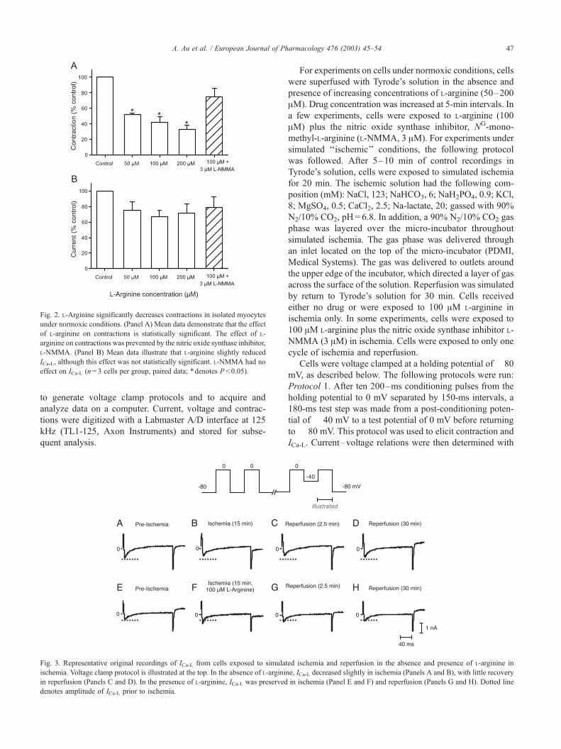
Fig. 2. L-Arginine significantly decreases contractions in isolated myocytes
under normoxic conditions. (Panel A) Mean data demonstrate that the effect
of L-arginine on contractions is statistically significant. The effect of L-
arginine on contractions was prevented by the nitric oxide synthase inhibitor,
L-NMMA. (Panel B) Mean data illustrate that L-arginine slightly reduced
ICa-L, although this effect was not statistically significant. L-NMMA had no
effect on ICa-L (n= 3 cells per group, paired data; *denotes P < 0.05).
A. Au et al. / European Journal of Pharmacology 476 (2003) 45–54 47
to generate voltage clamp protocols and to acquire and
analyze data on a computer. Current, voltage and contrac-
tions were digitized with a Labmaster A/D interface at 125
kHz (TL1-125, Axon Instruments) and stored for subse-
quent analysis.
Fig. 3. Representative original recordings of ICa-L from cells exposed to simulat
ischemia. Voltage clamp protocol is illustrated at the top. In the absence of L-arginin
in reperfusion (Panels C and D). In the presence of L-arginine, ICa-L was preserved
denotes amplitude of ICa-L prior to ischemia.
For experiments on cells under normoxic conditions, cells
were superfused with Tyrode’s solution in the absence and
presence of increasing concentrations of L-arginine (50–200
AM). Drug concentration was increased at 5-min intervals. In
a few experiments, cells were exposed to L-arginine (100
AM) plus the nitric oxide synthase inhibitor, NG-mono-
methyl-L-arginine (L-NMMA, 3 AM). For experiments under
simulated ‘‘ischemic’’ conditions, the following protocol
was followed. After 5–10 min of control recordings in
Tyrode’s solution, cells were exposed to simulated ischemia
for 20 min. The ischemic solution had the following com-
position (mM): NaCl, 123; NaHCO3, 6; NaH2PO4, 0.9; KCl,
8; MgSO4, 0.5; CaCl2, 2.5; Na-lactate, 20; gassed with 90%
N2/10% CO2, pH= 6.8. In addition, a 90% N2/10% CO2 gas
phase was layered over the micro-incubator throughout
simulated ischemia. The gas phase was delivered through
an inlet located on the top of the micro-incubator (PDMI,
Medical Systems). The gas was delivered to outlets around
the upper edge of the incubator, which directed a layer of gas
across the surface of the solution. Reperfusion was simulated
by return to Tyrode’s solution for 30 min. Cells received
either no drug or were exposed to 100 AM L-arginine in
ischemia only. In some experiments, cells were exposed to
100 AM L-arginine plus the nitric oxide synthase inhibitor L-
NMMA (3 AM) in ischemia. Cells were exposed to only one
cycle of ischemia and reperfusion.
Cells were voltage clamped at a holding potential of � 80
mV, as described below. The following protocols were run:
Protocol 1. After ten 200–ms conditioning pulses from the
holding potential to 0 mV separated by 150-ms intervals, a
180-ms test step was made from a post-conditioning poten-
tial of � 40 mV to a test potential of 0 mV before returning
to � 80 mV. This protocol was used to elicit contraction and
ICa-L. Current–voltage relations were then determined with
ed ischemia and reperfusion in the absence and presence of L-arginine in
e, ICa-L decreased slightly in ischemia (Panels A and B), with little recovery
in ischemia (Panel E and F) and reperfusion (Panels G and H). Dotted line
A. Au et al. / European Journal of Pharmacology 476 (2003) 45–5448
a similar protocol, but the test potential was changed in 10-
mV steps from � 40 to + 80 mV. This protocol was
repeated at 5-min intervals throughout the experiment.
Protocol 2. From the holding potential, sequential steps
were made to � 40 and + 20 mV for 300 ms each, followed
by a 900-ms hyperpolarization to test potentials between
� 100 and � 30 mV before returning to � 80 mV. This
protocol was used to detect the occurrence of the arrhyth-
mogenic transient inward current (ITI) and was repeated at
2.5-min intervals in early reperfusion.
2.3. Data measurement and analyses
Cell shortening was measured as the difference between
the peak contraction and the baseline preceding contraction.
Magnitude of ICa-L was measured as the difference between
peak inward current and net current 200 ms later on the
same test step. ITI incidence was counted as the number of
cells in which ITI was observed; incidence of aftercontrac-
tions was the number of cells in which aftercontractions
were observed. Data other than incidence are presented as
meansF S.E.M. Differences between means were assessed
with a Student’s t-test or with either one-or two-way
analysis of variance (SYSTAT v7.0.1, SPSS). The non-
parametric Chi-square test was used to determine whether
the incidences of ITI or aftercontractions were affected by
drug treatment in ischemia and reperfusion (Sigmastat,
Jandel Scientific). Differences were considered significant
for P < 0.05. No more than two cells from the same heart
were used for any experiment.
2.4. Chemicals
L-Arginine was purchased from the Sigma (St. Louis,
MO). L-NMMA was purchased from Calbiochem-Novabio-
chem (La Jolla, CA). Chemicals for buffer solutions were
purchased from BDH (Toronto, ON), Fisher Scientific
(Nepean, ON) and Sigma.
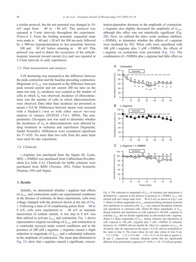
Fig. 4. The reduction in magnitude of ICa-L in ischemia and reperfusion is
abolished by L-arginine in the absence or presence of L-NMMA. ICa-L was
elicited with test voltage steps from � 40 to 0 mV, as shown in Figs 1 and
3. (Panel A) Mean magnitudes of ICa-L measured during simulated ischemia
and reperfusion in untreated cells. ICa-L was reduced throughout ischemia
and reperfusion in untreated cells. (Panel B) Mean magnitudes of ICa-Lduring ischemia and reperfusion in cells exposed to 100 AM L-arginine in
ischemia. ICa-L did not decline significantly in cells treated with L-arginine.
(Panel C) Mean magnitudes of ICa-L during ischemia and reperfusion in
cells exposed to 100 AM L-arginine plus 3 AM L-NMMA in ischemia.
Inclusion of L-NMMA did not abolish the effect of L-arginine on ICa-L. In
all panels, data are expressed as the meanF S.E.M. and are normalized to
the value at time 0. The mean values for raw data values at time 0 are
� 1.21F 0.06, � 1.15F 0.14 and � 1.03F 0.15 nA for data in panels A,
B and C, respectively. Asterisks illustrate points that are significantly
different from preischemic responses (P < 0.05; n= 10–19 cells per group).
3. Results
Initially, we determined whether L-arginine had effects
on ICa-L and contractions under our experimental conditions
in the absence of ischemia. In these experiments, cells were
voltage clamped with the protocol shown at the top of Fig.
1. Following a train of 10 conditioning pulses from � 80 to
0 mV, cells were repolarized to � 40 mV to maintain
inactivation of sodium current. A test step to 0 mV was
then utilized to activate ICa-L and contraction. Fig. 1 shows
representative original recordings of ICa-L and contraction in
a ventricular myocyte under control conditions and in the
presence of 200 AM L-arginine. L-Arginine caused a slight
reduction in magnitude of ICa-L and a substantial reduction
in the amplitude of contraction. The mean data illustrated in
Fig. 2A show that L-arginine caused a significant, concen-
tration-dependent decrease in the amplitude of contraction.
L-Arginine also slightly decreased the amplitude of ICa-L,
although this effect was not statistically significant (Fig.
2B). Next, we utilized the nitric oxide synthase inhibitor,
L-NMMA, to determine whether the effects of L-arginine
were mediated by NO. When cells were superfused with
100 AM L-arginine plus 3 AM L-NMMA, the effects of
L-arginine on contraction were prevented (Fig. 2A). The
combination of L-NMMA plus L-arginine had little effect on
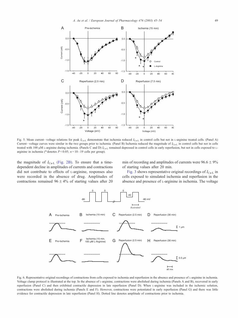
Fig. 5. Mean current–voltage relations for peak ICa-L demonstrate that ischemia reduced ICa-L in control cells but not in L-arginine treated cells. (Panel A)
Current–voltage curves were similar in the two groups prior to ischemia. (Panel B) Ischemia reduced the magnitude of ICa-L in control cells but not in cells
treated with 100 AM L-arginine during ischemia. (Panels C and D) ICa-L remained depressed in control cells in early reperfusion, but not in cells exposed to L-
arginine in ischemia (*denotes P < 0.05; n= 10–19 cells per group).
A. Au et al. / European Journal of Pharmacology 476 (2003) 45–54 49
the magnitude of ICa-L (Fig. 2B). To ensure that a time-
dependent decline in amplitudes of currents and contractions
did not contribute to effects of L-arginine, responses also
were recorded in the absence of drug. Amplitudes of
contractions remained 96F 4% of starting values after 20
Fig. 6. Representative original recordings of contractions from cells exposed to isch
Voltage clamp protocol is illustrated at the top. In the absence of L-arginine, contra
reperfusion (Panel C) and then exhibited contractile depression in late reperfu
contractions were abolished during ischemia (Panels E and F). However, contra
evidence for contractile depression in late reperfusion (Panel H). Dotted line den
min of recording and amplitudes of currents were 96.6F 9%
of starting values after 20 min.
Fig. 3 shows representative original recordings of ICa-L in
cells exposed to simulated ischemia and reperfusion in the
absence and presence of L-arginine in ischemia. The voltage
emia and reperfusion in the absence and presence of L-arginine in ischemia.
ctions were abolished during ischemia (Panels A and B), recovered in early
sion (Panel D). When L-arginine was included in the ischemic solution,
ctions were potentiated in early reperfusion (Panel G) and there was little
otes amplitude of contractions prior to ischemia.
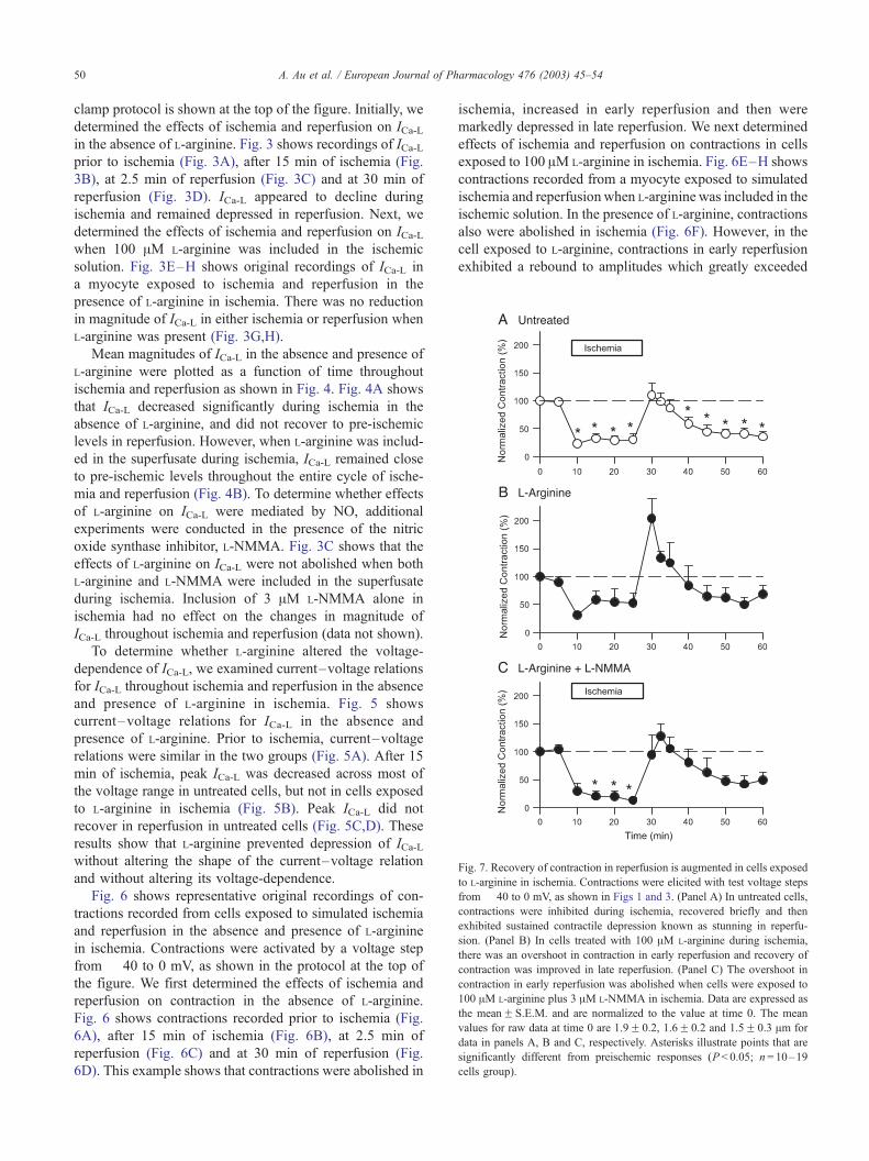
Fig. 7. Recovery of contraction in reperfusion is augmented in cells exposed
to L-arginine in ischemia. Contractions were elicited with test voltage steps
from � 40 to 0 mV, as shown in Figs 1 and 3. (Panel A) In untreated cells,
contractions were inhibited during ischemia, recovered briefly and then
exhibited sustained contractile depression known as stunning in reperfu-
sion. (Panel B) In cells treated with 100 AM L-arginine during ischemia,
there was an overshoot in contraction in early reperfusion and recovery of
contraction was improved in late reperfusion. (Panel C) The overshoot in
contraction in early reperfusion was abolished when cells were exposed to
100 AM L-arginine plus 3 AM L-NMMA in ischemia. Data are expressed as
the meanF S.E.M. and are normalized to the value at time 0. The mean
values for raw data at time 0 are 1.9F 0.2, 1.6F 0.2 and 1.5F 0.3 Am for
data in panels A, B and C, respectively. Asterisks illustrate points that are
significantly different from preischemic responses (P < 0.05; n= 10–19
cells group).
A. Au et al. / European Journal of Pharmacology 476 (2003) 45–5450
clamp protocol is shown at the top of the figure. Initially, we
determined the effects of ischemia and reperfusion on ICa-Lin the absence of L-arginine. Fig. 3 shows recordings of ICa-Lprior to ischemia (Fig. 3A), after 15 min of ischemia (Fig.
3B), at 2.5 min of reperfusion (Fig. 3C) and at 30 min of
reperfusion (Fig. 3D). ICa-L appeared to decline during
ischemia and remained depressed in reperfusion. Next, we
determined the effects of ischemia and reperfusion on ICa-Lwhen 100 AM L-arginine was included in the ischemic
solution. Fig. 3E–H shows original recordings of ICa-L in
a myocyte exposed to ischemia and reperfusion in the
presence of L-arginine in ischemia. There was no reduction
in magnitude of ICa-L in either ischemia or reperfusion when
L-arginine was present (Fig. 3G,H).
Mean magnitudes of ICa-L in the absence and presence of
L-arginine were plotted as a function of time throughout
ischemia and reperfusion as shown in Fig. 4. Fig. 4A shows
that ICa-L decreased significantly during ischemia in the
absence of L-arginine, and did not recover to pre-ischemic
levels in reperfusion. However, when L-arginine was includ-
ed in the superfusate during ischemia, ICa-L remained close
to pre-ischemic levels throughout the entire cycle of ische-
mia and reperfusion (Fig. 4B). To determine whether effects
of L-arginine on ICa-L were mediated by NO, additional
experiments were conducted in the presence of the nitric
oxide synthase inhibitor, L-NMMA. Fig. 3C shows that the
effects of L-arginine on ICa-L were not abolished when both
L-arginine and L-NMMA were included in the superfusate
during ischemia. Inclusion of 3 AM L-NMMA alone in
ischemia had no effect on the changes in magnitude of
ICa-L throughout ischemia and reperfusion (data not shown).
To determine whether L-arginine altered the voltage-
dependence of ICa-L, we examined current–voltage relations
for ICa-L throughout ischemia and reperfusion in the absence
and presence of L-arginine in ischemia. Fig. 5 shows
current–voltage relations for ICa-L in the absence and
presence of L-arginine. Prior to ischemia, current–voltage
relations were similar in the two groups (Fig. 5A). After 15
min of ischemia, peak ICa-L was decreased across most of
the voltage range in untreated cells, but not in cells exposed
to L-arginine in ischemia (Fig. 5B). Peak ICa-L did not
recover in reperfusion in untreated cells (Fig. 5C,D). These
results show that L-arginine prevented depression of ICa-Lwithout altering the shape of the current–voltage relation
and without altering its voltage-dependence.
Fig. 6 shows representative original recordings of con-
tractions recorded from cells exposed to simulated ischemia
and reperfusion in the absence and presence of L-arginine
in ischemia. Contractions were activated by a voltage step
from � 40 to 0 mV, as shown in the protocol at the top of
the figure. We first determined the effects of ischemia and
reperfusion on contraction in the absence of L-arginine.
Fig. 6 shows contractions recorded prior to ischemia (Fig.
6A), after 15 min of ischemia (Fig. 6B), at 2.5 min of
reperfusion (Fig. 6C) and at 30 min of reperfusion (Fig.
6D). This example shows that contractions were abolished in
ischemia, increased in early reperfusion and then were
markedly depressed in late reperfusion. We next determined
effects of ischemia and reperfusion on contractions in cells
exposed to 100 AM L-arginine in ischemia. Fig. 6E–H shows
contractions recorded from a myocyte exposed to simulated
ischemia and reperfusion when L-arginine was included in the
ischemic solution. In the presence of L-arginine, contractions
also were abolished in ischemia (Fig. 6F). However, in the
cell exposed to L-arginine, contractions in early reperfusion
exhibited a rebound to amplitudes which greatly exceeded
A. Au et al. / European Journal of Pharmacology 476 (2003) 45–54 51
pre-ischemic levels (Fig. 3G) and contractile depression later
in reperfusion was minimal (Fig. 3H).
The mean magnitude of contraction in the absence and
presence of L-arginine was plotted as a function of time
throughout ischemia and reperfusion as shown in Fig. 7.
Fig. 7A shows that, in the absence of L-arginine, contrac-
tions decreased in ischemia, recovered transiently in reper-
fusion and then exhibited contractile depression or stunning
(Louch et al., 2000, 2002). Fig. 7B shows that contractions
also were depressed in ischemia when L-arginine was includ-
ed in the superfusate. However, recovery of contractions in
early reperfusion was greatly potentiated in cells exposed to
L-arginine, and stunning was not significant in reperfusion
(Fig. 7B). To determine whether effects of L-arginine on
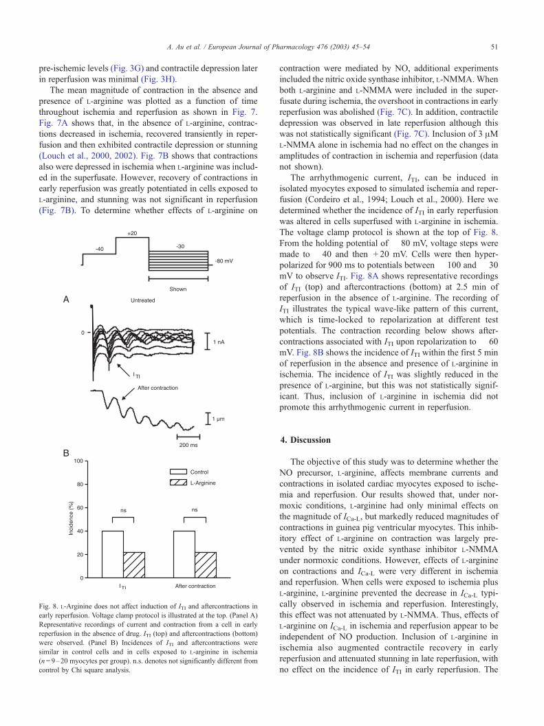
Fig. 8. L-Arginine does not affect induction of ITI and aftercontractions in
early reperfusion. Voltage clamp protocol is illustrated at the top. (Panel A)
Representative recordings of current and contraction from a cell in early
reperfusion in the absence of drug. ITI (top) and aftercontractions (bottom)
were observed. (Panel B) Incidences of ITI and aftercontractions were
similar in control cells and in cells exposed to L-arginine in ischemia
(n= 9–20 myocytes per group). n.s. denotes not significantly different from
control by Chi square analysis.
contraction were mediated by NO, additional experiments
included the nitric oxide synthase inhibitor, L-NMMA.When
both L-arginine and L-NMMA were included in the super-
fusate during ischemia, the overshoot in contractions in early
reperfusion was abolished (Fig. 7C). In addition, contractile
depression was observed in late reperfusion although this
was not statistically significant (Fig. 7C). Inclusion of 3 AML-NMMA alone in ischemia had no effect on the changes in
amplitudes of contraction in ischemia and reperfusion (data
not shown).
The arrhythmogenic current, ITI, can be induced in
isolated myocytes exposed to simulated ischemia and reper-
fusion (Cordeiro et al., 1994; Louch et al., 2000). Here we
determined whether the incidence of ITI in early reperfusion
was altered in cells superfused with L-arginine in ischemia.
The voltage clamp protocol is shown at the top of Fig. 8.
From the holding potential of � 80 mV, voltage steps were
made to � 40 and then + 20 mV. Cells were then hyper-
polarized for 900 ms to potentials between � 100 and � 30
mV to observe ITI. Fig. 8A shows representative recordings
of ITI (top) and aftercontractions (bottom) at 2.5 min of
reperfusion in the absence of L-arginine. The recording of
ITI illustrates the typical wave-like pattern of this current,
which is time-locked to repolarization at different test
potentials. The contraction recording below shows after-
contractions associated with ITI upon repolarization to � 60
mV. Fig. 8B shows the incidence of ITI within the first 5 min
of reperfusion in the absence and presence of L-arginine in
ischemia. The incidence of ITI was slightly reduced in the
presence of L-arginine, but this was not statistically signif-
icant. Thus, inclusion of L-arginine in ischemia did not
promote this arrhythmogenic current in reperfusion.
4. Discussion
The objective of this study was to determine whether the
NO precursor, L-arginine, affects membrane currents and
contractions in isolated cardiac myocytes exposed to ische-
mia and reperfusion. Our results showed that, under nor-
moxic conditions, L-arginine had only minimal effects on
the magnitude of ICa-L, but markedly reduced magnitudes of
contractions in guinea pig ventricular myocytes. This inhib-
itory effect of L-arginine on contraction was largely pre-
vented by the nitric oxide synthase inhibitor L-NMMA
under normoxic conditions. However, effects of L-arginine
on contractions and ICa-L were very different in ischemia
and reperfusion. When cells were exposed to ischemia plus
L-arginine, L-arginine prevented the decrease in ICa-L typi-
cally observed in ischemia and reperfusion. Interestingly,
this effect was not attenuated by L-NMMA. Thus, effects of
L-arginine on ICa-L in ischemia and reperfusion appear to be
independent of NO production. Inclusion of L-arginine in
ischemia also augmented contractile recovery in early
reperfusion and attenuated stunning in late reperfusion, with
no effect on the incidence of ITI in early reperfusion. The
A. Au et al. / European Journal of Pharmacology 476 (2003) 45–5452
effect of L-arginine on contractile recovery in early reperfu-
sion was reversed by L-NMMA. These observations suggest
that effects of L-arginine on recovery of contraction in
ischemia and reperfusion are likely mediated, at least in
part, by NO.
In the present study, we found that L-arginine caused
only a slight decrease in the magnitude of ICa-L and a large
decrease in amplitudes of contractions under normoxic
conditions. Further, we found that the effect of L-arginine
on contractions could be reversed by L-NMMA, which
suggests that this effect is mediated by NO production.
These observations are in general agreement with the
results of earlier studies of the effects of NO supplementa-
tion on isolated cardiac myocytes. Previous studies have
reported that NO inhibits cell shortening in isolated cardiac
myocytes (Brady et al., 1992, 1993; Kojda et al., 1996). In
addition, although previous studies have shown that NO
supplementation inhibits ICa-L when current is augmented
by ß-adrenoceptor stimulation and in cells from trans-
planted hearts undergoing rejection (Wahler and Dollinger,
1995; Ziolo et al., 2001a), it has little effect on ICa-L under
basal conditions (Wahler and Dollinger, 1995). Interesting-
ly, our results demonstrate that L-arginine markedly re-
duced amplitudes of contractions with only minimal
inhibition of ICa-L. ICa-L is believed to initiate contraction
in heart by release of Ca2 + from the sarcoplasmic reticulum
(Bers, 2001). As the effect of L-arginine on contractions is
much larger than its effect on ICa-L, our results suggest that
L-arginine might alter the coupling between ICa-L and
sarcoplasmic reticulum Ca2 + release, at least under nor-
moxic conditions. However, this remains to be demonstrat-
ed conclusively.
Previous studies have shown that the magnitude of ICa-Lis decreased in ischemia and reperfusion (Cordeiro et al.,
1994; Louch et al., 2000, 2002) and by metabolic inhibition
(Lederer et al., 1989). In the present study, we found that
inclusion of L-arginine in the ischemic solution prevented
this decrease in ICa-L in ischemia and reperfusion. This is
surprising, as L-arginine actually caused a slight decrease in
ICa-L under normoxic conditions. Thus, the results of this
study show that effects of L-arginine on ICa-L in ischemia
differ from effects under normoxic conditions.
It is unlikely that the effects of L-arginine in ischemia are
mediated by production of NO, as the actions of L-arginine
on ICa-L were not affected by the nitric oxide synthase
inhibitor L-NMMA. Therefore, our results demonstrate that
L-arginine has effects on ICa-L that are independent of NO
synthesis. The mechanism by which L-arginine prevents the
decline in ICa-L in ischemia and reperfusion is not known.
Cytosolic Ca2 + levels increase in ischemia (Nayler et al.,
1979; Tani and Neely, 1989) and elevated free intracellular
Ca2 + inhibits ICa-L (Hofer et al., 1997; Schuhmann et al.,
1997). Thus, it is possible that L-arginine prevents depres-
sion of ICa-L by reducing cytosolic Ca2 + levels in ischemia,
although there is no direct evidence for this at the present
time.
Earlier studies have shown that contractions associated
with activation of ICa-L rapidly decline in ischemia, recover
transiently in reperfusion and then exhibit contractile de-
pression later in reperfusion (Cordeiro et al., 1994; Louch et
al., 2000, 2002). Here we found that contractions also were
inhibited in ischemia in the presence of L-arginine. Howev-
er, recovery of contractions in early reperfusion was greatly
potentiated in cells exposed to L-arginine in ischemia. In
addition, L-arginine attenuated stunning in reperfusion.
Interestingly, under normoxic conditions, L-arginine signif-
icantly reduced the amplitudes of contractions. Thus, the
results of this study demonstrate that the effects of L-
arginine on contraction in ischemia cannot be predicted
from its effects in the absence of ischemia.
The mechanism by which L-arginine potentiates recov-
ery of contractile function in reperfusion is not yet known.
It is possible that L-arginine improves contractile recovery,
at least in part, because it prevents the decrease in ICa-L in
ischemia. If there is an increase in availability of ICa-L in
ischemia and reperfusion, there would be more current
available to trigger sarcoplasmic reticulum Ca2 + release
(Bers, 2001). However, effects of L-arginine on ICa-L were
independent of NO synthesis, while effects of L-arginine on
the overshoot in contraction are abolished by L-NMMA
and likely involve NO. It is possible that NO might
sensitize one or more components involved in excita-
tion–contraction coupling in the cell to augment contractile
recovery in reperfusion. Indeed, there is some evidence that
NO might affect sarcoplasmic reticulum Ca2 + release.
Studies have shown that NO can increase or decrease
sarcoplasmic reticulum Ca2 + release depending upon the
concentration of NO and the ambient level of ß-adreno-
ceptor activation (Zahradnikova et al., 1997; Ziolo et al.,
2001b). Thus, modulation of sarcoplasmic reticulum Ca2 +
release by NO also might augment recovery of contraction
in reperfusion.
In earlier studies with our cellular model of simulated
ischemia and reperfusion, we reported that both ITI and
aftercontractions can occur in early reperfusion (Cordeiro et
al., 1994; Louch et al., 2000). ITI is thought to arise as a
consequence of intracellular Ca2 +-overload (Lederer and
Tsien, 1976; Kass et al., 1978), which gives rise to the
oscillatory release of Ca2 + from the sarcoplasmic reticulum
and triggers cardiac arrhythmias (Ferrier et al., 1973). In the
present study, we found that L-arginine had little effect on
the incidence of ITI or aftercontractions observed in early
reperfusion. Indeed, the incidence of ITI was slightly de-
creased in the presence of L-arginine, despite increased
contractile activity in reperfusion. Thus, L-arginine pre-
serves contractile activity without promoting this mecha-
nism of arrhythmia. A number of previous studies have
demonstrated that NO has significant antiarrhythmic effects
in various models of ischemia and reperfusion (reviewed by
Pagliaro et al., 2001). The results of the present study
demonstrate that antiarrhythmic effects of NO in ischemia
and reperfusion are not likely due to inhibition of ITI.
A. Au et al. / European Journal of Pharmacology 476 (2003) 45–54 53
In summary, the results of this study demonstrate that
inclusion of L-arginine in ischemia prevents the decrease in
ICa-L typically observed in ischemia and reperfusion, and
improves recovery of contractile function in reperfusion.
However, L-arginine had no effect on the incidence of ITI in
early reperfusion. In contrast, under normoxic conditions, L-
arginine slightly inhibited ICa-L and markedly reduced
magnitudes of contractions in guinea pig ventricular myo-
cytes. Thus, an important finding in the present study is that
the effects of L-arginine under normoxic conditions are very
different from its effects in ischemia and reperfusion. In
addition, our results demonstrate that effects of L-arginine
on recovery of contraction in reperfusion appear to be
mediated, at least in part, by NO. However, L-arginine has
marked effects on ICa-L in ischemia and reperfusion that are
independent of NO synthesis.
Acknowledgements
The authors would like to thank Peter Nicholl and Cindy
Mapplebeck for their excellent technical assistance. This
work was supported by grants from the Heart and Stroke
Foundation of Nova Scotia and the Canadian Institutes of