The present study was designed to examine whether LA positivity is associ ated with an increased prevalence of retinopathy in pashytients with diabetes in India
(DM) 44 with type 1 and 41 with type 2 classified according to the National Diabetes Data Group criteria 14 were studied They inshy
cluded 64 males (27 with type 1 37 with type 2) and 21 females (1 7 with type 1 4 with type 2) Those with good glycaemic control (HbA I clt7) and no hypertension (gt 14090) hypertriglyceridaemia (fasting triglyceride gt 19 mmollL) or hypercholesterolaemia (toshytal cholesterol gt56 mmollL) were included Most patients were on multiple injections of subcutaneous insulin (Isophane insulin alone or mixed with soluble insulin 7030) with or without additional drugs such as metformin andlor glitazone All patients were nonshysmokers and had not taken any drugs known to affect hemostasis for at least 4 weeks beshyfore the study
Retinopathy was assessed by clinical exshyamination by a consultant ophthalmologist fundus photography and fluorescein anshygiography Diabetic retinopathy was categoshyrized into proliferative (PDR) and nonshyproliferative (NPDR) types as per ADA guidelines 15 Complete blood counts includshying platelet counts were performed in an automated hematology cell counter Plasma samples were examined for prothrombin time (PT) and activated pal1ial thromboplastin time (APTT) LA was diagnosed according to the criteria of the International Society of Hemostasis and Thrombosis16
mmollL buffered citrate solution in a 19 propol1ion After centrifugation at 4000g for 20 minutes at 4degC platelet-poor plasma was used immediately for coagulation assays The following coagulation tests were used for the laboratory detection of LA Kaolin-clotting time (KCT) confirmed by dilute Russels Vishyper Venom time (dRVVT) Normal values were obtained from the normal pooled plasma for the day of test All tests were pershyformed on the patients plasma and normal pool plasma Mixing studies were evaluated by the ratio of clotting time mixture clotting time of normal pool for KCT index and dRVVT index A positive result was indishycated by a ratio greater than 10 in both KCT and dRVVT
positive (14 1) LA positivity was higher in type I DM (20 5) than in type 2 (73) The results are shown in Table 1
age ranged from 75 to 38 years with a meshydian of 17 years Retinopathy was present in 9 cases (205 proliferative in 2 and nonshyproliferative in 7) LA was positive in 3 of them (333) Of the 35 without retinopathy 6 were positive for LA (1 71 ) Thus in type 1 DM LA was positive in 9 of the 44 cases (20 5) Conversely retinopathy was present in 33 3 of the LA -posiitivee patients but only in 187 of the LA-negative patients
age ranged from 22 years to 74 years with a median of 52 years Retinopathy was present in 31 (855 proliferative in 15 and non prol iferative in 16) Of these only 2 were
19 for
was
The
the
ting
Vishylues )led
pershy
mal ated
ting
and
ndishy
eCT
er m
3)
heir
meshy
]t in
oonshy3 of
tthy
type
ases
sent
but
heir
ith a
sent
non vere
LA Positivity in Diabetes with Retinopathy 35
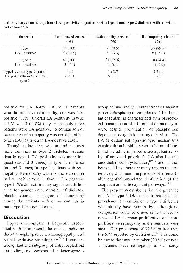
Table 1 Lupus anticoagulant (LA) positivity in patients with type 1 and type 2 diabetes with or withshyout retinopathy
Diabetics Total no of cases Retinopathy present Retinopathy absent () () ()
T)pe I 44 (100) 9 (205) 35 (795) LA -positive 9 (205) 3 (333 ) 6 (171)
Type 2 41 (100) 31 (756) 10 (244) LA -posit ive 3 (73) 2 (64) 1 (100)
Type I versus type 2 (ratio) 1 I 1 37 32 1 LA positivity in type 1 vs 29 1 52 1 17 I
type 2
positive for LA (64) Of the 10 patients
who did not have retinopathy one was LAshy
positive (10) Overall LA positivity in type
2 DM was 3 (73) only Since only three
patients were LA positive no comparison of
occurrence of retinopathy was considered beshy
tween LA-positive and LA-negative cases
Though retinopathy was around 4 times
more common in type 2 diabetes patients
than in type I LA positivity was more freshy
quent (around 3 times) in type I more so
(around 5 times) in type 1 patients with retishy
nopathy Retinopathy was also more common
in LA positive type 1 than in LA negative
type 1 We did not find any significant differshy
ence for gender ratio duration of diabetes
platelet counts or degree of retinopathy
among the patients with or without LA in
both type 1 and type 2 cases
Discussion Lupus anticoagulant is frequently associshy
ated with thromboembolic events including
diabetic nephropathy macroangiopathy and retinal occlusive vasculopathy7ll Lupus anshy
ticoagulant is a subgroup of antiphospholipid
antibodies and consists of a heterogenous
International Journal of Endocrinology and Metabolism
group of IgM and IgG autoantibodies against
prote inphospholipid complexes The lupus
anticoagulant is characterized by a paradoxishy
cal phenomenon of a thrombotic tendency in
vivo despite prolongation of phospholipid
dependent coagulation assays in vitro The
LA dependent pathophysiologic mechanisms
causing thrombophilia seem to be multifuncshy
tional including impaired anticoagulant activshy
ity of activated protein C LA also induces endothelial cell dysfunction8 917 and in diashy
betes mellitus there are many reports that exshy
tensively document the presence of a remarkshy
able endothelium-related dysfunction of the coagulant and anticoagulant pathways 1819
The present study shows that the presence
of LA in type I DM is not infrequent The
prevalence is even higher in type 1 diabetics
who already have retinopathy although no
comparison could be drawn as to the occurshy
rence of LA between proliferative and nonshy -proliferative retinopathy as the numbers were
small Our prevalence of 333 is less than
the 60 reported by Guisti et a1 13 This could
be due to the smaller number (205) of type
I patients with retinopathy in our study
36 S Dash et al
group while retinopathy was documented In
30 of cases in their study The precise role played by LA in retinopashy
thy in type 1 diabetes is speculative In view of increasing evidence of immune abnormalishyties in the pathogenesis of type 1 diabetes and the possible role of LA in endothelial dysfunction LA positivity should be consid-
References l Klein R Klein BE Moss SE Davis MD DeMets
DL The Wisconsin epidemiologic study of diashybetic retinopathy III Prevalence and risk of diashybetic retinopathy when age at diagnosis is 30 or more years Arch Ophthalmol 1984 Apr 1 02 (4)527-32
2 Klein R Klein BE Moss SE Epidemiology of proliferative diabetic retinopathy Diabetes Care 1992 Dec15 (12)1875-91
3 Fuller JH Keen H Jarrett RJ Omer T Meade TW Chakrabarti R et al Haemostatic variables associated with diabetes and its complications Br MedJ19790ct202(6196)964-6
4 Haefliger [0 Meyer P Flammer J Luscher TF The vascular endothelium as a regulator of the ocular circulation a new concept in ophthalmolshyogy Surv Ophthalmol 1994 Sep-Oct39 (2)123shy32
5 Kohner EM Patel V Rassam SM Role of blood flow and impaired autoregulation in the pathoshygenesis of diabetic retinopathy Diabetes 1995 Jun44 (6)603-7
6 Andreani D Malignant microangiopathy Diabeshytologia 1980 Mar 18 (3)255
7 Jones DB Wallace R Frier BM Vascular endoshythelial cell antibodies in diabetic patients Association with diabetic retinopathy Diabetes Care 1992 Apr15 (4)552-5
8 Lechner K Pabinger-Fasching 1 Lupus anticoagushylants and thrombosis A study of 25 cases and reshyview of the literature Haemostasis 198515 (4) 254-62
9 Triplett DA Obstetrical complications associated with antiphospholipid antibodies In Coulama BC Faulk WP Mclntyre lA ed Immunological Obstetrics New York WW Norton 1992 p 377shy403
10 Montehermoso A Cervera R Font J RamosshyCasals M Garcia-carrasco M Formiga F et al Association of anti phospholipid antibodies with retinal vascular disease in systemic lupus erytheshy
ered as an additional risk factor To clarifY and support this issue studies in larger multishyethnic patient groups are necessary not only to reach a better understanding of the pathoshygenesis of diabetic retinopathy but also to prevent its onset andor progression with apshypropriate treatment
matosus Semin Arthritis Rheum 1999 Apr28 (5)326-32
11 Galtier-Dereure F Biron C Vies M Bourgeois V Schved JF Bringer J Vascular complications of diabetes mellitus what role for phospholipidshybinding antibodies Lupus 19987 (7)469-74
12 Wiechens B Schroder JO Potzsch B Rochels R PrimalY anti phospholipid antibody syndrome and retinal occlusive vasculopathy Am J Ophthalmol 1997 Jun 123 (6)848-50
13 Giusti C Schiaffini R Bosco D Ciampalini P Pantaleo A Vingolo EM et al Lupus anticoagushylant positivity in insulin dependent diabetic pashytients an additional risk factor in the pathogenesis of diabetic retinopathy Br J Ophthalmol 2000 May84 (5)531-3
14 National Diabetes Data Group Classification of diabetes mellitus and other categories of glucose intolerance Diabetes 1970 28 1039-57
15 American Diabetes Association Diabetes Care 2000 23 (suppl)
16 Brandt JT Triplett DA Alving B Scharrer I Crishyteria for the diagnosis of lupus anticoagulants an update On behalf of the Subcommittee on Lupus AnticoagulantAntiphospholipid Antibody of the Scientific and Standardisation Committee of the ISTH Thromb Haemost 1995 Oct 74 (4) 1185shy90
17 Meroni PL Papa ND Beltrami B Tincani A Balestrieri G Krilis SA Modulation of endotheshylial cell function by antiphospholipid antibodies Lupus 1996 Oct5 (5)448-50
18 Dash S Alterations in haemorrheological and coshyagulation parameters in Diabetes In Dash RJ editor New Vistas in type 2 Diabetes 2000 p61shy72
19 Lorenzi M Cagliero E Pathobiology of endotheshylial and other vascular cells in diabetes mellitus Call for data Diabetes 1991 Jun40 (6)653-9
~
~
t
I
l
~
I (
t s
-(
1
International Journal of Endocrinology and Metabolism
C