Received 23rd February 2018,Accepted 26th May 2018
DOI: 10.1039/c8lc00202a
rsc.li/loc
Real-time observation of leukocyte–endotheliuminteractions in tissue-engineered blood vessel†
Z. Chen,‡ab M. Tang,‡a D. Huang,a W. Jiang,a M. Li, a H. Ji,a J. Park,a B. Xu,c
L. J. Atchison,d G. A. Truskeyd and K. W. Leong *a
Human cell-based 3D tissue constructs play an increasing role in disease modeling and drug screening. In-
flammation, atherosclerosis, and many autoimmune disorders involve the interactions between immune
cells and blood vessels. However, it has been difficult to image and model these interactions under realistic
conditions. In this study, we fabricated a perfusion and imaging chamber to allow the real-time visualiza-
tion of leukocyte perfusion, adhesion, and migration inside a tissue-engineered blood vessel (TEBV). We
monitored the elevated monocyte adhesion to the TEBV wall and transendothelial migration (TEM) as the
TEBV endothelium was activated by the inflammatory cytokine TNF-α. We demonstrated that treatment
with anti-TNF-α or an NF-kB signaling pathway inhibitor would attenuate the endothelium activation and
reduce the number of leukocyte adhesion (>74%) and TEM events (>87%) close to the control. As the first
demonstration of real-time imaging of dynamic cellular events within a TEBV, this work paves the way for
drug screening and disease modeling in TEBV-associated microphysiological systems.
Introduction
Many vascular diseases, such as inflammation, atherosclero-sis, and cancer metastasis,1–4 involve the interactions betweenleukocyte and the endothelium of blood vessel. The numberof leukocytes that aggregate near a lesion, adhering to, ortransmigrating into the blood vessel, can serve as an impor-tant biomarker for vascular disease or drug screening. How-ever, as blood vessels are opaque and mostly embeddedwithin the human body, conventional optical or confocalmicroscopy cannot be applied, except to the microvessels.Other imaging modalities such as computed tomography (CT)scan and magnetic resonance imaging (MRI), which has aspatial resolution at hundreds of microns, cannot observe atthe cellular level.5,6 In short, poor access to human blood ves-sels and the difficulty to image cellular interactions withinthem hinder the understanding of many vascular diseasemechanisms.
Advances in vascular tissue engineering offer an alterna-tive to the accessibility of human blood vessels. Human cell-based tissue-engineered blood vessels (TEBV) have been dem-onstrated as predictive in vitro models to facilitate drug toxic-ity/efficacy testing and disease modelling.7–11 Differing fromearlier generations of artificial blood vessels fabricated fromsynthetic polymers like expanded polyetrafluoroethylene(ePTFE) and polyethylene terephthalate fibre, these TEBV aremade with human cells and ECM proteins, thus can mimicmany native blood vessel functions such as vaso-contractility,constriction, dilation, and endothelium-dependent NO re-lease. Recent studies demonstrated the feasibility of studyingdrug responses in those TEBV.12–14 However, another impor-tant feature of blood vessel function – leukocyte–endotheliuminteractions – has not been characterized. Only limited stud-ies have tackled the leukocytes interaction with TEBV, relyingon “end-stage” observations in which TEBV are fixed and cutopen at the end of the experiment to characterize the loca-tion of the leukocytes on and in the TEBV.13,14 The most de-sired information – the dynamics of leukocyte adhesion andtransendothelial migration into the TEBV – has not beenavailable. There are two primary obstacles that currently hin-der the live imaging of TEBV, one is the lack of an optimallydesigned bioreactor that is compatible with microscopy re-garding working distance of the microscope objectives. Theother is the opaqueness of TEBV – it is not possible to lookdeep into the TEBV with common microscopic techniques.15
In this work, we address both problems by developing aperfusion and imaging chamber (PIC), that coupled with
deep-penetrating two-photon laser microscopy can observedynamically the interactions of leukocyte with TEBV. Wecaptured the leukocyte behaviour inside of TEBV under flowconditions and observed leukocyte–TEBV endothelium inter-actions such as leukocyte adhesion, migration, and trans-endothelial migration. We further investigated drug-inducedalteration in leukocyte behaviour. As a first demonstration ofreal-time imaging of leukocyte adhesion on TEBV, this workpaves the way for studying the mechanisms of immune cell-related vascular disease, offering a potential platform toscreen anti-inflammatory drugs.
Materials and methodsCell culture
hEPC: human umbilical cord blood derived endothelial pro-genitor cells (hEPCs) were isolated as previously described.12
In short, umbilical cord blood was obtained from the Caro-lina Cord Blood Bank. All patient identifiers were removedprior to receipt. The protocol for the collection and the usageof human blood in this study was approved by the Duke Uni-versity Institutional Review Board. Cells were maintained inEGM2 (Lonza) in a humidified 5% CO2 incubator. Mediumwas changed every day. After reaching 90% confluency, cellswere trypsinized (0.05% trypsin/EDTA; Gibco) and split 1 : 4.Cells were used at passages 6–9 for TEBV lumen perfusion toform the endothelium. UASMC: umbilical artery smooth mus-
cle cells (UASMCs) were purchased from Lonza and culturedin SmGM (Lonza) in a humidified 5% CO2 incubator. Afterreaching 90% confluence, cells were trypsinized (0.25% tryp-sin/EDTA; Gibco) and split 1 : 6. Cells were used at passages5–6 to make the collagen tubular-TEBV. Leukocytes: monocyte-like HL-60 cells were purchased from ATCC and cultured inRPMI-1640 with 15% FBS (Gibco) in a humidified 5% CO2 in-cubator. Cells were split 1 : 8 when reach 2 × 10⁶ cell per mL.
Fabrication of UASMC–TEBV in collagen scaffold
Collagen-TEBV: collagen TEBV was fabricated similarly aspreviously described.12 Briefly, 5 000 000 UASMCs wereembedded in 3 mL (1.5 mg mL−1) rat-tail collagen I (BD Bio-sciences) in a 3 cc syringe (BD) with a closed two-way Luer-Lok stopcock (Cole-Parmer) attached. After gelation for 30minutes, TEBVs were gently dehydrated to remove the waterand increase the collagen fibre density.12 The outer and innerdiameters of the TEBV were decided by the size of themandrel and the mold, which was 2.5 mm and 0.8 mm re-spectively. TEBVs were then cultured in DMEM with 1.1 g L−1
glucose L-glutamine and 110 mg L−1 sodium pyruvatesupplemented with 5% heat inactivated-fatal bovine serum(Gibco) in a rotating bioreactor for two days. The TEBV lu-men was then coated with EPCs (2 × 10⁶ cell per mL) byinjecting EPCs into the lumen of each TEBV and rotated at10 rotations per hour (rph) on a custom-made rotation
Fig. 1 Schematic illustration of immune cell interactions with endothelium in blood vessels during inflammation caused by stimuli such asexternal bacterial infection or internal cell necrosis. Human cell-based tissue-engineered blood vessel (TEBV) is proposed as a model for diseasemodelling or drug screening in the vascular system.
platform for 2 minutes at 37 °C to allow for cell adhesion.The TEBV was cultured for extra two weeks in a perfusion re-actor with perfusion at a speed of 2 mL min−1. Peristalticpump (Ismatec) was used to create flow through the TEBV.Flow circuit for TEBV was created using Tygon LMT-55Tubing (Ismatec) and reservoirs. The circuit contained 35 mLof flow media (DMEM with 1.1 g L−1 glucose L-glutamine, and110 mg L−1 sodium pyruvate supplemented with 3.3% heatinactivated-fatal bovine serum (Gibco), 1× NEAA, 100 UPen-Strep). Media was replaced every 2–3 days.
Imaging
DIC/phase/epi-fluorescence imaging: DIC and phase contrastimaging was taken using an Olympus IX81 inverted micro-scope with a 10× or 20× objective. Images were capturedusing an air-cooled SensiCam QE CCD camera (Cooke Corp.,Romulus, MI) driven by Metamorph (Molecular Devices/MetaImaging, Downingtown, PA).
Two-photon-/multi-photon confocal microscopy: confocalimages were taken using Nikon A1RMP laser scanning systemon an Eclipse Ti stand equipped with a 25×/NA1.1 ApoLWD water-immersion objective. For two-photon imaging,samples were excited at 890 nm with infrared light producedby a Chameleon Vision II tunable laser (Coherent, SantaClara, CA).
The second harmonic generated (SHG) signal from colla-gen fibres was detected with a non-descanned detector (NDD)using a 400–450 nm bandpass filter. The green and red fluo-rescence signal from cells was detected as well with NDDusing a 470–550 nm and a 570–640 nm bandpass filter,respectively.
Leukocyte adhesion and trans-endothelial (TEM) migrationassay
a. 2D-assay and DIC imaging. The confluent layer of endo-thelial progenitor cells (EPCs) was treated with or w/o TNF-alpha (200 U mL−1) for 4 hours before 1 mL of monocytes likeHL-60 was added to the confluent layer of the endothelial cellin 6-well plate at a concentration of 2 × 10⁵ cell per mL. Leu-kocytes were labelled with 1 μM CMFDA for 30 minutes. Afterincubation for 1 hour, images serials were taken for 5minutes at a 30 s-interval. Cells that underwent trans-endothelial migration (TEM) – which significantly changedtheir shape – were recorded. After that, the dish was gentlywashed with PBS w/ Ca2+ and Mg2+ three times before fixa-tion. Both phase images and fluorescent images were takento analyze the adherent and transmigrated leukocyte. Thenumber of cells attached to ECs was calculated by AnalyzeParticle with ImageJ software. The number of cells trans-migrated was manually counted as their morphology was
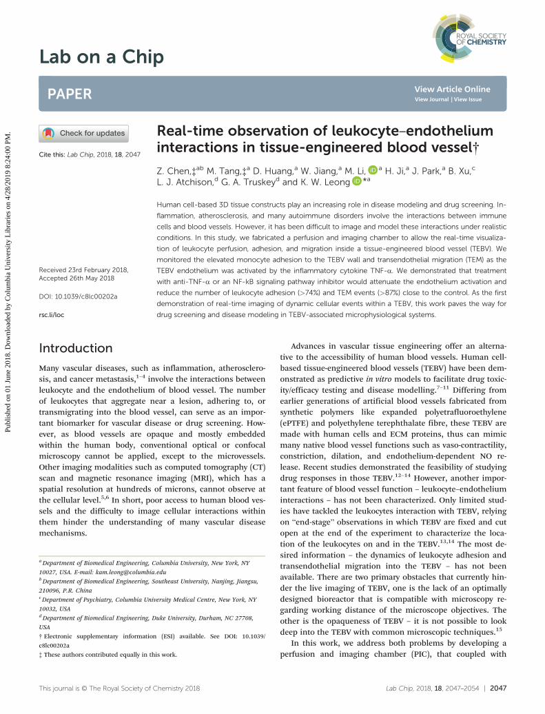
Fig. 2 Fabrication of human cell-based tissue-engineered blood vessel (TEBV). (A) Schematic illustration of the fabrication process of a TEBV. (B) ATEBV in perfusion inside of a bioreactor. (C) Image of TEBV with scanning electron microscopy (SEM). (D) Image of TEBV with H&E staining. (E) Im-munostaining of TEBV showing the blood vessel wall with an intact endothelium (red: CD31 as a marker for endothelial cells) and vascular smoothmuscle cells (green: smooth muscle actin as a marker for smooth muscle cells). Scale bars are indicated in each panel.
significantly different from other fluorescence-labelled mono-cytes. In each experiment, the cell number was counted andaveraged from three microscopic views. Results were fromfour independent experiments.
b. 3D-assay and two-photon imaging. Perfusion assay:monocyte-like HL-60 cells were labelled with red CMTPX(1 μM) for 45 minutes. Endothelialized TEBV was perfusedwith 200 U mL−1 TNF-α-containing DMEM medium for 4hours. The leukocyte (2 × 10⁵ cell per mL) was then perfusedin TEBV at a speed of 0.2 mL min−1 (inducing a shear stressof ∼0.5 dyne per cm2). Images/movies were taken using thetwo-photon confocal microscopy with a 25×/NA1.1 Apo LWDwater-immersion objective. The 2D side-view of the TEBV forthe perfusion of leukocyte, or the 3D-view of TEBV endothe-lium for the migration of leukocyte in TEBV was performed.Bi-directional imaging mode was used for leukocyte perfu-sion study to reach a scanning speed at 1 frame per s (FPS)(512 × 512 pixel). For leukocyte migration study, the stack offive images at different z position was imaged every minute(one z-stack per min) to achieve a better imaging quality(1024 × 1024 pixel). An 890 nm infrared laser was used to illu-minate the second harmonic signal of collagen and cellslabelled with green or red fluorescence signals. For the leuko-cyte perfusion study, the TEBV was perfused with leukocyte-containing medium continuously. For the leukocyte migra-tion study, the TEBV was perfused with HL-60 cell-containingDMEM medium for 20 minutes, and then followed with me-dium without the leukocytes. All images were taken in atime-lapse mode, and played back at different speed as indi-cated in the figure/movie captions. 3D images were 3D-reconstructed by Imaris 7.7. Results were calculated from 60leukocytes in three experiments.
Leukocyte adhesion and TEM in TEBV (End-pointImaging): monocyte-like HL-60 cells were labelled with redCMTPX (1 μM), while EPCs, UASMCs in TEBV were labelledgreen CMFDA (1 μM) for 45 minutes. TEBV were perfusedwith 200 U mL−1 TNF-α-containing DMEM medium for 4hours and then perfused with monocyte-like HL-60 cells at aconcentration of 5 × 10⁶ cell per mL for 1 hour. The TEBVswere then washed with medium, fixed with 4% PFA, and thecell nucleus labelled with DAPI and imaged with two-photonmicroscopy. The TEBV cells (in green) and leukocytes (in red)could be distinguished by their fluorescent colour and theirrelative 3D positions – on the lumen of TEBV or in the bloodvessel wall – were 3D-reconstructed by Imaris 7.7. The num-bers of cells that adhered on the lumen of TEBV or migratedinto the wall of TEBV were first filtered using Imaris 7.7 withsize and fluorescent intensity filters and then counted manu-ally. Data are averaged from three independent experiments.
Data analysis and statistics
Data was expressed as mean ± SEM. Statistically significantdifference was determined by either Student t-tests or one-way ANOVA and Tukey's post hoc test, statistical significancewas set at p < 0.05.
Results
The process of a leukocyte interaction with TEBV is shownschematically in Fig. 1. To perform the real-time imaging inTEBV, we first fabricated the TEBV and the perfusion andimaging chamber for TEBV imaging.
Fabrication of collagen scaffold-based tissue-engineeredblood vessel (TEBV)
Human umbilical artery smooth muscle cells (UASMC) weremixed within type I collagen hydrogel scaffold and embeddedin a tubular mould to construct the vessel wall of the TEBV(details in Materials and methods “TEBV-tubular method”).The cell-laden TEBVs were condensed and transferred to amicro-gravity bioreactor. After microgravity culture for twodays, TEBVs were mounted on to a perfusion bioreactor andendothelialized by perfusion of endothelial progenitor cells(EPC) through the TEBV lumen (details in Materials andmethods “TEBV-endothelialization”). The endothelializedTEBV continued to be perfused in the bioreactor for anothertwo weeks for maturation before further analysis. The overallprocess of fabrication and culture of TEBV is schematicallyshown in Fig. 2A. Fig. 2B to E are images of a TEBV in perfu-sion (2B), an SEM image of a TEBV (2C), an H&E staining of a
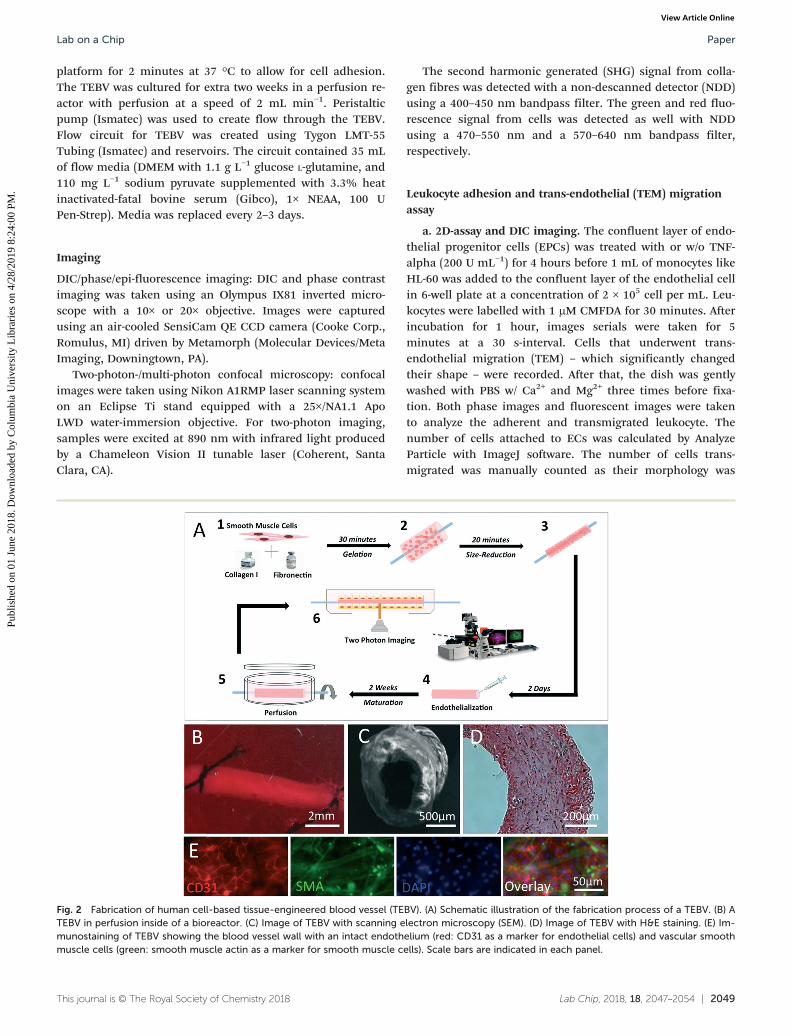
Fig. 3 The PIC chamber and the imaging system setup. (A)Components of a PIC chamber; numbers described in (C). (B)Illustration of a TEBV inside a PIC chamber. (C) the TEBV–PIC chamberin actual imaging condition. Major components are labelled: topobservation window (1), cover (2), chamber (3), TEBV perfusion inlet/outlet (4/4′), chamber medium perfusion inlet/outlet (5′), connector forair filter (6), bottom observation window (7), TEBV (8). (D) The setup ofPIC perfusion on the confocal microscope. Inserted figures are the keycomponents for TEBV imaging: PIC chamber (1), NIKON 25× LWD (2.0mm) objective (2), two-photon laser (3).
TEBV (2D) (Fig. S1†), and an immune-fluorescent staining im-age of a TEBV (2E), respectively.
Design and fabrication of perfusion and imaging chamber(PIC) for two-photon imaging of TEBV
To facilitate both perfusion and imaging for TEBV, we spe-cially designed the PIC chamber. The base of the chamberwas 3D-printed with an inner diameter (I.D.) of 50 mm formicroscopic stage mounting, and large observation windowson both the chamber lids and chamber bottom to facilitatetransmitted or inverted imaging. The central line of the inlet/outlet grips (in Fig. 3A) in PIC were designed to be 1.5 mmaway from the observation window to meet the microscopyobjective working distance specification (2 mm). Two otherside-channels in PIC are designed for chamber medium ex-change (details in Materials and methods). The main compo-nents of PIC are shown schematically in Fig. 3A and B andthe fabricated PIC with TEBV inside is shown in Fig. 3C. Thekey components in the integrated PIC-two photon imagingsetup are shown as inserts in Fig. 3D.
The main text of the article should appear here with head-ings as appropriate.
Leukocyte interaction with endothelial cells on 2D and in 3DPIC system
Activation of vascular endothelium is one of the initial andessential steps in vascular inflammatory responses.3 Activatedendothelial cells (ECs) express surface adhesion molecules torecruit leukocytes, e.g. monocytes and neutrophils, to the in-
flammatory sites, and induce their attachment, aggregation,migration, and transendothelial migration (TEM). We firstperformed a 2D assay in the conventional Transwell configu-ration. In the 2D model, the monocyte adhesion and transmi-gration on confluent endothelial cell layer was significantlyincreased for >70 folds after the endothelial cells were acti-vated by TNF-α (Fig. 4). The complete process of TEM, whichtypically transpire at a time scale of tens of seconds, wasrecorded (Movie S1†) and the montage of this process isshown in Fig. 4B. During TEM, the monocytes underwent(orange arrow) a significant morphological change, from aspherical shape (yellow arrow) to a flattened shape (redarrow), as they moved under the endothelial cells.
To observe the monocyte-endothelium interactions inTEBV, the PIC system was installed onto the Nikon A1R con-focal microscopy stage with a 25× L.W.D. objective (W.D. = 2mm, N.A. = 1.10) and using a two-photon imaging module.Monocytes were pre-labelled with red-fluorescent markerCMPTX. To minimize fluorescence background and possiblefluorescence bleed through, we did not stain the blood vesselwall but used the second harmonic mode to image the colla-gen fibres in the TEBV vessel wall. After screening, weselected wavelength of 890 nm as the best condition to ob-tain the second harmonic signals from collagen fibres(Fig. S2†). With this setup, we could directly image the leuko-cytes inside the TEBV with a perfusion flow rate of 0.2 mLmin−1. The imaging focal plane is schematically shown asFig. 5A upper left panel; and a full view of the microscopicimage is shown in Fig. 5A upper right panel. The blue signalindicates the dense collagen meshwork in TEBV wall, and the
Fig. 4 Imaging of monocyte adhesion and transmigration on 2D-cultured endothelial cells. (A) Time-lapse imaging of monocyte-like HL-60 cellsattached to TNF-α-activated endothelial progenitor cells (EPC). Arrows indicate monocytes attached to ECs. Yellow arrow: attached monocytes.Orange arrow: monocytes undergoing transendothelial migration. Red arrow: monocytes after transendothelial migration. Cell morphologieschanged after transmigration, as they were squeezed and trapped between ECs and the substrate. (B) A montage of monocytes undergoing trans-endothelial migration. The transmigration event happened during 100–160 s. (C) Comparison of the monocytes interaction with EPCs with or with-out TNF-α treatment. Adhesion: control (330 ± 133/cm2) vs. TNF-α (23 200 ± 1276/cm2); transmigration: control (not observed) vs. TNF-α (640 ±
332/cm2). In each experiment, the cell number was counted and averaged from 3 microscopic views. Data are shown as mean ± SEM from four in-dependent experiments. Significance was determined by Student t-test; * p < 0.05.
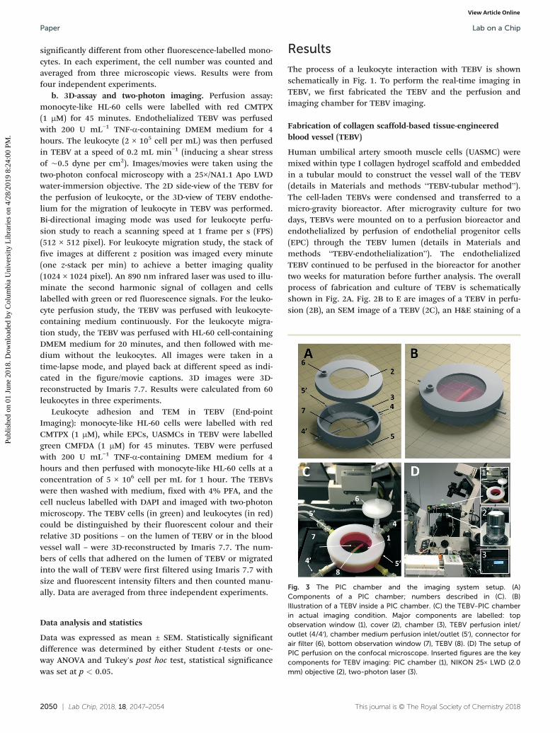
red signals are the stained monocytes. Monocytes in perfu-sion showed up as an elongated particle (green arrow) as theywere fast moving across the whole imaging plane during theimaging. Leukocytes that firmly attached onto the endothe-lium of TEBV showed as red dots (yellow arrow) (Fig. 5B;Movie S2†). In the movie, the monocyte highlighted by thearrow attached to the TEBV endothelium and graduallymoved out of the confocal focal plan, indicating a TEM event(Movie S2′0″–2′37″†); next, another monocyte (green arrow)approached and attached at the same region (Movie S2′48″†).The montage of the entire process was shown in Fig. 5C.
To investigate the leukocyte migration on the TEBV endo-thelium, we performed time-lapse multiple-layer Z-scanningat a higher resolution. The focal plane for imaging is sche-matically shown in Fig. 5B upper left panel; and a micro-scopic image is shown in Fig. 5B upper right panel. Wepre-labelled monocytes with the green-fluorescent marker(CMFDA) and SMCs/EPCs with the red-fluorescent marker(CMTPX). The second harmonic imaging was used to displaycollagen fibres in TEBV wall. Monocytes attached to the endo-thelium and migrated on its surface were imaged and dem-onstrated as a montage in Fig. 5A lower panel. Movie S3† wasa movie made from 3D-reconstructed image series to demon-strate the monocyte migration on the TEBV lumen. Themonocyte migration speed and directionality were analysedand demonstrated in Fig. 5C and the insert panel.
Inhibition of leukocyte–endothelium interaction in TEBV
To explore if this model can be used for future drug testing,we used two anti-inflammatory drugs, TNF-α-neutralizingantibody and Bay-11-7082 (an NF-kB signalling pathway in-hibitor), to attenuate the endothelium activation.16–18 Usingthe same cell-labelling scheme shown in Fig. 5, the monocyteadhesion or TEM in the TNF-α activated TEBV was imaged inFig. 6A and documented in 6B. We first confirmed in the 2Dmodel that both drugs could reduce the monocyte adhesionto the EC layer (Fig. S3†). We selected the anti-TNF-α anti-body at 10 nM, or Bay-11-7082 at 1 mM for the TEBV perfu-sion experiment. The anti-TNF-α significantly reduced themonocyte adhesion and TEM by 80.2% and 74.7%, (p < 0.05)respectively; similarly, addition of Bay-11-7082 to the perfu-sion reduced the monocyte adhesion and TEM by 87.3% and89.9% ( p < 0.05), respectively (Fig. 6B).
Discussion
The real-time observation of leukocyte behaviour in humanblood vessel, which would be valuable for drug screening anddisease modelling, has not been reported in the literaturedue to the difficulties of imaging such interactions. Althoughmany different bioreactors for TEBV have been developed inthe past, few of them are suitable for imaging. They aremostly made of Plexiglas with a thickness in millimetres,thus does not meet the working distance of most advancedmicroscopy objective.12–13 The opaqueness of TEBV is an-other challenge; the thick TEBV wall blocks the light penetra-tion and prevents the cells deep in the wall of the TEBV frombeing illuminated and distinguished. In this work, we spe-cially designed a PIC system to combine with two-photonmicroscopy to render the study of the TEBV lumen at the sin-gle cell level.
On one hand, this PIC imaging system can monitor the in-teractions between leukocytes and blood vessels under morebiomimetic conditions than those in existing flow chamber-based leukocyte-adhesion assays. Flow chambers usually haveendothelial cells cultured on glass or polystyrene, with a stiff-ness six orders of magnitude larger than that of physiological
Fig. 5 Real-time imaging of monocyte perfusion, adhesion andmigration inside TEBV. (A) Monocyte perfusion and adhesion in TEBV.Upper left panel, a schematic illustration of the imaging focal-plane.Upper right panel, a full view of monocyte perfusion in TEBV. Yellowarrow: a monocyte adhered to endothelium during its transmigration.Green arrow: a new monocyte attachment event. White arrow:monocyte in perfusion. Lower panel, montage of the monocyteattachment and transmigration events (Movie S2†). (B) Monocytemigration on the TEBV endothelium. Upper left panel, a schematicillustration of the imaging focal-plane. Upper-right panel, a full view ofmonocyte migration on TEBV endothelium. Lower panel, montage ofleukocyte migration on TEBV endothelium (Movie S3†). (C) Quantifica-tion of leukocyte migration speed distribution on TEBV endotheliumand their migration directionality with rose diagram (inserted panel).The flow direction is set as degree zero. Results were calculated from60 leukocytes in three experiments. Data are shown as mean ± SEM.
blood vessels. This difference in substrate stiffness could af-fect endothelial cell functions, leading to correspondingchanges in leukocyte behaviour. Moreover, once transmigra-tion occurs, the leukocytes will be trapped between the ECsand the solid chamber substrate and became flat (Fig. 4),whereas leukocytes in real physiological conditions would in-filtrate into tissue (Fig. 6). 2D-flow chamber studies also lack3D structured ECM, and the possible interactions amongECs, SMCs, and other cells. As a result, our model provides amore biomimetic microphysiological environment. These bio-mimetic features of the TEBV–PIC may be valuable for futureuse of TEBV as disease models, such as an inflammationmodel, or atherosclerosis model, which involves crosstalkamong SMCs, ECs, leukocytes, and foam cells.2,19
On the other hand, the possibility of monitoring leukocyteperfusion (Fig. 5A), adhesion, migration along the lumen,and transmigration (Fig. 5B, C and 6) in an TEBV offers re-searchers a tool to quantitatively measure and compare cellnumber in different inflammation conditions and/or with dif-ferent drug treatment (Fig. 6). This quantitative analysis ofleukocyte number and behaviour has many important appli-cations. Firstly, it could serve as a measure of biological sig-nals that could not previously be easily detected. For in-stance, endothelial activation can be classified into differentstages with different phenotypes at each stage.16–18 The adhe-sion and transmigration of leukocytes on ECs indicate stage-II EC activation, which involves EC hypertrophy as well as ex-pression and release of leukocyte adhesion proteins such asE-selectin, ICAM-1 and VCAM-1. The leukocyte number couldserve as a marker to indicate the level of adhesive protein ex-pression on EC. Secondly, the quantitative analysis of leuko-
cyte number and behaviour could serve as an indicator/marker for drug efficacy. Anti-inflammatory drugs play an im-portant role in treatment for immune system-related diseasessuch as arthritis, systemic lupus erythematous, and athero-sclerosis.18 These drug treatment efficacies can now be evalu-ated quantitatively in vitro, as we demonstrated in Fig. 6,demonstrated as reducing the monocytes adhesion and trans-migration of leukocyte. The further study on the measure-ment of the number of attached and transmigrated leuko-cytes over time may provide important information on thepharmacodynamics of a drug (or a drug candidate) – when itstarts to take effect as well as when it reaches maximum ef-fect in blood vessels.
Conclusions
We have developed a TEBV perfusion and imaging system tomonitor real-time the interactions of leukocytes with the ves-sel wall. This study paves the way for using TEBV as an im-portant platform for modelling immune system-related vascu-lar diseases and for screening drugs that can regulate theinflammation cascade in the vascular system.
Conflicts of interest
There are no conflicts to declare.
Acknowledgements
This research was supported by NIH UH3TR000505 (NCATSand the NIH Common Fund for the Microphysiological Sys-tems Initiative) and UG3TR002142 (NCATS and NIAMS).
Fig. 6 Effect of inflammatory cytokine and anti-inflammatory drugs on leukocyte–endothelium interactions. (A) A side view of the 3D-reconstructed two-photon confocal images in which monocytes attached to or transmigrated into activated TEBV endothelium in a TNF-α acti-vated TEBV (perfused with medium containing TNF-α, 200 U mL−1, 4 h). B) Quantification of cell attachment and transmigration in TEBV with dif-ferent treatments. TEBV was treated with i) null, ii) 200 U mL−1 TNF-α, iii) 200 U mL−1 TNF-α +20 ng mL−1 TNF-α neutralizing antibody, and iv) 200U mL−1 TNF-α + 1 μM Bay117082. N = 4 for each group. Data are shown as mean ± SEM. Significance was determined by one-way ANOVA andTukey's post hoc test; * p < 0.05 vs. number of cells adhered or transmigrated in control group, respectively; # p < 0.05 vs. number of cells ad-hered or transmigrated in TNF-α treated group.
1 D. Vestweber, F. Wessel and A. F. Nottebaum, Semin.Immunopathol., 2014, 36, 177.
2 I. Tabas, G. García-Cardeña and G. K. Owens, J. Cell Biol.,2015, 209, 13.
3 C. M. Steyers and F. J. Miller, Int. J. Mol. Sci., 2014, 15,11324.
4 J. Cedervall, A. Dimberg and A.-K. Olsson, MediatorsInflammation, 2015, 418290.
5 H. Machida, I. Tanaka, R. Fukui, Y. Shen, T. Ishikawa, E.Tate and E. Ueno, Radiographics, 2015, 35, 991.
6 D. Stucht, K. A. Danishad, P. Schulze, F. Godenschweger, M.Zaitsev and O. Speck, PLoS One, 2015, 10, e0133921.
7 W. J. Zhang, W. Liu, L. Cui and Y. Cao, J. Cell. Mol. Med.,2007, 11, 945.
8 C. E. Fernandez, H. E. Achneck, W. M. Reichert and G. A.Truskey, Curr. Opin. Chem. Eng., 2014, 3, 83.
9 D. G. Seifu, A. Purnama, K. Mequanint and D. Mantovani,Nat. Rev. Cardiol., 2013, 10, 410.
10 N. L'Heureux, S. Pâquet, R. Labbé, L. Germain and F. A.Auger, FASEB J., 1998, 12, 47.
11 L. Gui, B. C. Dash, J. Luo, L. Qin, L. Zhao, K. Yamamoto, T.Hashimoto, H. Wu, A. Dardik, G. Tellides, L. E. Niklasonand Y. Qyang, Biomaterials, 2016, 102, 120.
12 H. Ji, L. Atchison, Z. Chen, S. Chakraborty, Y. Jung, G. A.Truskey, N. Christoforou and K. W. Leong, Biomaterials,2016, 85, 180.
13 Y. Jung, H. Ji, Z. Chen, H. Fai Chan, L. Atchison, B.Klitzman, G. Truskey and K. W. Leong, Sci. Rep., 2015, 5,15116.
14 J. Robert, B. Weber, L. Frese, M. Y. Emmert, D. Schmidt, A.von Eckardstein, L. Rohrer and S. P. Hoerstrup, PLoS One,2013, 8, e79821.
15 J. Zhang, A. F. DeFelice, J. P. Hanig and T. Colatsky, Toxicol.Pathol., 2010, 38, 856.
16 F. H. Bach, W. W. Hancock and C. Ferran, Immunol. Today,1997, 18, 483.
17 J. Mestas and K. Ley, Trends Cardiovasc. Med., 2008, 18, 228.18 G. Nakazawa, S. K. Yazdani, A. V. Finn, M. Vorpahl, F. D.
Kolodgie and R. Virmani, J. Am. Coll. Cardiol., 2010, 55,1679.
19 L. A. Micol, M. Ananta, E.-M. Engelhardt, V. C. Mudera, R. A.Brown, J. A. Hubbell and P. Frey, Biomaterials, 2011, 32, 1543.