29
Lab.11 molecular detection for viruses By Assist. Lect. Mays Jassim

Lab.11 molecular detection for viruses
By
Assist. Lect. Mays Jassim

Beginning in 1990s, PCR based methods have proven to offer a better diagnostic alternative than T.C and serology based methods. Such molecular techniques not only improve laboratories’ sensitivity for detection of the microorganism, but are also more suitable, quicker and of course, more economic comparably with other assays.
20 cycles of PCR give million of PCR products
(genes)
PCR is DNA replication in a
test tube

Types of PCR 1. Conventional PCR. 2. Real time PCR (RT-qPCR) 3. Advanced PCR: A- reverse transcription PCR (RT-PCR). B- multiplex PCR. C- Nested PCR.
Application of PCR 1- Detection of mutations. 2- Detection of infection. 3- Forensics sciences. 4- Paternity testing. 5- Research applications: (DNA sequencing, molecular cloning, Genome mapping and Evolutionary analysis, etc.). PCR Problems: 1. Contamination. 2. Theoretically one molecule can amplify but practically it needs more
molecules (>10-20 molecules) to get the best results.

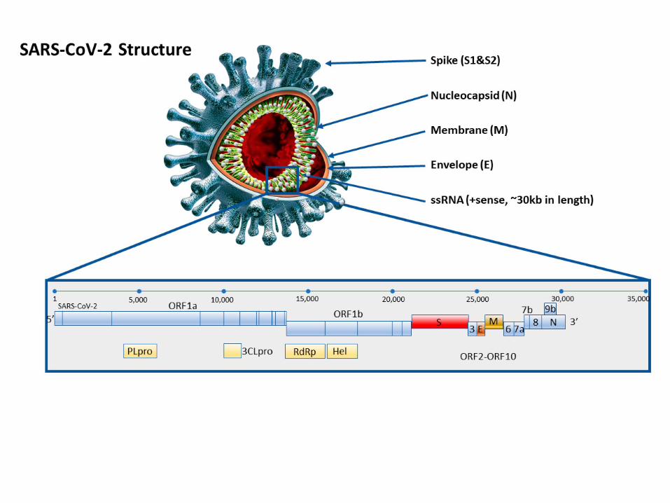
• All the viruses can be detected by using PCR method.
• First, the virus is extracted from the sample (blood, saliva, sputum, CSF, stool…etc).
• Second, the DNA or RNA of the virus then isolated from the virus to be used as a template for the next PCR procedure.
• Single or multiple genes are selected from the viral genome to be
the target that would be diagnosed by the PCR. so that two set of primers (forward and reverse) are designed to be complementary for the target gene of the virus.

Conventional PCR (Used to amplify DNA and RNA molecules)

A- Conventional PCR for DNA amplification:
PCR components of DNA
1- DNA template; (Extracted from cells or tissues of the sample).
2- Primers; (forward & reverse) .
3- Nucleotides; Deoxynucleotide triphosphate (dNTP): Mixture of dATP, dTTP, dCTP and dGTP.
4- Taq DAN Polymerase; enzyme for DNA amplification.
5- Buffer; Tris HCl, KCl or MgCl2.
Optimization of [Mg2+] is necessary, because its cofactor of Taq DNA polymerase.


(DNA- PCR)

Temperature Time Period The principle
95℃ 1 min. Initial denaturation
95℃ 30- 45 sec denaturation
50- 60 ℃ * 30- 45 sec Annealing of primer
72℃ 30 sec- 1 min. Initial extension of
nucleotides
72℃ 7 min Final extension of the
nucleotide
4℃ (hold) 24 hr Hold the product
Thermal Cycler program for DNA- PCR
30- 40 cycle


B- Conventional PCR for RNA amplification:
PCR components of RNA
The same components of DNA- PCR are used except;
1- RNA is used as a template which is extracted from the patient sample.
2- two enzymes are used in the procedure instead of one;
• Reverse transcriptase (RT) enzyme (used to convert RNA into complementary DNA (c DNA)).
• taq DNA polymerase (Taq DNAP) , that used to amplify cDNA .
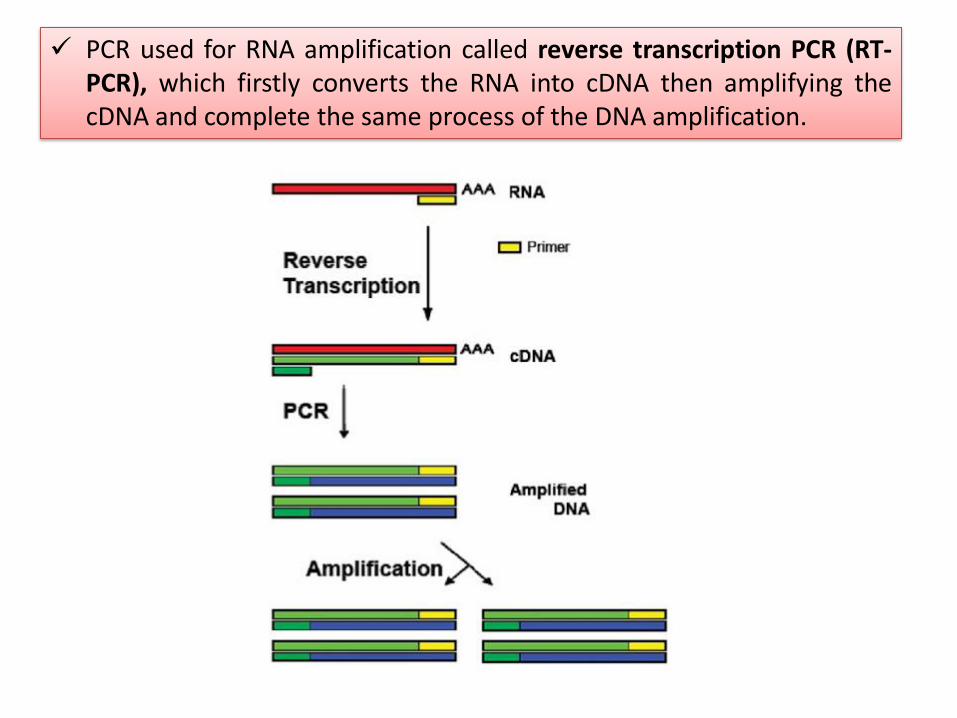
PCR used for RNA amplification called reverse transcription PCR (RT- PCR), which firstly converts the RNA into cDNA then amplifying the cDNA and complete the same process of the DNA amplification.

Temperature Time
Period
The principle
50℃ 30 min For reverse transcription
95℃ 15 min to stop RT enzyme reaction
95℃ 30 sec denaturation
50- 60 ℃ * 45 sec Annealing of primer
72℃ 45 sec Initial extension of nucleotides
72℃ 7 min Final extension of the nucleotide
4℃ (hold) 24 hr Hold the product
35- 40 cycle
Thermal Cycler program for RNA- PCR
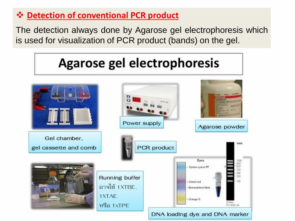
Detection of conventional PCR product
The detection always done by Agarose gel electrophoresis which
is used for visualization of PCR product (bands) on the gel.


Real-Time quantitative Polymerase Chain Reaction (RT-qPCR)

• Real Time
▫ signals (generally fluorescent) are monitored as they are generated and are tracked throughout the program.
• Quantitative
▫ Quantitatively measures the amplification of template
• Polymerase Chain Reaction
▫ Method dependent on thermo cycling and enzymes allowing for amplification of small starting material of DNA
Real-Time quantitative Polymerase Chain Reactoin (RT-qPCR)
From all the available quantification techniques, RT-qPCR has the highest;
sensitivity, reproducibility, simplicity and dynamic range.

the PCR components of DNA or RNA amplification for real time PCR are the same for the conventional PCR except that in RT-qPCR the detection process depends on signals need to be added to the reaction tube.
two types of signals are used:

A- TaqMan probe method

B- Cyber green method

Thermal Cycler program for real time- PCR


Detection of the real time PCR product (interpretation of the results)

Detection of the real time PCR product (interpretation of the results)
• The increase in the fluorescent signal intensity of Taqman probe or cyber green dye is captured by the Sequence Detection instrument and displayed as an Amplification Plot in the software screen.
• The amount of fluorescent signal increase is proportional to the amount
of product being produced for a given sample.

Positive samples
Negative samples

In cyber green method, there is a chance for getting false positive results due a non specific binding of the fluorescent dye with the double stranded DNA molecules. So that there is a specific melt temperature (tm) for each primer (The temperature at which 50% of DNA is denatured is known as the melting temperature). A melting curve chart shows the change in fluorescence observed when double-stranded DNA (dsDNA) with incorporated dye molecules dissociates, or “melts” into single-stranded DNA (ssDNA) as the temperature of the reaction is raised..

Thank you


