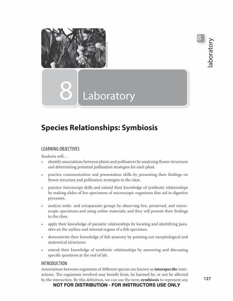
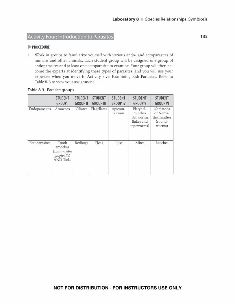
127 laboratory 8 8 Laboratory Robert Johnson/Shutterstock.com LEARNING OBJECTIVES Students will…. • identify associations between plants and pollinators by analyzing flower structures and determining potential pollination strategies for each plant. • practice communication and presentation skills by presenting their findings on flower structure and pollination strategies to the class. • practice microscopy skills and extend their knowledge of symbiotic relationships by making slides of live specimens of microscopic organisms that aid in digestive processes. • analyze endo- and ectoparasite groups by observing live, preserved, and micro- scopic specimens and using online materials; and they will present their findings to the class. • apply their knowledge of parasitic relationships by locating and identifying para- sites on the surface and internal organs of a fish specimen. • demonstrate their knowledge of fish anatomy by pointing out morphological and anatomical structures. • extend their knowledge of symbiotic relationships by answering and discussing specific questions at the end of lab. INTRODUCTION Associations between organisms of different species are known as interspecific inter- actions. e organisms involved may benefit from, be harmed by, or not be affected by the interaction. By this definition, we can use the term symbiosis to represent any Species Relationships: Symbiosis NOT FOR DISTRIBUTION - FOR INSTRUCTORS USE ONLY
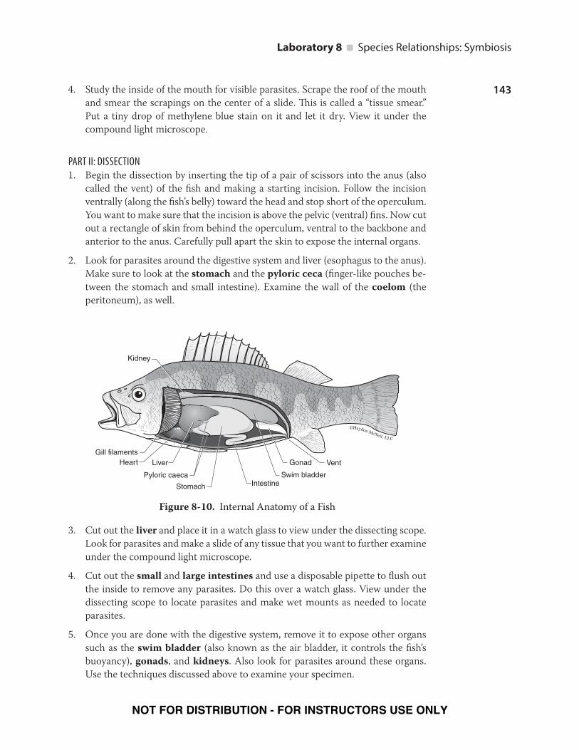
Transcript
127
lab
ora
tory
8
8 Laboratory R
ober
t Jo
hnso
n/S
hutt
erst
ock
.com
LEARNING OBJECTIVES
Students will….
• identify associations between plants and pollinators by analyzing flower structures
and determining potential pollination strategies for each plant.
• practice communication and presentation skills by presenting their findings on
flower structure and pollination strategies to the class.
• practice microscopy skills and extend their knowledge of symbiotic relationships
by making slides of live specimens of microscopic organisms that aid in digestive
processes.
• analyze endo- and ectoparasite groups by observing live, preserved, and micro-
scopic specimens and using online materials; and they will present their findings
to the class.
• apply their knowledge of parasitic relationships by locating and identifying para-
sites on the surface and internal organs of a fish specimen.
• demonstrate their knowledge of fish anatomy by pointing out morphological and
anatomical structures.
• extend their knowledge of symbiotic relationships by answering and discussing
specific questions at the end of lab.
INTRODUCTIONAssociations between organisms of different species are known as interspecific inter-
actions. The organisms involved may benefit from, be harmed by, or not be affected
by the interaction. By this definition, we can use the term symbiosis to represent any
Species Relationships: Symbiosis
NOT FOR DISTRIBUTION - FOR INSTRUCTORS USE ONLY
128
Laboratory 8 Species Relationships: Symbiosis
association between organisms, excluding interactions between members of the
same species, or intraspecific interactions. Symbiotic relationships exist between all
types of organisms including bacteria, protozoans, fungi, plants, and animals.
Various types of symbioses, whether beneficial or harmful, are described by the terms
mutualism, commensalism, and parasitism. In these relationships, we refer to the
symbiont as the organism that lives inside (endoparasites) or on (ectoparasites)
another organism, the host. In symbioses where the organisms interact with each
other, either living inside or on the other, both organisms are termed symbionts.
In mutualistic relationships, both partners benefit equally. Most commonly, organ-
isms enable the acquisition of nutrients for one another. An example of a mutualistic
relationship is the one between Aiptasia pallida, a small sea anemone found in the
Caribbean and along the east coast of the United States, and a dinoflagellate algae.
The dinoflagellate is called an endosymbiont, because it lives inside the sea anemone,
its host. The sea anemone receives oxygen and photosynthetic products from the
dinoflagellate, whereas the dinoflagellate receives protection and molecules for pho-
tosynthesis from the sea anemone.
The protozoans found in the stomach of herbivores, which help the animals digest
food, receiving nutrients in the process, represent another mutualistic relationship.
Protozoans also inhabit the gut of termites, helping them digest wood material. You
will have the opportunity to observe these protozoans when you complete the nutri-
tion lab. Mutualistic relationships also occur between plants and fungi. Fungi called
Mycorrhizae form an association with the roots of a plant, in which they help plants
to extract nutrients from nutrient-poor soils and in exchange receive organic com-
pounds from the plant’s photosynthetic processes. Another important symbiotic
relationship involves a fungi and a photosynthetic organism like an algae, which un-
dergoes a remarkable change during their association, resulting in a new entity called
a lichen.
An association in which the symbiont benefits while the host organism is neither
harmed nor benefited is called commensalism. The relationship between the tube
worm Chaetopterus and pea crabs is an example of a commensalistic relationship.
In this relationship, the worm shares its tube-like dwelling with a crab. The crab gets
protection from the tube and receives food and oxygen from the water that passes
into the tube. The shark and sucker fish Echeneis is another example of commensal-
ism. The sucker fish rides along with the shark by sticking to its underside with a
modified dorsal fin shaped like a suction disc. Close proximity to the shark allows the
sucker fish to scavenge bits of food left over from the shark’s meal.
Parasitism is a symbiosis in which the symbiont benefits at the expense of the host.
As in most symbiotic relationships, the driving force behind parasitic associations
is usually food/nutrients, since the parasite obtains its food from the host. Parasitic
relationships affect the host to varying degrees. Some parasites are so patheno-
genic (disease-causing) that they cause symptoms in the host almost immediately
after infection. In these cases, the host may die, but most parasites do not kill their
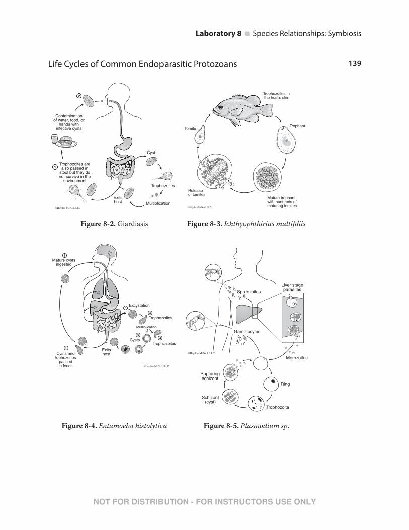
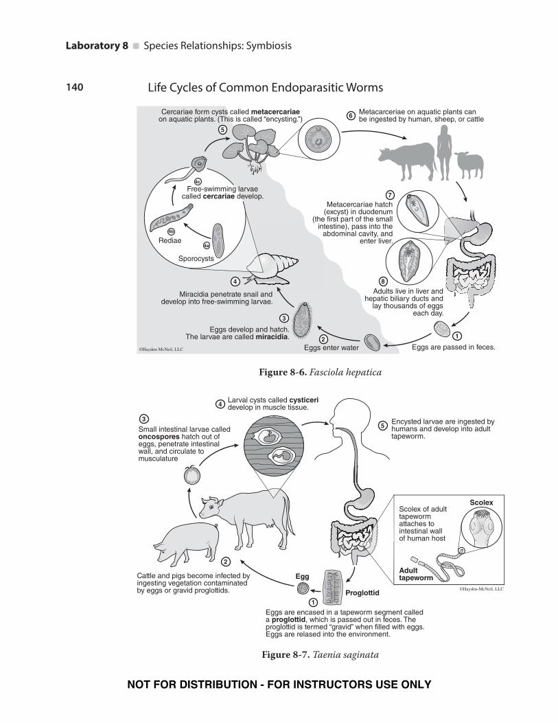
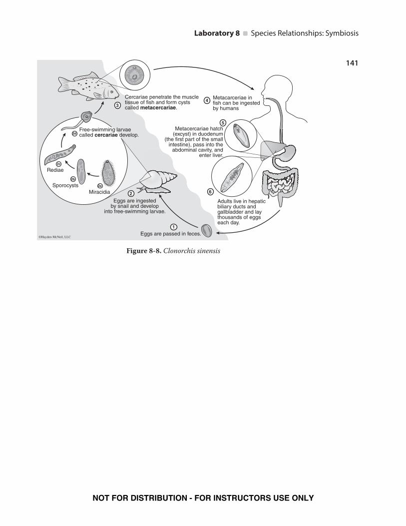
host until they have reproduced and completed their life cycles (see life cycle Figures
8-2 through 8-8). Some parasites need more than one host. Hosts are then either
NOT FOR DISTRIBUTION - FOR INSTRUCTORS USE ONLY
129
Laboratory 8 Species Relationships: Symbiosis
intermediate or final. You will have a chance to observe some of these parasites in the
laboratory. Hosts can also serve as reservoirs for a parasite (a breeding ground and
source of infection for another host) without showing signs of infection themselves,
or they can be vectors—carriers of the parasite to a final host. Plants can also be
parasitic. There are thousands of parasitic plant species, ranging from trees to small,
herbaceous plants. The best-known group of parasitic plants is mistletoe. There are
about 800 species, most occurring in the tropics and subtropics (Paracer and Ah-
madjian 2000). Mistletoe parasitize tree branches, but the giant mistletoe, Nuylsia,
forms a tree that grows as high as 10 meters and parasitizes roots of nearby grasses
and plants.
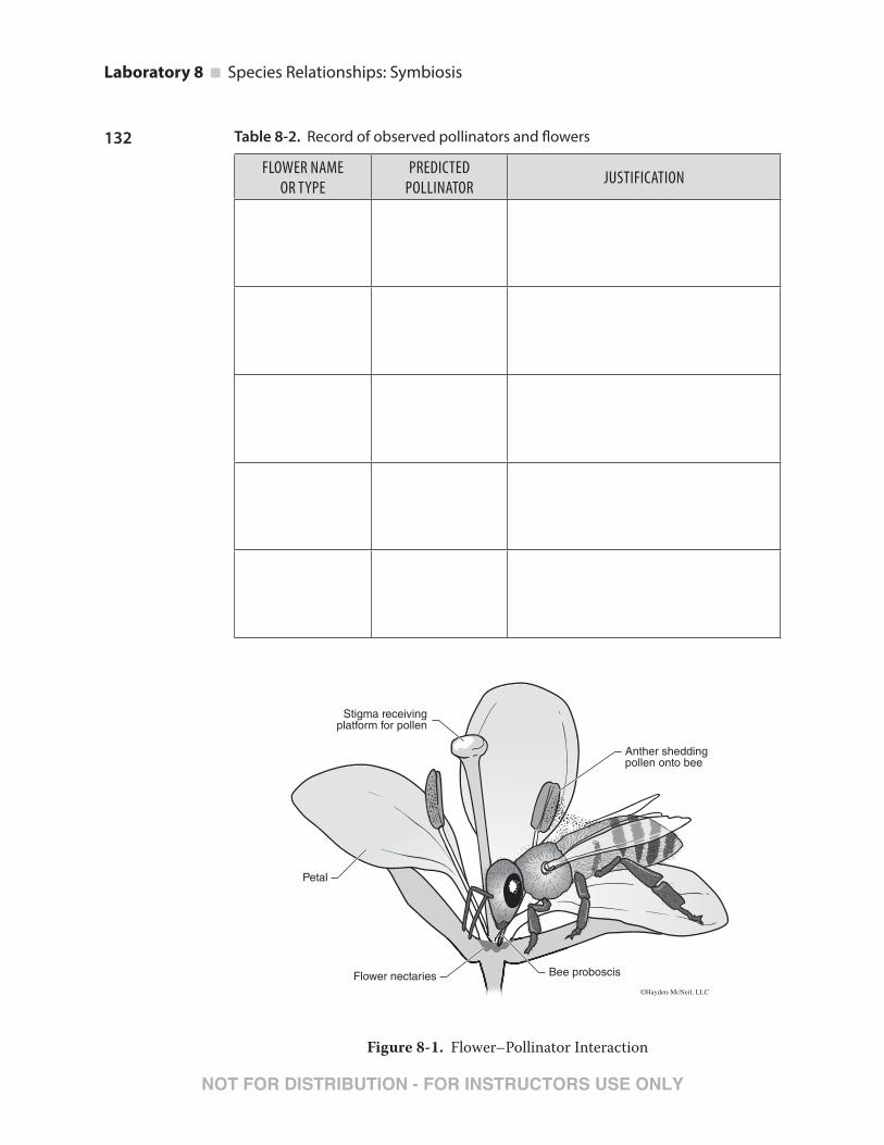
Symbioses between plants and their pollinators (Figure 8-1) are considered a prime
example of coevolution of these two groups of organisms over the past 200 million
years. So remarkable is the “fit” between pollinator and flower that a fairly novice
observer can predict the type of pollinator for which a flower is adapted by examining
the flower’s color, shape, scent, and other characteristics. Similarly, specialized struc-
tures on a pollinator, like the shape and length of the proboscis (the tubular feeding
organ) closely match the flower’s anatomy. Pollinators, usually insects or some other
animal, carry pollen from the anther of one plant where it is produced to the stigma of
another plant, while plants provide the animal with a food source in the form of nectar
or pollen (see Figure 8-1). Nearly 70% of flowering plants rely on insects for pollination
and 30% of our food comes from bee-pollinated crops (Kearns and Inouye 1997).
Symbiotic relationships between animals and microorganisms are also important in
the process of nutrient acquisition (you will learn more about specific nutritional
adaptations in the nutrition lab). Ruminants and other animals rely on certain spe-
cies of bacteria and protists within their digestive tracts for digesting tough, cellulose
material. The most advanced fiber processing digestive tract, which is found in graz-
ing types of mammals, is the ruminant system. Cows, sheep, and deer, among many
others, are ruminants. These animals are often described as having four stomachs,
because the stomach is partitioned into four chambers that each have a specific func-
tion for digesting plant material before reaching the small intestine. In order, the
chambers are: rumen, reticulum, omasum, and abomasum. Symbiotic bacteria and
protists in the first two processing chambers use enzymes to degrade the plant ma-
terial and yield large quantities of a waste product called volatile fatty acids through
fermentation reactions. These fatty acids are absorbed into the blood and are trans-
ported to the liver where they are converted to sugars that are used in metabolism.
The fluid from these chambers is often referred to as “rumen fluid,” since the rumen
is the largest chamber that contains microorganisms. The third chamber of the ru-
minant stomach acts as a particle sieve keeping the larger, less degraded particles in
the first two chambers. It also reabsorbs water. The final chamber acts as the “true
stomach,” with acids and enzymes that break down materials just as they do in the
stomach of an animal with a monogastric system.
The alimentary canals of animals also possess symbiotic bacteria housed in spe-
cialized intestinal structures, such as the cecum. In herbivores, the cecum can be a
large fermentation chamber. For example, the koala has a long, tubular cecum with
abundant symbiotic bacteria. Bacteria in the cecum use enzymes to break down the
NOT FOR DISTRIBUTION - FOR INSTRUCTORS USE ONLY
130
Laboratory 8 Species Relationships: Symbiosis
fibrous eucalyptus leaves, which are the sole food source of the koala. On the other
hand, carnivores have a small cecum, since plant material is not common in their
diets. Regardless of the anatomical structures present, most animals possess gut mi-
croorganisms collectively known as the “gut flora.” In addition to helping with the
digestion of dietary fiber, these microorganisms perform other important roles. For
instance, in mammals, beneficial intestinal Escherichia coli synthesize vitamin K. A
healthy “gut flora” is also essential for maintaining a healthy gastrointestinal system
and plays an important role in the immune system.
In the invertebrates, termites are a classic example of a type of organism that has a
coevolutionary relationship with gut microorganisms. Termites rely on bacteria and
protists to digest cellulose from the wood they consume. Termites and their diverse
community of microorganisms form obligate symbiotic relationships in which one
cannot live without the other. In this lab, you will have the opportunity to analyze the
content of a termite gut and find some of these microorganisms. The most common
organism you will find is a protist called, Trychonympha spp. You will also observe
rumen fluid from a cow to view the gut flora.
Activity One: Plants and Pollinators
PROCEDURE
1. Read Table 8-1. It describes various pollinator and flower characteristics (you
may also refer to pictures of flowers seen in Appendix F). You will also watch a
series of video clips on pollination.
2. Work in groups at your laboratory bench during this activity to try to predict the
type of pollinator for the flowers you observe in the laboratory. Fill out Table 8-2
as you make your observations. Each group should present their findings to the
class.
NOT FOR DISTRIBUTION - FOR INSTRUCTORS USE ONLY
131
Laboratory 8 Species Relationships: Symbiosis
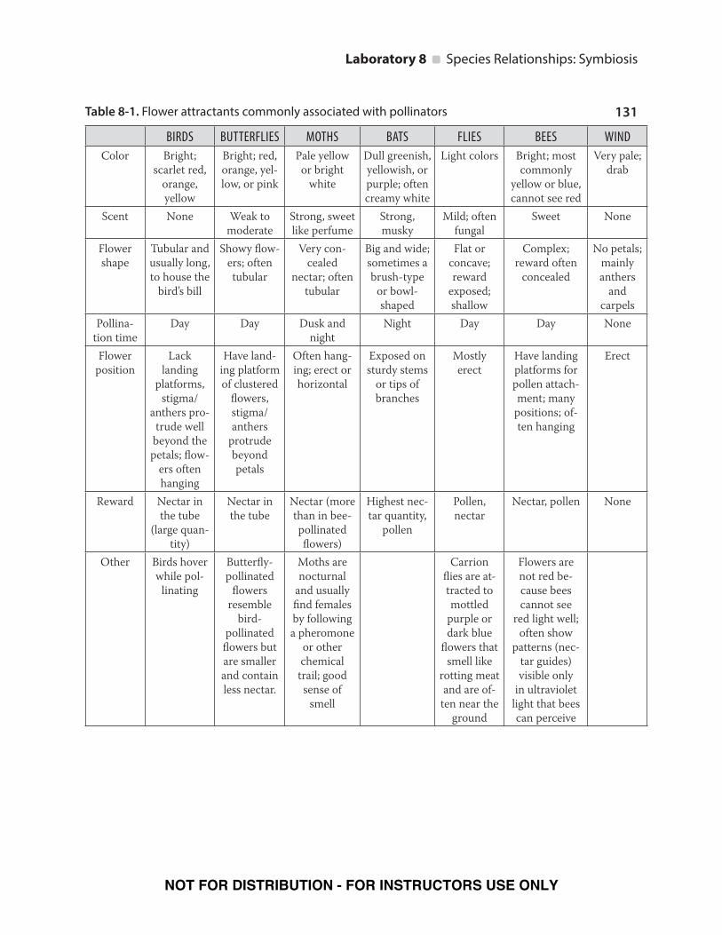
Table 8-1. Flower attractants commonly associated with pollinators
BIRDS BUTTERFLIES MOTHS BATS FLIES BEES WIND
Color Bright;
scarlet red,
orange,
yellow
Bright; red,
orange, yel-
low, or pink
Pale yellow
or bright
white
Dull greenish,
yellowish, or
purple; often
creamy white
Light colors Bright; most
commonly
yellow or blue,
cannot see red
Very pale;
drab
Scent None Weak to
moderate
Strong, sweet
like perfume
Strong,
musky
Mild; often
fungal
Sweet None
Flower
shape
Tubular and
usually long,
to house the
bird’s bill
Showy flow-
ers; often
tubular
Very con-
cealed
nectar; often
tubular
Big and wide;
sometimes a
brush-type
or bowl-
shaped
Flat or
concave;
reward
exposed;
shallow
Complex;
reward often
concealed
No petals;
mainly
anthers
and
carpels
Pollina-
tion time
Day Day Dusk and
night
Night Day Day None
Flower
position
Lack
landing
platforms,
stigma/
anthers pro-
trude well
beyond the
petals; flow-
ers often
hanging
Have land-
ing platform
of clustered
flowers,
stigma/
anthers
protrude
beyond
petals
Often hang-
ing; erect or
horizontal
Exposed on
sturdy stems
or tips of
branches
Mostly
erect
Have landing
platforms for
pollen attach-
ment; many
positions; of-
ten hanging
Erect
Reward Nectar in
the tube
(large quan-
tity)
Nectar in
the tube
Nectar (more
than in bee-
pollinated
flowers)
Highest nec-
tar quantity,
pollen
Pollen,
nectar
Nectar, pollen None
Other Birds hover
while pol-
linating
Butterfly-
pollinated
flowers
resemble
bird-
pollinated
flowers but
are smaller
and contain
less nectar.
Moths are
nocturnal
and usually
find females
by following
a pheromone
or other
chemical
trail; good
sense of
smell
Carrion
flies are at-
tracted to
mottled
purple or
dark blue
flowers that
smell like
rotting meat
and are of-
ten near the
ground
Flowers are
not red be-
cause bees
cannot see
red light well;
often show
patterns (nec-
tar guides)
visible only
in ultraviolet
light that bees
can perceive
NOT FOR DISTRIBUTION - FOR INSTRUCTORS USE ONLY
132
Laboratory 8 Species Relationships: Symbiosis
Table 8-2. Record of observed pollinators and flowers
Scolex of adulttapewormattaches tointestinal wallof human host
1
2
4
Eggs are encased in a tapeworm segment called a proglottid, which is passed out in feces. The proglottid is termed “gravid” when filled with eggs. Eggs are relased into the environment.
Cattle and pigs become infected by ingesting vegetation contaminated by eggs or gravid proglottids.
Larval cysts called cysticeri develop in muscle tissue.
3
Small intestinal larvae called oncospores hatch out of eggs, penetrate intestinal wall, and circulate to musculature