47
Plant virus Symptoms P.N. Sharma Department of Plant Pathology, CSK HPKV, Palampur (H.P.)
Plant virus Symptoms
P.N. Sharma
Department of Plant Pathology,
CSK HPKV, Palampur (H.P.)

Plant virus symptoms
Important for virus identification
Used to name the disease
Solely can not be used to
characterize the virus as affected by
many factors
Environment, virus strain/ mixed
infections, host varieties, nutrition,
age, stage of infection

Types of Symptoms
Macroscopic symptoms
Microscopic/ Histological chnages

Macroscopic Symptoms
Macroscopic Symptoms
Local symptoms
Develops near the site of entry of the virus,
Important in biological assay
Size vary from small pinpoint spots to large necrotic areas
Generally produced in mechanical inoculaions
Chlorotic lesions
Necrotic lesions
Ring spots

Local symptoms
local lesions: necrotic, chlorotic
Local lesions BCMNV
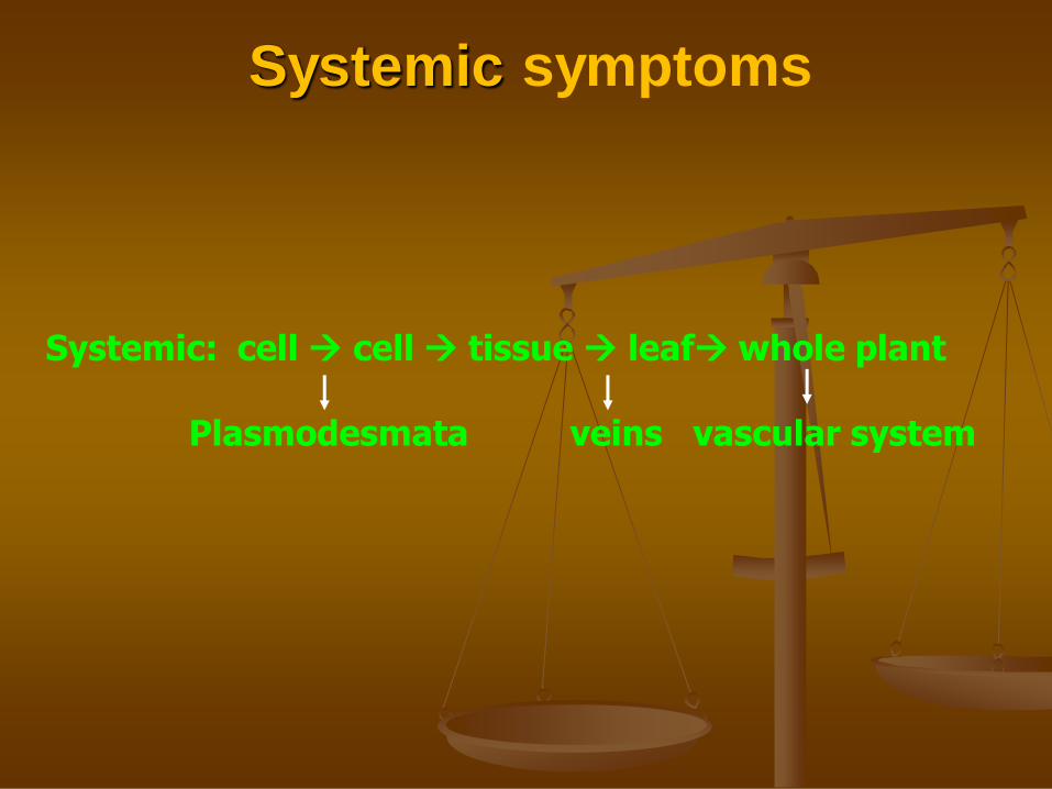
Systemic symptoms
Systemic: cell cell tissue leaf whole plant
Plasmodesmata veins vascular system

Systemic symptoms
Appear in different patterns depending upon virus-host combinations
Appear in sequential patterns and
May comprise of different symptoms

1. Mosaic
2. Mottle
3. Yellowing
4. Chlorosis
5. Vein clearing
6. Vein banding 1. Green vein banding
2. Yellow vein banding
7. Leaf roll
8. Leaf curl
9. Streak
10. Blistering
Plant virus Symptoms 1. Phyllody
2. Enation
3. Witches broom
4. Proliferations
5. Stunting
6. Ringspot
7. Wilt
8. Tumors/ galls
9. Necrosis

1. Effect on plant size
Stunting
Occurs due to reduction in leaf size and inter-nodal length of the infected plant
Depend upon stage of infection of the host. And severity of the symptoms
Stunting may affect all parts of plants
Type of host e.g. in perennial plants – takes more time; in vegetative plants effects are evident over years

2. Mosaic patterns and related symptoms
Most common effect of virus infection in the form of colour patterns on leaves
Various colour patterns like green & dark green; green& yellow and green & golden etc.
Generally depends on different host-virus combinations
Borders between green and dark green areas may be distinct or diffused
the patterns appears in systematic sequences
Mosaic appears at very early stages or some time chlorosis also occurs.
Other patterns includes
vein banding alongwith vein banding.
Stripes
Streaks
Variegation or breaking in petal color (consist of flecks, streaks, or diff.
colors segments. In petals genearlly due to loss of anthocyanin
Patterns on fruits e.g cucumber –CMV (mottle)

Mosaic
BCMV
yellow mosaic
MYMV
Golden mosaic CpGMV
Mosaic patterns
Common mosaic
Yellow mosaic
Golden mosaic

KATTE DISEASE of cardamom

Mosaic on infected fruits: CMV
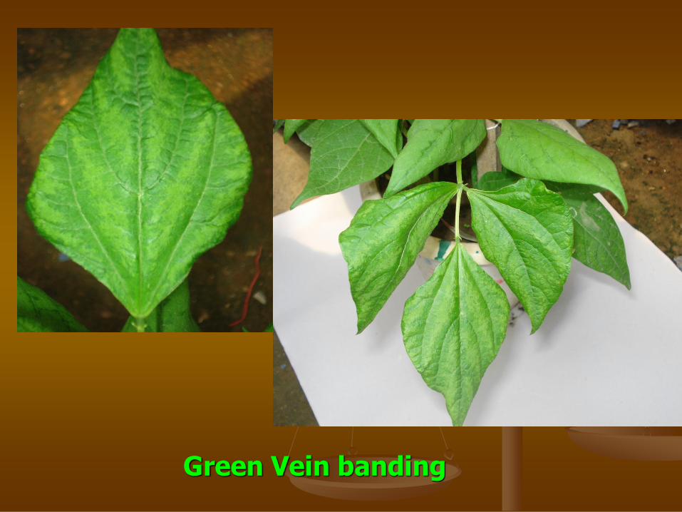
Green Vein banding

Tulip variegation

Variegation

Chlorosis:
The loss of chlorophyll from the tissues of a plant, resulting from microbial infection,
e.g. viral infection, the action of certain phytotoxins, the lack of light, to magnesium or iron deficiency, etc. Chlorotic tissues commonly appear yellowish
Chlorosis & Chlorotic ring spot by BCMV

3. Yellows
Induced by some viruses e.g. sugarbeeet, peach yellow,
May be sight or severe; covers whole leaf or some times sectors of yellow and normal colour are formed
Yellowing: A symptom characterized by the turning yellow of plant tissues that were once green

Yellow vein mosaic Okra
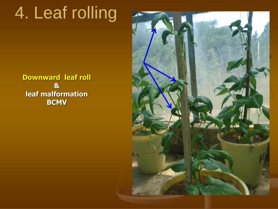
4. Leaf rolling
Downward leaf roll &
leaf malformation BCMV

Typical Leaf roll of lower leaves
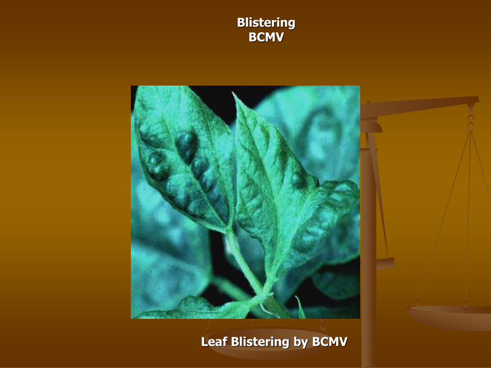
Blistering BCMV
Leaf Blistering by BCMV

Leaf Crinkle- Urdbean (ULCV)

Enations: Pea enation mosaic virus (PEMV)

Leaf curl of tomato

Vein clearing BCMV
Chlorotic ringspot BCMV
5. Ring spot diseases
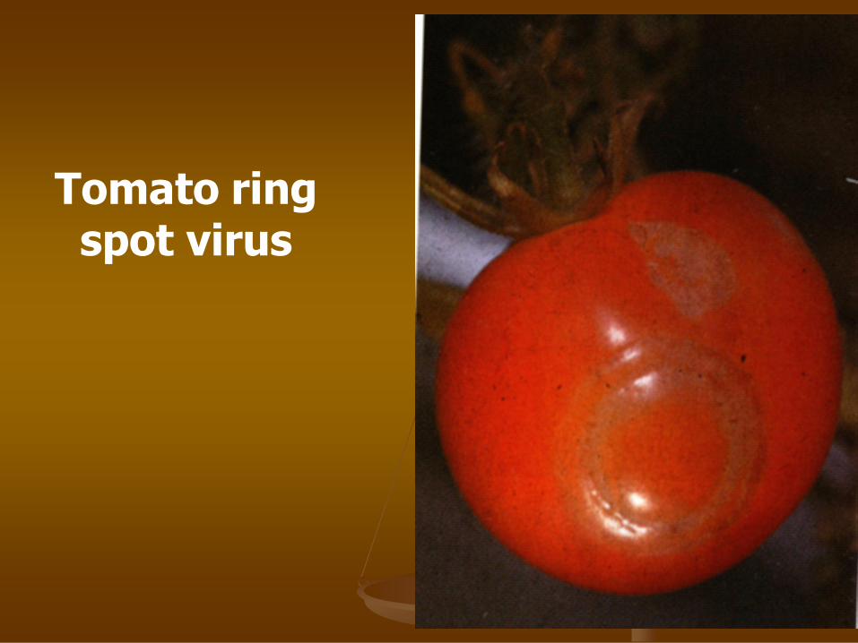
Tomato ring spot virus

6. Necrotic diseases
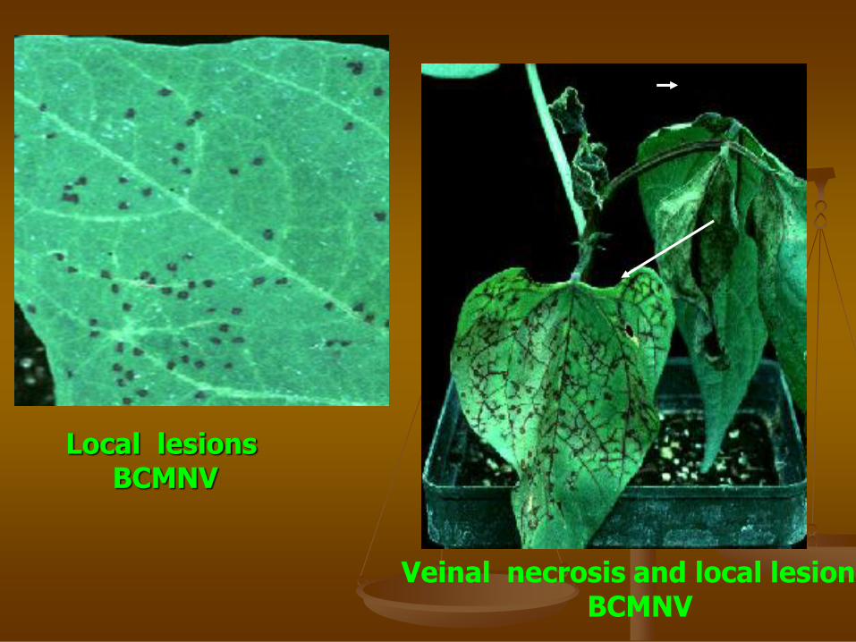
Local lesions BCMNV
Veinal necrosis and local lesions BCMNV

Veinal necrosis
BCMV

BANANA DIE BACK
7. Abnormalities of plants
Galls
Tumors
Leaf deformation
Shoe stringing
Stem pitting
w

Crown gall/
tumor
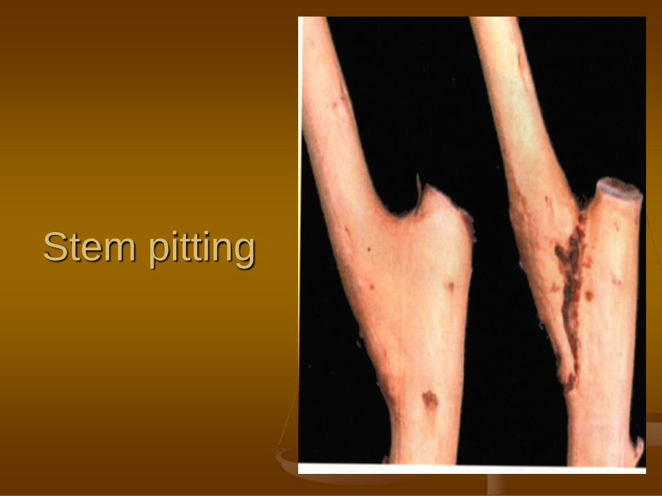
Stem pitting
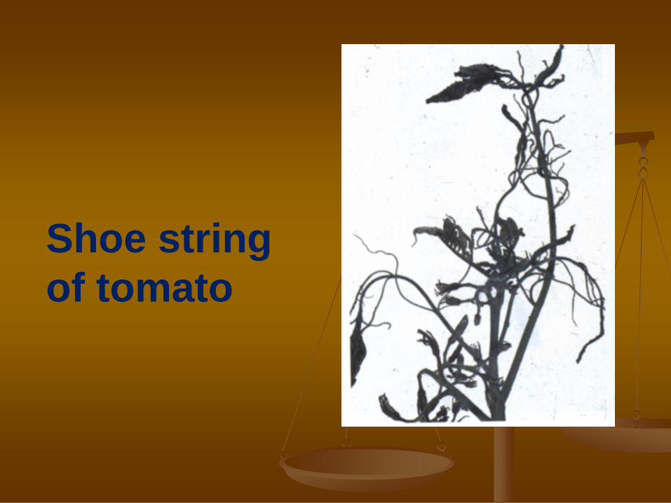
Shoe string
of tomato

BANANA BUNCHY TOP
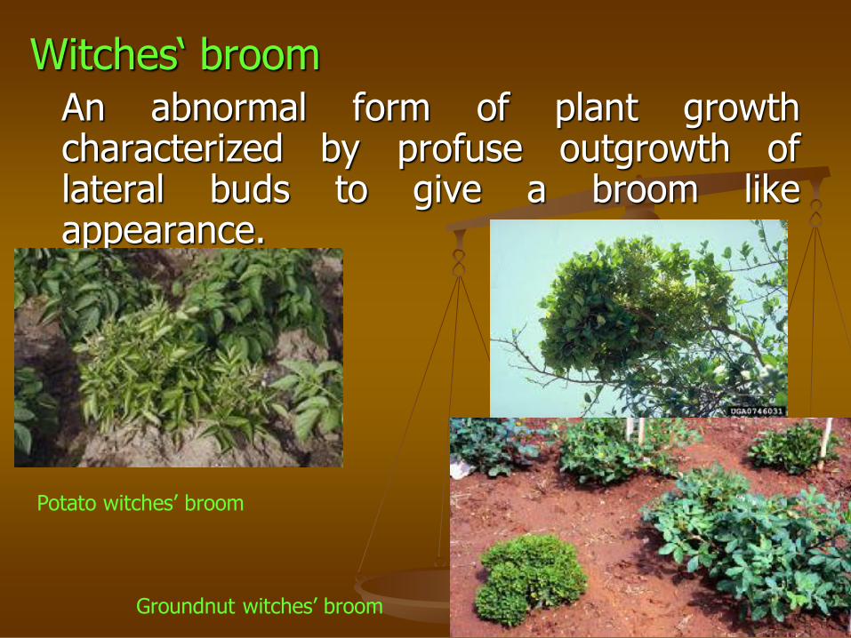
Witches‘ broom An abnormal form of plant growth
characterized by profuse outgrowth of lateral buds to give a broom like appearance.
Potato witches’ broom
Groundnut witches’ broom

Rosette:
An abnormal condition in which the
leaves form a radial cluster on the
stem.
Groundnut rosettee caused by: Groundnut rosette umbravirus

8. Wilting

Microscopic/ Histological changes
Expressed as anatomical and histological changes in the affected plants.
Generally as necrosis, hypoplasia and hyperplasia
Formation of inclusion bodies

INTERNAL SYMPTOMS
Expressed as anatomical and histological changes in
the affected plants.
Generally as necrosis, hypoplasia and hyperplasia
In mosaic infected plants, generally hypoplasia in the
yellow or light green areas is noticed.
Cytological changes Virus infection affect various cell organelles like nucleus, chloroplasts, and
mitochondria etc.
Nucleus: induces formation of nuclear inclusions, affect nucleolus
Nucleus becomes granular, chromatins may be reduced, disintegrated
Chloroplasts show disintegration, swelling or clumping etc.
Mitochondria show aggregation in virus infected cells, formation of vesicles
Cell wall show thickening, deposition of electron dense material between cell
wall and plasma membrane, callus deposition

Inclusion Bodies Formation of inclusion bodies
These are intracellular structures formed due to
the virus infection
Major cytological effect of virus infection
some may be seen with ordinary microscope
May contains virus particles, virus related
material or degenerate conditions
May be nuclear or cytoplasmic
These may be amorphous, crystalline or pin-
wheel
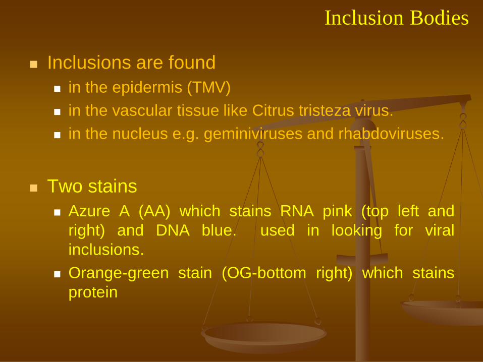
Inclusion Bodies
Inclusions are found
in the epidermis (TMV)
in the vascular tissue like Citrus tristeza virus.
in the nucleus e.g. geminiviruses and rhabdoviruses.
Two stains
Azure A (AA) which stains RNA pink (top left and
right) and DNA blue. used in looking for viral
inclusions.
Orange-green stain (OG-bottom right) which stains
protein

Crystalline bodies
These are in the form of virus aggregates arranged in orderly fashion giving three dimensional appearance
Mostly accumulates in cell cytoplasm
Commonly seen in the epidermal and hair cells in the cytoplasm
May be in the form of rods, helical, icosahedral or crystalline array of infected cells.

Pinwheel inclusions
Characteristic of infection
by Potyviridae family of
viruses
PVY, BCMV, and Soybean
mosaic virus.
Others are scrolls,
laminated aggregates etc.
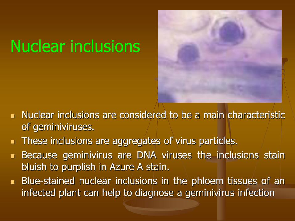
Nuclear inclusions are considered to be a main characteristic of geminiviruses.
These inclusions are aggregates of virus particles.
Because geminivirus are DNA viruses the inclusions stain bluish to purplish in Azure A stain.
Blue-stained nuclear inclusions in the phloem tissues of an infected plant can help to diagnose a geminivirus infection
Nuclear inclusions

Acknowledgements
I gratefully acknowledge the use of some very important contents of the text book “Matthew’s Plant Virology” by Roger Hull.
This book serves as one of the important and essential source of learning of plant virology.
I also acknowledge the scientists who spent valuable time in generating information on various aspects of Virology and displayed the same on internet for use by teachers and researchers
PN Sharma


