Microbio – Virology – Lec1 - General Properties of Viruses Dr. Boromeo A Brief History of Virology By the late 1800s Jacob Henle (German) hypothesized the existence of infectious agents too small to be seen with a light microscope At this time, Louis Pasteur (French) and Robert Koch (German) had developed methods for sterile culture and bacterial isolation Koch formulated Koch’s Postulates There must be an association between an infectious disease and a particular microbe The microbe must be isolated from all patients with the disease When experimentally inoculated into a susceptible host, the microbe must cause the disease The microbe must be re-isolated from the experimental animals Adolf Mayer (German) described a non- bacterial, non-fungal cause of tobacco mosaic disease Showed it was not a toxin by limiting dilution (serial passaged to extinction) Filtered infected plant material through a Chamberland filter (unglazed porcelain) Walter Reed discovered the first filterable human infectious agent (yellow fever virus) in Cuba (1901) After that, many new viruses were discovered, including bacteriophages, viruses that infect bacteria The Early Period of Virology (1938-1965) Electron microscopy was developed and was instrumental in conclusively showing the existence of viruses Penicillin was discovered and allowed animal cell cultures free of bacterial contamination DNA was shown to be the hereditary material of life WWII led to the end of Germany’s leadership in biological sciences Vaccines for many viral diseases were developed Polio (US: 21,000 ➞ zero) Measles (US: 500,000 ➞ dozens) Smallpox (Global: Millions ➞ eradicated in 1978) The Modern Period of Virology (1965-Present) Virology led to discoveries in basic biology Revolutionized molecular biology Understand gene regulation Understand genetic recombination Understand cancer Virus discovery Thousands of new viruses were isolated and identified Recombinant DNA used for production of vaccines for unsafe viruses Reverse genetics technology developed for producing infectious DNA clones of RNA viruses Morphologic Properties of Viruses INTRODUCTION Viruses are complex macromolecules They are obligate intracellular parasites They are not alive; thus you cannot kill a virus, only inactivate it They have either DNA or RNA genomes All species are susceptible to viruses There are thousands known, and probably many thousands unknown Viruses are usually species-specific; that is, they can infect one or a few species only They range in size from about 20 nm to about 300 nm in diameter The nucleic acid is protected by a protein polymer shell Viruses are inert outside of a cell Diversity of Viruses Terms and Definitions in Virology Capsid - the protein shell that protects the viral nucleic acid Capsomere - morphologic unit of capsid Defective virus - a virus particle that is deficient such that it is incapable of productive infection Envelope - phospholipid bilayer that surrounds some viruses; formerly part of the cell’s membrane in which the virus arose Peplomer - glycoproteins that protrude outward from some viral envelopes, which mediate cellular entry. Often called spike proteins Evolutionary Origins of Viruses Completely unknown Retroviruses may have arisen from transposable elements Some may have arisen from eukaryotic parasites that lost most of their genomes Classification of Viruses The International Committee for the Taxonomy of Viruses ICTV http://www.ncbi.nlm.nih.gov/ICTVdb/ index.htm Basis of Classification Morphology (usually by EM) DNA or RNA Physicochemical properties Protein properties Genome organization and replication Antigenic properties (serology) An antigen is any substance that elicits an immune response Serology is the use of serum antibodies that react to microbial products
Transcript
Microbio – Virology – Lec1 - General Properties of VirusesDr. Boromeo
A Brief History of Virology By the late 1800s Jacob Henle (German) hypothesized the existence of
infectious agents too small to be seen with a light microscope At this time, Louis Pasteur (French) and Robert Koch (German) had
developed methods for sterile culture and bacterial isolation Koch formulated Koch’s Postulates
There must be an association between an infectious disease and a particular microbe
The microbe must be isolated from all patients with the disease When experimentally inoculated into a susceptible host, the
microbe must cause the disease The microbe must be re-isolated from the experimental animals Adolf Mayer (German) described a non-bacterial, non-fungal
cause of tobacco mosaic disease Showed it was not a toxin by limiting dilution (serial
passaged to extinction) Filtered infected plant material through a Chamberland filter
(unglazed porcelain) Walter Reed discovered the first filterable human infectious agent
(yellow fever virus) in Cuba (1901) After that, many new viruses were discovered, including
bacteriophages, viruses that infect bacteria The Early Period of Virology (1938-1965)
Electron microscopy was developed and was instrumental in conclusively showing the existence of viruses
Penicillin was discovered and allowed animal cell cultures free of bacterial contamination
DNA was shown to be the hereditary material of life WWII led to the end of Germany’s leadership in biological
sciences Vaccines for many viral diseases were developed
Polio (US: 21,000 ➞ zero) Measles (US: 500,000 ➞ dozens) Smallpox (Global: Millions ➞ eradicated in 1978)
The Modern Period of Virology (1965-Present) Virology led to discoveries in basic biology Revolutionized molecular biology Understand gene regulation Understand genetic recombination Understand cancer
Virus discovery Thousands of new viruses were isolated and identified Recombinant DNA used for production of vaccines for unsafe
viruses Reverse genetics technology developed for producing infectious
DNA clones of RNA viruses
Morphologic Properties of Viruses
INTRODUCTION Viruses are complex macromolecules They are obligate intracellular parasites They are not alive; thus you cannot kill a virus, only inactivate it They have either DNA or RNA genomes All species are susceptible to viruses There are thousands known, and probably many thousands unknown Viruses are usually species-specific; that is, they can infect one or a few
species only They range in size from about 20 nm to about 300 nm in diameter The nucleic acid is protected by a protein polymer shell Viruses are inert outside of a cell
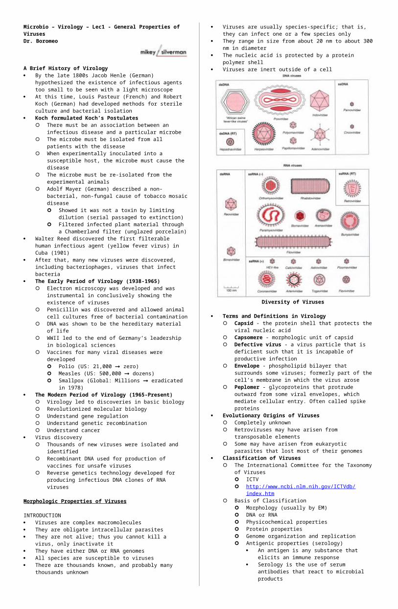
Diversity of Viruses
Terms and Definitions in Virology Capsid - the protein shell that protects the viral nucleic acid Capsomere - morphologic unit of capsid Defective virus - a virus particle that is deficient such that it is
incapable of productive infection Envelope - phospholipid bilayer that surrounds some viruses;
formerly part of the cell’s membrane in which the virus arose Peplomer - glycoproteins that protrude outward from some viral
envelopes, which mediate cellular entry. Often called spike proteins
Evolutionary Origins of Viruses Completely unknown Retroviruses may have arisen from transposable elements Some may have arisen from eukaryotic parasites that lost most of
their genomes Classification of Viruses
The International Committee for the Taxonomy of Viruses ICTV http://www.ncbi.nlm.nih.gov/ICTVdb/index.htm
Basis of Classification Morphology (usually by EM) DNA or RNA Physicochemical properties Protein properties Genome organization and replication Antigenic properties (serology)
An antigen is any substance that elicits an immune response
Serology is the use of serum antibodies that react to microbial products
Biological properties (e.g., host range, mode of transmission, etc.)
Universal System of Virus Taxonomy Suffix -viridae for family (italicized) names (e.g. Flaviviridae) Suffix -virus for genus (italicized) names (e.g. Flavivirus) Species name (NOT italicized) usually based upon geographic
region of discovery (e.g., West Nile virus discovered in Uganda)
See Table 29-1 Other terms
Strain - different wild isolates of the same virus The WNV isolate from New York is called WNV-NY
Type - Distinct strains of the same virus based upon known genetic differences HIV type 1 vs. HIV type 2
Variant - a virus that behaves differently in the lab than the wild-type, but for unknown reasons
Structural unit - two or more bound non-identical subunits (e.g., VP1, VP2, VP3, VP4 of poliovirus) that form a larger building block
Assembly unit - multiple structural units Morphological unit (aka capsomere) - clusters protruding
outward from the virus surface Capsid - the entire protein shell (or coat) Nucleocapsid - capsid and enclosed nucleic acid Envelope - phospholipid bilayer membrane that surrounds
some viruses Virion = entire virus particle
Cubic (spherical) symmetry Most animal viruses are spherical Icosahedron - closed shell composed of 20 facets
(equilateral triangles) Spontaneously assembles into a sphere For viruses, these facets are the morphologic units are
usually encoded by three assembly units
Helical symmetry (Fig 29-3) Found in filamentous viruses (e.g. Ebola viruses) The protein subunits assemble only in the presence of
nucleic acid (unlike cubic) The nucleocapsid assembles in a helical manner, with the
nucleic acid constrained to a spiral conformation within the capsid
Complex structures Some viruses have pleiomorphic shapes
Poxviruses are brick-shaped with a dumbbell structure in the center
Chemical Composition of Viruses Proteins
All have structural proteins Some have enzymes Viral proteins are principal targets of the immune response
Nucleic Acids DNA or RNA Single or double-stranded Plus or negative strand (ss RNA viruses only) Contiguous or segmented
Lipids Some viruses have lipid envelopes acquired from the cell in
which the virus was produced Glycoproteins
Some viral proteins, particularly those that protrude outward, have carbohydrate groups
These groups often mediate virus attachment to susceptible cells
Cultivation and Assay of Viruses Cultivation
Viruses are obligate intracellular parasites, thus require living cells for propagation
Cultivation of viruses ranges from simple (cell culture, embyonated eggs), difficult (whole animal) to impossible (unculturable)
Cell culture methods Primary cell culture
Uses freshly explanted tissues from animal Cells are dissociated from one another by trypsin
a pancreatic protease cleaves at arginine and lysine residues does not cross cell membranes, thus remains
extracellular results in a single cell suspension
Cells can be plated (or seeded) in a variety culture vessels
Primary cells will grow to confluence (aka monolayer), then stop dividing (termed contact inhibition)
After passage (or splitting) of cultures, cells will resume division
Normal primary cells are sometimes referred to as diploid cells
Primary cell cultures eventually undergo senescence (natural death)
Cultivation Cell culture methods
Transformed cell lines Immortal Often easier to grow than primary lines Often aneuploid Cannot be used for vaccine production
Embyonated chicken eggs Specific pathogen free (SPF) live chicken eggs Inoculation of chorioallantoic membrane (CAM) or yolk
Live animal If virus cannot be propagated in cell culture or eggs, it must
be grown in a living animal After virus replicates, the target organ must be collected,
pureed and centrifuged to collect virus homogenate Cannot be used for vaccines (except vaccinia virus for
smallpox) Detection of viral infection
Cytopathic effect (CPE) occurs with most viruses Cell death Irregularly-shaped cells Syncytia formation - fusion of cell membranes resulting in
multinucleated giant cells Inclusion bodies - viral proteins aggregate within cells and
become visible under the microscope Cytoplasmic or nuclear Virus factories
Relies upon the ability of a virus to induce CPE in cell culture or embryonated eggs
Uses limiting dilution (dilution to extinction) to determine virus numbers
The dilution that infects (or kills) 50% of cells is termed tissue culture infectious dose-50 (TCID50)
For eggs, EID50 For animals ID50 or LD50 (lethal dose)
These assays all provide a titer of the virus Titer is the unit used to describe the greatest dilution of a
substance (e.g., virus, antibody) that contains biological activity It is usually reported as the reciprocal of this dilution (10-6 activity
has a titer of 6) The Reed-Muench Method for calculating TCID50 (1938)
Make log10 dilution series of a virus prep Add 100 µl of dilutions in 5 replicates on 96 well plate containing
confluent susceptible cells Incubate cells until CPE is complete Score the wells for infection (yes or no) Identify the two adjacent dilutions where more than 50% of the
wells are infected, and less than 50% are infected Determine the TCID50 using following formula:
Reed-Muench Method
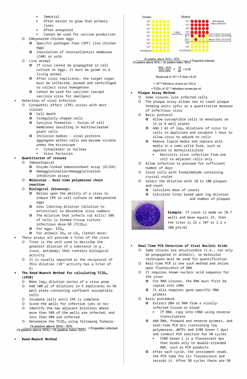
Plaque Assay Method Some viruses lyse infected cells The plaque assay allows one to count plaque forming units (pfu)
as a quantitative measure of infectious virus Basic protocol
Allow susceptible cells to monolayer on 12 or 6 well plates Add 1 ml of log10 dilutions of virus to cells in duplicate and
incubate 1 hour to allow virus to adsorb to cells Remove liquid media and replace with media in a semi-solid
form, such as agarose or methylcellulose Restricts virus infection from one cell to adjacent cells
only Allow infection to proceed for sufficient number of days Stain cells with formaldehyde containing crystal violet Select the dilution with 10 to 100 plaques and count
Calculate mean of counts Calculate titer based upon log dilution and number of
plaques
Example: If count is made on 10-7 wells and mean equals 22, then the titer is 22 x 107 or 2.2 x 10⁸ pfu/ml
Real-Time PCR Detection of Viral Nucleic Acids Some viruses are unculturable (i.e., can only be propagated in
animals), so molecular techniques must be used for quantification Real-time PCR is one such method and relies upon fluorescence of
DNA It requires known nucleic acid sequence for the virus
For RNA viruses, the RNA must first be copied into cDNA It also requires gene-specific DNA primers
Basic procedure Extract DNA or RNA from a virally-infected tissue or blood
If RNA, copy into cDNA using reverse transcriptase Add DNA, forward and reverse primers, and real-time PCR
mix (containing Taq polymerase, dNTPs and SYBR Green I dye) and conduct PCR reaction for 50 cycles SYBR Green 1 is a fluorescent dye that binds only to
double-stranded DNA, such as PCR products After each cycle, the instrument reads the PCR tube for its
fluorescence and records it. After 50 cycles there are 50 data points that are used to generate a graph of DNA abundance
Real-Time PCR Detection of Viral Nucleic Acids
Determining copy number by real-time PCR In separate tubes, include plasmid standards with the target viral
gene in known copy numbers (e.g. 102, 104, 106 copies) These provide data for generating a standard curve, which can
then be used to quantify copy number in test samples using statistical algorithms, such as linear regression
Purification and Identification of Viruses A common method of virus purification is precipitation with
polyethylene glycol at 4° C overnight Once precipitated, the virus can be washed with a buffer to
remove PEG Identification of an unknown virus isolate
Morphology by EM DNA or RNA? Enveloped? Reactivity against defined antisera
CDC and other institutions have panels of neutralizing antisera raised against hundreds of known viruses
These antibodies can be used to identify viruses in cell culture assays
Laboratory Safety Laboratory safety is essential when working with pathogenic microbes Safety is based upon three levels of practice that ensure containment
Proper technique Proper equipment
Biosafety cabinets Centrifuge containment
Policies and procedures for preventing and managing breaks of containment
Aerosols and punctures are the principal threats Ingestion and splashes are less common
Biosafety practices Training in aseptic technique No mouth pipetting No eating, drinking, smoking or application of makeups or balms
in the laboratory Use appropriate protective clothing and equipment (lab coat or
gown, gloves, mask) Sterilization of infectious wastes Use of biosafety cabinets Immunization if necessary
Reaction to Physical and Chemical Agents Heat and cold - heat inactivates some viruses, while cold usually
preserves them Stabilization by salts - unknown how this works pH - some viruses are resistant to dramatic changes in pH Radiation - damages nucleic acids; crosslinks viral proteins Photodynamic inactivation (e.g., neutral red) renders some viruses
susceptible to visible light Ether susceptibility - ether is an organic solvent, thus damages envelop
membranes of viruses Detergents - amphipathic, thus solublizes membranes and can
dissociate noncovalent bonds between viral proteins Formaldehyde - cross-links nucleic acids and proteins
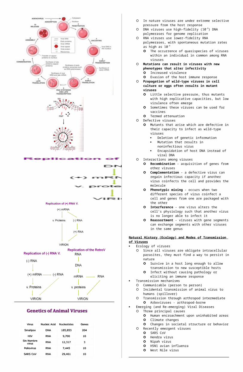
Replication of Viruses: An Overview Attachment, penetration and uncoating
Viruses attach to susceptible cells be interacting with specific molecules on the target cell’s surface HIV’s gp41 spike protein binds to CD4 on target cell
Penetration is mediated differently by viruses HIV gp120 interacts with a chemokine receptor on the cell in
such a was as to induce membrane fusion of the virus envelope with the cell’s plasma membrane
Other viruses exploit receptor mediated endocytosis to gain access to the cell’s cytoplasm
Once inside the cell, uncoating of the virus capsid occurs, liberating viral nucleic acids and enzymes into the cytosol
Expression of viral genome and synthesis of viral components Once liberated, the virus must make polypeptides and replicate its
genome DNA genomes are used similarly to that of cellular DNA Some RNA viruses use their RNA genome as template (plus
strand) for mRNA synthesis Other RNA viruses’ genomes are already mRNA (negative
strand) Some RNA viruses encode their own RNA-dependent RNA
polymerases
Once liberated, the virus must make polypeptides and replicate its genome (cont.) Early transcription and translation lead to polypeptide
synthesis using the cell’s protein synthesis machinery to gain control of the cell
Viruses have highly economic genomes Some genes encode polypeptides that are cleaved into
two or more functional proteins Some viruses have genes that encode overlapping
mRNA or mRNA in both directions DNA synthesis, late transcription and late translation are
directed at producing new macromolecules for progeny virions
Morphogenesis and release Viral macromolecules accumulate in compartments
(condensation) in the cytoplasm and undergo assembly Viruses exit cells (release) in one of two ways
Lysis of the cell, liberating progeny virus into the extracellular environment
Budding, where assembled nucleocapsids push through a membrane, taking part of it with the progeny virus as its envelope
In nature viruses are under extreme selective pressure from the host response
DNA viruses use high-fidelity (10-9) DNA polymerases for genome replication
RNA viruses use lower-fidelity RNA polymerases, with spontaneous mutation rates as high as 10-4 The occurrence of quasispecies of viruses within an
individual in common among RNA viruses Mutations can result in viruses with new phenotypes that alter
infectivity Increased virulence Evasion of the host immune response
Propagation of wild-type viruses in cell culture or eggs often results in mutant viruses Little selective pressure, thus mutants with high replicative
capacities, but low virulence often emerge Sometimes these viruses can be used for vaccines Termed attenuation
Defective viruses Mutants that arise which are defective in their capacity to
infect as wild-type viruses Deletion of genetic information Mutation that results in noninfectious virus Encapsidation of host DNA instead of viral DNA
Interactions among viruses Recombination - acquisition of genes from other viruses Complementation - a defective virus can regain infectious
capacity if another virus coinfects the cell and provides the molecule
Phenotypic mixing - occurs when two different species of virus coinfect a cell and genes from one are packaged with the other
Interference - one virus alters the cell’s physiology such that another virus is no longer able to infect it
Reassortment - viruses with gene segments can exchange segments with other viruses in the same genus
Natural History (Ecology) and Modes of Transmission of Viruses Ecology of viruses
Since all viruses are obligate intracellular parasites, they must find a way to persist in nature Survive in a host long enough to allow transmission to new
susceptible hosts Infect without causing pathology or eliciting an immune
response Transmission mechanisms
Communicable (person to person) Incidental transmission of animal virus to humans (spillover) Transmission through arthropod intermediate