Copyright 2007, The Johns Hopkins University and Ronald Gray. All rights reserved. Use of these materials permitted only in accordance with license rights granted. Materials provided “AS IS”; no representations or warranties provided. User assumes all responsibility for use, and all liability related thereto, and must independently review all materials for accuracy and efficacy. May contain materials owned by others. User is responsible for obtaining permissions for use from third parties as needed. This work is licensed under a Creative Commons Attribution-NonCommercial-ShareAlike License . Your use of this material constitutes acceptance of that license and the conditions of use of materials on this site.
Transcript
Copyright 2007, The Johns Hopkins University and Ronald Gray. All rights reserved. Use of these materials permitted only in accordance with license rights granted. Materials provided “AS IS”; no representations or warranties provided. User assumes all responsibility for use, and all liability related thereto, and must independently review all materials for accuracy and efficacy. May contain materials owned by others. User is responsible for obtaining permissions for use from third parties as needed.
This work is licensed under a Creative Commons Attribution-NonCommercial-ShareAlike License. Your use of this material constitutes acceptance of that license and the conditions of use of materials on this site.
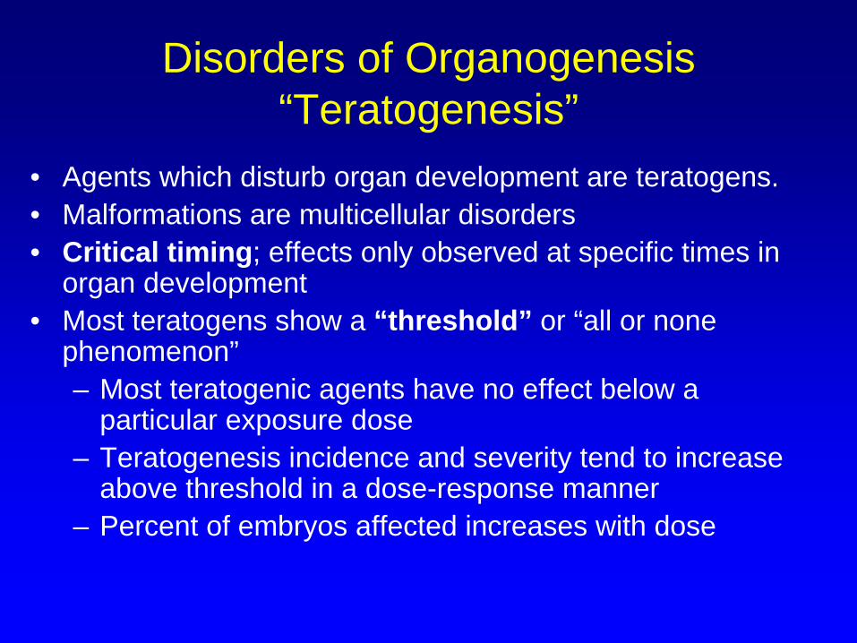
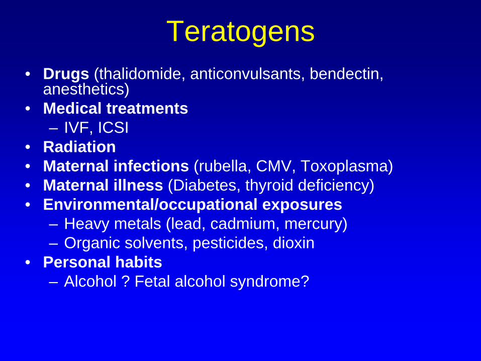
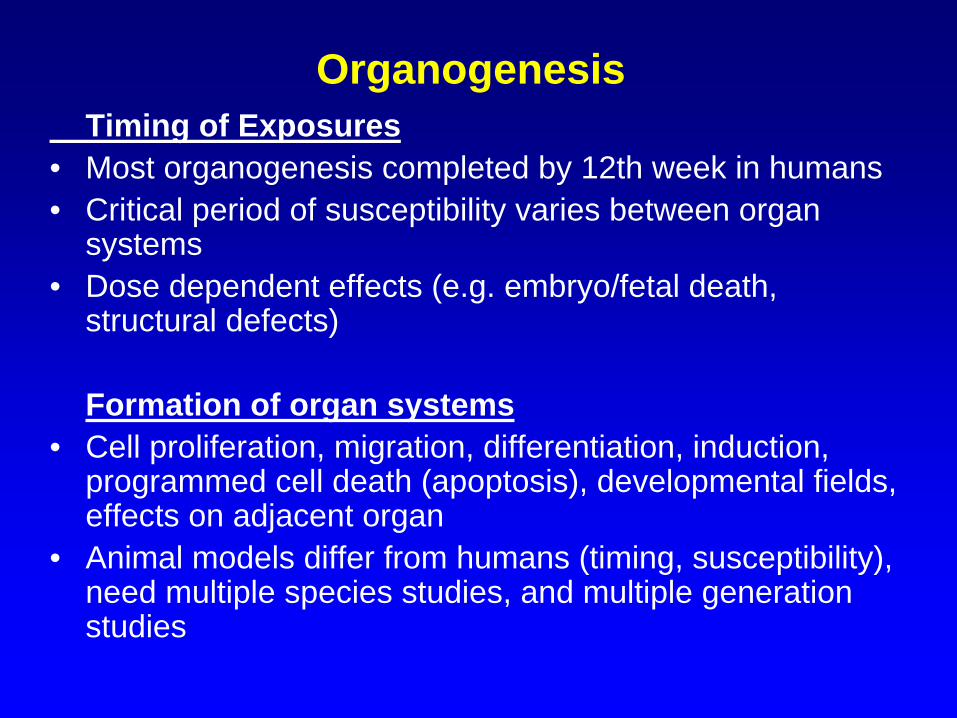
• Agents which disturb organ development are teratogens. • Malformations are multicellular disorders• Critical timing; effects only observed at specific times in
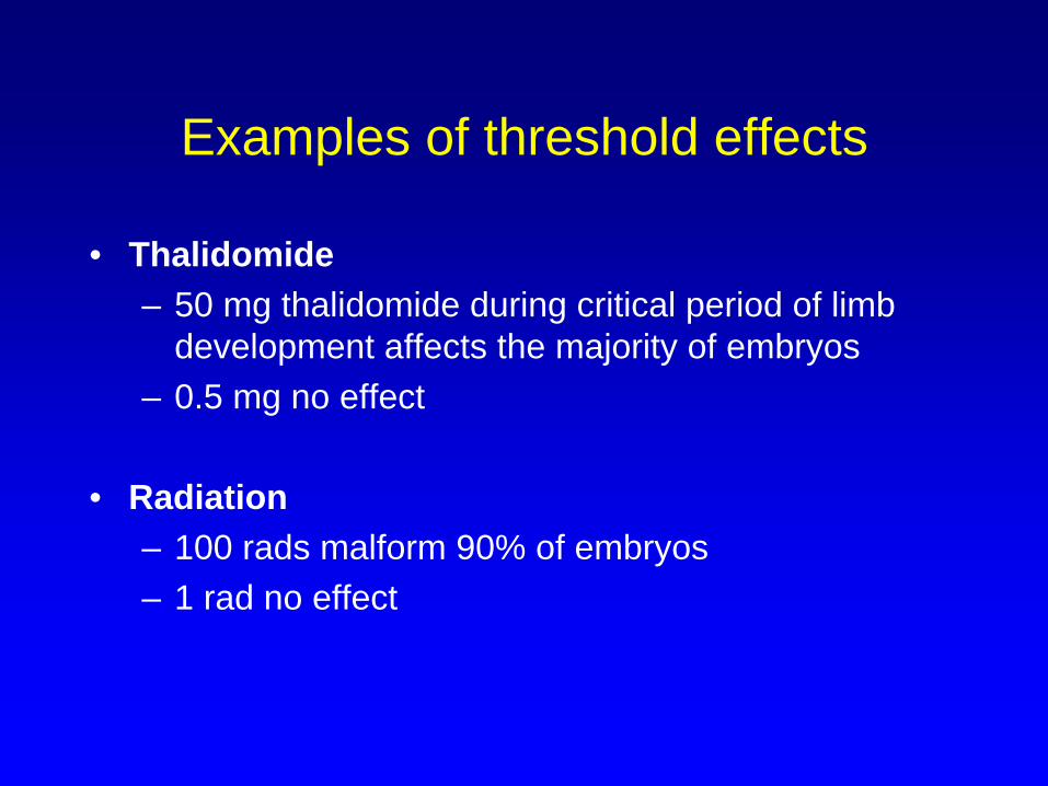
organ development• Most teratogens show a “threshold” or “all or none
phenomenon”– Most teratogenic agents have no effect below a
particular exposure dose– Teratogenesis incidence and severity tend to increase
above threshold in a dose-response manner– Percent of embryos affected increases with dose
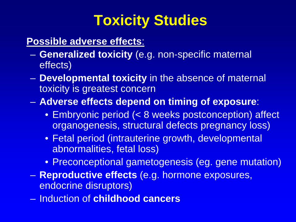
endocrine disruptors)– Induction of childhood cancers
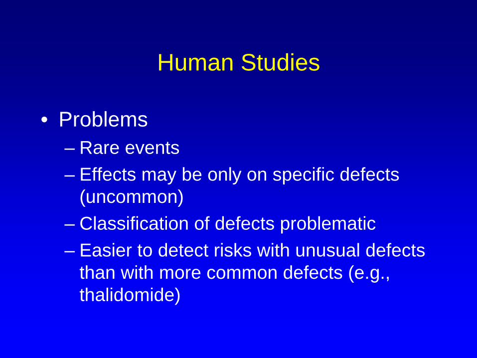
Human Studies
• Problems– Rare events– Effects may be only on specific defects
(uncommon)– Classification of defects problematic– Easier to detect risks with unusual defects
than with more common defects (e.g., thalidomide)
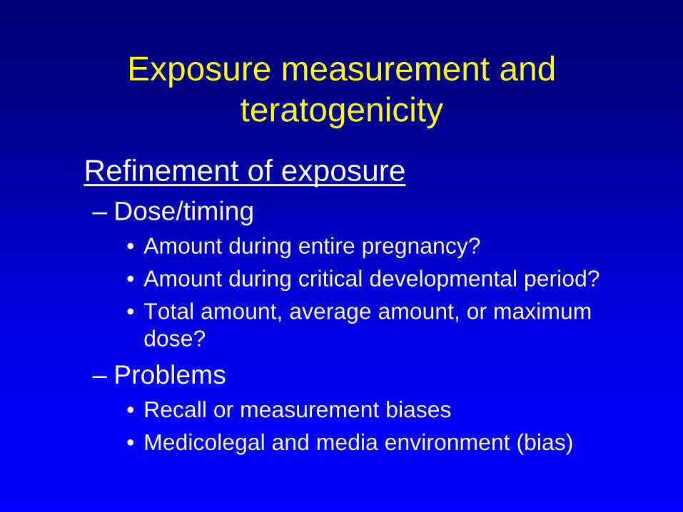
Exposure measurement and teratogenicity
Refinement of exposure– Dose/timing
• Amount during entire pregnancy?• Amount during critical developmental period?• Total amount, average amount, or maximum
dose?– Problems
• Recall or measurement biases• Medicolegal and media environment (bias)
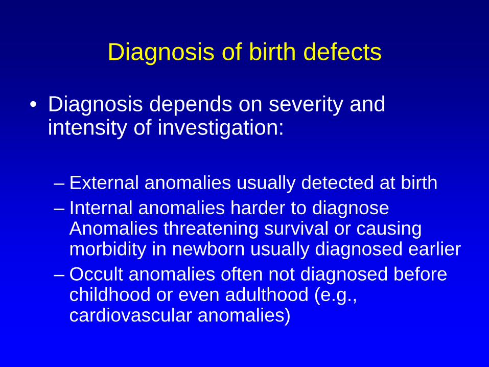
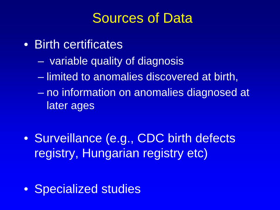
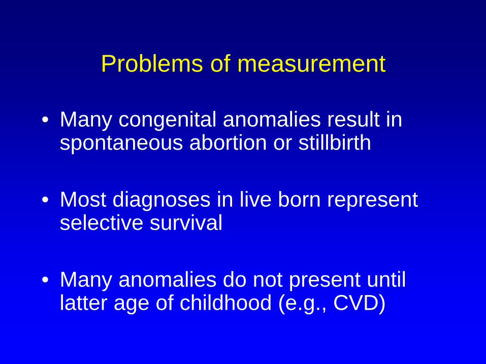
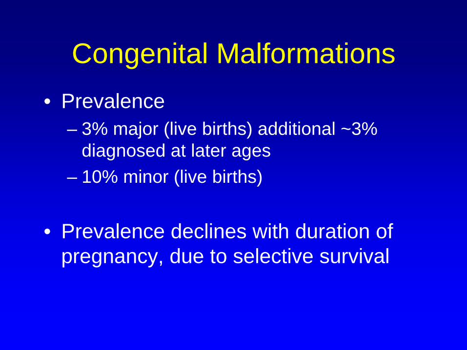
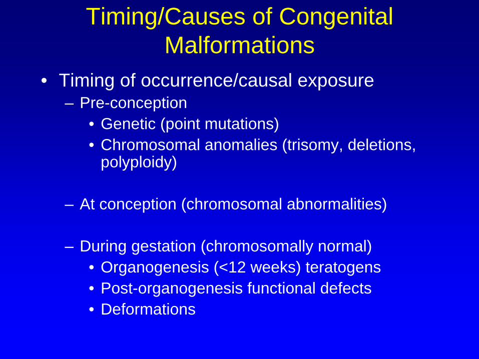
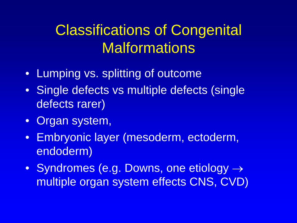
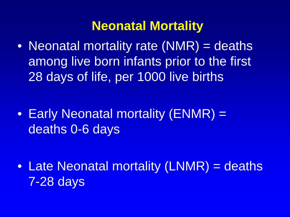
Congenital Malformations
Refinement of exposure– Stage of gestation
• Early blood glucose control with diabetic mother prevents adverse effects
• Folic acid and neural tube defects• Bendectin and pyloric stenosis
– problem one of the most commonly used drugs in early pregnancy in 1980s, massive publicity, selective recall and other bias, lack of consistency between studies
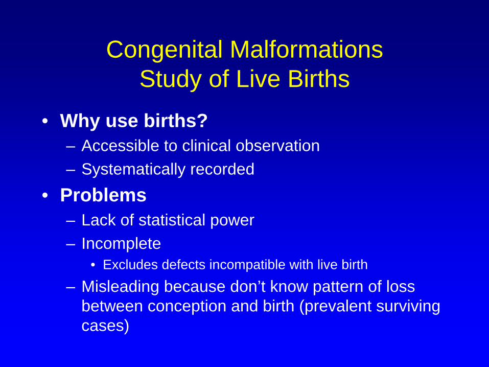
Congenital Malformations Study of Live Births
• Why use births?– Accessible to clinical observation– Systematically recorded
• Problems– Lack of statistical power– Incomplete
• Excludes defects incompatible with live birth
– Misleading because don’t know pattern of loss between conception and birth (prevalent surviving cases)
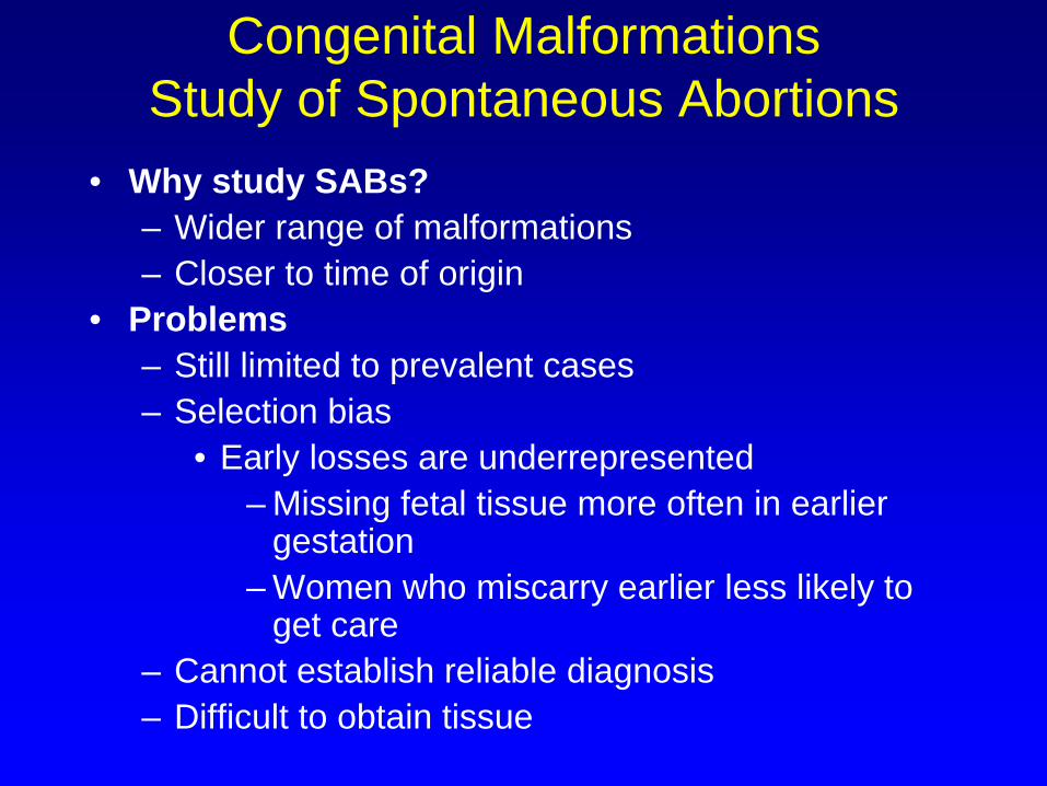
Congenital Malformations Study of Spontaneous Abortions
• Why study SABs?– Wider range of malformations– Closer to time of origin
• Problems– Still limited to prevalent cases– Selection bias
• Early losses are underrepresented– Missing fetal tissue more often in earlier
gestation– Women who miscarry earlier less likely to
get care– Cannot establish reliable diagnosis– Difficult to obtain tissue
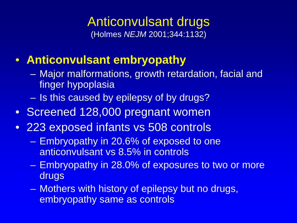
– Embryopathy in 20.6% of exposed to one anticonvulsant vs 8.5% in controls
– Embryopathy in 28.0% of exposures to two or more drugs
– Mothers with history of epilepsy but no drugs, embryopathy same as controls
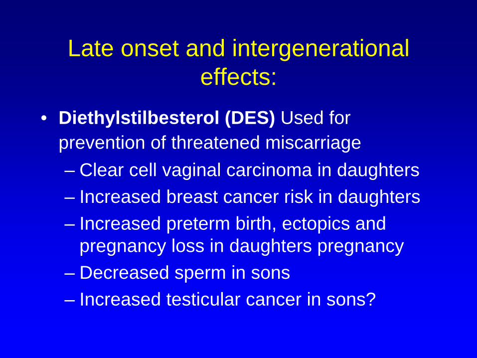
Late onset and intergenerational effects:
• Diethylstilbesterol (DES) Used for prevention of threatened miscarriage– Clear cell vaginal carcinoma in daughters– Increased breast cancer risk in daughters– Increased preterm birth, ectopics and
pregnancy loss in daughters pregnancy– Decreased sperm in sons– Increased testicular cancer in sons?
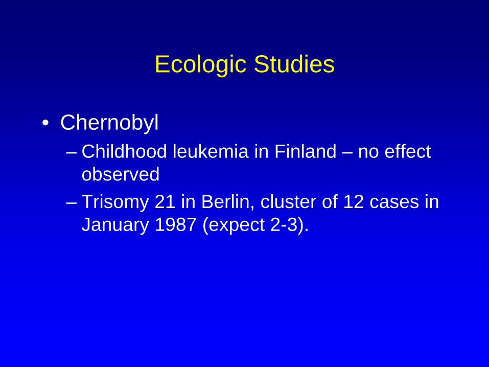
Ecologic Studies
• Chernobyl– Childhood leukemia in Finland – no effect
observed– Trisomy 21 in Berlin, cluster of 12 cases in
January 1987 (expect 2-3).
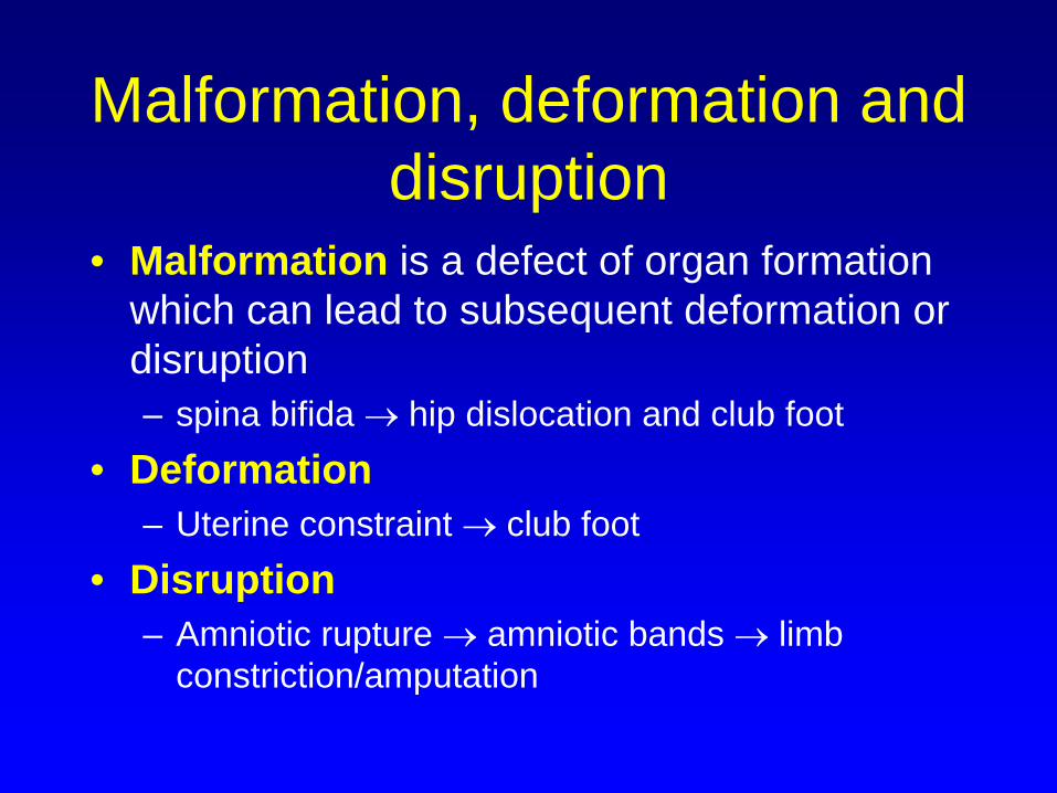
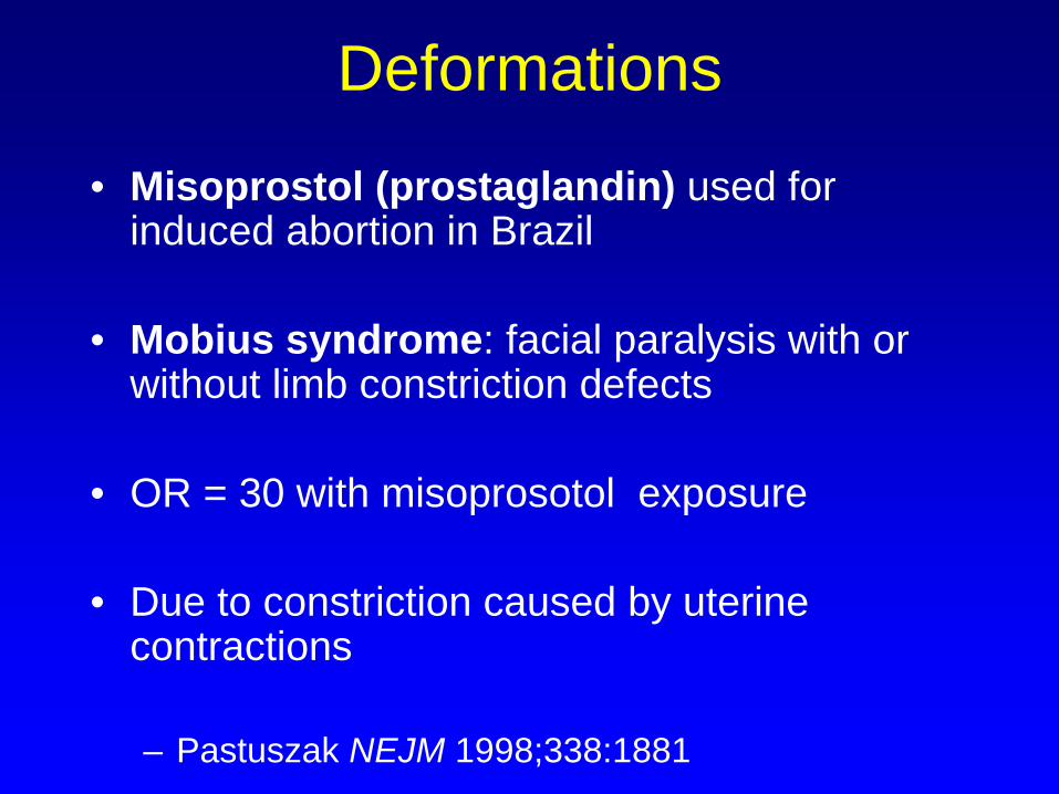
Deformations
• Misoprostol (prostaglandin) used for induced abortion in Brazil
• Mobius syndrome: facial paralysis with or without limb constriction defects
• OR = 30 with misoprosotol exposure
• Due to constriction caused by uterine contractions