Massachusetts Institute of Technology Massachusetts Institute of Technology Harvard Medical School Harvard Medical School Brigham and Women Brigham and Women ’ ’ s Hospital s Hospital VA Boston Healthcare System VA Boston Healthcare System 2.79J/3.96J/20.441/HST522J 2.79J/3.96J/20.441/HST522J TISSUE TYPES TISSUE TYPES M. Spector, Ph.D. M. Spector, Ph.D.
Transcript
Massachusetts Institute of TechnologyMassachusetts Institute of TechnologyHarvard Medical SchoolHarvard Medical School
Brigham and WomenBrigham and Women’’s Hospitals HospitalVA Boston Healthcare SystemVA Boston Healthcare System
OnOn--line Medical Dictionariesline Medical Dictionarieshttp://cancerweb.ncl.ac.uk/omd/http://cancerweb.ncl.ac.uk/omd/http://medicalhttp://medical--dictionary.thefreedictionary.com/dictionary.thefreedictionary.com/
Extracellular matrixExtracellular matrix continuous; and in somecontinuous; and in some tissues also cell continuoustissues also cell continuous
Cell continuousCell continuousMuscle and nerve cells areMuscle and nerve cells are surrounded by a basal laminasurrounded by a basal lamina Basal epithelial cells areBasal epithelial cells are attached to a basementattached to a basement membranemembraneBasal lamina=basementBasal lamina=basement membranemembrane
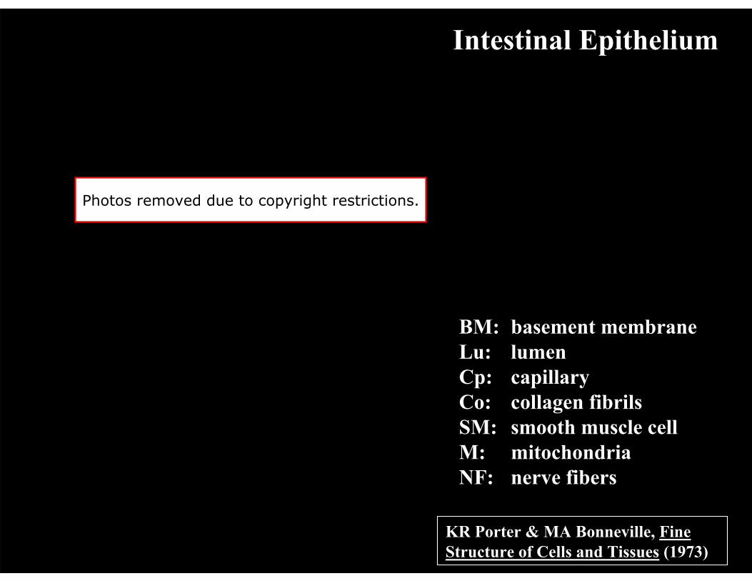
BASEMENT MEMBRANEBASEMENT MEMBRANE
Continuous sheet, 50Continuous sheet, 50--300 nm thick300 nm thickNo cells contained within the BM; it is aNo cells contained within the BM; it is a
nonliving structurenonliving structurePrincipal protein constituentsPrincipal protein constituents
LamininLamininType IV collagenType IV collagenType XVIII collagenType XVIII collagen
L. Jeng
Basement Membrane
Diagram removed due to copyright restrictions.
FASEB J 2005
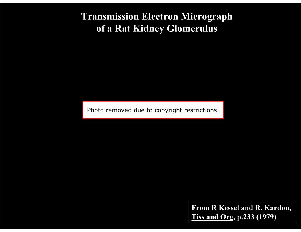
Image removed due to copyright restrictions. Diagrams of muscle, epithelial sheet, and kidney glomerulus structures.
B Alberts, et al, Mol Biol of the Cell, p. 819 (1989)
Scanning Electron Micrograph of the Cornea of a Chick Embryo
E: Epithelial cells BL: Basal lamina C: Collagen fibrils in
the underlying CT
Photo removed due to copyright restrictions.
B Alberts, et al, Mol Biol of the Cell, p. 819 (1989)
Connective Tissue
Image removed due to copyright restrictions.
Sketches from Illustrated Physiology, AB McNaught and R Callander, Williams and Wilkins, 1967
Connective Tissues
Image removed due to copyright restrictions. See http://cal.vet.upenn.edu/projects/histo/Index.htm
Loose and dense connective tissue from a cow's planum.
Image removed due to copyright restrictions. See http://cal.vet.upenn.edu/projects/histo/Index.htm
Fibrocartilage Cartilage identification is principally based on morphology; rounded cells in a lacuna; ECM is type I collagen (fibrocartilage) or type II cartilage (hyaline), with (elastic) or without elastin.
Simple Squamous Epithelium(chick blastodisc at about 33 hours of incubation )
Top View Cross-Sectional View
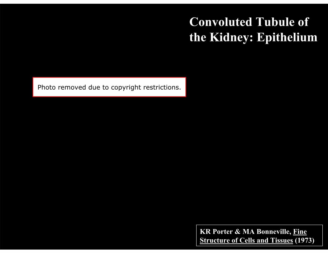
Simple squamous epithelium, which generally occurs as a thin sheet-like layer allowing for minimal resistance to diffusion, is also been called "pavement” epithelium, because it can look like like paving stones as seen from above. Examples include the linings of the peritoneal, pleural and pericardial cavities. Other places simple squamous epithelium can be found include: the glomerulus of the kidney, the walls of capillaries, and the alveoli of the lungs.
Since columnar cells are quite thick, they do not readily allow passive diffusion. As a result, these cells use active transport to move nutrients through them from the intestine to the blood. This is what we commonly call "absorption." To help with this, they have numerous microvilli on their apical (lumenal) surface, which increases their surface area to allow for greater absorption.
Image removed due to copyright considerations. See http://cal.vet.upenn.edu/projects/histo/Index.htm
This is a section through the edge of a gallbladder. There is a layer of simple columnar epithelium overlying the connective tissue as indicated by the arrows.
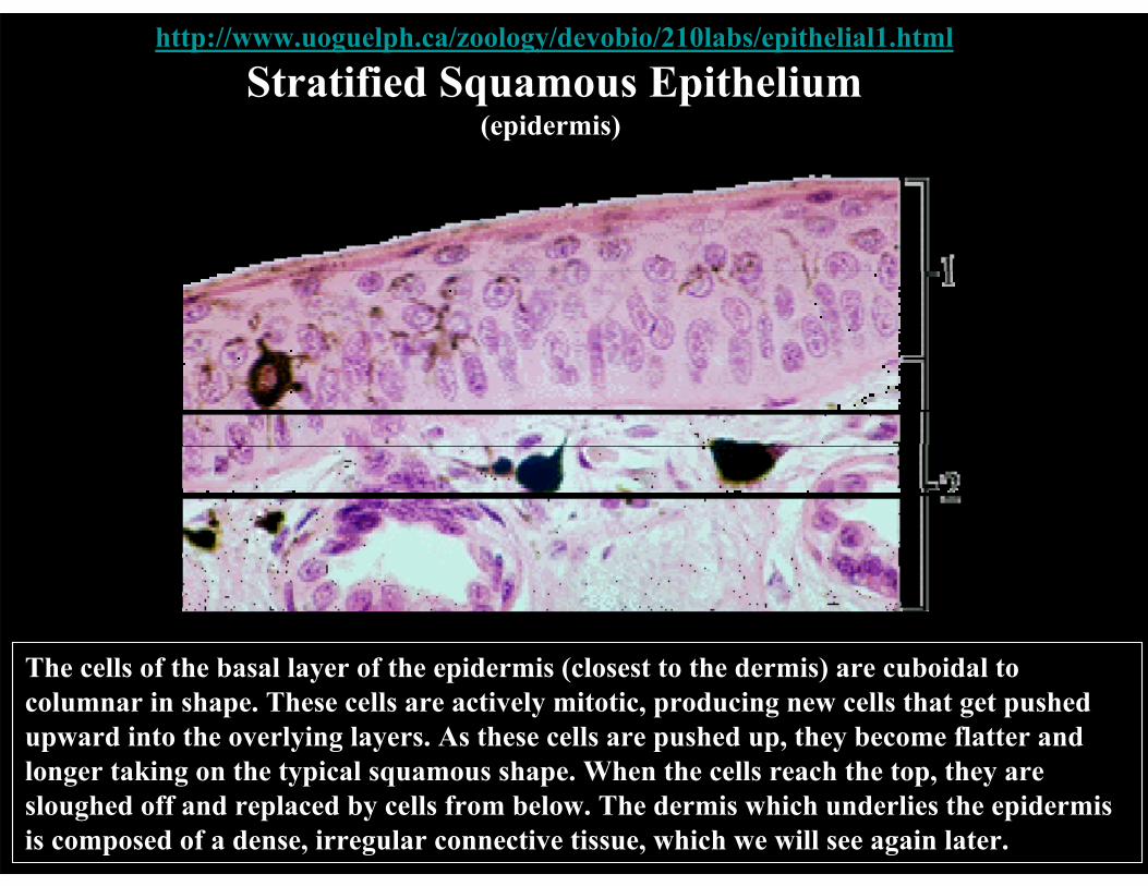
Stratified Squamous Epithelium
Image removed due to copyright restrictions. See http://cal.vet.upenn.edu/projects/histo/Index.htm
This is an example of stratified squamous epithelium from the esophagus of a cat. Arrows show nuclei of the outermost layer. This is normal for mucosa. Most stratified squamous cells in other areas, such as skin, lose their nuclei by the time they approach the outermost layers.
The cells of the basal layer of the epidermis (closest to the dermis) are cuboidal to columnar in shape. These cells are actively mitotic, producing new cells that get pushed upward into the overlying layers. As these cells are pushed up, they become flatter and longer taking on the typical squamous shape. When the cells reach the top, they are sloughed off and replaced by cells from below. The dermis which underlies the epidermis is composed of a dense, irregular connective tissue, which we will see again later.
Image removed due to copyright restrictions. See http://cal.vet.upenn.edu/projects/histo/Index.htm
This is an example of ciliated pseudostratified columnar epithelium from the trachea. The arrows indicate the layer of cilia on the surface of the pseudostratified columnar cell layer.
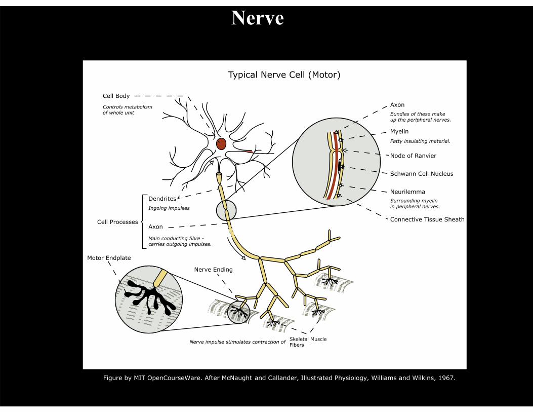
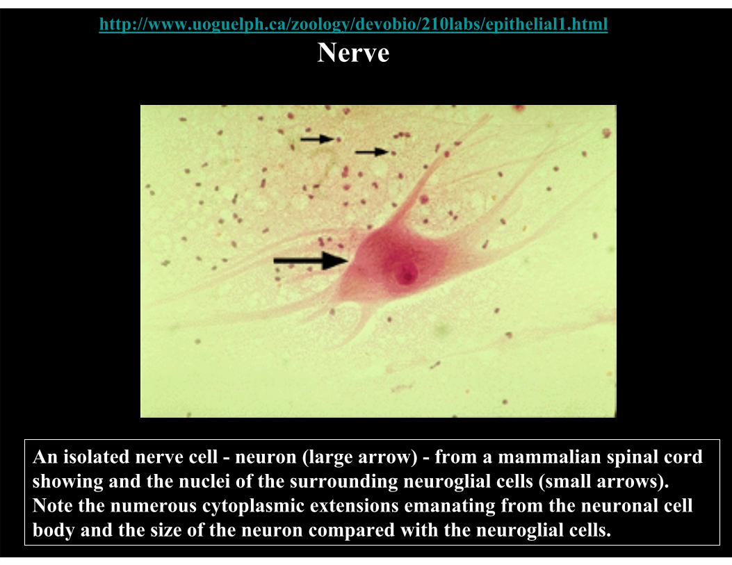
An isolated nerve cell - neuron (large arrow) - from a mammalian spinal cord showing and the nuclei of the surrounding neuroglial cells (small arrows). Note the numerous cytoplasmic extensions emanating from the neuronal cell body and the size of the neuron compared with the neuroglial cells.