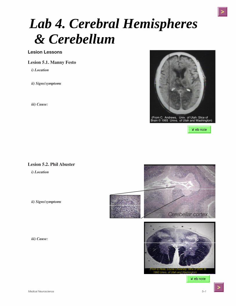
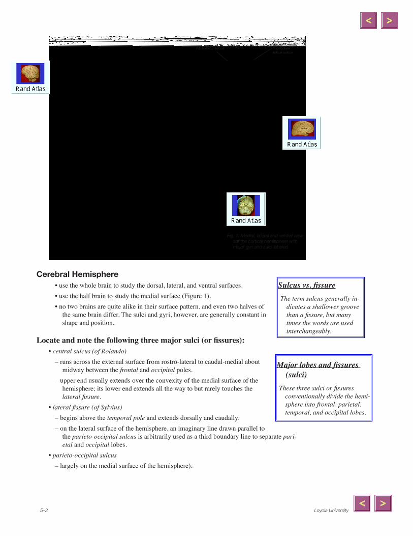
Cerebral Hemisphere• use the whole brain to study the dorsal, lateral, and ventral surfaces.• use the half brain to study the medial surface (Figure 1).• no two brains are quite alike in their surface pattern, and even two halves of
the same brain differ. The sulci and gyri, however, are generally constant in shape and position.
Locate and note the following three major sulci (or fissures):• central sulcus (of Rolando)
– runs across the external surface from rostro-lateral to caudal-medial about midway between the frontal and occipital poles.
– upper end usually extends over the convexity of the medial surface of the hemisphere; its lower end extends all the way to but rarely touches the lateral fissure.
• lateral fissure (of Sylvius)– begins above the temporal pole and extends dorsally and caudally.– on the lateral surface of the hemisphere, an imaginary line drawn parallel to
the parieto-occipital sulcus is arbitrarily used as a third boundary line to separate pari-etal and occipital lobes.
• parieto-occipital sulcus – largely on the medial surface of the hemisphere).
Major lobes and fissures (sulci)
These three sulci or fissures conventionally divide the hemi-sphere into frontal, parietal, temporal, and occipital lobes.
Sulcus vs. fissureThe term sulcus generally in-
dicates a shallower groove than a fissure, but many times the words are used interchangeably.
Fig. �. Medial, lateral and ventral view-sof the cortical hemisphere with major gyri and sulci labeled.
Paracentral sulcus Marginal branch
of cingulate sulcus sulcus
Medical Neuroscience 5–3
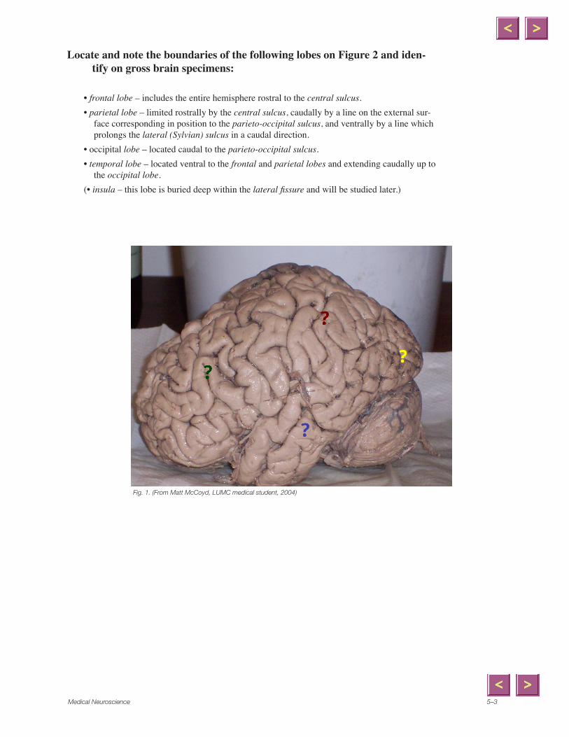
Locate and note the boundaries of the following lobes on Figure 2 and iden-tify on gross brain specimens:
• frontal lobe – includes the entire hemisphere rostral to the central sulcus.• parietal lobe – limited rostrally by the central sulcus, caudally by a line on the external sur-
face corresponding in position to the parieto-occipital sulcus, and ventrally by a line which prolongs the lateral (Sylvian) sulcus in a caudal direction.
• occipital lobe – located caudal to the parieto-occipital sulcus. • temporal lobe – located ventral to the frontal and parietal lobes and extending caudally up to
the occipital lobe.(• insula – this lobe is buried deep within the lateral fissure and will be studied later.)
Fig. �. (From Matt McCoyd, LUMC medical student, �004)
x
Polygon
x
Polygon
x
Polygon
x
Polygon
Loyola University5–4
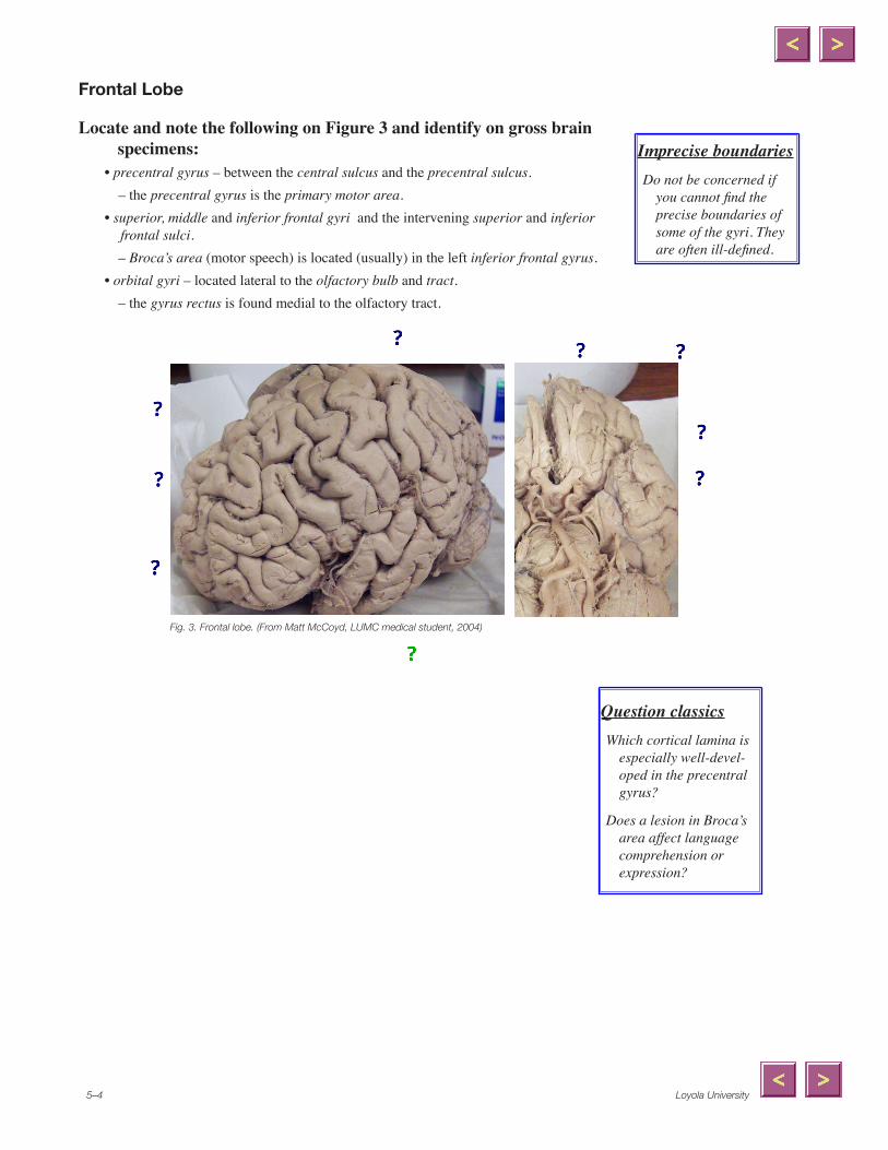
Frontal Lobe
Locate and note the following on Figure 3 and identify on gross brain specimens:
• precentral gyrus – between the central sulcus and the precentral sulcus.– the precentral gyrus is the primary motor area.
• superior, middle and inferior frontal gyri and the intervening superior and inferior frontal sulci.– Broca’s area (motor speech) is located (usually) in the left inferior frontal gyrus.
• orbital gyri – located lateral to the olfactory bulb and tract.– the gyrus rectus is found medial to the olfactory tract.
Fig. 3. Frontal lobe. (From Matt McCoyd, LUMC medical student, �004)
Imprecise boundariesDo not be concerned if
you cannot find the precise boundaries of some of the gyri. They are often ill-defined.
Question classicsWhich cortical lamina is
especially well-devel-oped in the precentral gyrus?
Does a lesion in Broca’s area affect language comprehension or expression?
x
Line
x
Line
x
Line
x
Line
x
Line
x
Polygonal Line
x
Line
x
Line
x
Polygonal Line
x
Polygonal Line
x
Line
x
Polygonal Line
x
Line
x
Oval
x
Polygonal Line
x
Line
Medical Neuroscience 5–5
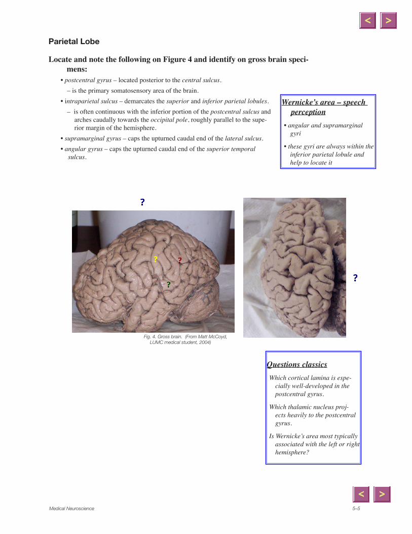
Parietal Lobe
Locate and note the following on Figure 4 and identify on gross brain speci-mens:
• postcentral gyrus – located posterior to the central sulcus.– is the primary somatosensory area of the brain.
• intraparietal sulcus – demarcates the superior and inferior parietal lobules.– is often continuous with the inferior portion of the postcentral sulcus and
arches caudally towards the occipital pole, roughly parallel to the supe-rior margin of the hemisphere.
• supramarginal gyrus – caps the upturned caudal end of the lateral sulcus.• angular gyrus – caps the upturned caudal end of the superior temporal
sulcus.
Fig. 4. Gross brain. (From Matt McCoyd, LUMC medical student, �004)
Wernicke’s area – speech perception
• angular and supramarginal gyri
• these gyri are always within the inferior parietal lobule and help to locate it
Questions classicsWhich cortical lamina is espe-
cially well-developed in the postcentral gyrus.
Which thalamic nucleus proj-ects heavily to the postcentral gyrus.
Is Wernicke’s area most typically associated with the left or right hemisphere?
x
Polygonal Line
x
Line
x
Oval
x
Oval
x
Oval
x
Line
Loyola University5–�
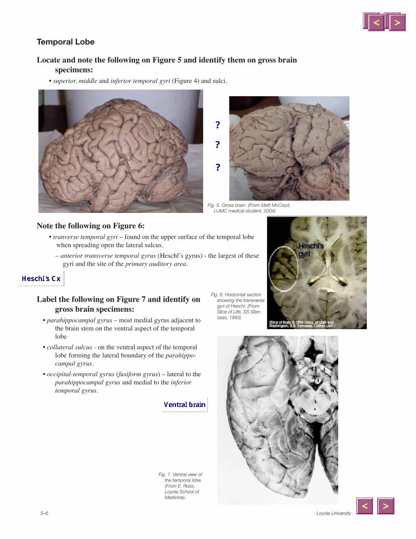
Temporal Lobe
Locate and note the following on Figure 5 and identify them on gross brain specimens:
• superior, middle and inferior temporal gyri (Figure 4) and sulci.
Note the following on Figure 6: • tranverse temporal gyri – found on the upper surface of the temporal lobe
when spreading open the lateral sulcus. – anterior transverse temporal gyrus (Heschl’s gyrus) - the largest of these
gyri and the site of the primary auditory area.
Label the following on Figure 7 and identify on gross brain specimens:
• parahippocampal gyrus – most medial gyrus adjacent to the brain stem on the ventral aspect of the temporal lobe
• collateral sulcus - on the ventral aspect of the temporal lobe forming the lateral boundary of the parahippo-campal gyrus.
• occipital-temporal gyrus (fusiform gyrus) – lateral to the parahippocampal gyrus and medial to the inferior temporal gyrus.
Fig. 5. Gross brain. (From Matt McCoyd, LUMC medical student, �004)
Fig. �. Horizontal section showing the transverse gyri of Heschl. (From Slice of Life, SS Sten-saas, �993)
Fig. 7. Ventral view of the temporal lobe. (From E. Ross, Loyola School of Medicine).
x
Polygonal Line
x
Polygonal Line
x
Polygonal Line
Medical Neuroscience 5–7
Insula• can be seen by gently pulling apart the borders (opercula) of the lateral fissure.• is cone-shaped portion of the cortex that is sometimes referred to as an additional lobe of the
cerebral cortex.
Label the following on Figure 8:• opercula – those portions of the frontal, parietal and temporal lobes that cover the insula. • limen insulae – apex or point of the insula
– is found ventrally where the insula meets the orbital surface of the frontal lobe.
• circular sulcus – surrounds the insula. • gyri breves and gyrus longus - the short and long gyri
of the insula.
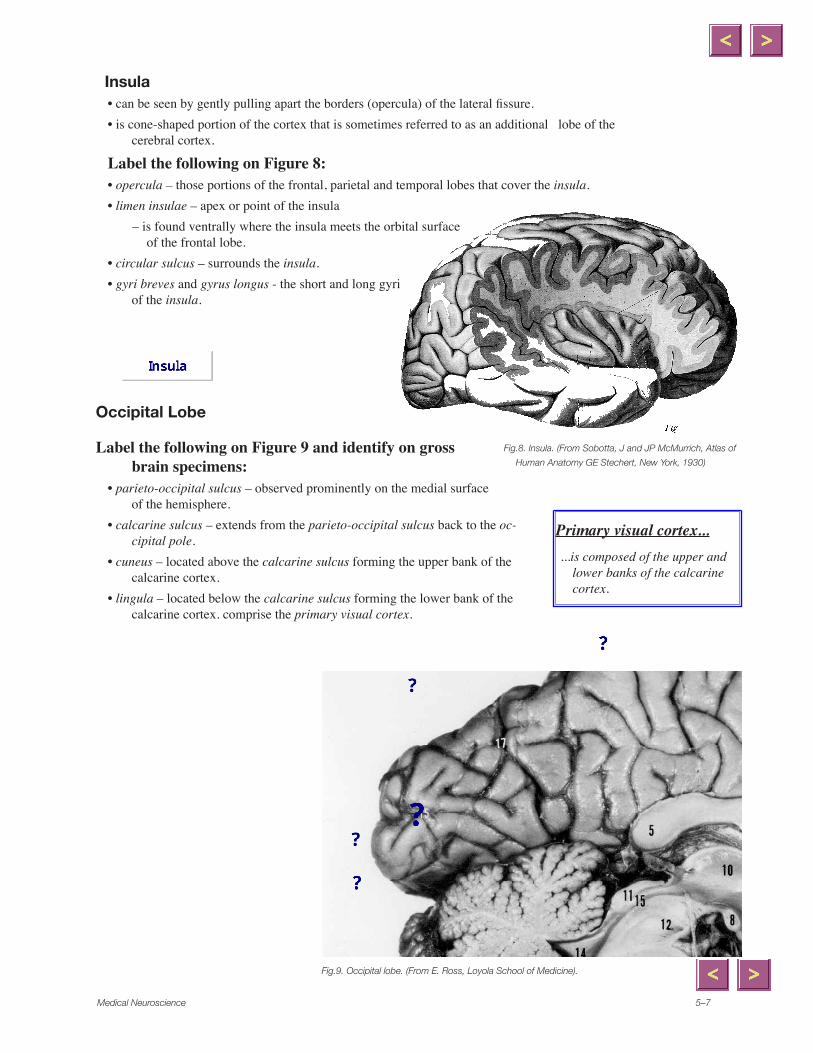
Occipital Lobe
Label the following on Figure 9 and identify on gross brain specimens:
• parieto-occipital sulcus – observed prominently on the medial surface of the hemisphere.
• calcarine sulcus – extends from the parieto-occipital sulcus back to the oc-cipital pole.
• cuneus – located above the calcarine sulcus forming the upper bank of the calcarine cortex.
• lingula – located below the calcarine sulcus forming the lower bank of the calcarine cortex. comprise the primary visual cortex.
Fig.9. Occipital lobe. (From E. Ross, Loyola School of Medicine).
Fig.8. Insula. (From Sobotta, J and JP McMurrich, Atlas of
Human Anatomy GE Stechert, New York, �930)
Primary visual cortex......is composed of the upper and
lower banks of the calcarine cortex.
x
Polygonal Line
x
Line
x
Polygonal Line
x
Line
x
Line
x
Polygonal Line
x
Polygon
x
Polygonal Line
Loyola University5–8
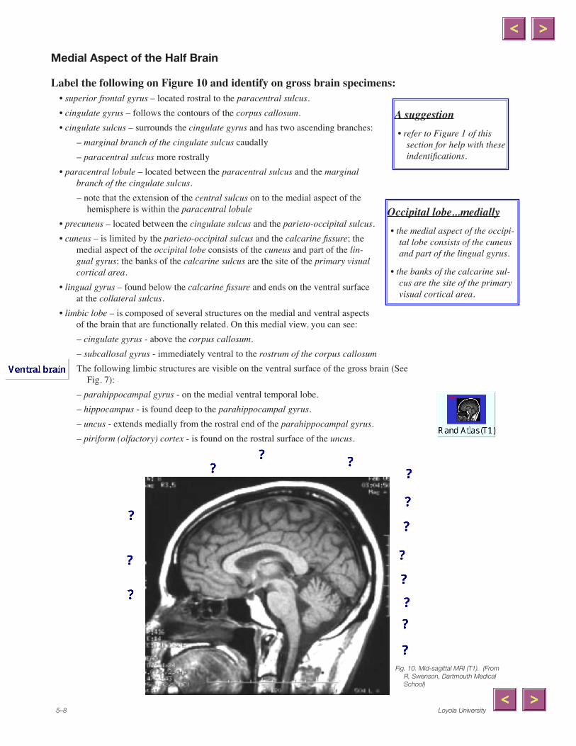
Medial Aspect of the Half Brain
Label the following on Figure 10 and identify on gross brain specimens: • superior frontal gyrus – located rostral to the paracentral sulcus.• cingulate gyrus – follows the contours of the corpus callosum.• cingulate sulcus – surrounds the cingulate gyrus and has two ascending branches:
– marginal branch of the cingulate sulcus caudally– paracentral sulcus more rostrally
• paracentral lobule – located between the paracentral sulcus and the marginal branch of the cingulate sulcus.– note that the extension of the central sulcus on to the medial aspect of the
hemisphere is within the paracentral lobule • precuneus – located between the cingulate sulcus and the parieto-occipital sulcus.• cuneus – is limited by the parieto-occipital sulcus and the calcarine fissure; the
medial aspect of the occipital lobe consists of the cuneus and part of the lin-gual gyrus; the banks of the calcarine sulcus are the site of the primary visual cortical area.
• lingual gyrus – found below the calcarine fissure and ends on the ventral surface at the collateral sulcus.
• limbic lobe – is composed of several structures on the medial and ventral aspects of the brain that are functionally related. On this medial view, you can see: – cingulate gyrus - above the corpus callosum.– subcallosal gyrus - immediately ventral to the rostrum of the corpus callosumThe following limbic structures are visible on the ventral surface of the gross brain (See
Fig. 7):– parahippocampal gyrus - on the medial ventral temporal lobe.– hippocampus - is found deep to the parahippocampal gyrus.– uncus - extends medially from the rostral end of the parahippocampal gyrus.– piriform (olfactory) cortex - is found on the rostral surface of the uncus.
Occipital lobe...medially• the medial aspect of the occipi-
tal lobe consists of the cuneus and part of the lingual gyrus.
• the banks of the calcarine sul-cus are the site of the primary visual cortical area.
A suggestion• refer to Figure 1 of this
section for help with these indentifications.
x
Polygonal Line
x
Polygonal Line
x
Polygonal Line
x
Line
x
Line
x
Line
x
Line
x
Polygonal Line
x
Polygonal Line
x
Polygonal Line
x
Polygonal Line
x
Polygonal Line
x
Line
x
Polygonal Line
x
Line
x
Polygonal Line
Medical Neuroscience 5–9
Brodmann’s Cytoarchitectural Numbering System
Fig. ��. From AJ Castro et al., Mosby �00� as derived from: Brodmann, K. Vergleichende Lokalisa-tionslehre der Grosshirnrinde in ihren Prinzipien dargestellt auf Grund des Zellenbaues (J. A. Barth, Leipzig, �909).
Add the Brodmann numbers to the cortical areas indicated below.
________ primary motor cortex (M1) ________ premotor cortex (M2)
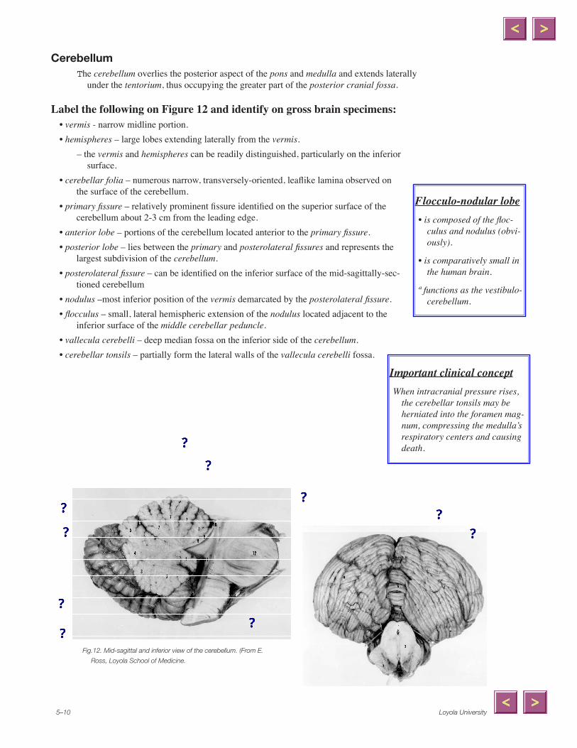
CerebellumThe cerebellum overlies the posterior aspect of the pons and medulla and extends laterally
under the tentorium, thus occupying the greater part of the posterior cranial fossa.
Label the following on Figure 12 and identify on gross brain specimens: • vermis - narrow midline portion.• hemispheres – large lobes extending laterally from the vermis.
– the vermis and hemispheres can be readily distinguished, particularly on the inferior surface.
• cerebellar folia – numerous narrow, transversely-oriented, leaflike lamina observed on the surface of the cerebellum.
• primary fissure – relatively prominent fissure identified on the superior surface of the cerebellum about 2-3 cm from the leading edge.
• anterior lobe – portions of the cerebellum located anterior to the primary fissure.• posterior lobe – lies between the primary and posterolateral fissures and represents the
largest subdivision of the cerebellum. • posterolateral fissure – can be identified on the inferior surface of the mid-sagittally-sec-
tioned cerebellum• nodulus –most inferior position of the vermis demarcated by the posterolateral fissure.• flocculus – small, lateral hemispheric extension of the nodulus located adjacent to the
inferior surface of the middle cerebellar peduncle.• vallecula cerebelli – deep median fossa on the inferior side of the cerebellum.• cerebellar tonsils – partially form the lateral walls of the vallecula cerebelli fossa.
Fig.��. Mid-sagittal and inferior view of the cerebellum. (From E.
Ross, Loyola School of Medicine.
Flocculo-nodular lobe• is composed of the floc-
culus and nodulus (obvi-ously).
• is comparatively small in the human brain.
ª functions as the vestibulo-cerebellum.
Important clinical conceptWhen intracranial pressure rises,
the cerebellar tonsils may be herniated into the foramen mag-num, compressing the medulla’s respiratory centers and causing death.
x
Polygonal Line
x
Line
x
Polygonal Line
x
Polygonal Line
x
Line
x
Polygonal Line
x
Line
x
Polygonal Line
x
Polygonal Line
x
Polygonal Line
x
Polygonal Line
x
Polygonal Line
x
Polygonal Line
x
Line
Medical Neuroscience 5–��
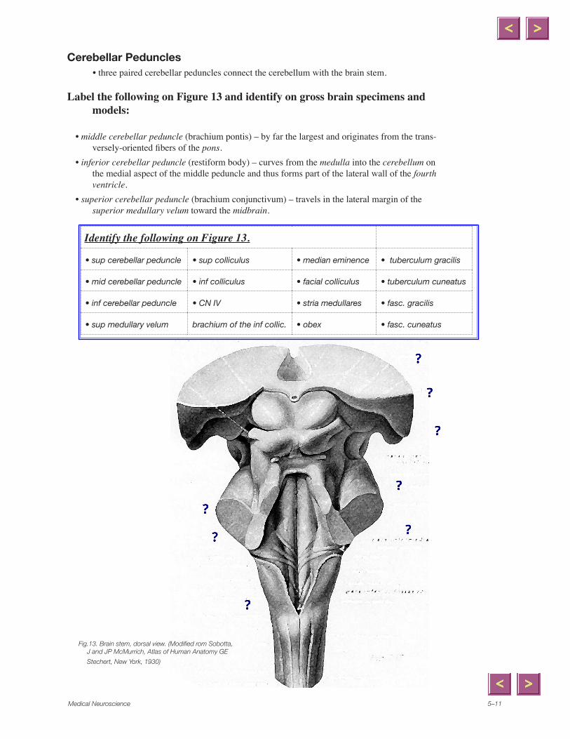
Cerebellar Peduncles• three paired cerebellar peduncles connect the cerebellum with the brain stem.
Label the following on Figure 13 and identify on gross brain specimens and models:
• middle cerebellar peduncle (brachium pontis) – by far the largest and originates from the trans-versely-oriented fibers of the pons.
• inferior cerebellar peduncle (restiform body) – curves from the medulla into the cerebellum on the medial aspect of the middle peduncle and thus forms part of the lateral wall of the fourth ventricle.
• superior cerebellar peduncle (brachium conjunctivum) – travels in the lateral margin of the superior medullary velum toward the midbrain.
Fig.13. Brain stem, dorsal view. (Modified rom Sobotta, J and JP McMurrich, Atlas of Human Anatomy GE
• sup medullary velum brachium of the inf collic. • obex • fasc. cuneatus
x
Polygonal Line
x
Polygonal Line
x
Line
x
Polygonal Line
x
Line
x
Line
x
Line
x
Polygonal Line
Loyola University5–��
Case break
Drops and Sways
A seven year old girl, Liz Tureen, always a “slow learner,” begins to drop things with her right hand, and sways when she walks. Examination shows dysarthric speech and dysmetria of the right upper and lower limbs on finger-nose-finger and heel-shin-knee maneuvers. She begins to have headaches, nausea, and lethargy over the next two weeks.
1. Where is her lesion?
2. What kind of lesion is most likely?
3. Why is she developing headaches, nausea, and lethargy?
4. If CSF flow is impaired, which ventricles would be enlarged or dilated on CT scan?
Medical Neuroscience 5–�3
Study Questions – Cerebral Cortex1. What are the Brodmann numbers of the... _______ primary motor cortex in the precentral gyrus? _______ primary somatosensory cortex in the postcentral gyrus? _______ primary auditory cortex in the transverse temporal gyri? _______ primary visual cortex in the banks of the calcarine fissure? 2. Where is the frontal eyefield?
3. What deficit would you expect with damage to the left inferior frontal gyrus in a right-handed individual? ...in a left-handed individual?
4. How could you test the laterality of language function in a patient?
5. How could temporal lobe lesions affect vision?
6. What is the topographic organization of bodily movements in the precentral gyrus?
7. Where is the striate cortex?
8. What deficits result from occlusion of the anterior cerebral artery? ...the middle cerebral artery? ...the posterior cerebral artery?
Loyola University5–�4
Study Questions – Cerebellum1. Identify the areas of the cerebellum associated with its functional subdivisions: ...vestibulocerebellum
...spinocerebellum
...neocerebellum.
2. What are climbing fibers? ...mossy fibers?
3. Name the component tracts of each cerebellar peduncle. inferior
middle
superior
4. What is the posterior inferior cerebellar artery (PICA) syndrome?
5. Where do Purkinje cells axons terminate? Are they inhibitory, facilitatory, or mixed?
Medical Neuroscience 5–�5
Label the following structures on Figure. 15.• sup, middle and inf frontal gyri • lateral fissure
• sup, middle and inf temporal gyri • insula
• lateral ventricles • internal carotid artery
• septum pellucidum • middle cerebral artery
• corpus callosum • superior sagittal sinus
Fig. �5. Coronal MRI (T�) through the frontal lobe. (From R, Swenson, Dartmouth Medical School)
Label the following struc-tures on Figure 16.
• frontal pole
• occipital pole
• genu and splenium of the corpus callosum
• lateral ventricles
• septum pellucidum
• insula
• superior sagittal sinus
Fig. ��. Axial MRI (T�) through the hemisphere. (From R, Swenson, Dartmouth Medical School)
MRI Review
x
Line
x
Line
x
Line
x
Line
x
Polygonal Line
x
Polygonal Line
x
Polygonal Line
x
Polygonal Line
x
Polygonal Line
x
Polygonal Line
x
Polygonal Line
x
Polygonal Line
x
Polygonal Line
x
Line
x
Line
x
Line
x
Line
x
Polygonal Line
x
Line
x
Line
x
Line
Note
None set by
Note
Unmarked set by
x
Line
Loyola University5–��
Label the following structures on Figure 17:• lateral ventricles • foramen of Monroe• fornix • temporal lobe• corpus callosum • thalamus• insula • internal capsule• sup sagittal sinus • caudate
Fig. �7. Axial MRI’s (T�) showing a more dorsal level of
the temporal lobe as compared to figure . (From R, Swenson, Dartmouth Medical School)
Label the following structures on Figure 18 • cerebellum • uncus• middle cerebral artery • mammillary body• cerebral aqueduct • uncus• pituitary stalk (infun-
dibulum)• orbital gyrus of the
frontal lobe
Related QuestionsOn the Figure 17, can you
identify the course of the optic radiations that project from the lateral geniculate at the caudal end of the thala-mus to the calcarine cortex in the occipital lobe?
Also, what part of the frontal lobe is visible.
MRI Review (cont’d)
Related questionsWhat cranial nerves emerge from
this level of the brain stem?
What do they innervate?
Can you trace their course? What part of the temporal lobe might they course by? (hint: uncus)
Fig. �8. Axial MRI’s (T�) showing a more ventral level of
the temporal lobe as compared to figure . (From R, Swenson, Dartmouth Medical School)
Medical Neuroscience 5–�7
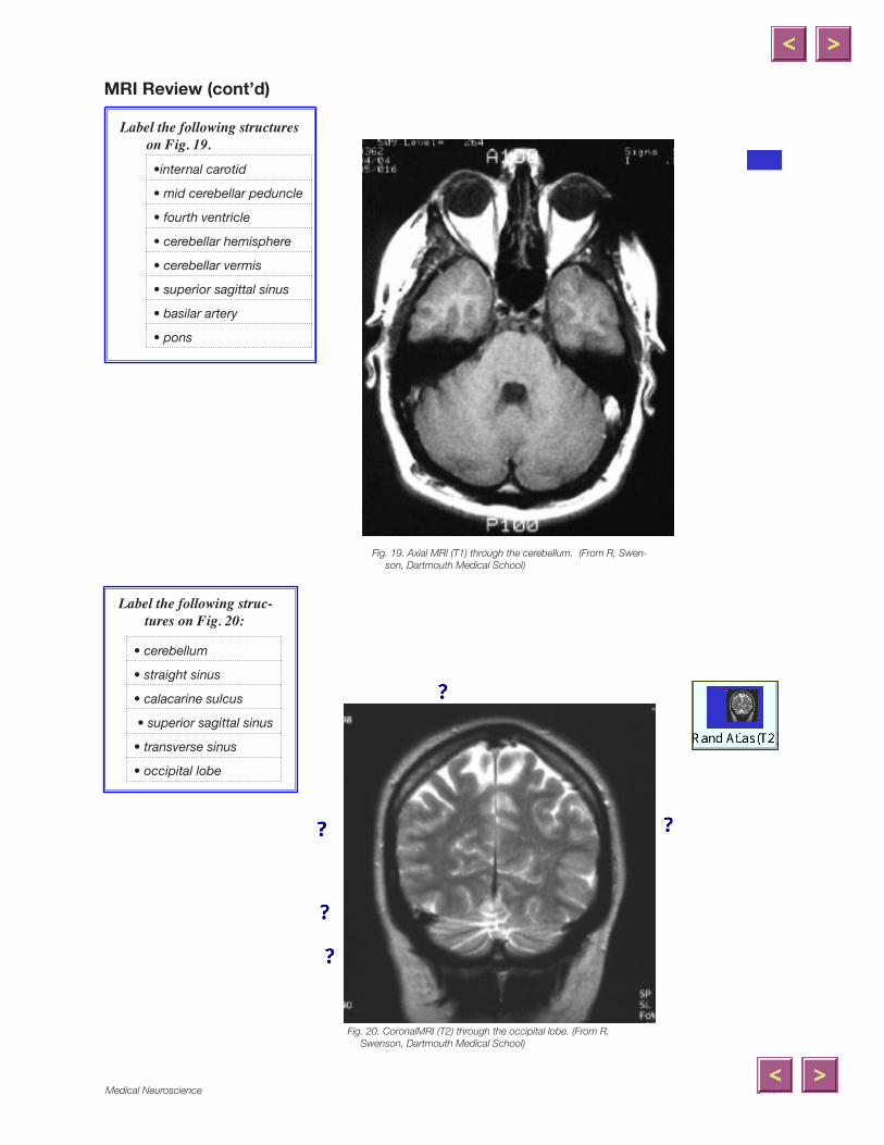
Fig. �9. Axial MRI (T�) through the cerebellum. (From R, Swen-son, Dartmouth Medical School)
Label the following structures on Fig. 19.
•internal carotid
• mid cerebellar peduncle
• fourth ventricle
• cerebellar hemisphere
• cerebellar vermis
• superior sagittal sinus
• basilar artery
• pons
Fig. �0. CoronalMRI (T�) through the occipital lobe. (From R, Swenson, Dartmouth Medical School)
Label the following struc-tures on Fig. 20:
• cerebellum
• straight sinus
• calacarine sulcus
• superior sagittal sinus
• transverse sinus
• occipital lobe
MRI Review (cont’d)
x
Polygonal Line
x
Polygonal Line
x
Polygonal Line
x
Line
x
Polygonal Line
Loyola University5–�8
Patient Puzzle
Patient 5.1. Case of the clumsy manPatient: Mr. Sam Anella Age: 62 Occupation: Lepidopterist
Signs and Symptoms:• Mr. Anella demonstrates a progressively worsening uncoordinated gate.• You observe clumsy arm movements as well.• He complains most of “losing his balance” and therefore often uses a
wheelchair.• No somatosensory loss is detected and he shows no paralysis.• You learn that he had a uncle who passed away with the same symptoms.
Diagnosis:1. Is this a vestibular nerve problem? If so, what else would you look for?
2. Do you suspect Sam had a stroke? What’s the evidence for and against?
3. What about his uncle? What does this suggest?
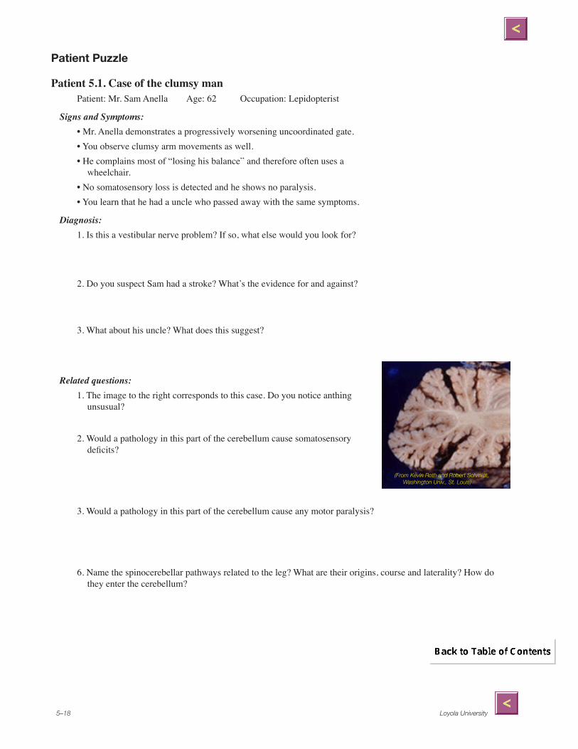
Related questions:1. The image to the right corresponds to this case. Do you notice anthing
unsusual?
2. Would a pathology in this part of the cerebellum cause somatosensory deficits?
3. Would a pathology in this part of the cerebellum cause any motor paralysis?
6. Name the spinocerebellar pathways related to the leg? What are their origins, course and laterality? How do they enter the cerebellum?
(From Kevin Roth and Robert Schmidt, Washington Univ., St. Louis)