504 Biomed Environ Sci, 2013; 26(6): 504-508 doi: 10.3967/0895-3988.2013.06.013 1.State Key Laboratory for Communicable Disease Prevention and Control, National Institute for Communicable Disease Control and Prevention, Beijing, 102206, China; 2.Hainan Provincial People’s Hospital, Sanya, 572000, China; 3. Guangdong Provincial Center for Disease Control and Prevention, Guangzhou, 510300, China Letter to the Editor Molecular Typing of Brucella Suis Collected from 1960s to 2010s in China by MLVA and PFGE LI Zhen Jun 1 , CUI Bu Yun 1 , CHEN Hai 2 , CHEN Jing Diao 3 , ZHAO Hong Yan 1 , PIAO Dong Ri 1 , JIANG Hai 1 , ZHANG Li 1 , TANG Xu 1 , KE Chang Wen 3 , YAO zhen 2 , and TIAN Guo Zhong 1,# Brucellosis is a bacterial anthropozoonosis usually caused by Brucella abortus, Brucella melitensis, Brucella suis and Brucella canis. Brucella suis, the causative agent of swine brucellosis, is classified into five biovars and preferentially infects different animal hosts [1] . In China, brucellosis is a national notifiable communicable disease both in animals and in human. In 2009, 35 816 brucellosis cases were reported. The annual incidence was 2.7 per 100 000 population. The brucellosis cases, including zoonotic and human infections caused by B. suis, were mainly distributed in rural areas in southern China, such as Guangdong, Guangxi, and Hainan provinces. In this study, the B. suis strains, which were collected from 1960s to 2010s in China were used in pulsed-field gel electrophoresis (PFGE) fingerprinting and multiple-locus variable-number tandem-repeat analysis (MLVA) to understand their molecular characteristics and evolution relationships among them. Thirty-two B.suis strains including 26 field strains, 1 vaccine strain S2 and 5 reference strains of B. suis biovar 1-5 (biovar 1: REF 1330S, biovar 2: REF Thomsen, biovar 3: REF 686, biovar 4: REF40, and biovar 5: REF 513) were analyzed in this study (Table 1). Sixteen strains were isolated from sick pigs, 6 were from sick cattle, 4 from brucellosis patients. Among 2 strains isolated from Guangdong, 16 strains from Guangxi and 2 strains from Hainan, no epidemiological link was found. All strains were biotyped according to the standard bacteriological procedures [1] . Genomic DNA of Brucella strains was extracted according to the manufacturer's protocol (QIAamp® DNA Mini Kit, Switzerland). Genomic DNA fingerprinting of PFGE was performed as previously described [2] but briefly revised in this study. Brucella DNA in agarose was digested with 20 U of XbaI for 4 h at 37 °C in the appropriate buffer according to the instructions of the manufacturer (Merck). Plug slices containing the digested DNA were placed in the wells of a 1% agarose gel (SeaKem Gold) in 0.5× TBE [Tris- Borate-EDTA] buffer (Invitrogen Life Technologies) and subjected to electrophoresis in a CHEF-DR III System (Bio-Rad Laboratories). Electrophoresis conditions were as follows: switch times of 0.5 and 8 s for 16 h, 10 and 56 s for 5 h, angle of 120°, gradient of 6 V/cm, temperature of 14 °C, and a linear ramping factor. After the electrophoresis, gels were stained with ethidium bromide (0.5 mg /mL) (AMRESCO, Inc.) for 30 min, followed by three 20 min cycles of destaining in water. The gels were then photographed under ultraviolet illumination (Gel Doc TM XR, USA). PFGE patterns were analyzed by using the BioNumerics version 4.0 software (Applied Maths, Applied Maths BVBA, Belgium). The TIFF images were normalized by using the PulseNet universal Salmonella enterica serotype Braenderup (H9812) size standard on each gel (16) against the reference in the database. PFGE profiles were compared by using the Dice coefficient and UPGMA (unweighted pair group method using arithmetic averages) clustering with a 1.5% band position tolerance window and 1.5% optimization. The method for selecting appropriate VNTRs has been previously described by Le Flèche and Maquart [3-4] . The sequences of 16 primer pairs were listed in Table 2. PCR amplification was performed using C1000 Thermal Cycler (BIO RAD) in a total volume of 25 μL containing 30 ng of DNA, 1× PCR reaction buffer, 1 U of Taq DNA polymerase (TaKaRa), 200 μmol/L of each deoxynucleotide triphosphate, and 0.8 μmol/L of each flanking primer. An initial denaturation step at 95 °C for 5 min was
Transcript
504 Biomed Environ Sci, 2013; 26(6): 504-508
doi: 10.3967/0895-3988.2013.06.013 1.State Key Laboratory for Communicable Disease Prevention and Control, National Institute for Communicable
Disease Control and Prevention, Beijing, 102206, China; 2.Hainan Provincial People’s Hospital, Sanya, 572000, China; 3. Guangdong Provincial Center for Disease Control and Prevention, Guangzhou, 510300, China
Letter to the Editor
Molecular Typing of Brucella Suis Collected from 1960s to 2010s in China by MLVA and PFGE
LI Zhen Jun1, CUI Bu Yun1, CHEN Hai2, CHEN Jing Diao3, ZHAO Hong Yan1, PIAO Dong Ri1, JIANG Hai1, ZHANG Li1, TANG Xu1, KE Chang Wen3, YAO zhen2, and TIAN Guo Zhong1,#
Brucellosis is a bacterial anthropozoonosis usually caused by Brucella abortus, Brucella melitensis, Brucella suis and Brucella canis. Brucella suis, the causative agent of swine brucellosis, is classified into five biovars and preferentially infects different animal hosts[1]. In China, brucellosis is a national notifiable communicable disease both in animals and in human. In 2009, 35 816 brucellosis cases were reported. The annual incidence was 2.7 per 100 000 population. The brucellosis cases, including zoonotic and human infections caused by B. suis, were mainly distributed in rural areas in southern China, such as Guangdong, Guangxi, and Hainan provinces. In this study, the B. suis strains, which were collected from 1960s to 2010s in China were used in pulsed-field gel electrophoresis (PFGE) fingerprinting and multiple-locus variable-number tandem-repeat analysis (MLVA) to understand their molecular characteristics and evolution relationships among them.
Thirty-two B.suis strains including 26 field strains, 1 vaccine strain S2 and 5 reference strains of B. suis biovar 1-5 (biovar 1: REF 1330S, biovar 2: REF Thomsen, biovar 3: REF 686, biovar 4: REF40, and biovar 5: REF 513) were analyzed in this study (Table 1). Sixteen strains were isolated from sick pigs, 6 were from sick cattle, 4 from brucellosis patients. Among 2 strains isolated from Guangdong, 16 strains from Guangxi and 2 strains from Hainan, no epidemiological link was found. All strains were biotyped according to the standard bacteriological procedures[1]. Genomic DNA of Brucella strains was extracted according to the manufacturer's protocol (QIAamp® DNA Mini Kit, Switzerland).
Genomic DNA fingerprinting of PFGE was performed as previously described[2] but briefly revised in this study. Brucella DNA in agarose was
digested with 20 U of XbaI for 4 h at 37 °C in the appropriate buffer according to the instructions of the manufacturer (Merck). Plug slices containing the digested DNA were placed in the wells of a 1% agarose gel (SeaKem Gold) in 0.5× TBE [Tris- Borate-EDTA] buffer (Invitrogen Life Technologies) and subjected to electrophoresis in a CHEF-DR III System (Bio-Rad Laboratories). Electrophoresis conditions were as follows: switch times of 0.5 and 8 s for 16 h, 10 and 56 s for 5 h, angle of 120°, gradient of 6 V/cm, temperature of 14 °C, and a linear ramping factor. After the electrophoresis, gels were stained with ethidium bromide (0.5 mg /mL) (AMRESCO, Inc.) for 30 min, followed by three 20 min cycles of destaining in water. The gels were then photographed under ultraviolet illumination (Gel DocTM XR, USA).
PFGE patterns were analyzed by using the BioNumerics version 4.0 software (Applied Maths, Applied Maths BVBA, Belgium). The TIFF images were normalized by using the PulseNet universal Salmonella enterica serotype Braenderup (H9812) size standard on each gel (16) against the reference in the database. PFGE profiles were compared by using the Dice coefficient and UPGMA (unweighted pair group method using arithmetic averages) clustering with a 1.5% band position tolerance window and 1.5% optimization.
The method for selecting appropriate VNTRs has been previously described by Le Flèche and Maquart[3-4]. The sequences of 16 primer pairs were listed in Table 2. PCR amplification was performed using C1000 Thermal Cycler (BIO RAD) in a total volume of 25 μL containing 30 ng of DNA, 1× PCR reaction buffer, 1 U of Taq DNA polymerase (TaKaRa), 200 μmol/L of each deoxynucleotide triphosphate, and 0.8 μmol/L of each flanking primer. An initial denaturation step at 95 °C for 5 min was
Biomed Environ Sci, 2013; 26(6): 504-508 505
followed by 30 cycles of denaturation at 95 °C for 30 s, annealing at 60 °C for 30 s, and elongating at 72 °C for 30 s. The final extension step was performed at 72 °C for 5 min. Three microliters of the amplification product were loaded in 2.0% standard agarose gel (Cambraex Bio Science Rockland) and the gel run under a voltage of 8 V/cm until the bromophenol blue dye had reached the 20 cm position(3 h or so). A 100-bp ladder were used as molecular size markers. Gels were stained with
ethidium bromide, visualized under UV light and photographed. The molecular weight of PCR products was managed using the BioNumerics software package (Version 4.0, Applied Maths BVBA, Belium). Band size estimates were converted to the tandem-repeat numbers using the BioNumerics software. A phylogenetic tree for 16 loci was constructed by BioNumerics software package using the UPGMA algorithm (Version 4.0, Applied Maths BVBA, Belium).
Table 1. Bacterium Strains and Genotyping by MLVA Assay and PFGE Fingerprinting
Origin of Strains (No. of Strains) MLVA PFGE Key
Strain ID Biovar Host Year Source Cluster Subcluster Genotypes Cluster Genotypes
Note. B.suis* expected PCR product size in the sequenced genomes, Brucella suis 1330.
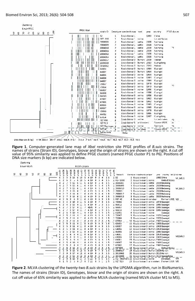
Digestion of Brucella genomic DNA with XbaI resulted in fingerprints, each consisted of 21-24 restriction fragments that ranged in size from 10 to 310 kb (Figure 1). The PFGE fingerprinting resolved 9 genotypes among the 32 isolates (genetic similarity: 100%) by BioNumerics software package. The genotying by PFGE was in general consistent with the biotyping. The PFGE cluster was defined by a cut off value of 95% similarity. Six PFGE clusters were identified (clusters P1-P6). In cluster P1, there were 8 strains (biovar 1), including 6 strains originated from cattle in Inner Mongolia, 1 vaccine strain S2 and 1 reference strain REF 1330S. In cluster P2, there were 20 strains (biovar 3), including 3 strains in genotype 3 collected from 2009 to 2010 in Hainan (2 strains) and Guangdong (1 strain), 14 strains in genotype 4 collected during 1960s (9 strains), 1970s (2 strains) and 1980s (3 strains) and 3 strains in genotype 5 collected during 1970s (1 strain) and 1980s (2 strains). The cluster P3 was REF 686 (biovar 3). The cluster P4 was REF 513 (biovar 5). The cluster P5 was REF Thomsen (biovar 2) and the cluster P6 was REF40 (biovar 4) (Figure1 and Table 1).
MLVA Analysis
The MLVA resolved 23 genotypes among the 32 strains. The strains which were isolated from different years present different MLVA genotypes. The MLVA cluster was defined by a cut off value of
65% similarity. Five MLVA clusters was identified (MLVA clusters M1 to M5), which represent five biovars of B.suis (Figures 2). Cluster M1 consisting of 8 strains could be divided again into two subclusters (M1.1 and M1.2). In subcluster M1.1, there were vaccine strain S2 and REF 1330 (biovar 1) and in subcluster M1.2, there were 6 strains (biovar 1) originated from cattle in Inner Mongolia. The cluster M2 was B.suis REF40 (biovar 4). The cluster M3 consisting of 18 strains could be divided again into three subclusters (M3.1, M3.2, and M3.3) (genetic similarity 74%). In subcluster M3.1, there were 8 strains collected from pigs from 1970s to 1980s. In subcluster M3.2, there were 10 strains collected from 8 pigs and 2 brucellosis patients during 1960s and REF 686 (biovar 3). In subcluster M3.3, there were 3 strains collected from brucellosis patients during 2009-2010, in which 2 strains were from Hainan, 1 strain was from Guangdong. The cluster M4 was REF Thomsen (biovar 2). The cluster P5 was REF 513 (biovar 5) (Figure 2 and Table 1).
Comparison of PFGE Fingerprinting and MLVA Analysis
The genotyping of PFGE and MLVA was highly relevant to the biological typing. The B.suis biovar 1 strains were mainly in PFGE cluster P1 and MLVA cluster M1. The B.suis biovar 3 strains were mainly in PFGE cluster P2 and MLVA cluster M3. And the results
Biomed Environ Sci, 2013; 26(6): 504-508 507
Figure 1. Computer-generated lane map of XbaI restriction site PFGE profiles of B.suis strains. The names of strains (Strain ID), Genotypes, biovar and the origin of strains are shown on the right. A cut off value of 95% similarity was applied to define PFGE clusters (named PFGE cluster P1 to P6). Positions of DNA size markers (k bp) are indicated below.
Figure 2. MLVA clustering of the twenty-two B.suis strains by the UPGMA algorithm, run in BioNumerics. The names of strains (Strain ID), Genotypes, biovar and the origin of strains are shown on the right. A cut off value of 65% similarity was applied to define MLVA clustering (named MLVA cluster M1 to M5).
508 Biomed Environ Sci, 2013; 26(6): 504-508
of MLVA genotyping were similar to that of PFGE fingerprinting. The 6 B.suis biovar 3 collected from cattle in Inner Mongolia showed same genotype (PFGE genotype 2 and MLVA genotype 3). The MLVA clustered into 23 genotypes, whereas PFGE clustered into 9 PFGE genotypes. And the discriminatory power of MLVA assay was greater than that of PFGE fingerprinting (Table 1).
Swine brucellosis has long been widespread throughout the world. B.suis tends to be more pathogenic to human than other five Brucella species. In addition, infected pigs tend to have higher levels of B.suis organisms in their tissues compared with cattle infected with B.abortus. Available evidence indicates that other Brucella species are not highly pathogenic to pigs. This means a higher risk on people exposed to infected pigs[5]. B.suis biovar 2 was commonly isolated in Europe[6]. And biovar 1 was most frequently isolated in Latin America and Americas[7-9]. But in China, the epidemic strains was B.suis biovar 3[10]. The strains collected from Inner Mongolia were induced by Vaccine S2 immunity. And this study was therefore important. The PFGE analysis was a useful tool for calculating genetic relatedness among the Brucella species and may be an indicator of clonal origin and genetic relatedness[2]. MLVA has proven to be highly discriminatory and was the most suitable assay for simultaneously identifying B.suis, tracing infection sources and investigating outbreaks[6], and suitable for typing analysis of a large number of strains, while providing a clustering in consistent with all methods that previously reported. Our present study firstly reported molecular characteristics of B.suis in China by PFGE and MLVA analysis. There were three kinds of epidemic strains with similar characteristics and distribution in three different epidemic periods: 1960s-1970s, 1980s, and 2009-2010. The close molecular relationship was found in 1960s and 1970s. Two strains collected from Hainan in 2009 and 1 strain isolated from Gongdong in 2010 had close genetic relatedness, but no epidemiological link was found between them. And they were different from the strains isolated during 1960s-1970s and in 1980s. Hainan province is an island which is separated from the mainland of China, so the isolation of 2 strains
there had therefore important epidemiological significance. The epidemic of brucellosis occurred in China in 1960s, 1970s, and 1980s. However, no strain was isolated in 1990s. In fact, brucellosis rarely occurred in China in 1990s[10]. And the findings from this study were in consistent with the brucellosis epidemiological situation in China.
#Correspondence should be addressed to TIAN Guo Zhong. Tel: 86-10-58900767. Fax: 86-10- 58900700. E-mail: [email protected]
Biographical note of the first author: LI Zhen Jun, male, born in 1975, Ph.D, majoring in research on pathogenic microorganism. E-mail: [email protected]
Received: August 3, 2012; Accepted: January 2, 2013
REFERENCES
1. Alton GG, Jones LM, Pietz DE. Laboratory techniques in brucellosis. Monogr Ser World Health Organ, 1975; 55, 1-163.
2. Jensen AE, Cheville NF, Thoen CO, et al. Genomic fingerprinting and development of a dendrogram for Brucella spp. isolated from seals, porpoises, and dolphins. J Vet Diagn Invest, 1999; 11, 152-7.
3. Le Flèche P, Jacques I, Grayon M, et al. Evaluation and selection of tandem repeat loci for a Brucella MLVA typing assay. BMC Microbiol, 2006; 6, 9.
4. Maquart M, Le Flèche P, Foster G, et al. MLVA-16 typing of 295 marine mammal Brucella isolates from different animal and geographic origins identifies 7 major groups within Brucella ceti and Brucella pinnipedialis. BMC Microbiol, 2009; 20(9), 145.
5. MacMillan AP. Brucellosis. In: B.E. Straw, S. D´Allaire, W.L. Mengeling and D.M. Taylor (eds), Diseases of swine. 8 edn. (Blackwell Science, Iowa), 1999; 385-93.
6. Abril C, Thomann A, Brodard I, et al. A novel isolation method of Brucella species and molecular tracking of Brucella suis biovar 2 in domestic and wild animals. Veterinary Microbiology, 2011; 150, 405-10.
7. Lucero NE, Ayala SM, Escobar GI, et al. Brucella isolated in humans and animals in Latin America from 1968 to 2006. Epidemiology and Infection, 2008; 136, 496-503.
8. Stoffregen WC, Olsen SC, Wheeler CJ, et al. Diagnostic characterization of a feral swine herd enzootically infected with Brucella. Journal of Veterinary Diagnostic Investigation, 2007; 19, 227-37.
9. Meirelles-Bartoli RB, Mathias LA, Samartino LE. Brucellosis due to Brucella suis in a swine herd associated with a human clinical case in the State of São Paulo, Brazil. Trop Anim Health Prod, 2012; 44, 1575-9.
10.Deqiu S, Donglou X, Jiming Y. Epidemiology and control of brucellosis in China. Vet Microbiol, 2002; 90, 165-82.