DEVELOPMENT 357 RESEARCH ARTICLE INTRODUCTION Neural circuits throughout the nervous system use a combination of fast-excitatory and fast-inhibitory neurotransmitters to regulate neural activity. In the vertebrate nervous system, fast inhibitory transmission is primarily mediated by two transmitters – GABA and glycine. The neurons that release these neurotransmitters express a number of genes that encode components of the inhibitory-neurotransmitter machinery. These include the vesicular inhibitory amino acid transporter (VIAAT, also known as Slc32a1 – Mouse Genome Informatics), which loads GABA and glycine into secretory vesicles (McIntire et al., 1997), and the glycine transporter, GlyT2, which is responsible for glycine reuptake and transport across the plasma membrane of glycinergic neurons (Liu et al., 1993). In addition, GABAergic neurons express two genes, Gad1 and Gad2, that encode glutamic acid decarboxylase – the enzyme that converts glutamate to GABA (Erlander and Tobin, 1991). During embryogenesis, the expression of these genes is activated in subsets of differentiating neurons, thus imbuing them with the necessary cellular machinery for fast inhibitory neurotransmission. The developmental programs that determine the neurotransmitter status of a neuron remain largely unknown. Whereas the pattern and levels of a particular neurotransmitter can be regulated under certain circumstances by neural activity and target-derived signals during development (Schotzinger and Landis, 1988; Borodinsky et al., 2004), a cells acquisition of a particular neurotransmitter ‘phenotype’ appears to be closely linked to the gene regulatory events that determine neuronal subtype identity. In the embryonic spinal cord, developing neurons fall into three fast neurotransmitter classes: cholinergic neurons, excitatory glutamatergic neurons and inhibitory neurons that use GABA and or glycine as their primary transmitters. Motor neurons are primarily cholinergic (Phelps et al., 1991), as are a small population of interneurons of unknown function that are located near the central canal (Barber et al., 1991). Glutamatergic excitatory interneurons include the early-born dI1-3, dI5, V2 and V3 neurons, as well as a population of late-born dorsal interneurons, the so-called dIL B neurons. The dI4, dI6, V0 and V1 classes of interneuron that are generated during the first wave of neurogenesis are inhibitory (Saueressig et al., 1999; Wenner et al., 2000; Lanuza et al., 2004; Glasgow et al., 2005), as are the late-born dIL A neurons that settle in the dorsal horn (Cheng et al., 2004; Cheng et al., 2005; Mizuguchi et al., 2006). Studies in the dorsal horn have begun to delineate the transcriptional mechanisms that control the neurotransmitter phenotype of spinal-interneuron cell types. Inhibitory neurons in the dorsal spinal cord are derived exclusively from cells that express the homeodomain transcription factor Lbx1 (Gross et al., 2002; Muller et al., 2002; Matise, 2002; Cheng et al., 2004). These cells are comprised of two early-born populations – dI4 and dI6 neurons – and late-born dIL A neurons that are generated during the second wave of neurogenesis, which begins at E12 in the mouse. All three classes of neuron express the paired-domain transcription factor Pax2 together with the LIM-homeodomain transcription factors Lhx1 and Lhx5 (Gross et al., 2002; Muller et al., 2002). A subset of Lbx1-expressing neurons, dI5 and dIL B neurons, also differentiate as glutamatergic neurons. These cells express the homeodomain transcription factors Tlx1, Tlx3 and Lmx1b. Tlx1 and Tlx3 function in a cell-autonomous manner to specify glutamatergic dI5- and dIL B -sensory neurons (Cheng et al., 2004), in part by over-riding an inhibitory differentiation program that is Lbx1-dependent (Cheng et al., 2005). Inactivation of Tlx1 and Tlx3 results in the loss of glutamatergic cell types in the dorsal horn, along with the concomitant upregulation of Pax2 and GABAergic markers, such as Viaat. Conversely, Tlx3 overexpression induces a switch from GABAergic to glutamatergic cell fate (Cheng et al., 2004; Cheng et al., 2005; Mizuguchi et al., 2006). Interestingly, the loss of Lmx1b does not alter the neurotransmitter status of dI5 and dIL B neurons (Ding et al., 2004). Lhx1 and Lhx5 maintain the inhibitory-neurotransmitter status of interneurons in the dorsal spinal cord Andrea Pillai 1,5 , Ahmed Mansouri 2 , Richard Behringer 3 , Heiner Westphal 4 and Martyn Goulding 5, * Lhx1 and Lhx5 are co-expressed in multiple interneuron cell types in the developing spinal cord. These include early-born dI4 and dI6 inhibitory interneurons, as well as late-born inhibitory dIL A neurons (dIL A ), all of which express the paired-domain transcription factor Pax2. Although it appears that Lhx1 and Lhx5 do not control the initial specification of the neuronal cell types in which they are expressed, we have found a cell-autonomous requirement for either Lhx1 or Lhx5 to maintain the expression of Pax2, Pax5 and Pax8 in dorsal inhibitory neurons at later developmental stages. Lhx1; Lhx5 double-knockout mice exhibit a downregulation of Gad1 and Viaat (Slc32a1) from E13.5 onwards that is closely associated with a decrease in Pax2 expression. Pax2 is a key factor for dorsal GABAergic identity, with the expression of Pax5 and Pax8 being differentially dependent on Pax2 in the dorsal horn. In summary, our findings support a model in which the differentiation of GABAergic interneurons in the dorsal cord depends on Pax2, with Lhx1 and Lhx5 helping to activate and maintain Pax2 expression in these cells. Lhx1 and Lhx5 therefore function together with Pax2, Pax5 and Pax8 to establish a GABAergic inhibitory-neurotransmitter program in dorsal horn interneurons. KEY WORDS: Lhx1, Lhx5, Pax2, Inhibitory neurons, Spinal cord, Mouse Development 134, 357-366 (2007) doi:10.1242/dev.02717 1 Biology Graduate Program, University of California, San Diego, La Jolla, CA 92037, USA. 2 Department of Molecular Cell Biology, Max Planck Institute for Biophysical Chemistry, Gottingen 37077, Germany. 3 Department of Molecular Genetics, University of Texas, M. D. Anderson Cancer Center, Houston, TX 77030, USA. 4 Laboratory of Mammalian Genes and Development, National Institute of Child Health and Human Development, Bethesda, MD 20892, USA. 5 Molecular Neurobiology Laboratory, The Salk Institute for Biological Studies, 10010 North Torrey Pines Road, La Jolla, CA 92037, USA. *Author for correspondence (e-mail: [email protected]) Accepted 25 October 2006 Development ePress online publication date 13 December 2006
Transcript
DEVELO
PMENT
357RESEARCH ARTICLE
INTRODUCTIONNeural circuits throughout the nervous system use a combinationof fast-excitatory and fast-inhibitory neurotransmitters to regulateneural activity. In the vertebrate nervous system, fast inhibitorytransmission is primarily mediated by two transmitters – GABAand glycine. The neurons that release these neurotransmittersexpress a number of genes that encode components of theinhibitory-neurotransmitter machinery. These include thevesicular inhibitory amino acid transporter (VIAAT, also knownas Slc32a1 – Mouse Genome Informatics), which loads GABAand glycine into secretory vesicles (McIntire et al., 1997), and theglycine transporter, GlyT2, which is responsible for glycinereuptake and transport across the plasma membrane of glycinergicneurons (Liu et al., 1993). In addition, GABAergic neuronsexpress two genes, Gad1 and Gad2, that encode glutamic aciddecarboxylase – the enzyme that converts glutamate to GABA(Erlander and Tobin, 1991). During embryogenesis, theexpression of these genes is activated in subsets of differentiatingneurons, thus imbuing them with the necessary cellular machineryfor fast inhibitory neurotransmission.
The developmental programs that determine the neurotransmitterstatus of a neuron remain largely unknown. Whereas the pattern andlevels of a particular neurotransmitter can be regulated under certaincircumstances by neural activity and target-derived signals duringdevelopment (Schotzinger and Landis, 1988; Borodinsky et al.,2004), a cells acquisition of a particular neurotransmitter‘phenotype’ appears to be closely linked to the gene regulatoryevents that determine neuronal subtype identity. In the embryonicspinal cord, developing neurons fall into three fast neurotransmitter
classes: cholinergic neurons, excitatory glutamatergic neurons andinhibitory neurons that use GABA and or glycine as their primarytransmitters. Motor neurons are primarily cholinergic (Phelps et al.,1991), as are a small population of interneurons of unknownfunction that are located near the central canal (Barber et al., 1991).Glutamatergic excitatory interneurons include the early-born dI1-3,dI5, V2 and V3 neurons, as well as a population of late-born dorsalinterneurons, the so-called dILB neurons. The dI4, dI6, V0 and V1classes of interneuron that are generated during the first wave ofneurogenesis are inhibitory (Saueressig et al., 1999; Wenner et al.,2000; Lanuza et al., 2004; Glasgow et al., 2005), as are the late-borndILA neurons that settle in the dorsal horn (Cheng et al., 2004;Cheng et al., 2005; Mizuguchi et al., 2006).
Studies in the dorsal horn have begun to delineate thetranscriptional mechanisms that control the neurotransmitterphenotype of spinal-interneuron cell types. Inhibitory neurons in thedorsal spinal cord are derived exclusively from cells that express thehomeodomain transcription factor Lbx1 (Gross et al., 2002; Mulleret al., 2002; Matise, 2002; Cheng et al., 2004). These cells arecomprised of two early-born populations – dI4 and dI6 neurons –and late-born dILA neurons that are generated during the secondwave of neurogenesis, which begins at E12 in the mouse. All threeclasses of neuron express the paired-domain transcription factorPax2 together with the LIM-homeodomain transcription factorsLhx1 and Lhx5 (Gross et al., 2002; Muller et al., 2002). A subset ofLbx1-expressing neurons, dI5 and dILB neurons, also differentiateas glutamatergic neurons. These cells express the homeodomaintranscription factors Tlx1, Tlx3 and Lmx1b. Tlx1 and Tlx3 functionin a cell-autonomous manner to specify glutamatergic dI5- anddILB-sensory neurons (Cheng et al., 2004), in part by over-riding aninhibitory differentiation program that is Lbx1-dependent (Cheng etal., 2005). Inactivation of Tlx1 and Tlx3 results in the loss ofglutamatergic cell types in the dorsal horn, along with theconcomitant upregulation of Pax2 and GABAergic markers, such asViaat. Conversely, Tlx3 overexpression induces a switch fromGABAergic to glutamatergic cell fate (Cheng et al., 2004; Cheng etal., 2005; Mizuguchi et al., 2006). Interestingly, the loss of Lmx1bdoes not alter the neurotransmitter status of dI5 and dILB neurons(Ding et al., 2004).
Lhx1 and Lhx5 maintain the inhibitory-neurotransmitterstatus of interneurons in the dorsal spinal cordAndrea Pillai1,5, Ahmed Mansouri2, Richard Behringer3, Heiner Westphal4 and Martyn Goulding5,*
Lhx1 and Lhx5 are co-expressed in multiple interneuron cell types in the developing spinal cord. These include early-born dI4 anddI6 inhibitory interneurons, as well as late-born inhibitory dILA neurons (dILA), all of which express the paired-domain transcriptionfactor Pax2. Although it appears that Lhx1 and Lhx5 do not control the initial specification of the neuronal cell types in which theyare expressed, we have found a cell-autonomous requirement for either Lhx1 or Lhx5 to maintain the expression of Pax2, Pax5 andPax8 in dorsal inhibitory neurons at later developmental stages. Lhx1; Lhx5 double-knockout mice exhibit a downregulation ofGad1 and Viaat (Slc32a1) from E13.5 onwards that is closely associated with a decrease in Pax2 expression. Pax2 is a key factor fordorsal GABAergic identity, with the expression of Pax5 and Pax8 being differentially dependent on Pax2 in the dorsal horn. Insummary, our findings support a model in which the differentiation of GABAergic interneurons in the dorsal cord depends on Pax2,with Lhx1 and Lhx5 helping to activate and maintain Pax2 expression in these cells. Lhx1 and Lhx5 therefore function together withPax2, Pax5 and Pax8 to establish a GABAergic inhibitory-neurotransmitter program in dorsal horn interneurons.
Development 134, 357-366 (2007) doi:10.1242/dev.02717
1Biology Graduate Program, University of California, San Diego, La Jolla, CA 92037,USA. 2Department of Molecular Cell Biology, Max Planck Institute for BiophysicalChemistry, Gottingen 37077, Germany. 3Department of Molecular Genetics,University of Texas, M. D. Anderson Cancer Center, Houston, TX 77030, USA.4Laboratory of Mammalian Genes and Development, National Institute of ChildHealth and Human Development, Bethesda, MD 20892, USA. 5MolecularNeurobiology Laboratory, The Salk Institute for Biological Studies, 10010 NorthTorrey Pines Road, La Jolla, CA 92037, USA.
Development ePress online publication date 13 December 2006
DEVELO
PMENT
358
Pax2-expressing neurons in the hindbrain and spinal cordpredominantly differentiate as inhibitory interneurons (Maricich andHerrup, 1999; Gross et al., 2002; Cheng et al., 2004; Glasgow et al.,2005; Mizuguchi et al., 2006; Wildner et al., 2006). In the Pax2-mutant cord, there is a marked loss of GABAergic markers in thedorsal horn, demonstrating that Pax2 functions as an obligatoryregulator of the inhibitory-neurotransmitter program in these cells(Cheng et al., 2004). These dorsal inhibitory interneurons, as well asthe ventrally-derived V0 and V1 inhibitory interneurons, also expressLhx1 and Lhx5 (Burrill et al., 1997; Moran-Rivard et al., 2001; Grosset al., 2002; Muller et al., 2002) (this study). This has led to thesuggestion that the co-expression of Pax2 and Lhx1 and/or Lhx5 mayprovide a transcription factor code for inhibitory neurons in thehindbrain and spinal cord. Although roles for Lhx1 and Lhx5 havebeen demonstrated in head, kidney and motor neuron development(Kobayashi et al., 2005; Kobayashi et al., 2004; Zhao et al., 1999;Kania et al., 2000), their overlapping expression in spinal interneurons(Sheng et al., 1997), coupled with the early embryonic lethalphenotype of the Lhx1 mutant (Shawlot and Behringer, 1995), hasimpeded analyzing their role(s) in spinal-interneuron development.
In this study, we set out to address three questions: (1) Do Lhx1and Lhx5 play a role in the early establishment of spinal-interneuronsubtypes in the spinal cord? (2) Do Lhx1 and Lhx5 function incombination with Pax2 to establish inhibitory-neurotransmitterphenotypes in the developing spinal cord? (3) Do Lhx1 and Lhx5have roles in maintaining the neurotransmitter status of inhibitoryinterneurons and, if so, how is this function executed? Our resultsshow that, although Lhx1 and/or Lhx5 are not required for thespecification of early-born interneurons that form at E10.5-E11, bothgenes are necessary for the proper development of late-borninhibitory dILA interneurons. Moreover, we find that a reciprocalregulatory relationship exists between Lhx1 and/or Lhx5 and Pax2genes in these cells. Lhx1;Lhx5 double mutants exhibit a selectiveloss of Pax2 protein expression in the dorsal horn that precedes thereduction in Gad1 and Viaat expression. As a result of this, late-bornPax2+ GABAergic neurons that settle in the lateral dorsal horn failto retain their GABAergic identity. Pax2 is also required to maintainLhx1, Lhx5, Pax5 and Pax8 expression in these cells, demonstratinga genetic interdependence between these two transcription factorclasses in late-born dorsal inhibitory neurons.
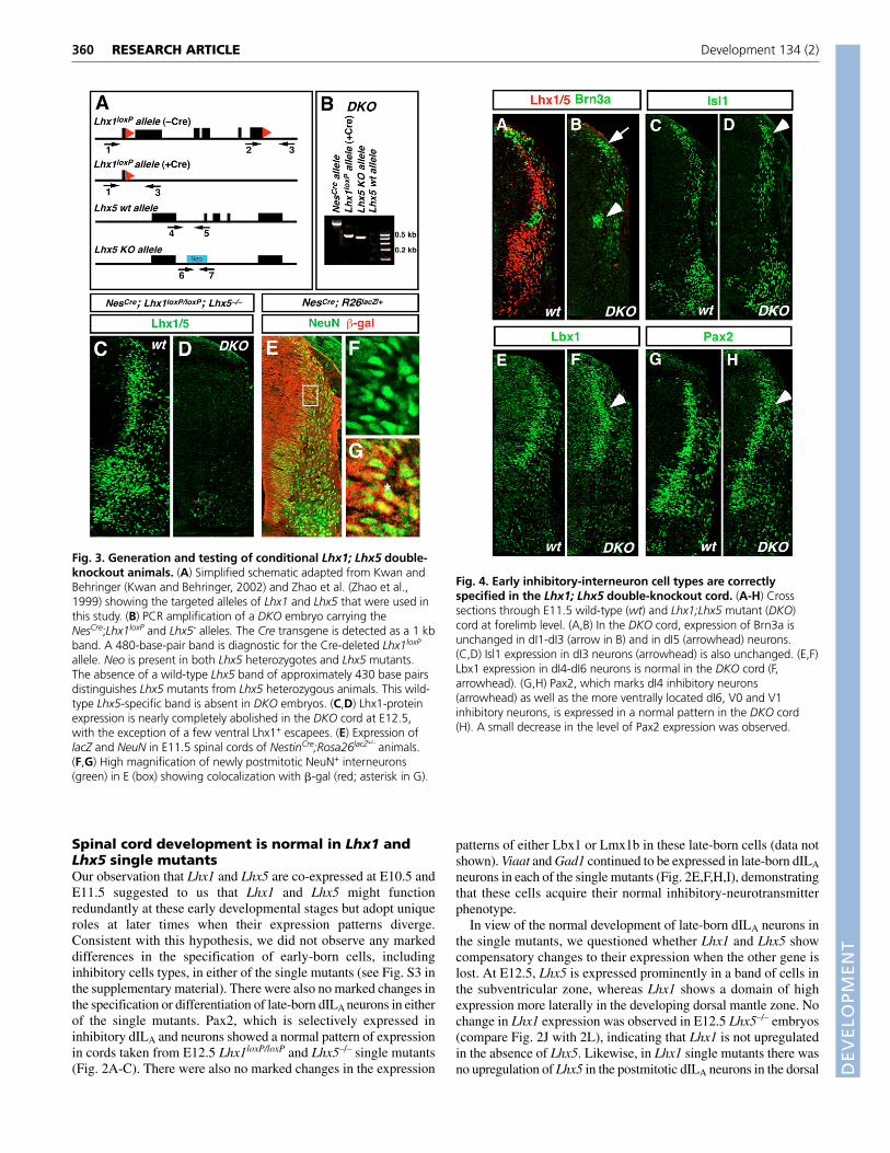
MATERIALS AND METHODSGeneration of Lhx1;Lhx5 double-knockout miceLhx5 mice, in which exons 2-4 of the targeted Lhx5 gene were replacedwith a neomycin-resistance gene, were obtained from H. Westphal (Zhaoet al., 1999) (see Fig. 3A). Lhx1 conditional-mutant mice (Lhx1loxP) werekindly provided by R. Behringer (M. D. Anderson Cancer Center,Houston, USA). The Lhx1 coding region is flanked by two loxP sites inthe Lhx1loxP conditional allele (Kwan and Behringer, 2002) (see Fig. 3A).Selective inactivation of Lhx1 in neurons was achieved by crossing aNestin-Cre (NesCre) transgenic line in which Cre-recombinase is under thecontrol of a nervous-system-specific enhancer present in the second intronof the rat nestin gene (Lendahl et al., 1990) into a Lhx1loxP/loxP
background. In this study, conditional Lhx1-mutant mice with thegenotype Lhx1loxP/loxP;NesCre are referred to as Lhx1–/– mutants. Doublemutants with the genotype Lhx1loxP/loxP;Lhx5–/–;NesCre are referred to asDKO mice. Lhx5–/–- and Lhx1–/–-mutant mice both die at birth.Lhx1loxP/loxP homologous strains are, however, healthy and fertile, and soDKO mice were generated by crossing parental lines comprised of geneticcombinations of Lhx5+/–;NesCre;Lhx1loxP/+ with Lhx1loxP/loxP;Lhx5+/– mice.Pax2–/– and Pax8–/– embryos were provided by A. Mansouri (Mansouri etal., 1998). Pax5–/– embryos were derived from the breedings ofheterozygous Pax5+/– mice (Urbanek et al., 1994). The primers forgenotyping all mutant animals are identical to those described in theaforementioned references.
Immunohistochemistry and in situ hybridizationMouse embryos were fixed for 1 hour in 4% paraformaldehyde inphosphate-saline buffer (PBS), cryoprotected in 25% sucrose, embedded inOCT (Tissue-Tec) and sectioned at 20 �m. Immunohistochemistry wasperformed on frozen sections as previously described (Burrill et al., 1997).The following antibodies were used in the study: monoclonal anti-Lhx1 andanti-Lhx5 (4F2-10, Developmental Hybridoma Studies Bank), monoclonalanti-NeuN (Chemicon International), rat anti-BrdU monoclonal antibody(Harlan), anti-Lbx1 (Gross et al., 2002), polyclonal anti-Pax2 (Zymed) andguinea-pig anti-Lmx1b (gift of T. Jessell, HHMI, Columbia University, NY,USA). Species-specific antibodies conjugated to Cy2, Cy3 or Cy5 were used(Jackson ImmunoResearch). In situ hybridizations were performed aspreviously described (Goulding et al., 1993). Pax2 immuno-in situ doublelocalization was performed according to Cheng et al. (Cheng et al., 2004).In situ hybridizations were preformed using probes specific for mouse Lhx1(Bertuzzi et al., 1996), Lhx5 (Zhao et al., 1999), Viaat and VGluT2, and ratGad1, as described previously by Mizuguchi et al. (Mizuguchi et al., 2006).
RESEARCH ARTICLE Development 134 (2)
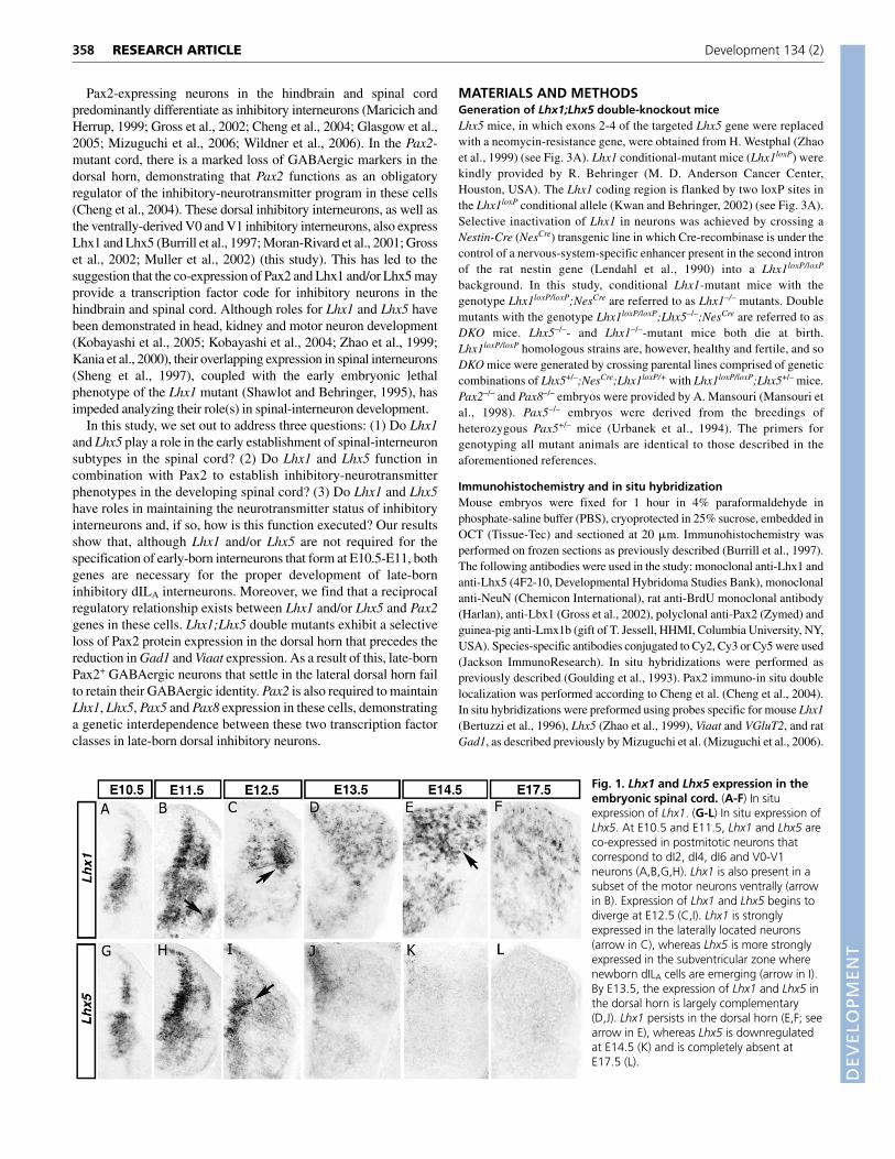
Fig. 1. Lhx1 and Lhx5 expression in theembryonic spinal cord. (A-F) In situexpression of Lhx1. (G-L) In situ expression ofLhx5. At E10.5 and E11.5, Lhx1 and Lhx5 areco-expressed in postmitotic neurons thatcorrespond to dI2, dI4, dI6 and V0-V1neurons (A,B,G,H). Lhx1 is also present in asubset of the motor neurons ventrally (arrowin B). Expression of Lhx1 and Lhx5 begins todiverge at E12.5 (C,I). Lhx1 is stronglyexpressed in the laterally located neurons(arrow in C), whereas Lhx5 is more stronglyexpressed in the subventricular zone wherenewborn dILA cells are emerging (arrow in I).By E13.5, the expression of Lhx1 and Lhx5 inthe dorsal horn is largely complementary(D,J). Lhx1 persists in the dorsal horn (E,F; seearrow in E), whereas Lhx5 is downregulatedat E14.5 (K) and is completely absent atE17.5 (L).
DEVELO
PMENT
HistologySections of 5 �m were cut from paraffin-embedded E17.5 spinal cords andstained with hematoxylin and Eosin, as described by Gross et al. (Gross etal., 2002). Cell counts were performed on three sections from three cords(i.e. nine sections each for wild-type and DKO embryos). For each section,cells in a single dorsal quadrant were counted twice in order to minimizecounting errors. Statistical differences in cell counts between wild-type andmutant cords were determined using the Student’s t-test.
Apoptotic cells in the developing spinal cord (E14.5-E17.5) werevisualized by TUNEL labeling using the ApopTag-plus Fluorescein In SituApoptosis Detection Kit (Chemicon International). Stainings wereperformed according to the manufacturer’s instructions. Counts for apoptoticcells were tabulated for both the dorsal and ventral halves of the cord.Apoptotic cell counts for each sample represents the average of six sections(three sections from two cords).
BrdU labelingPregnant dams were injected intraperitoneally with 50 mgbromodeoxyuridine (50 �g/ml dissolved in 0.9% saline) per gram of mousebodyweight at E12.5. E14.5 embryos were collected and processed forimmunohistochemistry sections, and stained with an antibody to Pax2followed by anti-BrdU mouse monoclonal antibody.
ImagingAntibody, TUNEL and BrdU staining was visualized on a Zeiss LSM 510confocal microscope. In situ hybridization images were captured on aZeiss Axiophot 2 microscope fitted with an Axiocam MRm camera. Allfigures were color-corrected and assembled using Photoshop and Canvassoftware.
RESULTSExpression of the Lhx1 and Lhx5 genes in thedeveloping spinal cordAs a first step towards analyzing the function of Lhx1 and Lhx5 in theembryonic spinal cord, we undertook a detailed analysis of the normalexpression profiles of Lhx1 and Lhx5 during development. Althoughprevious studies documented the early expression patterns of bothgenes in the nervous system (Sheng et al., 1997), these studies did notaddress the dynamic changes in the expression of Lhx1 and Lhx5 thatoccur at later developmental times. Previous studies by Gross et al.(Gross et al., 2002) using antibodies that recognize both Lhx1 andLhx5 had indicated that both proteins are co-expressed in late-borndILA neurons that populate the dorsal horn. However, although Lhx1had been shown to persist dorsally at later stages by in situhybridization (Muller et al., 2002), it was unclear whether Lhx5 wasalso expressed at later developmental times.
To clarify this issue, we used in situ hybridization to compare thedevelopmental expression profiles of Lhx1 and Lhx5 in theembryonic spinal cord. During the early phase of neurogenesis(E10.5-E11.5), Lhx1 and Lhx5 were found to be co-expressed inmultiple spinal-interneuron populations, including three dorsal celltypes – the dI2, dI4 and dI6 interneurons (Fig. 1A,B,G,H; also seeFig. S1 and Fig. S2 in the supplementary material). However, fromE12.5 onwards, the expression patterns of these two genes began todiverge, leading to complementary patterns of expression in dorsalinterneurons at later developmental times (Fig. 1C,I). In the E13.5dorsal horn, the highest level of Lhx5 transcripts was found medially,decreasing towards the lateral rim of the dorsal cord. Lhx1 wasexpressed in an inverse gradient, with cells closest to the ventricularzone expressing low levels of Lhx1 transcripts, while cells furtheraway from the ventricular zone expressed higher levels (Fig. 1D,J).Whereas Lhx1 continued to be expressed in a mosaic pattern in thedorsal horn neurons up to birth (Fig. 1E,F, and data not shown), littleor no Lhx5 expression was detected in the spinal cord from E14.5 toE17.5 (Fig. 1K,L).
The divergent expression patterns of Lhx1 and Lhx5 in the late-born dIL cells can be accounted for in two ways. First, thecomplementary expression patterns of Lhx1 and Lhx5 in late-borndILA cells may reflect high-level expression of Lhx1 in the dILA cellsthat are born first. These cells would be expected to accumulate inthe more lateral regions of the dorsal horn, whereas later-born dILA
cells that are located more medially might exhibit high Lhx5-lowLhx1 expression. Alternatively, differentiating dILA cells maydownregulate Lhx5 and upregulate Lhx1 as they migrate from thesubventricular zone into the dorsal horn. Support for the laterpossibility comes from the observation that Lhx5 begins to bedownregulated when dILA cells cease being generated at E13.5(Gross et al., 2002). For this reason, we favor a model in whichnewborn dILA cells express Lhx5 at high levels, while the moremature dILA neurons downregulate Lhx5 and upregulate Lhx1.
359RESEARCH ARTICLELhx1 and Lhx5 function in spinal inhibitory interneurons
Fig. 2. Dorsal interneuron development in Lhx1 and Lhx5 singlemutants. (A-C) Inactivation of either Lhx1 or Lhx5 does not alter thespecification of dILA interneurons. dILA interneurons express Pax2 andLhx5 in Lhx1–/– embryos (B), or express Pax2 and Lhx1 in Lhx5–/–
embryos (C). (D-I) In both Lhx1 and Lhx5 single mutants, dILA cellsretain their GABAergic identity, and express Viaat (D-F) and Gad1 (G-I).(J-O) Lhx1 expression is unchanged in the cord of Lhx5–/– mutants.There is no upregulation of Lhx1 mRNA at early (not shown) or later(arrows in J,L) stages. There is also no change in Lhx5 expression in thecord of Lhx1–/– mutants at E12.5 (arrows in M,N).
DEVELO
PMENT
360
Spinal cord development is normal in Lhx1 andLhx5 single mutantsOur observation that Lhx1 and Lhx5 are co-expressed at E10.5 andE11.5 suggested to us that Lhx1 and Lhx5 might functionredundantly at these early developmental stages but adopt uniqueroles at later times when their expression patterns diverge.Consistent with this hypothesis, we did not observe any markeddifferences in the specification of early-born cells, includinginhibitory cells types, in either of the single mutants (see Fig. S3 inthe supplementary material). There were also no marked changes inthe specification or differentiation of late-born dILA neurons in eitherof the single mutants. Pax2, which is selectively expressed ininhibitory dILA and neurons showed a normal pattern of expressionin cords taken from E12.5 Lhx1loxP/loxP and Lhx5–/– single mutants(Fig. 2A-C). There were also no marked changes in the expression
patterns of either Lbx1 or Lmx1b in these late-born cells (data notshown). Viaat and Gad1 continued to be expressed in late-born dILA
neurons in each of the single mutants (Fig. 2E,F,H,I), demonstratingthat these cells acquire their normal inhibitory-neurotransmitterphenotype.
In view of the normal development of late-born dILA neurons inthe single mutants, we questioned whether Lhx1 and Lhx5 showcompensatory changes to their expression when the other gene islost. At E12.5, Lhx5 is expressed prominently in a band of cells inthe subventricular zone, whereas Lhx1 shows a domain of highexpression more laterally in the developing dorsal mantle zone. Nochange in Lhx1 expression was observed in E12.5 Lhx5–/– embryos(compare Fig. 2J with 2L), indicating that Lhx1 is not upregulatedin the absence of Lhx5. Likewise, in Lhx1 single mutants there wasno upregulation of Lhx5 in the postmitotic dILA neurons in the dorsal
RESEARCH ARTICLE Development 134 (2)
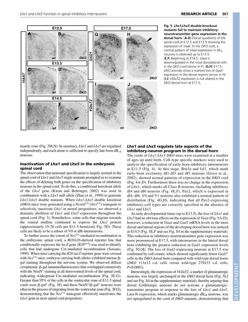
Fig. 4. Early inhibitory-interneuron cell types are correctlyspecified in the Lhx1; Lhx5 double-knockout cord. (A-H) Crosssections through E11.5 wild-type (wt) and Lhx1;Lhx5 mutant (DKO)cord at forelimb level. (A,B) In the DKO cord, expression of Brn3a isunchanged in dI1-dI3 (arrow in B) and in dI5 (arrowhead) neurons.(C,D) Isl1 expression in dI3 neurons (arrowhead) is also unchanged. (E,F)Lbx1 expression in dI4-dI6 neurons is normal in the DKO cord (F,arrowhead). (G,H) Pax2, which marks dI4 inhibitory neurons(arrowhead) as well as the more ventrally located dI6, V0 and V1inhibitory neurons, is expressed in a normal pattern in the DKO cord(H). A small decrease in the level of Pax2 expression was observed.
Fig. 3. Generation and testing of conditional Lhx1; Lhx5 double-knockout animals. (A) Simplified schematic adapted from Kwan andBehringer (Kwan and Behringer, 2002) and Zhao et al. (Zhao et al.,1999) showing the targeted alleles of Lhx1 and Lhx5 that were used inthis study. (B) PCR amplification of a DKO embryo carrying theNesCre;Lhx1loxP and Lhx5- alleles. The Cre transgene is detected as a 1 kbband. A 480-base-pair band is diagnostic for the Cre-deleted Lhx1loxP
allele. Neo is present in both Lhx5 heterozygotes and Lhx5 mutants.The absence of a wild-type Lhx5 band of approximately 430 base pairsdistinguishes Lhx5 mutants from Lhx5 heterozygous animals. This wild-type Lhx5-specific band is absent in DKO embryos. (C,D) Lhx1-proteinexpression is nearly completely abolished in the DKO cord at E12.5,with the exception of a few ventral Lhx1+ escapees. (E) Expression oflacZ and NeuN in E11.5 spinal cords of NestinCre;Rosa26lacZ+/–
animals.(F,G) High magnification of newly postmitotic NeuN+ interneurons(green) in E (box) showing colocalization with �-gal (red; asterisk in G).
DEVELO
PMENT
mantle zone (Fig. 2M,N). In summary, Lhx1 and Lhx5 are regulatedindependently, and each alone is sufficient to specify late-born dILA
neurons.
Inactivation of Lhx1 and Lhx5 in the embryonicspinal cordThe observation that neuronal specification is largely normal in thespinal cord of Lhx1 and Lhx5 single mutants prompted us to examinethe effects of deleting both genes on the specification of inhibitoryneurons in the spinal cord. To do this, a conditional knockout alleleof the Lhx1 gene (Kwan and Behringer, 2002) was used incombination with a Lhx5-null allele (Zhao et al., 1999) to generateLhx1;Lhx5 double mutants. When Lhx1;Lhx5 double knockout(DKO) mice were generated using a NestinCre (NesCre) transgene toselectively inactivate Lhx1 in neural progenitors, we observed adramatic abolition of Lhx1 and Lhx5 expression throughout thespinal cord (Fig. 3). Nonetheless, some cells that migrate towardsthe ventral midline continued to express the Lhx1 protein(approximately 15-20 cells per E11.5 hemicord, Fig. 3D). Thesecells are likely to be a subset of V0 or dI6 interneurons.
To further assess the extent of NesCre-mediated recombination inthe embryonic spinal cord, a ROSA26-derived reporter line thatconditionally expresses the lacZ gene (R26lacZ) was used to identifycells that had undergone Cre-mediated recombination (Soriano,1999). When mice carrying the R26-lacZ reporter gene were crossedwith NesCre mice, embryos carrying both alleles exhibited intense �-gal staining throughout the nervous system. We observed diffusecytoplasmic �-gal immunofluorescence that overlapped extensivelywith the NeuN+ staining at all dorsoventral levels of the spinal cord,indicating widespread Cre-mediated recombination (Fig. 3E-G).Greater than 95% of the cells in the ventricular zone of E11.5 spinalcords were �-gal+ (Fig. 3E) and these NeuN+/�-gal+ neurons wereoften in the process of migrating from the ventricular zone (Fig. 3F,G),demonstrating that the NesCre transgene effectively inactivates theLhx1 gene in most spinal cord progenitors.
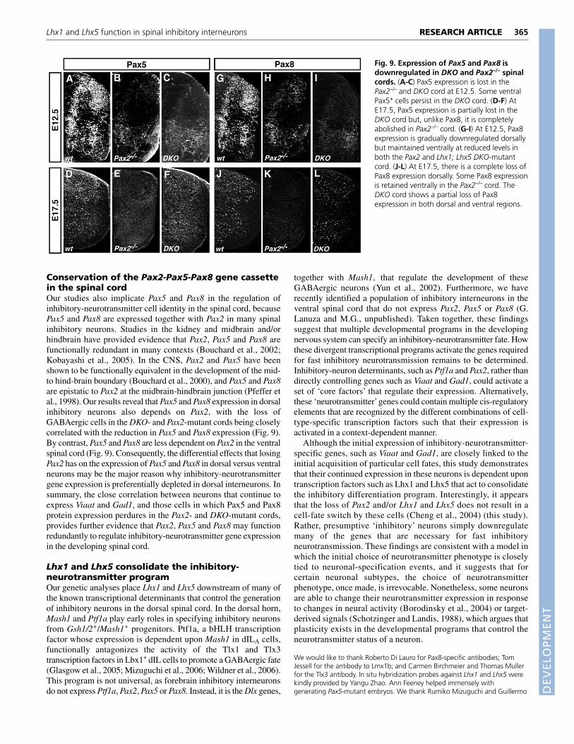
Lhx1 and Lhx5 regulate late aspects of theinhibitory-neuron program in the dorsal hornThe cords of Lhx1;Lhx5 DKO mice were examined at a numberof ages up until birth. Cell-type specific markers were used toanalyze the specification of early-born inhibitory interneuronsat E11.5 (Fig. 4). At this stage, Brn3a and Isl1, which markearly-born excitatory dI1-dI3 and dI5 neurons (Gross et al.,2002), showed normal patterns of expression in the DKO cord(Fig. 4A-D). Furthermore there was no change in the expressionof Lbx1, which marks all Class B neurons, including inhibitorydI4 and dI6 neurons (Fig. 4E,F). Pax2, which is expressed indI4, dI6, V0 and V1 neurons also exhibited a normal pattern ofdistribution (Fig. 4G,H), indicating that all Pax2-expressinginhibitory-cell types are correctly specified in the absence ofLhx1 and Lhx5.
At early developmental times (up to E13.5), the loss of Lhx1 andLhx5 had no obvious effects on the expression of Viaat (Fig. 5A-D);however, a reduction in Viaat and Gad1 mRNA levels in the mostdorsal and lateral regions of the developing dorsal horn was noticedat E14.5 (Fig. 5E,F and see Fig. S4 in the supplementary material).This reduction in inhibitory-neurotransmitter gene expression wasmore pronounced at E17.5, with interneurons in the lateral dorsalhorn exhibiting the greatest reduction in Gad1 expression levels(Fig. 5G,H). The loss of Gad1-expressing neurons at E17.5 wasconfirmed by cell counts, which showed significantly fewer Gad1+
cells in the DKO dorsal horn compared with wild-type dorsal horns(DKO 113±12 s.d. cells versus wild-type 279±33 s.d. cells;P<0.0001).
Interestingly, the expression of VGluT2, a marker of glutamatergicneurons, was largely unchanged in the DKO dorsal horn (Fig. 5I,Jand see Fig. S4 in the supplementary material), thereby arguing thatdorsal GABAergic neurons do not activate a glutamatergic-transmitter program in response to the loss of Lhx1 and Lhx5.Lmx1b expression, which marks glutamatergic dILB neurons, wasnot upregulated in the cord of DKO mutants, demonstrating that
361RESEARCH ARTICLELhx1 and Lhx5 function in spinal inhibitory interneurons
Fig. 5. Lhx1;Lhx5 double-knockoutmutants fail to maintain inhibitory-neurotransmitter gene expression in thedorsal horn. (A-D) Dorsal quadrants of thespinal cord at E12.5 and E13.5 showing theexpression of Viaat. In the DKO cord, anormal pattern of Viaat expression in dILA
neurons is observed up to E13.5.(E,F) Beginning at E14.5, Viaat isdownregulated in the most-dorsolateral cellsin the DKO cord (arrow in F). (G,H) E17.5DKO animals show a marked loss of Gad1expression in the dorsal regions (arrow in H).(I,J) VGluT2 expression is not altered in theDKO dorsal horn at E17.5.
DEVELO
PMENT
362
dILA neurons do not acquire a dILB fate (see Fig. 6). The DKOphenotype thus resembles that seen in the Pax2-mutant spinal cord,where the selective loss of inhibitory markers in the dorsal horn isnot accompanied by an upregulation of excitatory markers such asVGluT2 (Cheng et al., 2004). Although the loss of Gad1 was mostpronounced in the dorsal horn, some loss of Gad1 was noted in theventral horn, suggesting that Lhx1 and Lhx5 also have a similar rolein maintaining inhibitory-gene expression in ventral neurons.
Lhx1 and Lhx5 maintain Pax2 expression in dorsalinhibitory interneuronsIn view of the parallels between the spinal cord phenotypes of the Lhx1and Lhx5 DKO and Pax2 mutants, we investigated whether the alteredexpression of Viaat and Gad1 in the cord of Lhx1 and Lhx5 DKOmutants might be caused by a reduction in Pax2 expression. At E12.5,a moderate reduction in the expression level Pax2 in dIL neurons wasseen in the DKO cord (Fig. 6A,B); by E14.5, this reduction was evenmore pronounced (Fig. 6C,D). This loss of Pax2 expression was mostprominent at the lateral margins of the dorsal horn, where Gad1 andViaat expression are reduced the most (Fig. 6C,D asterisk, and see Fig.S4 in the supplementary material). Interestingly, no significantaccumulation of Pax2+ cells was noticed medially, which would haveindicated a defect in dILA cell migration. Instead, it appears that thedILA neurons fail to maintain Pax2 expression as they migrate andsettle in the lateral dorsal horn. As noted previously, the dILA cells inthe DKO dorsal horn do not switch to a glutamatergic Lmx1b+ dILB
fate; there was no increase in Lmx1b+ cell numbers (Fig. 6E-H) orVGluT2 expression (Fig. 6I-L) in the dorsal horn.
To further investigate the nature of the loss of Pax2-expressingcells in the DKO dorsal horn, we investigated whether a normalcomplement of dIL neurons are generated. Spinal cords were pulsedwith BrdU at E12.5, when late-born dILA neurons are in the midstof being born (Gross et al., 2002), and these cords were analyzed at
E14.5. No difference in BrdU labeling in the dorsal spinal cord ofDKO embryos compared to their wild-type counterparts wasobserved (Fig. 7A,B), nor was there any marked change in thedistribution of these BrdU-labeled dIL neurons. Whereas the grossmigration of late-born neurons in the DKO spinal cord appeared tobe largely unaffected, some small differences in their settlingpatterns were noticed (Fig. 7C-F).
Cell counts at E17.5 revealed no significant difference in cellnumbers in the dorsal horns of wild-type and DKO mice (Fig. 7C-F;wild type 1173±93 s.d. versus DKO 1153±77 s.d., P<0.001).TUNEL assays were also used to assess whether the Pax2-expressing neurons in the dorsal horn undergo prematureprogrammed cell death. There was no increase in apoptotic cellnumbers between E14.5 and E17.5 in the DKO cord (see Fig. S5 inthe supplementary material), nor was there an increase in activatedcaspase-3 expression in the DKO cord, thereby arguing that the dILneurons do not undergo programmed cell death when Lhx1 and Lhx5are absent. These data demonstrate that the reduction in Gad1expression at E17.5 in the cord of DKO mutants is unlikely to arisefrom a loss of dILA neurons. Instead, our data support a model inwhich Lhx1 and Lhx5 are required to maintain the expression ofPax2 and Gad1 in late-born dILA neurons.
Reciprocal genetic interactions between Lhx1 andLhx5 with Pax2 in the developing spinal cordThe similarity in the deficits in inhibitory-neurotransmitter geneexpression that occur in Pax2- and Lhx1;Lhx5-mutants led us toinvestigate whether there are genetic interactions between these twoclasses of genes. Whereas the expression of the Lhx1 and Lhx5proteins in the cord of Pax2–/– mutants was initially unchanged atearly developmental times (E11.5-E12.5; Fig. 8A-D), by E14.5 therewas a marked loss of Lhx1 and Lhx5 expression in the dorsal horn(Fig. 8E-H, arrows). In view of previous findings showing that Gad1
RESEARCH ARTICLE Development 134 (2)
Fig. 6. Lhx1 and Lhx5 regulate late aspects ofdevelopment in Pax2 inhibitory interneurons.(A-D) Dorsal horn quadrants showing thedownregulation of Pax2 in dILA neurons. Thisdownregulation begins at E12.5 in DKO animals (asterisk,B) and is more pronounced at E14.5 (asterisk, D).(E-H) Lmx1b+ dILB neurons are specified normally andsettle in the superficial dorsal horn in DKO animals atsimilar stages to wild type. (I-L) VGluT2 is notupregulated in the superficial dorsal horn (arrows)indicating that GABAergic Pax2+ cells do not adopt a dILB
glutamatergic phenotype in the DKO cord. (M,N) E17.5DKO animals exhibit a loss of Pax2 expression bothdorsally and ventrally at E17.5. (O) Cell counts of Pax2+
and Lmx1b+ cells at E14.5 show a >60% reduction in thenumber of dorsal Pax2+ cells without any increase in thenumber of Lmx1b+ dILB cells.
DEVELO
PMENT
expression is lost in the cord of Pax2–/– mutants (Cheng et al., 2004),we analyzed in more detail the temporal changes in Viaat expressionthat occur when Pax2 is absent. A reduction in Viaat expressionlevels in the dorsal spinal cord was seen as early as E12.5 (Fig.8K,L), even though Lhx1 and Lhx5 continued to be expressed in thecord of Pax2–/– mutants at these times (Fig. 8B,D). Consequently,the loss of Viaat expression in the Pax2–/– mutant cord precedes thatof Lhx1 and Lhx5, indicating that the regulation of Viaat by Pax2 atE12.5 is Lhx1 and/or Lhx5 independent. At E14.5, the loss of Viaatexpression in the Pax2–/– spinal cord was more apparent (Fig.8M,N), which is consistent with what has previously been reportedfor Gad1 expression (Cheng et al., 2004).
The preferential loss of inhibitory-neurotransmitter-specific geneexpression in late-born dorsal neurons is a common feature of theLhx1;Lhx5 DKO and Pax2–/– cords (see above). This raises thequestion as to why are early-born neurons, particularly those locatedin the ventral spinal cord, largely unaffected by the loss of thesegenes? One clue comes from the previous demonstration that Pax2together with Pax5 and Pax8 form a subfamily of highlyhomologous Pax genes (Walther et al., 1991), which are, in manyinstances, functionally equivalent (Bouchard et al., 2000). Pax2,
Pax5 and Pax8 are expressed in the developing neural tube inoverlapping domains (Nornes et al., 1990; Plachov et al., 1990;Asano and Gruss, 1992; Schwarz et al., 1997), with all three proteinsbeing co-expressed with Lhx1 and Lhx5 in dI4 and dI6 neurons, andin dILA neurons (data not shown). We therefore investigated whetherPax5 and Pax8 might continue to be expressed in ventral but notdorsal regions of the Pax2–/–-mutant spinal cord, thus compensatingfor the loss of Pax2 expression in neurons that continue to expressinhibitory-neurotransmitter-specific genes, such as Viaat.
Sections from Pax2-mutant cords were stained using antibodies thatrecognize Pax5 and Pax8. In the Pax2–/–-mutant cords, we observed acomplete absence of Pax5 in the dorsal spinal cord at early stages(E12.5; Fig. 9B) and at E17.5 (Fig. 9E). Pax8 was transientlyexpressed up to E12.5, albeit at reduced levels (Fig. 9H). However,from E14.5 onwards, Pax8 was also completely absent from the dorsalhorn (Fig. 9K, data not shown). Because Pax2, Pax5 and Pax8 arelikely to function redundantly, the pronounced loss of Pax5 and Pax8in the dorsal cord of Pax2–/– mutants could account for the reducedexpression of Viaat and Gad1 in this domain. Moreover, the continuedexpression of Pax5 and Pax8 ventrally at E12.5 may explain thepersistence of ventral inhibitory neurons in the Pax2–/– cord (Fig. 8N).There is also a population of GABAergic neurons in the ventral hornthat do not express Pax2, Pax5 or Pax8 (G. Lanuza and M.G.,unpublished), and these cells may contribute to the residual Viaatexpression that is seen in the ventral Pax2–/– cord.
By E17.5, few, if any, neurons in the Pax2–/– spinal cord expressPax5 and Pax8, with only a few ventral neurons continuing toexpress Pax8 (Fig. 9E,K). This late reduction in Pax5 and Pax8 isconsistent with the loss of Viaat and Gad67 expression in ventralneurons that occurs at later times in the Pax2–/– cord. Notably, thePax5 and Pax8 single-mutant mice do not exhibit any inhibitory-neuron phenotype, nor is there a concomitant loss of Pax2expression in these animals. Pax5 and Pax8 are therefore epistatic toPax2 in the dorsal spinal cord.
The observation that Pax2 is required for the continued expressionof Pax8 and Pax5 prompted us to examine whether Pax8 and Pax5are similarly dependent on Lhx1 and/or Lhx5 for their maintenanceat E12.5. Although Pax5 and Pax8 expression in the dorsal spinalcord was markedly reduced at E12.5 in the DKO cord (Fig. 9C,I),which is in line with the reduction of Pax2 at this time, expressionof both proteins persisted up until E17.5 in some cells scatteredthroughout the ventral and dorsal horn (Fig. 9F,L). This residualexpression of Pax5 and Pax8 in the dorsal horn might explain whysome dorsal horn interneurons in the Lhx1;Lhx5 DKO cord continueto express Viaat and Gad1. In summary, our analyses reveal thatLhx1 and Lhx5 play a crucial role in maintaining the expression ofnot only Pax2, but also that of Pax5 and Pax8 in the dorsal inhibitoryneurons. The downregulation of Pax5 and Pax8 in the Pax2–/– cordalso suggests that the loss of Pax5 and Pax8 in the Lhx1;Lhx5 DKOis mediated in part by the loss of Pax2.
DISCUSSIONThis study demonstrates a key role for the LIM-homeodomaintranscription factors Lhx1 and Lhx5 in inhibitory-neurondevelopment in the dorsal spinal cord. Lhx1 and Lhx5 are co-expressed together with Pax2 in the majority of differentiatinginhibitory neurons in the spinal cord, where they function togetherto maintain Pax2 expression in subsets of spinal inhibitoryinterneurons and establish a stable GABAergic differentiationprogram in these cells. Inactivating Lhx1 and Lhx5 in the embryoniccord results in the loss of Pax2 expression in dorsal neurons, whichis followed by the downregulation of the inhibitory neuronal markers
363RESEARCH ARTICLELhx1 and Lhx5 function in spinal inhibitory interneurons
Fig. 7. BrdU pulse-chase and histological analysis of late neurondevelopment in DKO spinal cord. (A,B) Late-born interneurons werepulsed with BrdU at E12.5, and were analyzed at E14.5 when thedownregulation of Gad1 is first observed in the dorsal horn. Thedistribution of BrdU+ cells in wild-type (wt) and DKO spinal cord issimilar, suggesting that there are no major defects in cell birth or cellmigration in the DKO cord. (C-F) Anatomical analysis indicates nomarked loss of neurons in the superficial dorsal horn. The dorsalfuniculus is reduced in size (arrowheads) in the lumbar spinalenlargement of the DKO cord. E and F: enlargements of the boxedregions in C and D.
DEVELO
PMENT
364
Gad1 and Viaat. Thus, Lhx1 and Lhx5, together with Pax2, formpart of a transcriptional network that generates and maintains thedifferentiated phenotype of inhibitory neurons in the dorsal spinalcord.
Lhx1, Lhx5 and neuronal cell-type specification inthe spinal cordMash1 (also known as Ascl1 – Mouse Genome Informatics), Ptf1a,Lbx1 and Pax2 all play crucial roles in the development of dorsalinhibitory neurons (Gross et al., 2002; Muller et al., 2002; Cheng etal., 2004; Glasgow et al., 2005; Mizuguchi et al., 2006). Ptf1a andMash1 are expressed in the precursors of dI4 and dILA neurons, andthey are required for the initial specification of each cell type. Lbx1is expressed in postmitotic Class B neurons, where it functionsupstream of Pax2, Lhx1 and Lhx5 in specifying dI4 and dILA
inhibitory neurons. In analyzing the Lbx1-mutant phenotype, Grosset al. (Gross et al., 2002) proposed a model in which Lhx1 and Lhx5would function in establishing the identity of dI2 and dI4 neurons.In testing this postulate with Lhx1;Lhx5 DKO mice, we found nodeficits in the initial specification of dI2 and dI4 neurons (Fig. 4),demonstrating that Lhx1 and Lhx5 do not confer subtype identity oneither of these two dorsal cell types. Although Lhx1 and Lhx5 arelargely dispensable for the specification of dorsally derived dI4 anddILA neurons, both genes may play roles in other aspects of dI4 anddILA development. Interestingly, we observed some loss ofventrally-derived V1 neurons in the DKO cord. The exact functionof Lhx1 and Lhx5 in these cells is not clear and needs to beinvestigated further.
Lhx1, Lhx5 and Pax2 coordinately regulateGABAergic-interneuron developmentAlthough Lhx1 and Lhx5 do not regulate the initial choice betweeninhibitory dILA and excitatory dILB cell fates in the dorsal horn (Fig.4), one or other gene is needed for dILA neurons to maintain their
differentiated inhibitory phenotype, and for the full induction ofPax2 in newborn dILA neurons. The observation that some spinalneurons continue to express inhibitory-neurotransmitter markerswhen Lhx1 and Lhx5 are inactivated argues that both genes are notobligatory determinants for inhibitory neurotransmission, and thatthey are thus unlikely to directly control the transcription ofinhibitory-neurotransmitter-specific genes, such as Viaat, Gad1,Gad2 and GlyT2. This conclusion is also consistent with the gradualloss of Viaat and Gad1 transcripts that occurs in the cord ofLhx1;Lhx5 DKO mutants (Fig. 5).
Our study did not precisely define the time period when Lhx1 andLhx5 are required for inhibitory-neuron differentiation; however, thereduced expression of Pax2 at E12.5 in the DKO cord suggests thatthere may be a critical period up to E12.5 when either Lhx1 or Lhx5is needed to consolidate Pax2 expression and the inhibitory program.Further support for the idea that the Lhx genes are required at earlyrather than later times comes from the observation that Lhx1expression after E13.5 is apparently not necessary for continuedPax2 expression, or for the maintenance of Viaat and Gad1, becauseall three inhibitory markers continue to be expressed in the Lhx1-mutant cord after E13.5.
In spite of the strong similarities in the spinal cord phenotypes ofthe Lhx1;Lhx5 DKO and Pax2–/– mutants, there are differences.Although these dissimilarities most likely reflect temporal differencesin Pax2 expression in the DKO versus Pax2–/– cord, it is nonethelesspossible that Lhx1, Lhx5 and Pax2 have distinct roles in GABAergic-neuron development. For instance, Lhx1 and Lhx5 might regulateinhibitory markers at later developmental times in a manner that isindependent of its role in maintaining Pax2. Alternatively, the transientexpression of Pax2 that occurs at E10.5-E11.5 in the Lhx1;Lhx5 DKOcord might be sufficient for the initiation of Viaat and Gad1expression, and for its persistence in some neurons even after Pax2 isdownregulated, thus accounting for any differences in Viaat and/orGad1 expression between the two mutants.
RESEARCH ARTICLE Development 134 (2)
Fig. 8. Pax2 maintains Lhx1and Lhx5 expression in dILA
interneurons. (A-D) Expressionof Lhx1 and Lhx5 in the Pax2–/–
cord. Lhx1 and Lhx5 antibodystainings showing Lhx1 and Lhx5expression in the dorsal neurons isPax2-independent at E11.5 (A,B)and E12.5 (C,D). (E-H) In situhybridization analysis of Lhx1 (E,F)and Lhx5 (G,H) at E14.5 showsthat both genes depend on Pax2for maintenance in the dILA
population. (I-N) Viaat expressionis correctly initiated in the Pax2–/–-mutant cord (I,J), but begins to bedownregulated dorsally at E12.5.The dILA population shows adownregulation of Viaat at E12.5(arrow in L). Viaat expression inthe dorsal horn is largely missingby E14.5 (arrow in N) (see alsoCheng et al., 2004). Notice thatViaat is downregulated dorsally,but remains largely intact in theventral cord.
DEVELO
PMENT
Conservation of the Pax2-Pax5-Pax8 gene cassettein the spinal cordOur studies also implicate Pax5 and Pax8 in the regulation ofinhibitory-neurotransmitter cell identity in the spinal cord, becausePax5 and Pax8 are expressed together with Pax2 in many spinalinhibitory neurons. Studies in the kidney and midbrain and/orhindbrain have provided evidence that Pax2, Pax5 and Pax8 arefunctionally redundant in many contexts (Bouchard et al., 2002;Kobayashi et al., 2005). In the CNS, Pax2 and Pax5 have beenshown to be functionally equivalent in the development of the mid-to hind-brain boundary (Bouchard et al., 2000), and Pax5 and Pax8are epistatic to Pax2 at the midbrain-hindbrain junction (Pfeffer etal., 1998). Our results reveal that Pax5 and Pax8 expression in dorsalinhibitory neurons also depends on Pax2, with the loss ofGABAergic cells in the DKO- and Pax2-mutant cords being closelycorrelated with the reduction in Pax5 and Pax8 expression (Fig. 9).By contrast, Pax5 and Pax8 are less dependent on Pax2 in the ventralspinal cord (Fig. 9). Consequently, the differential effects that losingPax2 has on the expression of Pax5 and Pax8 in dorsal versus ventralneurons may be the major reason why inhibitory-neurotransmittergene expression is preferentially depleted in dorsal interneurons. Insummary, the close correlation between neurons that continue toexpress Viaat and Gad1, and those cells in which Pax5 and Pax8protein expression perdures in the Pax2- and DKO-mutant cords,provides further evidence that Pax2, Pax5 and Pax8 may functionredundantly to regulate inhibitory-neurotransmitter gene expressionin the developing spinal cord.
Lhx1 and Lhx5 consolidate the inhibitory-neurotransmitter programOur genetic analyses place Lhx1 and Lhx5 downstream of many ofthe known transcriptional determinants that control the generationof inhibitory neurons in the dorsal spinal cord. In the dorsal horn,Mash1 and Ptf1a play early roles in specifying inhibitory neuronsfrom Gsh1/2+/Mash1+ progenitors. Ptf1a, a bHLH transcriptionfactor whose expression is dependent upon Mash1 in dILA cells,functionally antagonizes the activity of the Tlx1 and Tlx3transcription factors in Lbx1+ dIL cells to promote a GABAergic fate(Glasgow et al., 2005; Mizuguchi et al., 2006; Wildner et al., 2006).This program is not universal, as forebrain inhibitory interneuronsdo not express Ptf1a, Pax2, Pax5 or Pax8. Instead, it is the Dlx genes,
together with Mash1, that regulate the development of theseGABAergic neurons (Yun et al., 2002). Furthermore, we haverecently identified a population of inhibitory interneurons in theventral spinal cord that do not express Pax2, Pax5 or Pax8 (G.Lanuza and M.G., unpublished). Taken together, these findingssuggest that multiple developmental programs in the developingnervous system can specify an inhibitory-neurotransmitter fate. Howthese divergent transcriptional programs activate the genes requiredfor fast inhibitory neurotransmission remains to be determined.Inhibitory-neuron determinants, such as Ptf1a and Pax2, rather thandirectly controlling genes such as Viaat and Gad1, could activate aset of ‘core factors’ that regulate their expression. Alternatively,these ‘neurotransmitter’ genes could contain multiple cis-regulatoryelements that are recognized by the different combinations of cell-type-specific transcription factors such that their expression isactivated in a context-dependent manner.
Although the initial expression of inhibitory-neurotransmitter-specific genes, such as Viaat and Gad1, are closely linked to theinitial acquisition of particular cell fates, this study demonstratesthat their continued expression in these neurons is dependent upontranscription factors such as Lhx1 and Lhx5 that act to consolidatethe inhibitory differentiation program. Interestingly, it appearsthat the loss of Pax2 and/or Lhx1 and Lhx5 does not result in acell-fate switch by these cells (Cheng et al., 2004) (this study).Rather, presumptive ‘inhibitory’ neurons simply downregulatemany of the genes that are necessary for fast inhibitoryneurotransmission. These findings are consistent with a model inwhich the initial choice of neurotransmitter phenotype is closelytied to neuronal-specification events, and it suggests that forcertain neuronal subtypes, the choice of neurotransmitterphenotype, once made, is irrevocable. Nonetheless, some neuronsare able to change their neurotransmitter expression in responseto changes in neural activity (Borodinsky et al., 2004) or target-derived signals (Schotzinger and Landis, 1988), which argues thatplasticity exists in the developmental programs that control theneurotransmitter status of a neuron.
We would like to thank Roberto Di Lauro for Pax8-specific antibodies; TomJessell for the antibody to Lmx1b; and Carmen Birchmeier and Thomas Mullerfor the Tlx3 antibody. In situ hybridization probes against Lhx1 and Lhx5 werekindly provided by Yangu Zhao. Ann Feeney helped immensely withgenerating Pax5-mutant embryos. We thank Rumiko Mizuguchi and Guillermo
365RESEARCH ARTICLELhx1 and Lhx5 function in spinal inhibitory interneurons
Fig. 9. Expression of Pax5 and Pax8 isdownregulated in DKO and Pax2–/– spinalcords. (A-C) Pax5 expression is lost in thePax2–/– and DKO cord at E12.5. Some ventralPax5+ cells persist in the DKO cord. (D-F) AtE17.5, Pax5 expression is partially lost in theDKO cord but, unlike Pax8, it is completelyabolished in Pax2–/– cord. (G-I) At E12.5, Pax8expression is gradually downregulated dorsallybut maintained ventrally at reduced levels inboth the Pax2 and Lhx1; Lhx5 DKO-mutantcord. (J-L) At E17.5, there is a complete loss ofPax8 expression dorsally. Some Pax8 expressionis retained ventrally in the Pax2–/– cord. TheDKO cord shows a partial loss of Pax8expression in both dorsal and ventral regions.
DEVELO
PMENT
366
Lanuza for their advice on this project and Sonja Kriks for help with thesupplementary data. A.P. was supported by a fellowship from ASTAR(Singapore) and M.G. was supported by grants from the National Institutes forHealth (NS 31978, NS 31249).
Supplementary materialSupplementary material for this article is available athttp://dev.biologists.org/cgi/content/full/134/2/357/DC1
ReferencesAsano, M. and Gruss, P. (1992). Pax-5 is expressed at the midbrain-hindbrain
boundary during mouse development. Mech. Dev. 39, 29-39.Barber, R. P., Phelps, P. E. and Vaughn, J. E. (1991). Generation patterns of
immunocytochemically identified cholinergic neurons at autonomic levels of therat spinal cord. J. Comp. Neurol. 311, 509-519.
Bertuzzi, S., Sheng, H. Z., Copeland, N. G., Gilbert, D. J., Jenkins, N. A., Taira,M., Dawid, I. B. and Westphal, H. (1996). Molecular cloning, structure, andchromosomal localization of the mouse LIM/homeobox gene Lhx5. Genomics36, 234-239.
Borodinsky, L., Root, C. M., Cronin, J. A., Sann, S. B., Gu, X. and Spitzer, N. C.(2004). Activity-dependent homeostatic specification of transmitter expression inembryonic neurons. Nature 429, 523-530.
Bouchard, M., Pfeffer, P. and Busslinger, M. (2000). Functional equivalence ofthe transcription factors Pax2 and Pax5 in mouse development. Development127, 3703-3713.
Bouchard, M., Souabni, A., Mandler, M., Neubuser, A. and Busslinger, M.(2002). Nephric lineage specification by Pax2 and Pax8. Genes Dev. 16, 2958-2970.
Burrill, J. D., Moran, L., Goulding, M. D. and Saueressig, H. (1997). PAX2 isexpressed in multiple spinal cord interneurons, including a population of EN1+interneurons that require PAX6 for their development. Development 124, 4493-4503.
Cheng, L., Arata, A., Mizuguchi, R., Qian, Y., Karunaratne, A., Gray, P. A.,Arata, S., Shirasawa, S., Bouchard, M., Luo, P. et al. (2004). Tlx3 and Tlx1are post-mitotic selector genes determining glutamatergic over GABAergic cellfates. Nat. Neurosci. 7, 510-517.
Cheng, L., Samad, O., Xu, Y., Mizuguchi, R., Luo, P., Shirasawa, S., Goulding,M. and Ma, Q. (2005). Lbx1 and Tlx3 are opposing switches in determiningGABAergic versus glutamatergic transmitter phenotypes. Nat. Neurosci. 8, 1510-1515.
Ding, Y. Q., Yin, J., Kania, A., Zhao, Z. Q., Johnson, R. L. and Chen, Z. F.(2004). Lmx1b controls the differentiation and migration of the superficial dorsalhorn neurons of the spinal cord. Development 131, 3693-3703.
Erlander, M. G. and Tobin, A. J. (1991). The structural and functionalheterogeneity of glutamic acid decarboxylase: a review. Neurochem. Res. 16,215-226.
Glasgow, S. M., Henke, R. M., Macdonald, R. J., Wright, C. V. and Johnson, J.E. (2005). Ptf1a determines GABAergic over glutamatergic neuronal cell fate inthe spinal dorsal horn. Development 132, 5461-5469.
Goulding, M., Sterrer, S., Fleming, J., Balling, R., Nadeau, J., Moore, K. J.,Brown, S. D., Steel, K. P. and Gruss, P. (1993). Analysis of the Pax-3 gene inthe mouse mutant splotch. Genomics 17, 355-363.
Gross, M. K., Dottori, M. and Goulding, M. (2002). Lbx1 specifies somatosensoryassociation interneurons in the dorsal spinal cord. Neuron 34, 535-549.
Kania, A., Johnson, R. L. and Jessell, T. M. (2000). Coordinate roles for LIMhomeobox genes in directing the dorsoventral trajectory of motor axons in thevertebrate limb. Cell 102, 161-173.
Kobayashi, A., Shawlot, W., Kania, A. and Behringer, R. R. (2004).Requirement of Lim1 for female reproductive tract development. Development131, 539-549.
Kobayashi, A., Kwan, K. M., Carroll, T. J., McMahon, A. P., Mendelsohn, C. L.and Behringer, R. R. (2005). Distinct and sequential tissue-specific activities ofthe LIM-class homeobox gene Lim1 for tubular morphogenesis during kidneydevelopment. Development 132, 2809-2823.
Kwan, K. M. and Behringer, R. R. (2002). Conditional inactivation of Lim1function. Genesis 32, 118-120.
Lanuza, G. M., Gosgnach, S., Pierani, A., Jessell, T. and Goulding, M. (2004).Genetic identification of neurons that coordinate left-right motor activity duringlocomotion. Neuron 42, 375-386.
Lendahl, U., Zimmerman, L. B. and McKay, R. D. (1990). CNS stem cells expressa new class of intermediate filament protein. Cell 60, 585-595.
Liu, Q. R., Lopez-Corcuera, B., Mandiyan, S., Nelson, H. and Nelson, N.
(1993). Cloning and expression of a spinal cord- and brain-specific glycinetransporter with novel structural features. J. Biol. Chem. 268, 22802-22808.
Mansouri, A., Chowdhury, K. and Gruss, P. (1998). Follicular cells of the thyroidgland require Pax8 gene function. Nat. Genet. 19, 87-90.
Maricich, S. M. and Herrup, K. (1999). Pax-2 expression defines a subset ofGABAergic interneurons and their precursors in the developing murinecerebellum. J. Neurobiol. 41, 281-294.
Matise, M. (2002). A dorsal elaboration in the spinal cord. Neuron 34, 491-493.McIntire, S. L., Reimer, R. J., Schuske, K., Edwards, R. H. and Jorgensen, E.
M. (1997). Identification and characterization of the vesicular GABA transporter.Nature 389, 870-876.
Mizuguchi, R., Kriks, S., Cordes, R., Gossler, A., Ma, Q. and Goulding, M.(2006). Ascl1 and Gsh1/2 control inhibitory and excitatory cell fate in spinalsensory interneurons. Nat. Neurosci. 9, 770-778.
Moran-Rivard, L., Kagawa, T., Saueressig, H., Gross, M. K., Burrill, J. andGoulding, M. (2001). Evx1 is a postmitotic determinant of V0 interneuronidentity in the spinal cord. Neuron 29, 385-399.
Muller, T., Brohmann, H., Pierani, A., Heppenstall, P. A., Lewin, G. R., Jessell,T. M. and Birchmeier, C. (2002). The homeodomain factor Lbx1 distinguishestwo major programs of neuronal differentiation in the dorsal spinal cord. Neuron34, 551-562.
Nornes, H. O., Dressler, G. R., Knapik, E. W., Deutsch, U. and Gruss, P. (1990).Spatially and temporally restricted expression of Pax2 during murineneurogenesis. Development 109, 797-809.
Pfeffer, P. L., Gerster, T., Lun, K., Brand, M. and Busslinger, M. (1998).Characterization of three novel members of the zebrafish Pax2/5/8 family:dependency of Pax5 and Pax8 expression on the Pax2.1 (noi) function.Development 125, 3063-3074.
Phelps, P. E., Barber, R. P. and Vaughn, J. E. (1991). Embryonic development ofcholine acetyltransferase in thoracic spinal motor neurons: somatic andautonomic neurons may be derived from a common cellular group. J. Comp.Neurol. 307, 77-86.
Plachov, D., Chowdhury, K., Walther, C., Simon, D., Guenet, J. L. and Gruss,P. (1990). Pax8, a murine paired gene expressed in the developing excretorysystem and thyroid gland. Development 110, 643-651.
Saueressig, H., Burrill, J. and Goulding, M. (1999). Engrailed-1 and netrin-1regulate axon pathfinding by association interneurons that project to motorneurons. Development 126, 4201-4212.
Schotzinger, R. J. and Landis, S. C. (1988). Cholinergic phenotype developed bynoradrenergic sympathetic neurons after innervation of a novel cholinergictarget in vivo. Nature 335, 637-639.
Schwarz, M., Alvarez-Bolado, G., Urbanek, P., Busslinger, M. and Gruss, P.(1997). Conserved biological function between Pax2 and Pax5 in midbrain andcerebellum development: evidence from targeted mutations. Proc. Natl. Acad.Sci. USA 94, 14518-14523.
Shawlot, W. and Behringer, R. R. (1995). Requirement for Lim1 in head-organizer function. Nature 374, 425-430.
Sheng, H. Z., Bertuzzi, S., Chiang, C., Shawlot, W., Taira, M., Dawid, I. andWestphal, H. (1997). Expression of murine Lhx5 suggests a role in specifyingthe forebrain. Dev. Dyn. 208, 266-277.
Soriano, P. (1999). Generalized lacZ expression with the ROSA26 Cre reporterstrain. Nat. Genet. 21, 70-71.
Urbanek, P., Wang, Z. Q., Fetka, I., Wagner, E. F. and Busslinger, M. (1994).Complete block of early B cell differentiation and altered patterning of theposterior midbrain in mice lacking Pax5/BSAP. Cell 79, 901-912.
Walther, C., Guenet, J.-L., Simon, D., Deutsch, U., Jostes, B., Goulding, M.,Plachov, D., Balling, R. and Gruss, P. (1991). The murine Pax genes: amultigene family of paired-box containing genes. Genomics 11, 424-434.
Wenner, P., O’Donovan, M. and Matise, M. P. (2000). Topographical andphysiological characterization of the interneurons that express engrailed-1 in theembryonic chick spinal cord. J. Neurophysiol. 84, 2651-2657.
Wildner, H., Muller, T., Cho, S.-H., Brohl, D., Cepko, C. L., Guillemot, F. andBirchmeier, C. (2006). dILA neurons in the dorsal spinal cord are the product ofterminal and non-terminal asymmetric divisions, and require Mash1 for theirdevelopment. Development 133, 2105-2113.
Yun, K., Fischman, S., Johnson, J., Hrabe de Angelis, M., Weinmaster, G. andRubenstein, J. L. (2002). Modulation of the Notch signaling by Mash1 andDlx1/2 regulates sequential specification of progenitor cell types in thesubcortical telencephalon. Development 129, 5029-5040.
Zhao, Y., Sheng, H. Z., Amini, R., Grinberg, A., Lee, E., Huang, S., Taira, M.and Westphal, H. (1999). Control of hippocampal morphogenesis and neuronaldifferentiation by the LIM homeobox gene Lhx5. Science 284, 1155-1158.