31
London Cancer Guidelines for Renal Cancer June 2014 Review June 2015 Version 1.0

London Cancer
Guidelines for Renal Cancer
June 2014
Review June 2015
Version 1.0

Contents
1. Introduction …………………………………………………………………………………………………… 03
2. Referral Pathways from Primary and Secondary Care………………………………...…… 04
3. Fast-Track Pathway for Suspected Renal Cancer …………………………………………..… 05
4. Renal Cancer MDT Referral Notes ……………………………………………………………..…… 05
5. Renal Cancer MDT checklist ………………………………………………………………………..… 09
6. Renal Cancer MDT Review – Imaging Outcomes…………………………………………..… 10
7. Guidelines for the Use of MRI contrast in Renal Cancer……………………………..…… 11
8. Renal Mass Biopsy Guidelines …………………………………………………………………..…… 12
9. Surgical Pathways for Unilateral Tumours in patients with normal renal function
(eGFR>60) ………………………………….………………………………………………………………..…… 15
10. Surgical Pathways for Bilateral tumours, tumours in solitary functioning kidneys
and patients with impaired renal function ……………………………………………………… 16
11. Protocol for patients wih ≤ CKD 3 for NSS / nephrectomy …………………………...… 17
12. Post nephrectomy follow-up schedule for RCC……………………………….…………….… 18
13. Kidney cancer guidelines for referral and follow up of patients considered for
ablation of a small tumour ………………………………………………………………………..…… 19
14. Kidney cancer Oncological pathways……………………………………………..…………..…… 22
15. Quality Performance Indicators and Outcomes…………………………………………..…… 24
16. Clinical Trials……………………………………………………………………………………………….…… 32
1. Introduction
These guidelines are intended to direct the treatment of patients with Renal cancer. Theyhave been developed by Royal Free London NHS Foundation Trust in conjunction with alltrusts with London Cancer. The guidelines should be read and used in conjunction withother guidelines covering the investigation and surgical management of Renal cancer.

Referral Pathways from Primary and Secondary Care
It is recognised that the classical presentation of renal malignancy (loin pain / haematuria /
mass) occurs only in a minority of cases. The commonest presentation is now as an
incidental finding on imaging which may have been requested as part of an inpatient or
outpatient by primary or secondary care.
Possible renal
mass detected on
imaging in
primary care
Possible renal
mass detected on
imaging in
secondary care
Local Urology
centre
sMDT
Urgent suspectedcancer referral massdetected on imaging
Direct referral to sMDT if
imaging complete

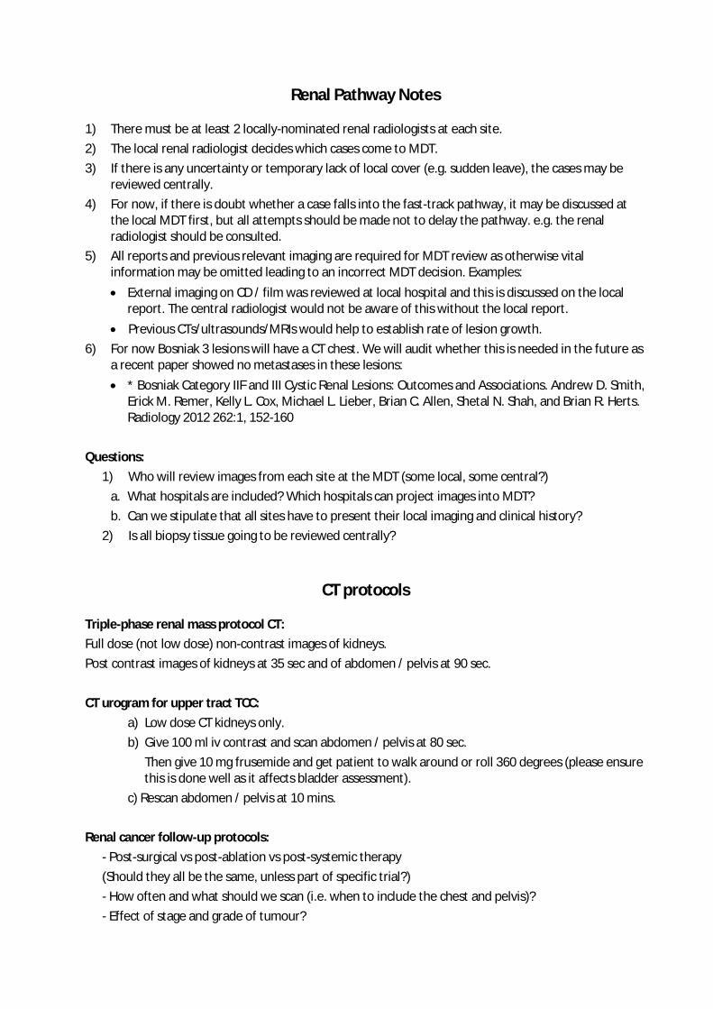
Renal Pathway Notes
1) There must be at least 2 locally-nominated renal radiologists at each site.
2) The local renal radiologist decides which cases come to MDT.
3) If there is any uncertainty or temporary lack of local cover (e.g. sudden leave), the cases may bereviewed centrally.
4) For now, if there is doubt whether a case falls into the fast-track pathway, it may be discussed atthe local MDT first, but all attempts should be made not to delay the pathway. e.g. the renalradiologist should be consulted.
5) All reports and previous relevant imaging are required for MDT review as otherwise vitalinformation may be omitted leading to an incorrect MDT decision. Examples:
External imaging on CD / film was reviewed at local hospital and this is discussed on the localreport. The central radiologist would not be aware of this without the local report.
Previous CTs/ultrasounds/MRIs would help to establish rate of lesion growth.
6) For now Bosniak 3 lesions will have a CT chest. We will audit whether this is needed in the future asa recent paper showed no metastases in these lesions:
* Bosniak Category IIF and III Cystic Renal Lesions: Outcomes and Associations. Andrew D. Smith,Erick M. Remer, Kelly L. Cox, Michael L. Lieber, Brian C. Allen, Shetal N. Shah, and Brian R. Herts.Radiology 2012 262:1, 152-160
Questions:
1) Who will review images from each site at the MDT (some local, some central?)
a. What hospitals are included? Which hospitals can project images into MDT?
b. Can we stipulate that all sites have to present their local imaging and clinical history?
2) Is all biopsy tissue going to be reviewed centrally?
CT protocols
Triple-phase renal mass protocol CT:
Full dose (not low dose) non-contrast images of kidneys.
Post contrast images of kidneys at 35 sec and of abdomen / pelvis at 90 sec.
CT urogram for upper tract TCC:
a) Low dose CT kidneys only.
b) Give 100 ml iv contrast and scan abdomen / pelvis at 80 sec.
Then give 10 mg frusemide and get patient to walk around or roll 360 degrees (please ensurethis is done well as it affects bladder assessment).
c) Rescan abdomen / pelvis at 10 mins.
Renal cancer follow-up protocols:
- Post-surgical vs post-ablation vs post-systemic therapy
(Should they all be the same, unless part of specific trial?)
- How often and what should we scan (i.e. when to include the chest and pelvis)?
- Effect of stage and grade of tumour?
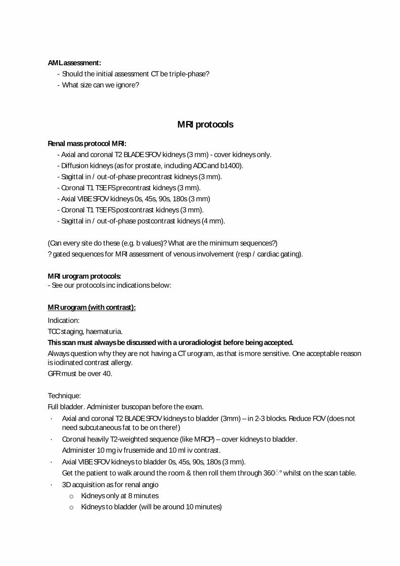
AML assessment:
- Should the initial assessment CT be triple-phase?
- What size can we ignore?
MRI protocols
Renal mass protocol MRI:
- Axial and coronal T2 BLADE SFOV kidneys (3 mm) - cover kidneys only.
- Diffusion kidneys (as for prostate, including ADC and b1400).
- Sagittal in / out-of-phase precontrast kidneys (3 mm).
- Coronal T1 TSE FS precontrast kidneys (3 mm).
- Axial VIBE SFOV kidneys 0s, 45s, 90s, 180s (3 mm)
- Coronal T1 TSE FS postcontrast kidneys (3 mm).
- Sagittal in / out-of-phase postcontrast kidneys (4 mm).
(Can every site do these (e.g. b values)? What are the minimum sequences?)
? gated sequences for MRI assessment of venous involvement (resp / cardiac gating).
MRI urogram protocols:- See our protocols inc indications below:
MR urogram (with contrast):
Indication:
TCC staging, haematuria.
This scan must always be discussed with a uroradiologist before being accepted.
Always question why they are not having a CT urogram, as that is more sensitive. One acceptable reasonis iodinated contrast allergy.
GFR must be over 40.
Technique:
Full bladder. Administer buscopan before the exam.
· Axial and coronal T2 BLADE SFOV kidneys to bladder (3mm) – in 2-3 blocks. Reduce FOV (does notneed subcutaneous fat to be on there!)
· Coronal heavily T2-weighted sequence (like MRCP) – cover kidneys to bladder.
Administer 10 mg iv frusemide and 10 ml iv contrast.
· Axial VIBE SFOV kidneys to bladder 0s, 45s, 90s, 180s (3 mm).
Get the patient to walk around the room & then roll them through 360 ◌۫° whilst on the scan table.
· 3D acquisition as for renal angio
o Kidneys only at 8 minutes
o Kidneys to bladder (will be around 10 minutes)

MR urogram (without contrast):
Indication:
TCC staging, haematuria.
This scan must always be discussed with a uroradiologist before being accepted. The radiologist willalso decide if the patient should have 10 mg iv frusemide, 10 minutes prior to the scan (no need forfrusemide if baggy PC system).
Always question why they are not having a CT urogram, as that is more sensitive. One acceptable reasonis low GFR (=< 40, hence not suitable for contrast).
Technique:
Full bladder. Administer 10 mg iv frusemide and buscopan before the exam.
Axial and coronal T2 BLADE SFOV kidneys to bladder (3mm) – in 2-3 blocks.
Coronal heavily T2-weighted sequence (like MRCP) – cover kidneys to bladder.
To Be Decided
1) AML Pathway
2) Discuss what we have read:
a. Bosniak:
i. Good paper re what constitutes each stage
ii. Malignancy and metastasis rates + follow-up protocols
b. Cancer:
i. Metastatic rates for different cancer stages / grades / tumour types
c. Renal cancer follow-up protocols
d. Evidence for MRI protocols

Checklist for new case discussion at Renal SMDT
For Local MDT co-ordinators
Renal mass CT or renal mass MRI performed
Should not be referred with ultrasound only, unless there is a very good reason
CT / MRI reviewed by local renal radiologist
Local renal radiologist must agree that case is suitable for MDT
If Bosniak 3 / 4 cyst or solid lesion, needs urgent CT chest locally
If this will delay referral by a week, discuss with RFH MDT co-ordinator (? RFH slot available)
All relevant imaging sent by IEP to The Royal Free
Including current and previous imaging of kidneys and CTs/MRIs of the body
All imaging reports for the scans sent to RFH
Either via IEP (if your PACS allows this) or via email to RFH MDT co-ordinator

Renal Cancer MDT Review – Imaging Outcomes
Definite primarytumour or Bosniak
3/4 cyst
? Upper tract TCCConsideration forbiopsy
Bosniak 2f cyst Possible renal vein /IVC involvement
Equivocalenhancement of
lesionExamples:
•Too small to assess•15-20 HU
enhancement
Review at central Renal MDTWith all relevant images AND reports
Consideration fordefinitive treatment
“2f Pathway”
1) Lesiondocumented oncentral database
(including reasonswhy lesion called 2f)
2) 5-year imagingfollow-up locally
3) Nurse-ledtelephone clinic
review suggested
4) Referral back toMDT if any change
in imaging
Consider local USSIf still equivocal:
1) MRI
2) Contrast USS(refer to sites which
can perform this)
Indications:
1) Uncertainty renature of lesion(e.g. lymphoma,metastasis, lipid-
poor AML)
2) Prior to systemictherapy
(i.e. metastatic)
3) Prior to ablativetherapy
4) Prior to lesionsurveillance
(e.g. Small masses,lipid-poor AML,
oncocytoma)
Consider:
1) CT venogram
2) IVC MRI(with gatedsequences)
CT urogram +cystoscopy
June 27, 2014

Guidelines for the Use of MRI contrast in renal cancer
These guidelines are based on ESUR 1 and RANZCR 2 guidelines, following consultation with The Nephrology Unit at The Royal Free Hospital(many thanks to Dr. Robin Woolfson).
Newer agents are believed to be more stable and less prone to cause NSF, but we have taken a slightly more cautious approach, especially inextremely low eGFR / haemodialyisis / peritoneal dialysis.
Always use the lowest possible contrast dose and avoid high-risk agents if possible (even if not contraindicated).
MRI requiring iv contrast(eg for renal mass characterisation, in
lieu of triple-phase renal CT)
Consider Risks vs Benefits of Contrast
• Consider other imaging modalities• Consider postponing until renal function
stabilised / improved• Use low-risk agent, if contrast MRI required
YES
Risk Factors for Chronic KidneyDisease (CKD)?
1. Known renal disease / dialysis2. Family history of renal disease3. Age of 60 or over4. Diabetes mellitus5. Vascular disease (MI / stroke)6. Hypertension7. BMI of 30 or over8. Smoker
NO
Unstable renal function?
Formal measurement of creatinine / eGFR
eGFR > 60 : Proceed with MRI contrasteGFR 30 – 60 : Try to avoid high-risk agents
eGFR 15-29 : High-risk agents contraindicatedeGFR < 15 : No contrast
Haemodialysis : No contrastPeritoneal dialysis : No contrast
YES
Proceed with iv contrastUse lowest dose and lowest risk agent
NO
Risk of NSF Trade Name Generic Name
High
Omniscan gadodiamide
Magnevist gadopentetate
Optimark gadoversetamide
Medium
Primovist gadoxetate
Ablavar gadofosveset
MultiHance gadobenate
Low
Dotarem gadoterate
Gadovist gadobutrol
ProHance gadoteridol
REFERENCES:1) Eur Radiol (2013) 23:307–318.
2) http://www.ranzcr.edu.au/component/docman/doc_download/553-revised-college-guidelines-for-gadolinium-containing-mri-contrast-agents-

Renal Mass Biopsy Guidelines
The most up-to-date recommendations are the EAU 2013 renal cancer guidelines 1. Thenetwork guidelines are based on this document.
Indications
1) When there is uncertainty regarding the nature of a renal lesion, especially if suspicionof:
a. Lymphomab. Metastasis
2) To obtain tissue (in order to select optimal systemic therapy) in cases of metastaticdisease.
3) Prior to ablative therapya. At a separate sittingb. Or at the start of the ablation procedure
4) To select patients with small renal masses for surveillance approachesa. Suspected lipid-poor AMLb. Suspected oncocytomac. Other lesions that are for active surveillance
Preparation
1) All patients should have recent routine bloods including FBC, U&E’s and clotting screen(INR and APTT).
2) If Hb < 10 or biopsy at high risk of bleed (e.g. very vascular lesion, anti-clotting therapy),needs Group & Save.a) If Hb < 8, consider preprocedure transfusion.
3) If on anti-coagulation / anti-platelet therapy, this will need to be stopped pre-procedure.a) Always consult the team that instituted the therapy (e.g. cardiologist, stroke
physician) or the haematology team regarding cessation.b) Aspirin - stopped for 7 days.c) Clopidogrel - stopped for 10 days 2.
i) If on combination aspirin / clopidogrel therapy, aspirin therapy may need to becontinued.(1) Higher risk should be explained to the patient.(2) Consider delaying biopsy if combination therapy is for a limited period (e.g.
6 months / 1 year).d) LMWH - omitted the night before and the day of the procedure.e) Warfarin – bridging plan from haematology team.f) Newer anticoagulants (e.g. rivaroxaban) – haematology review.

Technique
1) If the lesion cannot be confidently identified on ultrasound (especially if the lesion isendophytic), then CT guidance should be used.
a. Prebiopsy, contrast injection should be strongly considered to help targetenhancing regions of the lesion.
2) Core biopsies should be obtained rather than FNA.a. A co-axial needle should be used (thought to reduce risk of seeding).b. The biopsy needle should be at least 18G.c. At least 2 samples should be taken.
3) Non-necrotic (i.e. enhancing) areas of the tumour should be targeted – most often, theperiphery of the lesion is the best site on larger tumours.
a. If there is any concern regarding adequate sampling, the co-axial needle may berepositioned and further samples taken (i.e. multiple sites as well as multiplebiopsies).
4) If samples are to be used for cytogenetic studies or tissue banking, they most likelycannot be fixed in formalin.
a. See below.
Histological Analysis(Thanks to Drs. A Bates & S El Sheikh)
1) All the material received will be histologically examined and initial H&E sections cut.Immunohistochemistry will be undertaken as a panel at the pathologist’s discretion.a) Appropriate clinical information should be provided on the pathology request form.
2) Samples showing necrotic tissue only will be examined at multiple levels before the finalreport is issued as inadequate biopsy.
3) Fuhrman grading to be attempted, particularly as high grade (3-4) versus low grade (1-2), with the limitation of sampling error highlighted.
4) Oncocytic tumours may be present in the form of hybrids (particularly hybridoncocytoma-chromophobe carcinoma).a) The distinction between the two tumours on biopsy may be evident on H&E
supported by immunohistochemistry but tumour heterogeneity is a caveat thatmust be recognised.
5) Cytogenetic studies or tissue banking require fresh tissue in the majority of cases andcores taken for this purpose cannot be fixed in formalin.a) The problems anticipated are related to the rapid freezing and facilities required and
the possible erroneous sampling (for example a core of benign renal tissue or anentirely necrotic core).
b) Protocols must be in place before this is attempted.
6) Normal parenchyma present may be further assessed by special stains if renal function iscompromised.

References
1) http://www.uroweb.org/gls/pdf/10_Renal_Cell_Carcinoma_LR.pdf
2) Li C, Hirsh J, Xie C, Johnston MA, Eikelboom JW. Reversal of the anti-platelet effectsof aspirin and clopidogrel. J Thromb Haemost. 2012 Apr;10(4):521-8.
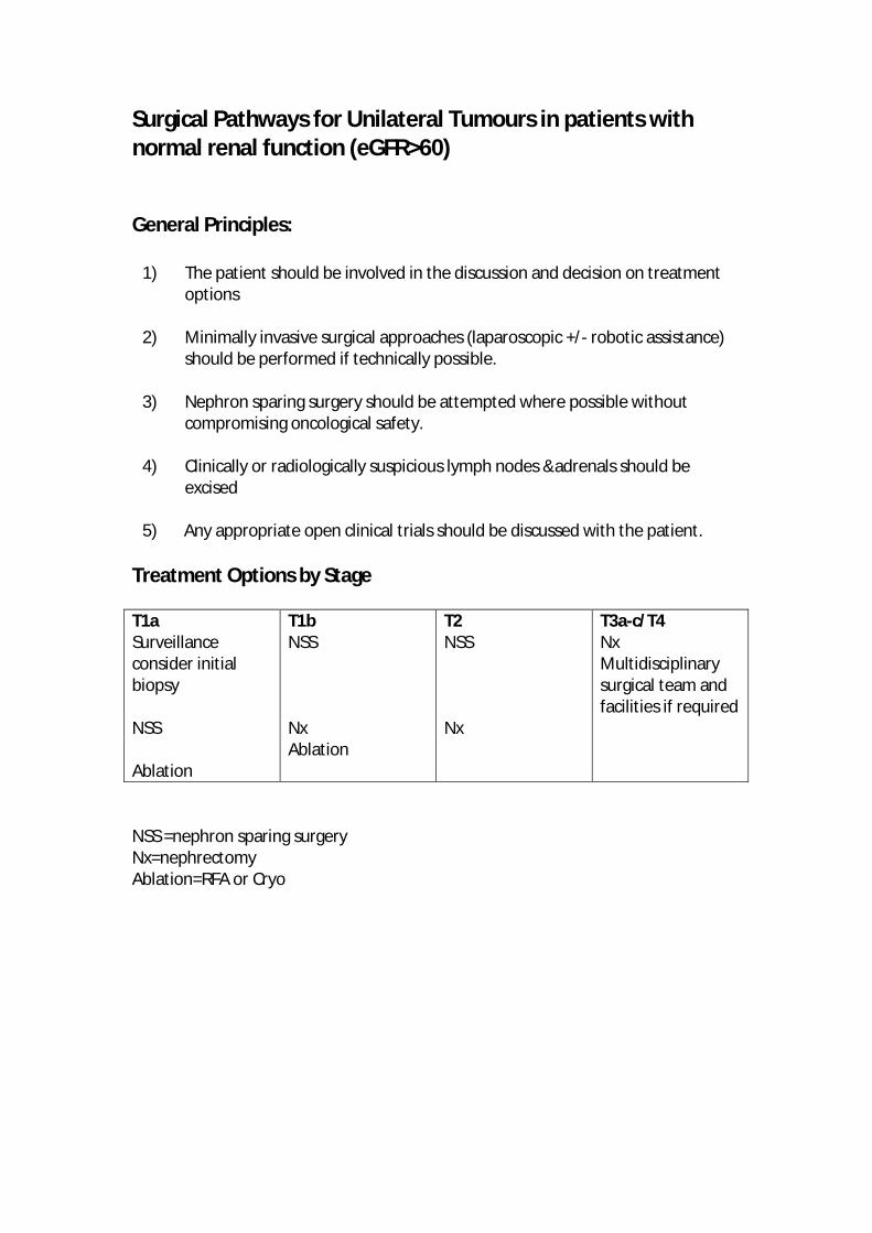
Surgical Pathways for Unilateral Tumours in patients withnormal renal function (eGFR>60)
General Principles:
1) The patient should be involved in the discussion and decision on treatmentoptions
2) Minimally invasive surgical approaches (laparoscopic +/- robotic assistance)should be performed if technically possible.
3) Nephron sparing surgery should be attempted where possible withoutcompromising oncological safety.
4) Clinically or radiologically suspicious lymph nodes &adrenals should beexcised
5) Any appropriate open clinical trials should be discussed with the patient.
Treatment Options by Stage
T1a T1b T2 T3a-c/T4Surveillanceconsider initialbiopsy
NSS NSS NxMultidisciplinarysurgical team andfacilities if required
NSS Nx Nx
AblationAblation
NSS =nephron sparing surgeryNx=nephrectomyAblation=RFA or Cryo

Surgical Pathways for bilateral tumours, tumours in solitaryfunctioning kidneys and patients with impaired renal function.
General Principles
1) The patient should be involved in the discussion and decision on treatmentoptions including the risk of cancer related mortality against the risks of surgeryand renal replacement therapy.
2) The aim of treatment is to achieve oncological cure whilst maintaining as muchrenal function as possible.
3) These are complex cases which may require a multimodality (ablative & surgical)and multidisciplinary approach (surgical/radiological/nephrological)
4) Additional investigations (differential renal function/biopsy)may be required tobest inform treatment decisions
5) Treatment pathways need to be decided on a case by case basis
6) The protocol for Renal Surgery in patients with CKD should be followed

Post nephrectomy follow up schedule for RCC
MAYO-Leibovichscore
6 weeks post opF/U
6 months 6-24 months Annually years2-5
5-10 yrs
Low risk 0-2RR: 7.5%@ 10 yrsRN/PN
Operative site andgen wellbeing andFeedback BloodsBP, Urine DipGFR
CT CAPBloodsBP, Urine DipGFR
US KUBCXRBloodsBP, Urine DipGFR-annual
US KUBCXRBloodsBP, Urine DipGFR
Dischargeafter 5 yrs
Intermediate risk3-5RR: 36%@ 10 yrsAll treatments
Operative site andgen wellbeing andFeedbackBloodsBP, Urine DipGFR
CT CAPBloodsBP, Urine DipGFR
CT CAPBloodsBP, Urine DipGFREvery 6 months
US KUBCXRBloodsBP, Urine DipGFR every yr.
US KUBCXRBloodsBP, Urine DipGFR yrly
High risk>6RR: 76%@ 10 yrsAll treatments
Operative site andgen wellbeing,Feedback and anytrial enrolmentdiscussionBloodsBP, Urine DipGFR
CT CAPBloodsBP, Urine DipGFR
CT CAPBloodsBP, Urine DipGFREvery 6 months
CT CAPBloodsBP, Urine DipGFR
CT andUS/CXRAlternate yrsBloodsBP, Urine DipGFR every yr
Individually tailored follow up for Bilateral and Familial disease
The metastatic risk may differ with histology other than clear cell

Kidney Cancer guidelines for referral and follow up of patients
considered for ablation of a small renal tumour.
Background
• Different expert societies have guidelines surrounding ablation of small renal masses.
• In the UK, the National Institute for Health and Clinical Excellence has also issued guidelines.
• Recommendations must be made for the referral of patients for ablation, the type of
ablation offered and follow up after ablation in the London Cancer Renal Cancer Pathway.
Methods
• Expert society and national guidelines have been reviewed and summarized below.
• More comprehend excerpts from the guidelines for each society are given in the appendices;
AUA (appendix 1), EAU 2010 update (appendix 2), BAUS (appendix 3), NICE – radiofrequency
ablation & cryoablation (appendix 4).
Results
Pre-ablation biopsy
AUA Recommended before ablation
EAU Recommended before ablation
BAUS Not specified
NICE Not specified, but noted that interpretation of data difficult withoutdefinitive histology
Referral for ablation
AUA ‘substantial comorbidities’
EAU Small, incidentally found renal cortical lesions in elderly patient,patients with a genetic predisposition for developing multiple tumours,those with bilateral tumours, and patients with a solitary kidney whoare at high risk of complete loss of renal function following NSS… Smalltumours and/or significant comorbidity who are unfit for surgery.
BAUS Stage T1 T2 disease
Life expectancy >1Small [<5] peripheral [cortical] tumoursGenetic predisposition to multiple tumoursA solidary kidneyBilateral tumours
NICE Not specified
?

Type of ablation & follow-up
AUA Not specified
EAU Cryoablation, as less risk of local recurrence
BAUS Not specified
NICE Less risk of local recurrence with cryoablation, but slightly increased riskof haemorrhage
Follow up after ablation
AUA CT or MR @ 3 & 6 months, then yearly. CXR yearly if low rick RCC oroncocytoma
EAU Intermediate risk – CT @ 6 month, 2 & 5 years. CXR & US @ 1, 3 & 4years. High risk – CT @ 6 months and the yearly
BAUS Not specified
NICE Not specified
Considerations
Given the paucity of long term data on ablative techniques, entry into national & international
registries and clinical trials should be encouraged
Most studies require a biopsy proven malignancy to be an inclusion criterion
Recommendations
• Biopsy
– A percutaneous renal biopsy should be undertaken before ablation to confirm the
diagnosis of malignancy
– This should be performed sufficiently in advance of the ablation so that an
inconclusive biopsy may be repeated
– Histological diagnosis of an oncocytoma will require further discussion at the MDT
given that oncocytoma and RCC may coexist
• Indications
– Nephron sparing surgery should be considered as first line intervention for stage T1a
renal tumours
– Ablation should be considered more favourably if:
• The patient does not wish to undergo surgery
• The patient is deemed unfit / high risk for surgery
• There is a solitary kidney or poor renal function

• There are bilateral tumours or a genetic predisposition to multiple tumours
– The tumour must be deemed suitable for ablation by at least two interventional
consultants.
– Chronological age is not necessarily an indication for ablation
– The MDT must record the suggested order of interventional options on the
information available (for instance: surgery first, ablation second & surveillance
third). However, the final decision on the type of intervention offered must be made
in the renal cancer MDT clinic with consensus between the surgeon and
interventional oncologist
• Type of ablation
– Percutaneous cryoablation should be offered in the first instance
– Radiofrequency ablation may be offered if there is an increased risk of bleeding at
the time of the procedure
• Follow up
– A contrast enhanced CT of the kidneys will be performed at 3, 6 and 12 months and
then yearly after ablation
– Where the renal function is poor or there is a contrast allergy, a contrast enhanced
MRI of the kidneys may be performed
– A non-contrast CT scan of the chest will be performed yearly to exclude metastatic
disease

Kidney Cancer Oncological Pathways
Commencing first line therapy in fit patients with metastatic/advanced renal cancer
Sequencing of systemic therapy
Powles et al. BJC 2010.
Targetedtherapy Disease
progression
Observation
il-2 metasectomyRFA/cyberknife
Symptomatic
Asymptomatic
Asymptomatic
MSKCC poor riskdisease
MSKCCintermediaterisk disease
MSKCC good riskdisease
Multiple organinvolvement
Multiple sites insingle organ
Single site insingle organ
Pazopanibor
sunitinib
Axitinibor
EverolimusEverolimus
STARor
PDL-1study
ZEBRAor
Meteor
Standard targetedtherapy
ZEBRAor
AZTECClinical trial strategy
Non clear celldisease
sunitinibor
pazopanib
Clinical trial referencesRECORD 1RECORD 3COMPARZAXISINTROSECTGOLD

The role of nephrectomy in metastatic disease*
MSKCC risk Tumour burden from primary Role of Nephrectomy1
GoodLow Indicated
High Indicated
IntermediateLow Questionable
High Indicated
Poor Low Not recommended
High Questionable
Bex A et alExpert Rev Anticancer Ther.2012 Jun;12(6):787-97.
1If primary is symptomatic then nephrectomy may be indicated
* Clinical trial include SURTIME and CARMENA

Royal Free London NHS Foundation Trust
Renal Cancer Specialist Service
Michael Aitchison, Renal Cancer Service Director & Consultant Urological Surgeon
Professor Tom Powles, Lead Renal Cancer Oncologist
UPDATE ON OUTCOMES
April 2014
1 INTRODUCTION AND GENERAL COMMENTS
In our previous paper “Update on outcomes December 2013 “we presented our outcomes for the first 9
months of 2013 against our quality performance indicators. This paper outlines progress made over the
course of the first quarter of 2014 in measuring and reporting on our kidney cancer outcomes.
Our aspiration is to provide World Class Surgical and Oncological care. Measuring and improving
outcomes is a key element of our plan for delivery of the specialist renal cancer surgical service and
leading improvement in renal cancer across London Cancer. The Quality performance indicators have
been reviewed and amended to bench mark the centre against world leading international centres in
terms of surgical and oncological outcomes.
Robotically assisted nephrectomy and partial nephrectomy were introduced in March 2014 with 15
cases being performed in a 4 week period.
Prior to 2013, measurement of activity and outcomes was largely ad hoc, retrospective and therefore of
doubtful accuracy and limited use in driving improvement. Although the data outlined in this summary
are incomplete, the establishment of a process for prospective collection of surgical outcome data and a
process for aligning data collection with the patient pathway is a significant achievement. While we
recognise that the process needs to be strengthened to ensure all patient data are captured, we feel
that we now have a robust process in place for capture of this data and production of reports that can
inform service improvement.
2 DATA SOURCES
The main data sources used to compile this report are: -
• Prospective collection of surgical outcome data (submitted to national BAUS Nephrectomy Audit).
The process for this data collection is outlined in figure 1. Data capture is now reviewed on a
weekly basis at the Renal Cancer Surgical Planning meeting and any missing data collected at this
point so it is anticipated that there will be minimal missing data in the future
• Cerner reports for process measures (e.g. LoS, time from referral etc.)
• Manual reports from renal cancer team (e.g. date of MDT from MDT co-ordinator)
• Patient satisfaction survey. Telephone follow up is currently underway as the postal survey yielded
a low response rate
• Morbidity and mortality meetings for details of major complications

Figure 1. Process for Recording Surgical Outcome Data
3 ACTIVITY
3.1 Volume of Cases
From 1 January 2013 to date, we have carried out 133 nephrectomies and partial nephrectomies. The
case load continues to grow (figure 2). And the projected figure for 2014 given the activity in the first
quarter of 2014 is shown.
Figure 2. Growth in Renal Surgery Case Load 2013 and projected load 2014
0
45
90
135
180
2012 2013 2014
60
133
172
annual cases

3.2 Referral Map
The geographical origin of the patients referred for surgery is summarised in figure 3.
Figure 3. Map of Nephrectomy Referrals

4 PERFORMANCE AGAINST QUALITY PERFORMANCE INDICATORS
Performance against our quality performance indicators is summarised in figure 4. Data for survival
measures are not yet available so these indicators are currently shown in grey. Green, amber or red
ratings have been assigned to each indicator. Targets will be reviewed on an annual basis to ensure
they reflect best contemporary standards.
The source of the data and period measured are referenced in the “COMMENTS” column.

Figure 4. Performance Against Quality Performance Indicators
MEASURE DESCRIPTION EXCLUSIONS TARGET OUTCOME COMMENTS
1 Time from referralreceived to first treatment< 62 days
Numerator = number of patients whoreceive first treatment within 62 days ofentry to the pathway
Patients who refusetreatment
85% 88% A few waiters over 62days might be excluded,data source Open Exeter(Oct12 - Sep13)
Denominator = all patients Patients who die beforetreatment
Patients unfit / unsuitablefor treatment
2 TNM Staging Numerator = number of patients diagnosedwith renal cell cancer who were clinicallyTNM staged before first treatment
100% 99% Process to be establishedto capture TNM stagingat first MDT discussionand submission to BAUSaudit
Denominator = all patients
3 Patient Satisfaction Numerator = number of patients ratingtheir overall satisfaction with the service asgood, very good or excellent in the annualpatient satisfaction survey
Patients who do not returnsurvey
75% 100% 795 return rate forpatient experiencesurvey of sMDT clinicand in-patient stay
Denominator = all patients surveyed
4 Nephron Sparing Surgeryin T1a Disease
Numerator = number of patients withT1aN0M0 tumours undergoing nephronsparing surgery as first treatment
Patients who refusetreatment
40% 57% BAUS
Patients who receiveRFA/cryotherapy
Patients receivingsupportive care only

MEASURE DESCRIPTION EXCLUSIONS TARGET OUTCOME COMMENTS
Denominator = number of patients withT1aN0M0 tumours undergoing surgery asfirst treatment
Patients receiving activesurveillance
Patients who died beforetreatment
5 Reoperation within 30days
Numerator = Number of patientsundergoing second surgical procedurewithin 30 days of primary surgery
3% 2.2% 2 splenectomy, 1completion nephrectomy
Denominator = Number of patientsundergoing surgery as first treatment
6 30 Day Mortality AfterSurgery or Ablation
Numerator = Number of patients whoundergo minimally invasive or operativetreatment as first treatment who die within30 days
Emergency surgery < 5% 0 Data source - Cerner(subject to DOD beingrecorded on Cerner) Jan-Sep13, M&M data Jan -Sep 13
Denominator = All patients who undergominimally invasive or operative treatmentas first treatment
7 Proportion of patientsundergoing minimalaccess rather than opensurgery
Numerator = number of patientsundergoing surgery as first treatment whohave minimal access surgery
Emergency surgery 65% 82% Data source - BAUS
Denominator = number of patientsundergoing surgery as first treatment
8 Proportion of patientsrequiring perioperative orpostoperative renalreplacement therapy
Numerator = number of patients requiringperioperative or postoperative renalreplacement therapy
Patients on renalreplacement therapy pre-operatively
<5% 3% Data source –BAUS /VitalData
Denominator = all patients undergoingsurgery

MEASURE DESCRIPTION EXCLUSIONS TARGET OUTCOME COMMENTS
9 Number of surgeonscarrying out fewer than 20nephrectomies or partialnephrectomies per annum(open / laparoscopic /robotic)
0 0
10 Mean change in eGFRfollowing partialnephrectomy(laparoscopic or robotic)at 6 months
eGFR= eGFR at diagnosis – eGFR at 6months / eGFR at diagnosis
Patients on renalreplacement therapy pre-operatively
10% 7.4% BAUS, eGFR on Cerner.NB Small numbers incurrent data set.
Mean eGFR for all patients undergoingpartial nephrectomy for whom at least 6months have elapsed since surgery
11 TRIFECTA rate in partialnephrectomyT1a tumours
Numerator = number of patients with T1atumour undergoing partial nephrectomywho have warm ischaemic time less than25 mins.,negative surgical margins and nocomplications ((CLAVIEN 3 or above)
Denominator = all patients with T1Aundergoing partial nephrectomy
60% 76% Data source - BAUS forprocedure type,ischaemic time, margins,complications, Cerner forLOS, RENAL score notrecorded Jan - Sep 2013
Cleveland Clinicbenchmark
12 Clinical trials Numerator = number of newly diagnosedrenal cancer patients presented to MDTentering investigational/translationalclinical trial
Denominator = number of newly diagnosedrenal cancer patients presented to MDT
7.5% NCRI benchmarkData not yet available

MEASURE DESCRIPTION EXCLUSIONS TARGET OUTCOME COMMENTS
13 Oncology Clinical trials% of patients who receivea systemic therapy whoare enrolled in clinicaltrials. This includes 1st,2nd, 3rd line and beyond.
Numerator = number of systemic therapiesgiven within the context of a clinical trialsin the population.
Denominator = number of new systemictherapies started in the population.
50% - Data not yet available
14 2 Year Survival MetastaticKidney Cancer from thetime of starting systemictherapy
Numerator = number of patients withmetastatic cancer at diagnosis for whom atleast 2 years have elapsed since diagnosiswho are alive 2 years after diagnosis
50% - Data not yet available
Denominator = number of patients withmetastatic cancer at diagnosis for whom atleast 2 years have elapsed since diagnosis
15 MSKCC Score Numerator = number of patients withbiopsy proven metastatic renal cell cancerwho are assigned an MSKCC score prior tostarting therapy.
100% - Data not yet available
Denominator = all patients with biopsyproven Renal cell cancer starting systemictherapy.
16 1 Year Survival T1 KidneyCancer(includes surveillance,ablation and surgery)
Numerator = number of patients with T1cancer at diagnosis for whom at least oneyear has elapsed since diagnosis who arealive one year after diagnosis
97% - Data not yet available
Denominator = all patients with T1 cancerat diagnosis for whom at least one year haselapsed since diagnosis

Clinical Trials
Key Principles
Potential recruitment into suitable trials to be discussed with all patients
Explanation of clinical trials included in patient information
All patients to be approached regarding biobanking of urine, serum and tissue
sMDT discussion and output to identify potential patients for current open clinical
trials


