32
MODULE 3.0 Chronic Venous Insufficiency and Venous Leg Ulcers (Approx 30 mins)
MODULE 3.0Chronic Venous Insufficiency
and Venous Leg Ulcers
(Approx 30 mins)
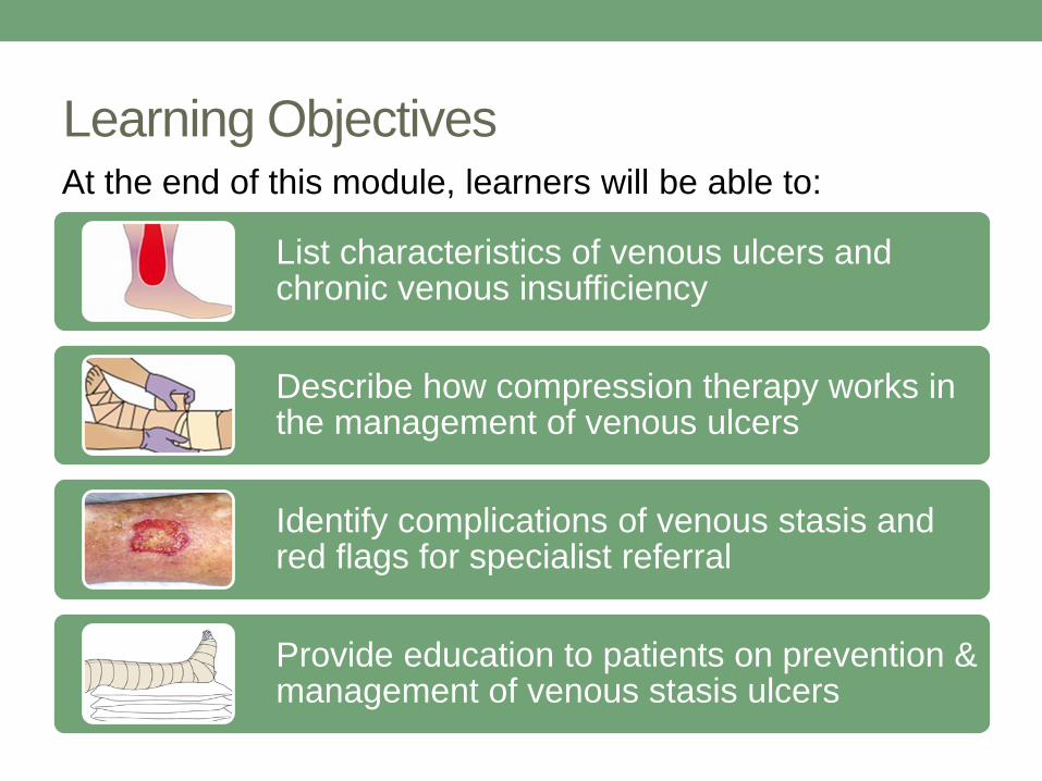
Learning Objectives
List characteristics of venous ulcers and chronic venous insufficiency
Describe how compression therapy works in the management of venous ulcers
Identify complications of venous stasis and red flags for specialist referral
Provide education to patients on prevention & management of venous stasis ulcers
At the end of this module, learners will be able to:

Chronic Venous Insufficiency (CVI)
• Chronic venous insufficiency or venous
stasis results from incompetent valves in the
veins of the lower leg.
• In healthy leg veins, one-way valves keep
blood flowing up to the heart, against gravity.
• When valves become damaged or
“stretched,” venous blood refluxes backward
down the veins into a congested leg.
• Fluid leaks out of veins leading to swelling
(edema), irritation of the skin, and eventually
skin breakdown.

• Lack of exercise and lack of
physical activity involving the
lower legs make CVI worse.
• A collection of veins are
located deep inside the lower
leg, supported by powerful calf
muscles that help push blood
back up to the heart with every
contraction.
• When veins are stretched or
unhealthy, or the calf muscle
pump is not working well, it
can lead to venous
hypertension in the lower leg
veins.

Skin inflammation
Stasis dermatitis
Skin breakdown
Leg ulcer
High intravascular pressure results in extravascular
fluid collection (edema) in the lower leg, resulting in
impaired blood flow and nutrition to the skin.
If not corrected, venous hypertension leads to:

Risk factors for CVI
• Older age (>50 years)
• Obesity
• Previous DVT
• Damage to lower leg veins – e.g. surgery (such as
saphenous vein harvest for CABG), trauma
• Varicose veins and incompetent venous valves
• Physical inactivity
• Family history

Features of Venous Leg Ulcers (VLU)
and Stasis Dermatitis due to CVI
Location on lower leg
• between knee and ankle
• “gaiter” or sock distribution –
between lower third of calf and 1 inch
below malleolus
• most commonly anteromedial calf
Lower leg edema
• typically worse by end of day
• less with leg elevation
• pedal pulses may be difficult to feel
due to edema

Stasis dermatitis:
acute phase
• erythematous
• maculopapular rash
• skin edema
• vesicles or bullae if
marked
• edema
• pruritis
Stasis dermatitis: chronic phase
• post-inflammatory skin changes/
scarring
• hemosiderin deposition in skin
• atrophie blanche (smooth ivory
white scarring of skin stippled with
telangiectasia and surrounded by
hyperpigmentation)
• hyperkeratosis (dry, thickened/
scaling skin)
• mild erythema
• dependant edema
• hypopigmented, atrophic skin
change at site of previous ulceration

Mild erythema
Atrophie blanche
Hemosiderin
deposition/staining
Telangiectasia
Chronic venous stasis skin changes

Chronic venous stasis and ulcer skin changes
Post-inflammatory scarring
Dry, scaling/lichenified skin

More features of venous leg ulcer
• shallow ulcer base
• dark red granulation tissue with yellow
adherent slough
• irregular border
• large amount of wound exudate
/drainage when leg edematous
• relatively painless – achy, dull pain
worsening as day progresses;
increased pain if infected
• surrounding skin features of acute or
chronic stasis dermatitis – may mimic
cellulitis if acute
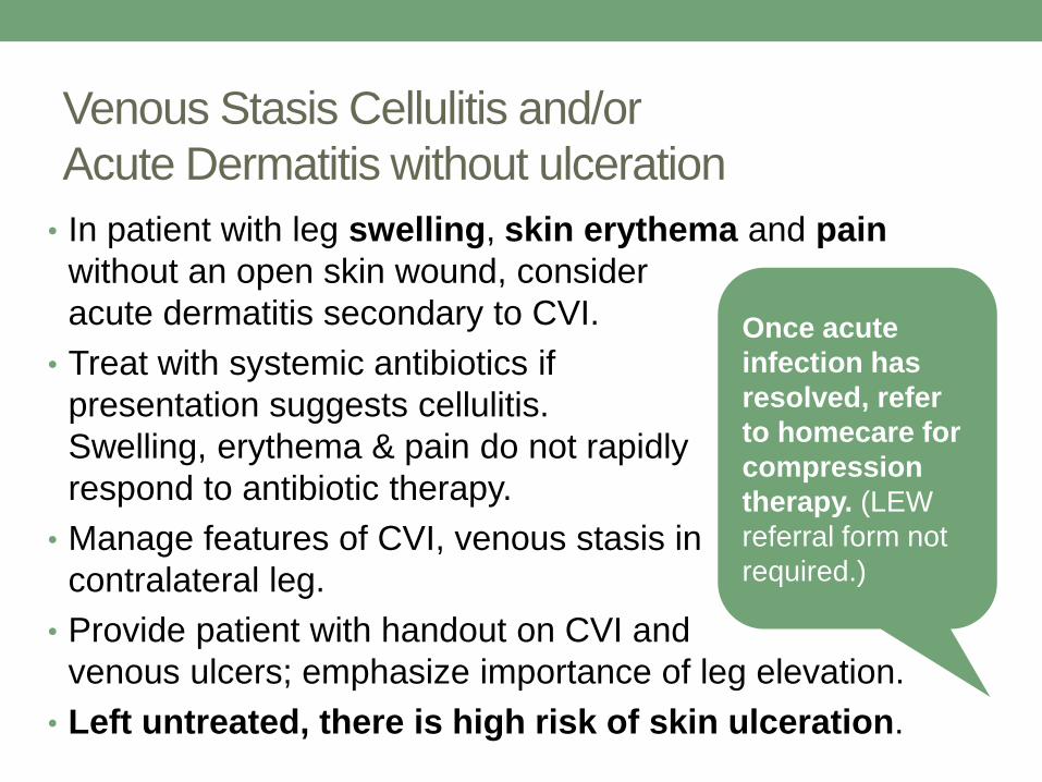
Venous Stasis Cellulitis and/or
Acute Dermatitis without ulceration
• In patient with leg swelling, skin erythema and pain
without an open skin wound, consider
acute dermatitis secondary to CVI.
• Treat with systemic antibiotics if
presentation suggests cellulitis.
Swelling, erythema & pain do not rapidly
respond to antibiotic therapy.
• Manage features of CVI, venous stasis in
contralateral leg.
• Provide patient with handout on CVI and
venous ulcers; emphasize importance of leg elevation.
• Left untreated, there is high risk of skin ulceration.
Once acute
infection has
resolved, refer
to homecare for
compression
therapy. (LEW
referral form not
required.)

Venous Leg Ulcer Management
- physician/NP role
1) Use the LEW Pathway referral form to make a non-
urgent referral to the homecare nurses.
• Specific orders are not required
• Treatment will be initiated according to pathway
protocols for venous wound management

Venous Leg Ulcer Management
- physician/NP role
2) While patient is waiting for wound care nursing assessment and management, MD/NP may apply a simple non-adherent gauze dressing with adequate padding to absorb exudate, and wrap lower leg with an elastic/Kling bandage.
3) Treat with oral antibiotics if cellulitis or wound infection.
4) Prescribe topical corticosteroid such as Clobetasolcream if acute dermatitis surrounding ulcer; wound care nurse will apply this when doing dressings.
5) Encourage patient to keep lower leg elevated as much as possible and perform calf muscle exercises.
6) Provide patient information handout – from LEW Pathway website.

To open a PDF of the
patient hand-out What
is a venous leg ulcer?
click on the link in the
sidebar.

Venous Leg Ulcer Treatment
1) Leg and wound assessment
• Nurses perform comprehensive assessment
• Details of leg and wound are documented
2) Wound dressing
• Skin emollients are applied to protect the peri-wound skin -perfume and lanolin-free*
• A topical moderate-high potency corticosteroid (e.g. clobetasol) may be applied if acute dermatitis of surrounding skin
• An absorbent dressing is applied to the wound
• Leg is wrapped – with compression if arterial flow adequate (requires ABPI prior to compression bandaging)
*High risk of skin sensitization in these patients from lanolin, topical antibiotics, antiseptics, preservatives in dressings, resins, latex

More on wound dressing
• A venous ulcer usually produces heavy exudate, especially once compression is applied.
• Dressings must absorb exudate produced by the ulcer and protect the peri-ulcer skin.
• The goal is to maintain a moist wound bed while managing exudate.
• A typical dressing is changed twice a week.
• For an uncomplicated wound, a nurse will select an alginate or foam dressing from the formulary
Dressings are selected according to wound characteristics such as:
amount of exudate
location of wound
skin condition
condition of wound bed
presence or absence of infection

Venous Leg Ulcer Treatment
3) Compression bandaging
• The cornerstone of treatment for venous
insufficiency is compression therapy.
• Compression removes excessive
extravascular fluid from the limb.
• Graduated compression (tighter at the
ankle) also aims to restore the normal
flow of venous blood up the leg
• Before applying compression, a wound
care nurse needs to exclude significant
peripheral arterial disease by performing
ABPI test.

More on compression bandaging
• Graduated high compression wrapping
may be applied if there is adequate
arterial flow to feet (ABPI ≥ 0.8). This is
part of standard protocols; separate
order from MD/NP is not required.
• If mild to moderate arterial obstruction
(ABPI 0.5-.79), modified compression
can be considered. MD/NP may be
consulted.
• If inadequate arterial flow (ABPI ≤ 0.49)
compression is not recommended.
MD/NP should refer patient to vascular
specialist.
Graduated
compression
stockings should
not be used for
treating VLU or
an edematous leg
with dermatitis.
Stockings are
used to prevent
edema, not
reduce it.

How to perform an ABPI
To open a two-minute
video Ankle Brachial
Index Procedure Using a
Handheld Doppler from
the New England Journal
of Medicine, click on the
link in the sidebar.

Interpretation of ABPI/TBPI in Determining Compression
ABPI Value Interpretation/Clinical Significance Compression Therapy
>1.3 Abnormally high range; TBPI indicated Incompressible arteries
0.8 – 1.3 Compressible (normal range)High compression (up to 30-40 mm)
0.51 – 0.79 Mild to moderate obstruction/ peripheral arterial disease
Modified compression (20-30 mm)
<0.5 Significant ischemiaCompress only if ordered by specialist
TBPI Value Interpretation/Clinical Significance Compression Therapy
> 0.7 Normal High compression
.41-0.69 Mild to moderate peripheral arterial disease Modified compression
< 0.4 Severe ischemia Contraindicated
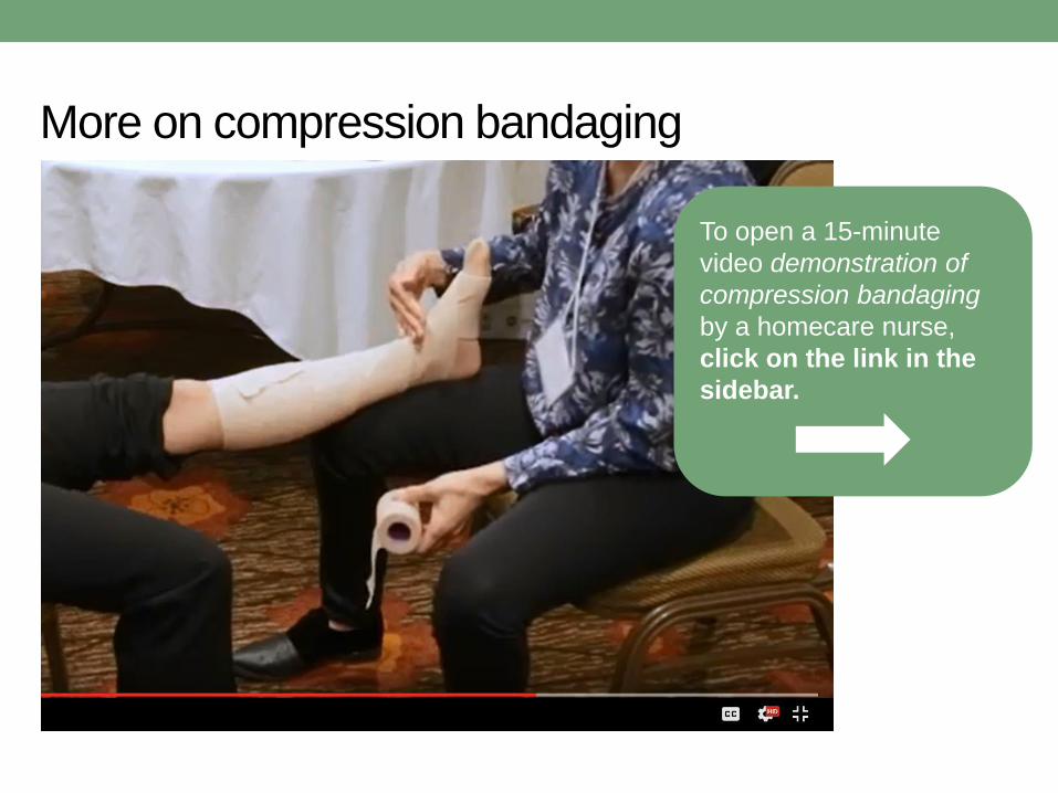
To open a 15-minute
video demonstration of
compression bandaging
by a homecare nurse,
click on the link in the
sidebar.
More on compression bandaging

Venous Leg Ulcer Treatment
4) Oral antibiotics if ulcer infected/cellulitis
• Avoid topical antibiotics – risk of skin sensitization
5) Other drugs to consider
• statin*
• pentoxifylline^
*Evangelista MT, Casintahan MF, Villafuerte LL. Simvastatin as a novel therapeutic agent for venous ulcers: a
randomized, double-blind, placebo-controlled trials. Br J Dermatol 2014;170(5):1151-1157.
^Jull A, Arroll B, Parag V, Waters J. Pentoxifylline for treating venous leg ulcers. Cochrane Database Syst Rev
2007;3:CD001733.

Practical tips about compression therapy
• Although nurses aim for a level of compression that is
tolerable to the patient, there may be some discomfort.
• Compression bandaging must be kept dry; patients can
coordinate with nurses to bathe on the day that wound
dressing is changed.
• For patients in remote locations without frequent
homecare, a self-applied product may be available. This
will be organized by wound care nurse.
• Patients will be advised to elevate the compressed leg
frequently to avoid swelling in the foot, and to walk as
much as possible.
• Nurses may take photos of a leg wound on a patient’s
phone, so patient can show this to physician.

When to refer for specialist assessment
• Clinical features of peripheral arterial
disease (PAD) and low ABPI –
preventing use of compression therapy
(See Module 5 for skin and nail features
of chronic arterial ischemia/PAD)
• Ulcer not healing adequately after 12
weeks of appropriate compression
therapy
• Suspicion of skin malignancy
• Dermatitis not responding to topical
steroids and compression therapy
• Frequent recurrence of VLU
As per LEW
pathway protocols,
the wound care
nurse will notify
the referring
physician/NP if
any of the above
occur, so that a
specialist referral
can be initiated.

Monitoring and surveillance
• Most venous stasis ulcers close in 6 months with optimal
care.
• Once the wound has healed, there is a high risk that an
ulcer will recur. 50% of VLU recur in 10 years.
• Encourage patients to be aware of increasing edema or
skin changes.
• Encourage patients to engage in preventative
management including lifetime use of compression
garments.
• Primary care providers can play an important role in
promoting adherence to compression therapy.

Compression therapy – long term use of compression stockings
• Once the venous ulcer has healed, the
wound care team will fit the patient for
compression stockings.
• Most patients will fit into standard stockings,
but some will require custom fitting by a
physiotherapist (for very large limbs).
• This will be arranged by the homecare team.
• Compression stockings are to be
put on early in the morning and
removed at bedtime.
• Aids are available for patients who have
difficulty applying the stockings.

Compression stockings – how much compression?
• 18-25 mmHg: low compression for varicose veins and
mild swelling
• 20-30 mmHg: moderate compression for prevention/long-
term management of edema related to venous
insufficiency
• 30-40 mmHg: high compression for post-thrombotic
venous insufficiency
• 50+ mmHg: control of lymphedema

Coverage for compression stockings
• Compression stockings are covered by the SAIL
program if they are ordered by a PT or
OT, wound specialist nurse or diabetes
nurse (arranged by the homecare team).
• A patient who has had a venous leg
ulcer is covered for 2 pairs of
compression stockings every six
months, for life.
• Compression stockings are also covered
by most private insurance plans –
prescription required from physician/NP.
• Only medical garments (>20 mmHg
compression) are covered.
For patients with
private insurance,
medical-grade
compression
garments are fitted
and sold at
medical supply
stores and many
local pharmacies.
Patients can call
ahead to ensure
that a trained fitter
is on staff.
Resources for patients and providers
The Lower Extremity Wound Pathway provides a web
platform to house resources for providers and for patients.
On provider pages you can find the referral form,
treatment tools and protocols for nurses.
On patient pages there are hand-outs and links to other
sources of information about caring for wounds and high-
risk legs and feet.
www.sasksurgery.ca/patient/lowerextremitywound.htmlTo open the Lower Extremity Wound
Pathway web pages, click on the link
in the sidebar.

References
• Scottish Intercollegiate Guidelines Network (2010).
Management of chronic venous leg ulcers: a national
clinical guideline.
• O’Donnell, Thomas F. et al. Management of venous leg
ulcers: Clinical practice guidelines of the Society for
Vascular Surgery and the American Venous Forum. 2014:
Journal of Vascular Surgery , Volume 60 , Issue 2 , 3S -
59S

END OF MODULEProceed to Module 3 Quiz


