Page 1
Lung radiology in the tropics
Eli Tumba Tshibwabwa, MD, PhDa,b,*,Jonathan L. Richenberg, MRCP, FRCRc, Zelena-Anne Aziz, MRCPd
aDepartment of Radiology, McMaster University Medical Centre, 1200 Main Street West, Room 2S40, Hamilton,
Ontario L8N 3Z5, CanadabFather Sean O’Sullivan Research Centre, St. Joseph’s Healthcare, 50 Charlton Avenue East,
Hamilton, Ontario L8N 4A6, CanadacDepartment of Radiology, The Royal Sussex County Hospital, Eastern Road, Brighton BN2 5BE, UK
dDepartment of Radiology, King’s College Hospital, Denmark Hill, London, UK
This article describes the radiologic findings of the
more common conditions that affect the thorax in the
tropics. Inevitably, the bulk of the discussion focuses
on infectious diseases. The intention, however, is not
simply to list salient imaging features out of context.
There are other important dimensions to lung radi-
ology in the tropics, and they are the physical,
political, and economic settings. The first part of this
article considers the broader issues of imaging serv-
ices in the tropics; the second part concentrates on the
radiology of infectious and other disease states.
Imaging services in the tropics
Radiologic evaluation is fundamental in the diag-
nosis of lung disease; a radiography unit is arguably
as important as a stethoscope in the assessment of
pulmonary pathology. In North America and else-
where in the developed world, high-quality and
timely imaging is taken for granted. Unfortunately, in
the tropical health care system, modern, dependable
imaging is a scarce luxury. The affluent areas in
Europe and North America have generous budgets
for radiologic equipment. Contrast this with the often-
impoverished regions in the tropics, which lack
resources, equipment, and personnel. (There is, for
example, no resident radiologist in the 800-bed
teaching hospital in Kumasi, Ghana’s second largest
city, which serves more than 1 million people.)
The differential is exacerbated by a hostile topog-
raphy and climate. Many areas are remote and
sparsely populated, such that in many tropical coun-
tries, resources are often concentrated in a few urban-
ized areas [1–4]. Clinicians who care for patients with
pulmonary symptoms in the tropics frequently must
accept suboptimal (or even no) imaging or send their
patients long distances to receive services. In turn, the
radiologist rarely has access to bronchoscopic or
lavage findings. The opportunity for multidisciplinary
meetings to review imaging with clinical and pathol-
ogy experts, a keystone to lung radiology in many
developed countries, is severely limited. How can
tropical lung radiology services be optimized?
The key to delivering a workable lung radiology
service within the tropics is to match provision to
demand. Imaging should be geared for the investiga-
tion and treatment of infection (eg, percutaneous
image-guided empyema drainage). Plain radiography
and ultrasound must form the core of any realistic
imaging service [2,4,5]. CT plays a limited role and is
found only in major centers.
Whenever possible, equipment should be cheap
and portable yet reliable and durable. Small machines
may be used in field hospitals with rapid transmission
of the images to large yet remote hospitals, where
interpretation is possible. The quality of the images
generated by these machines is good. Alternatively,
0272-5231/02/$ – see front matter D 2002, Elsevier Science (USA). All rights reserved.
PII: S0272 -5231 (02 )00008 -4
* Corresponding author. Father Sean O’Sullivan Re-
search Center, St. Joseph’s Healthcare, 50 Charlton Avenue
East, Hamilton, Ontario L8N 4A6, Canada.
E-mail address: [email protected]
(E.T. Tshibwabwa).
Clin Chest Med 23 (2002) 309–328
Page 2
the World Health Organization basic radiologic sys-
tem often could be a suitable basis for a basic but not
rudimentary imaging service in the tropics. With
respect to ultrasonography, which is helpful in the
diagnosis and treatment of pleural disease, there are
several high-quality yet compact and affordable units
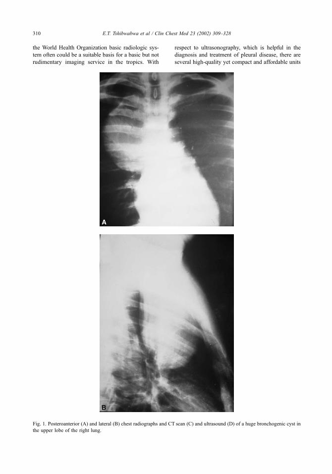
Fig. 1. Posteroanterior (A) and lateral (B) chest radiographs and CT scan (C) and ultrasound (D) of a huge bronchogenic cyst in
the upper lobe of the right lung.
E.T. Tshibwabwa et al / Clin Chest Med 23 (2002) 309–328310
Page 3
developed in the ironically high-tech world of North
American intensive care units that would function
admirably in the intensive environments experienced
in a tropical hospital.
Image-guided fine-needle aspiration and biopsy
may be crucially important in the management of
patients in the tropics. Ultrasound provides a rel-
atively cheap real-time method for guiding interven-
tional procedures. Aspirates may provide samples
from which organisms can be isolated so that valu-
able antibiotics can be husbanded and only used
where appropriate. After all, a pleural collection on
ultrasound is simply that: aspirate obtained by insert-
ing a needle under image guidance into the abscess,
which may reveal the pathogen [5].
Only 40% of countries in the sub-Saharan region
have any CT scanners. This is in contrast to the
situation in the Northern African region and in the
Republic of South Africa, where academic radiology
departments and other privately owned departments
are better equipped and serviced. Currently, major
South African cities have hospitals that provide such
high-tech lung imaging to patients from the neighbor-
ing countries and even from as far as Central and
Eastern Africa, where the few existing CT scanners
cannot cope with the patient load.
Lung radiology in the wider context of health care
Thoracic imaging can be worthwhile only if it is
cooordinated with the pulmonary medical and pathol-
ogy services within the hospital or within the region. A
chest radiograph is only as good as the report it
generates, and the report has worth only when it helps
the physician whomanages the patient. In other words,
a radiograph (or ultrasound scan) of the highest quality
still needs intelligent and clinically relevant interpreta-
tion [2,5]. One must move away from the mindset that
this interpretation must be provided at the site where
the images have been acquired. Dedicated landlines
can be linked to inexpensive modems to permit trans-
mission of digital ultrasound data across vast distan-
ces. On a more global scale, the World Wide Web
offers potential for image transfer and storage. Tele-
medicine is coming of age. The film or study can be
moved from the ‘‘spoke’’ to a ‘‘hub.’’ Once the film
has been read by a trained radiologist at the hub, the
Fig. 1 (continued ).
E.T. Tshibwabwa et al / Clin Chest Med 23 (2002) 309–328 311
Page 4
report can be sent back by a landline connection to the
remote spoke. Such teleradiology is well established in
parts of Scandinavia that are sparsely populated and
isolated, especially in winter.
Disease profile of lung imaging in the tropics
The scarcity of manpower and resources and the
logistical nightmares that ensue mean that lung
Fig. 2. Black and white photograph of a 41-year-old patient (A) with a biopsy proven right-sided huge non-Hodgkin’s lymphoma
chest wall of the lung. Chest radiograph (B) and ultrasound (C) demonstrate features of the well-circumscribed soft tissue mass.
(Courtesy of Prof. M. Kawooya and Z. Muyinda, MD, Makerere University, Kampala, Uganda.)
E.T. Tshibwabwa et al / Clin Chest Med 23 (2002) 309–328312
Page 5
imaging in the tropics cannot be regarded as a variant
of pulmonary radiology in the developed world. The
other stark difference between tropical and temperate
radiology is the disease profile of the patients.
The ‘‘tropics’’ refers to that region of the earth
between the Tropic of Cancer and the Tropic of
Capricorn. Climatic conditions there are such that
pathogenic organisms, their vectors, and intermediate
hosts thrive [6]. Infectious diseases are a common
cause of pulmonary disease in these areas. Some of
the infections are seen in temperate radiology depart-
ments, others are peculiar to the tropics. The radio-
graphic features of the tropical diseases are beyond
the day-to-day experience of North American radiol-
ogists, and these are summarized later. More signifi-
cantly, however, the radiographic features of the
familiar conditions that affect the lungs are not
necessarily synonymous in the tropics and the tem-
perate regions, and these differences are emphasized
in the following discussion.
Infectious diseases account for most of the lung
pathology imaged. Sporadic infections in America are
endemic in the tropics. Tuberculosis (TB) is rife in
many of the poorer communities in the world, and the
radiographic findings are so common as to be regarded
as normal. The key is to be aware of the chest
radiograph appearances that distinguish old disease
from active or reactivated infection. The radiography
of TB is all the more complex in the tropics because of
the high coincidence of sarcoidosis in many regions.
Sarcoid is a great mimicker, with myriad radiologic
manifestations. To make matters still more difficult,
the classic findings of TB on chest radiographs may
not be seen in immunosuppressed patients. The specter
of HIV and AIDS is ever present in the tropics.
It is a brutal and chilling fact that HIV will
claim more lives than World War I and the Black
Death combined. HIV is endemic in large swathes
of Africa and in other regions of the tropics. There
is correspondingly a high incidence of Pneumocystis
pneumonia and thoracic Kaposi’s sarcoma visible
on tropical lung imaging. The coexistence of TB
and HIV has been alluded to, and HIV has a strong
association with lymphoproliferative disorders in
the tropics.
Other infections that target the lungs (among
other organs) are rare in the developed world yet
are commonplace in the tropics. These infections
include the parasitic infections of Echinococcus
(hydatid), amebiasis, Strongyloides, paragonimiasis,
and melioidosis. The radiology of these conditions
as they affect the lungs is reviewed in the follow-
ing discussion.
Lung tumors, although prevalent, do not have
the same relative importance as in the developed
world, both because of the differing economic
factors and the high incidence of infectious lung
disease. Their imaging features are comparable to
the findings in temperate regions. No further ref-
erence is made to lung cancer in this article, except
to note that the patients may present later so that the
radiographic findings are at the same time more
florid. Such an end-stage disease phenomenon is
true for many lung conditions in the tropics:
Fig. 2 (continued ).
E.T. Tshibwabwa et al / Clin Chest Med 23 (2002) 309–328 313
Page 6
patients delay seeking medical advice because of
poverty, traditional belief, and limited access to
health care facilities or because they cannot afford
to be sick.
Industrial lung disease, especially mining-related
lung disease, kills many workers in parts of the
tropics. There is less opportunity to safeguard per-
sonnel (and less legislation demanding the safe-
guards). The radiology of the pneumoconioses is
not peculiar to the tropics (Figs. 1–5), and there are
many worthy reviews of the radiologic evaluations of
coal, tin, and gold miners. The discussion that follows
is limited almost exclusively to the infectious dis-
eases that ravage the lungs of many people in the
tropics. By refining the content in this way, the
essence of the differences between tropical and tem-
perate lung radiology may be captured.
Pulmonary tuberculosis
Pulmonary TB is a global scourge, and it kills
more people than any other single infectious disease.
Currently, more than 90% of all cases of TB and 98%
Fig. 3. Chronic Pseudomonas infection in a 31-year-old patient with AIDS. CT scan through the lungs demonstrates dilated
bronchi with adjacent inflammation in the upper part of the lingula. Sputum smear and culture were positive for Pseudomonas;
CD4 level was 30 cells/mm3.
Fig. 4. Aspergilloma. CT scan through the lungs depicts a fungus ball in an old tuberculous cavity.
E.T. Tshibwabwa et al / Clin Chest Med 23 (2002) 309–328314
Page 7
of deaths caused by TB occur in developing coun-
tries. TB has adapted to the tropics admirably:
unknown in sub-Saharan Africa before the nineteenth
century, by the 1950s, infection caused by Mycobac-
terium tuberculosis could be found in up to 50% of
the adult population. Subsequently, socioeconomic
changes, rapid urbanization, and the HIV epidemic
have resulted in a 300% to 400% increase in TB cases
in sub-Saharan Africa [7].
In young children, the tuberculin skin reaction
may be depressed, and the chest radiograph be-
comes critical in making the diagnosis [8,9]. Al-
though no single pulmonary radiologic change is
pathognomonic of TB, certain changes are associ-
ated with proven cases. Most children in the Nige-
rian and the South African studies had multiple
pulmonary tuberculous lesions (compare with data
from developed countries). This type of disease
is probably caused by a combination of late pre-
sentation and the effect of malnutrition on their
response [8,10].
Primary TB may manifest radiographically in
five major ways: parenchymal consolidation, atelec-
tasis, lymphadenopathy, pleural effusions, and mili-
ary disease. The most frequent lesion seen in
children in a Nigerian study of chest radiographs
was mediastinal lymphadenopathy (79%), with
right-sided involvement being more common [8].
In a South African study that looked at children
with advanced TB when they had their first radio-
graph, the incidence of lymphadenopathy was lower
(43%) [9].
Overall, the frequency of lymphadenopathy
seems to be lower than that seen in the West and
may be related to the fact that children from less
affluent countries are malnourished and present at a
later stage of disease [11]. Segmental lesions that
consist of consolidation, collapse, and patchy in-
flammatory change are seen in more than two thirds
of cases [8,9], with the right lung, particularly the
right lower lobe, being most frequently involved.
Leung and Muller [11] also found that right-
sided changes were more common, although they
did not observe a particular zonal predominance.
The strong predilection of the right lobe (Fig. 6) as
the site of parenchymal change would support the
contention that the initial infection favors the right
lung [9].
Pleural effusions, usually right sided, are seen in
12%, but hardly ever as the sole radiologic mani-
festation [8]. Miliary nodulation (10%) usually occurs
in children younger than 5 years old [1,8]. In two
thirds of the cases it is associated with bronchogenic
disease, segmental consolidation, or effusion. Miliary
disease on a chest radiograph (Fig. 7) must be
considered TB and treated empirically, not least
because other causes, including histoplasmosis, coc-
cidioidomycosis, fibrocystic disease, and hemosid-
erosis, are rare in the tropics.
Cavitation, which is usually a feature of postpri-
mary TB, is seen in 5% to 13% of cases [8,9]. These
features are more common in the younger age group
and often indicate extrapulmonary TB. Calcification
is seen in older children and is related either to the
primary focus or to mediastinal lymphadenopathy.
Calcification is not an indication of inactive disease.
The chest radiograph of pulmonary TB in the
immunocompetent adult (whether HIV infected or
not) usually demonstrates the characteristic features
of postprimary TB, namely, parenchymal infiltrates
Fig. 5. Chest radiograph of ‘‘measles’’ pneumonia demonstrates respiratory complications made of pneumothorax and bullae
attributed to staphylococci in a child with measles seen at the University Teaching Hospital, Lusaka, Zambia. Measles seems to
be one of the most important causes of child mortality in the tropics.
E.T. Tshibwabwa et al / Clin Chest Med 23 (2002) 309–328 315
Page 8
and fibrosis that involves the apical regions with or
without cavitation [7]. The details of the radiographic
features of postprimary TB are beyond the scope of
this article and have been reviewed extensively. The
following section concentrates on the impact of HIV
on TB.
Tuberculosis and HIV
The impact of the HIV epidemic on the incidence
of TB is most evident in sub-Saharan Africa, where
for the 10-year period from 1990 to 1999, 15
million incident cases of TB were expected. Of
these cases, 3.9 million (25%) were attributable to
HIV infection. The number of new cases of TB per
year in this region is forecast to double by the end
of the decade [1]. Estimates of persons dually
infected with HIV and M tuberculosis in other
developing countries in the world in 1994 include
more than 1.15 million in Southeast Asia and
450,000 in the Caribbean and Latin America. These
figures may be conservative.
Several studies have documented the modifying
effect of HIV on TB and have compared the radio-
graphic features of pulmonary TB in HIV-positive
Fig. 7. Miliary tuberculosis. Diffuse miliary nodules with hilar and mediastinal adenopathy are seen bilaterally on the chest
radiograph in this 3-year-old patient.
Fig. 6. Chest radiograph in a 25-year-old HIV-negative patient with sputum that tested positive for acid fast bacilli (AFB) reveals
tuberculous segmental consolidation in the right upper and left lower lobes and ipsilateral right hilar adenopathy. This pattern
supports the contention that the initial tuberculosis infection favors the right lung.
E.T. Tshibwabwa et al / Clin Chest Med 23 (2002) 309–328316
Page 9
and HIV-negative patients [12–16]. The general
consensus is that patients with HIV are more likely
to exhibit the radiographic features of primary TB.
These manifestations (Table 1) include mediastinal
and hilar lymphadenopathy, pleural effusions, middle
and lower lung infiltrates, and miliary dissemination,
with less cavitation being described. The typical
radiographic appearances in HIV-negative individ-
uals are usually that of postprimary TB, namely,
cavitation, calcification, and upper lobe fibrotic
changes. Although the features of dual HIV and
TB infection are characteristic of primary TB, in
most of these patients the disease is believed to be
caused by reactivation as a result of cellular immu-
nodeficiency. These patients behave as immuno-
naive individuals and develop a ‘‘childhood’’ pattern
of TB [13].
Atypical mycobacterial infection is rare in Africa
despite its presence in the environment [7]. Even
in patients with AIDS, Mycobacterium avium-
intracellulare has been isolated infrequently, which
is in contrast to the North American experience [17].
It has been postulated that the reason for the
low prevalence is that disease caused by M avium-
intracellulare occurs late in the course of HIV-related
immunosuppression after the occurrence of more
virulent species.
Pulmonary complications in HIV infection
Several studies from different countries in Africa
have investigated HIV-positive patients who present
with symptoms of bronchopulmonary disease [16,18–
20]. The findings have highlighted several points
regarding the pulmonary manifestations of HIV-
positive patients in this region.
1. TB is the most frequently encountered pulmo-
nary complication that occurs in 23% to 49%
of cases. Pulmonary infection by atypical
Mycobacteria, M avium-intracellulare, is rare.
Mahomed et al [19] suggest, however, that
with increasing length of survival of HIV-
infected patients in Africa, this infection will
be found to be part of the spectrum as in the
rest of the world.
2. Infection with P carinii is less common in
African patients in contrast to patients with
AIDS in North America and Europe. Some
researchers have attributed the lower preva-
lence rate to the fact that African patients with
HIV infection die of diseases caused by more
virulent organisms than P carinii, (eg,
M tuberculosis) before P carinii pneumonia
can develop. There is, however, regional
Table 1
Chest radiograph patterns in tuberculosis seen in the tropics in HIV-positive and HIV-negative patients
Radiographic pattern HIV positive (%) HIV negative (%)
Batungwanayo et al [13] Mediastinal and/or hilar adenopathy 30 0
Pleural effusion 43 9
Upper lobe infiltrate 16 55
Cavitation 39 91
Miliary disease 25 9
Pozniak et al [14] Mediastinal and/or hilar adenopathy 31 16
Pleural effusion 26 13
Upper lobe infiltrate 43 67
Cavitation 40 64
Awil et al [12] Mediastinal and/or hilar adenopathy 26 6
Pleural effusion 23 11
Pneumonic infiltrate 46 26
Cavitation 18 57
Miliary disease 7 0
Saks and Posner [15] Mediastinal and/or hilar adenopathy 50 8
Pleural effusion 38 20
Miliary disease 8 0
Cavitation 38 82
Atelectasis 31 82
Tshibwabwa et al [37] Mediastinal and/or hilar adenopathy 26 13
Pleural effusion 16 6.8
Miliary disease 9.8 5
Cavitation 33 78
Atelectasis 12 24
E.T. Tshibwabwa et al / Clin Chest Med 23 (2002) 309–328 317
Page 10
variation in the incidence of P carinii pneu-
monia infection within the tropics (up to 40%
in a cohort from Zimbabwe [19]), and P carinii
pneumonia is ignored when reading a chest
radiograph with jeopardy. The problem is that
several radiographic patterns have been docu-
mented, and they are usually nonspecific. Fine
reticulonodular shadowing has been identi-
fied as being a strong independent predictor
of P carinii pneumonia [7,20]. Other radio-
graphic findings include alveolar or air space
consolidation, lobar disease, and cystic lesions
that result in pneumothorax. Some patients
with P carinii pneumonia may have a normal
chest radiograph. The impression of other
groups is that although P carinii pneumonia
occurs in Africa (Fig. 8), in a continent where
diagnostic facilities are generally unavailable,
Pneumocystis is unlikely to be a relevant di-
agnosis [18].
3. Bacterial infection is common and is often
found in association with other diseases.
Fig. 8. Pneumocystis carinii pneumonia. (A) Chest radiograph in a 27-year-old patient with AIDS depicts bilateral, coarse
reticulonodular infiltrates predominantly in the parahilar middle zone of both lungs. On both sides, the pulmonary lesions
radiate out from the hilar regions. Bronchoalveolar lavage (BAL) was positive for P carinii pneumonia. (B) Chest radiograph
in a 39-year-old patient with AIDS demonstrates extensive and fairly symmetrical alveolar nodules throughout the lungs. The
apices are relatively spared. The pulmonary lesions resolved after 30 days of treatment with trimethoprim and sulfameth-
oxazole (TMP/SMX) for P carinii pneumonia.
E.T. Tshibwabwa et al / Clin Chest Med 23 (2002) 309–328318
Page 11
Streptococcus pneumonia, Staphylococcus aur-
eus, Nocardia, Klebsiella, and Haemophilus
influenza are among the most common patho-
gens isolated (Fig. 9, see Fig. 3).
4. Fungal infections are rare, in most studies they
appear infrequently. A study conducted by
Batungwanayo et al [13] reported pulmonary
Cryptococcus in 13% of patients. The chest
radiographic patterns associated with crypto-
coccal pneumonitis include alveolar shadowing,
interstitial infiltrates, miliary pattern, hilar
adenopathy (Fig. 10), and even a normal chest
radiograph [21]. Aspergillosis also seems to be
the most common pulmonary fungal infection in
the authors’ experience of the East and Central
African setting (Fig. 11).
5. Inevitably, where laboratory and diagnostic
services are limited, nonspecific pneumonitis
is a common diagnosis (range:19.4–38%).
Kaposi’s sarcoma
Kaposi’s sarcoma has been reported to occur
fairly commonly in older patients in Central Africa,
and the endemic variety, which is not HIV related,
Fig. 9. Chest radiograph in a 30-year-old HIV-positive patient demonstrates an abscess cavity that contains fluid level in the mid
lobe of the right lung. Note smooth wall and absence of adjacent air space consolidation. No ipsilateral adenopathy is evident.
Sputum smear and culture were positive for Klebsiella pneumonia.
Fig. 10. Chest radiograph of a 26-year-old patient with AIDS with proven Cryptococcal infection depicts extensive right hilar
and mediastinal adenopathy and presence of parahilar airspace consolidation. The CD4 level was 41 cells/mm3.
E.T. Tshibwabwa et al / Clin Chest Med 23 (2002) 309–328 319
Page 12
is a slowly progressing tumor that presents with
chronic lymphedema of the limbs in association with
cutaneous and subcutaneous plaques and nodules
[14]. In patients with AIDS, the tumor is more
aggressive, often multicentric, and progresses more
rapidly than the endemic variety; bronchopulmonary
lesions are common. The incidence of Kaposi’s
sarcoma in HIV-positive individuals who present
with pulmonary disease is in the range of 6% to
16% [16]. The radiographic features of Kaposi’s
sarcoma are essentially indistinguishable from oppor-
tunistic infections and include alveolar, interstitial,
Fig. 12. Endemic variety of Kaposi’s sarcoma. Chest radiographs in a 25-year-old patient with grade IV-B Kaposi’s sarcoma
depict progression of shadows from (A) increased bronchovascular shadows to (B) reticulolinear , and finally (C) a faint nodular
pattern predominantly in both lung bases. (Courtesy of Prof. M. Kawooya, Makerere University, Kampala, Uganda, and
E. Katongole Mbidde, MD, Uganda Cancer Institute, Kampala, Uganda).
Fig. 11. Aspergillosis infection in a 22-year-old patient with AIDS. CT scan through the lungs reveals a right upper lobe irregular
thick-walled cavity with a crescent of air and internal soft tissue mass. The CD4 level was 31 cells/mm3.
E.T. Tshibwabwa et al / Clin Chest Med 23 (2002) 309–328320
Page 13
mixed alveolar-interstitial, and nodular patterns, with
the nodular pattern being the most common. Clinical
and bronchoscopic findings are central to the diag-
nosis (Figs. 12,13).
Amebiasis
Amebiasis is the third leading parasitic cause of
death in the world. The disease is endemic in Mex-
Fig. 12 (continued ).
E.T. Tshibwabwa et al / Clin Chest Med 23 (2002) 309–328 321
Page 14
ico, the western part of South America, South
Africa, Egypt, India, and Southeast Asia. Approx-
imately 500 million people are infected with Enta-
moeba histolytica.
Amebic colitis and liver abscess are the most
common intestinal and extraintestinal manifestations
of E histolytica infection. Pleuropulmonary compli-
cations occur almost exclusively in individuals with a
liver abscess, with a reported incidence between 4%
and 14%. Thoracic disease may involve the pleura,
lung parenchyma, or pericardium [22]. Rupture of an
amebic liver abscess into the pleural cavity leads to
an amebic empyema, and subsequent rupture into the
lung may produce an abscess or an area of consol-
idation. Other pleuropulmonary complications in-
clude right-sided sympathetic effusions and basilar
atelectasis. Bronchohepatic fistula is an unusual and
distinctive problem characterized by expectoration of
sputum that may resemble anchovy paste. Left hep-
atic abscesses occasionally produce left-sided pleuro-
pulmonary complications and may result in lethal
rupture into the pericardium.
Amebic invasion of the thorax also has been
reported to occur by way of the lymphatics from
beneath the diaphragm. The occasional lung abscesses
that occur with or without associated demonstrable
amebic liver abscess have been attributed to emboliza-
tion from a diseased liver or colon via the portal system
or hepatic veins, the valveless paravertebral veins, the
inferior vena cava, and through the thoracic duct and
subclavian vein.
The radiographic features of pleuropulmonary
amebiasis are not specific for the disease, but in
conjunction with the history, a physical examination
may suggest the diagnosis (Fig. 14). Elevation of the
right hemidiaphragm is a frequent finding and occurs
in approximately 50% of patients with amebic liver
abscess. Other features include areas of consolidation
adjacent to the diaphragm, which may contain a
cavity; occasionally a pulmonary abscess may be seen
distant from the liver and indicates hematogenous
dissemination [22,23]. Pleural effusions seen on chest
radiograph may be massive (29%) or small and
produce only blunting of the right costophrenic angle
(20%). Ultrasound of the pleural space (and image-
guided aspiration) provides a cheap and reliable
means of making the diagnosis (Fig. 15).
Hydatid disease
Hydatid disease is a worldwide zoonosis produced
by the larval stage of the Echinococcus tapeworm.
Humans may become intermediate hosts through
contact with a definitive host (usually a domesticated
dog) or ingestion of contaminated water or vegetables.
Hydatid disease primarily affects the liver; how-
ever, there are many potential local complications,
Fig. 13. Kaposi’s sarcoma pulmonary involvement. Chest radiograph depicts interstitial infiltrates consistent with interlobular
septal thickening in a 28-year-old patient with AIDS with mucocutaneous Kaposi’s sarcoma. This nodular thickening radiates
from both hila toward the lower lobes. Airspace consolidation and segmental atelectasis in the left lower lobe are evident.
E.T. Tshibwabwa et al / Clin Chest Med 23 (2002) 309–328322
Page 15
including transdiaphragmatic thoracic involvement.
The lung also may be involved via hematogenous
dissemination. In humans the liver is involved in
approximately 75% of cases, the lung in 15%, and
other anatomic locations in 10% [23].
Most cysts in the lung are acquired in childhood,
remain asymptomatic for many years, and are dis-
covered incidentally on ‘‘routine’’ chest radiographs.
The typical hydatid cyst on chest radiograph is a
well-defined homogenous nodule more than 3 cm in
Fig. 14. Chest radiograph of a 35-year-old patient demonstrates an amebic empyema tracking in the right oblique fissure. There
is elevation of the right dome diaphragm. This particular patient had an amebic abscess of the liver that ruptured through into
the pleural cavity.
Fig. 15. Pleuropulmonary amebiasis in a 27-year-old patient with Entamoeba histolytica intestinal manifestation. Subcostal
abdominal ultrasound demonstrates a large loculated pleural effusion with internal echoic debris. No associated demonstrable
amebic liver abscess was evident. Ultrasound-guided aspiration reveals characteristic chocolate-brown pus.
E.T. Tshibwabwa et al / Clin Chest Med 23 (2002) 309–328 323
Page 16
diameter, although they may vary from 1 to 20 cm
(Fig. 16). Centrally located cysts are usually round,
although more peripheral cysts may be oval or poly-
cyclic [21]. Cysts are multiple in 30% of cases,
bilateral in 20%, and located in the lower lobes in
60%. Calcification in pulmonary cysts is extremely
rare (0.7% of cases) [24], although it may be a feature
of pericardial, pleural, and mediastinal cysts [25,26].
A closed cyst is indistinguishable from other large
nodular lesions within the lung on chest radiograph
[27]. When cyst growth produces erosions in the
bronchioles, air may be introduced between the peri-
cyst and the ectocyst (laminated membrane). This air
manifests as a thin radiolucency in the upper part of the
cyst and is known as the crescent sign or meniscus sign
[21,23,24]. This sign, however, is not specific for
hydatid disease and is seen in cavities that contain a
fungus ball or tumor. If more air enters this space, the
parasitic membranes (endocyst) collapse further, and
an air-fluid level is seen. When it has collapsed
completely, the crumpled endocyst floats freely in
the cyst fluid, which is the water lily sign [21,24].
von Sinner et al [25] have outlined several newer
radiologic signs of hydatid disease on ultrasound,
CT, and MRI, which are summarized in Table 2. In
some cases, in which signs such as the ‘‘serpent’’ and
‘‘spin’’ signs, are characteristic, a confirmed diagnosis
of Echinococcus may be possible.
Table 2
Summary of the radiologic signs of hydatid disease seen on ultrasound, CT or MRI
Radiologic sign Diagnostic characteristics
Rim sign The presence of a low-signal intensity rim separating the parasitic cyst from the
patient’s tissue assumed to represent the pericyst. More conspicuous if it is
contiguous to the thoracic wall and less so if it is bordering lung parenchyma.
Serpent sign The ‘‘snake’’ appearance on ultrasound, CT, and MRI that results from collapse of
parasitic membranes.
Spin or whirl sign Collapsed parasitic membranes on MRI may have a twirled and twisted appearance.
Cyst wall sign Cyst wall can be visualized on ultrasound, CT and MRI.
Ring enhancement sign Ring enhancement of the pericyst following contrast, which occurs mainly in
infected cysts due to hypervascularization of the pericyst. On CT and MRI, the
ring enhancement is similar to that of an abscess.
Halo sign A dense halo sign may be seen surrounding pulmonary hydatid cysts in CT and
MRI. It is caused by allergic or inflammatory infiltrates or atelectatic lung.
From von Sinner WN, Rifai A, Te Strake L, Siek J. Magnetic resonance imaging of thoracic hydatid disease. Acta Radiol 1990;
31:59–62; with permission.
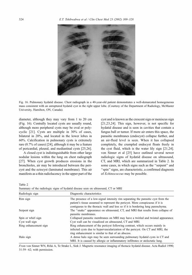
Fig. 16. Pulmonary hydatid disease. Chest radiograph in a 40-year-old patient demonstrates a well-demarcated homogeneous
mass consistent with an unruptured hydatid cyst in the right upper lobe. (Courtesy of the Department of Radiology, McMaster
University, Hamilton, ON, Canada).
E.T. Tshibwabwa et al / Clin Chest Med 23 (2002) 309–328324
Page 17
Pulmonary strongyloidiasis
Strongyloides stercoralis is a small nematode
endemic in tropical and subtropical regions. The
ova of the female nematode hatch into rhabditiform
(nonmigratory) larvae that are capable of maturing
into noninfectious adults or moulting into filariform
(infective) larvae. Initial invasion occurs when the
patient’s skin is exposed to contaminated soil or
feces. The filariform larvae penetrate the dermis and
migrate through the venous system to the lungs,
ascend the trachea, are swallowed into the digestive
tract, and infect the small intestine mucosa. Most
larvae penetrate the glandular epithelium into the
intestinal lumen and are excreted as feces. Some
larvae, however, reenter the blood stream and migrate
through the lungs without a soil cycle. This ability for
autoinfection means that infestation can be lifelong
and extremely heavy; massive autoinfection leads to
disseminated strongyloidiasis, the hyperinfection syn-
drome, which results in severe pulmonary disease
[28,29].
The primary migratory phase of the parasite
through the lung results in the larvae piercing the
pulmonary capillaries and entering the alveolar ducts.
During this transit from the vascular bed to the
respiratory tree, variable degrees of hemorrhage and
edema result along with desquamation of epithelial
cells and the migration of macrophages and inflam-
matory cells toward the parasites, which produces ill-
defined, patchy homogenous consolidation or, less
frequently, fine miliary nodulation on chest radio-
graphs. In patients with preexisting chronic lung
disease, the progress of the filariform larvae’s primary
migration through the lungs is retarded by excessive
bronchial secretions or inflammation, which causes a
moderate to severe pulmonary strongyloidiasis. This
infection may produce segmental or even lobar opac-
ities. Pulmonary opacities can be chronic, and serial
radiographs may show migration of the opacities
through the lungs.
The pulmonary manifestations of the hyperin-
fection syndrome include severe bronchospasm,
extensive pneumonia, pulmonary hemorrhage, and
the development of the adult respiratory distress
syndrome.
Pleural effusions are seen (40%) [28] more fre-
quently in patients with heavy Strongyloides infec-
tion. Secondary infections from bacteria or fungi are
common and are significantly associated with the
development of shock lung. Pulmonary cavitation
and abscess formation may occur, which suggests
superimposed bacterial infection.
Paragonimiasis
Paragonimiasis is an infestation caused by the
trematode parasite Paragonimus. The lungs are prim-
arily affected, although central nervous system
involvement does occur [30]. The radiographic fea-
tures are not specific and are easily confused with
Fig. 17. Paragonimiasis. Chest radiograph in this 35-year-old patient from Congo who presented with cough and brown
sputum demonstrates bilateral infiltrates with subtle foci of lucency, which suggest cavitation. Sputum was negative for acid
fast bacilli (AFB) but positive for paragonimiasis. Complete regression of both lesions was evident on follow-up after
therapy with praziquantel.
E.T. Tshibwabwa et al / Clin Chest Med 23 (2002) 309–328 325
Page 18
those of pulmonary TB. There is a high rate of a
normal chest radiograph in confirmed cases of para-
gonimiasis: 12.8% from a study in India [31] and
20% from a Nigerian study [30]. The most common
radiographic feature is multiple areas of patchy shad-
owing of low density with indistinct margins. There
is no lobar or segmental preponderance, but the
midzones are commonly affected with shadowing
that extends from the perihilar regions to the peri-
phery. Occasionally, cystic areas develop eccentri-
cally within the areas of opacity; these have smooth
outlines and have been likened to ‘‘bubbles’’ that de-
velop within the shadow [30]. Linear streaky shad-
ows are seen less often (2.6%) [31]. Other features
include pleural reaction or thickening (28%), with
pleural effusion seen in 10%. Although none of the
radiographic features is pathognomonic, a combina-
tion of these appearances should alert the radiologist
to the diagnosis in a patient from an endemic area
who presents with typical blood-stained, rusty, or
chocolate-colored sputum (Fig. 17).
Melioidosis
Melioidosis is endemic in Southeast Asia. The
organism is a gram-negative bacillus, Pseudomonas
pseudomallei, that infects humans via contaminated
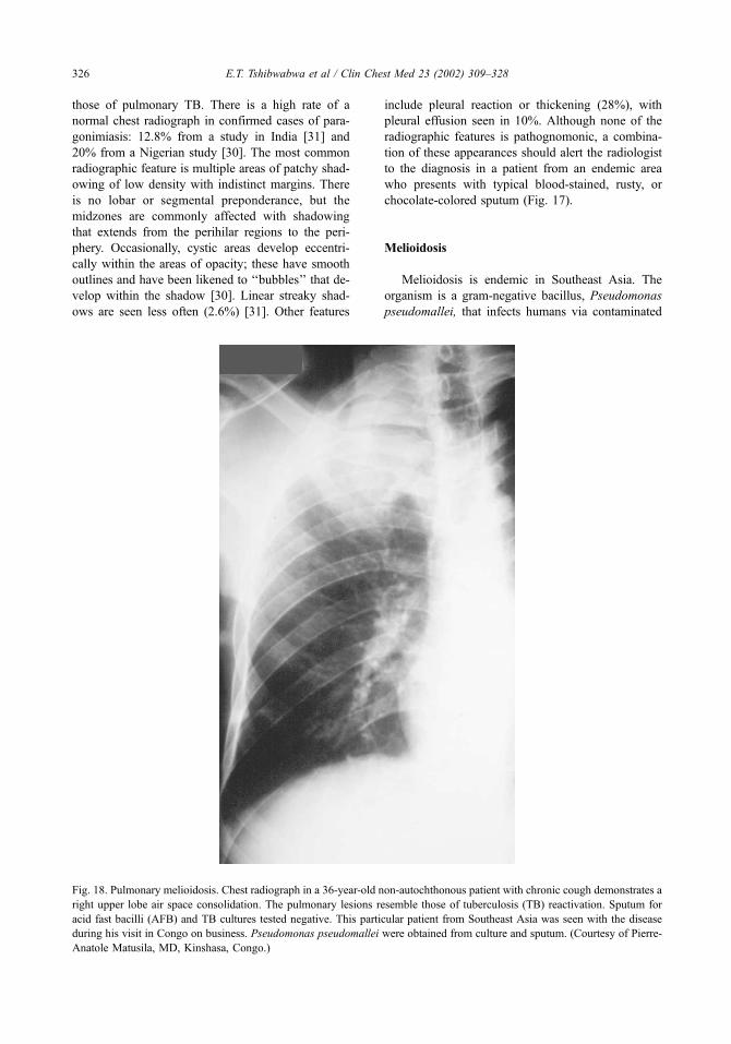
Fig. 18. Pulmonary melioidosis. Chest radiograph in a 36-year-old non-autochthonous patient with chronic cough demonstrates a
right upper lobe air space consolidation. The pulmonary lesions resemble those of tuberculosis (TB) reactivation. Sputum for
acid fast bacilli (AFB) and TB cultures tested negative. This particular patient from Southeast Asia was seen with the disease
during his visit in Congo on business. Pseudomonas pseudomallei were obtained from culture and sputum. (Courtesy of Pierre-
Anatole Matusila, MD, Kinshasa, Congo.)
E.T. Tshibwabwa et al / Clin Chest Med 23 (2002) 309–328326
Page 19
soil or dust that enters the respiratory or alimentary
tract or enters through a skin wound [32]. The
bacteria may remain quiescent in an infected person
for long periods and then become reactivated and
cause clinical symptoms [33,34]. Clinically, melioi-
dosis may manifest in four different ways [32].
� Patients with the acute form present with fever
and chills, and without antibiotics there is
usually rapid progression to overwhelming
septicemia. A diffuse pneumonitis develops
accompanied by multiple liver, spleen, and
subcutaneous abscesses. Acute respiratory
distress syndrome is a common sequela. The
most common radiographic appearance in
the acute form is the presence of multiple,
small, irregular densities that range in size
from 4 mm to 10 mm, which can simulate
disseminated TB. These nodules may coa-
lesce, which results in segmental or lobar
consolidation. Pleural effusion or empyema is
seen, but hilar adenopathy is rare.� The subacute form begins with a prodromal
period that eventually presents with chest
pain, low-grade fever, and weight loss. Chest
radiograph (Fig. 18) normally reveals a lobar
infiltrate, usually within the upper lobe, and
often shows cavitation.� In subclinical melioidosis, the patient is
asymptomatic, although serologic test results
are positive. Most infected persons fall into this
category [35]. The radiographic appearances
mimic those of TB with an upper lobe infiltrate
and cavity formation. These patients are at risk
of developing an acute exacerbation.� Chronic melioidosis is usually extrapulmo-
nary, in which skin lesions or osteomyelitis
represents the primary site of infection.
The most important factor involved in establishing
the diagnosis of melioidosis is a high index of
suspicion, and the diagnosis should be entertained
in patients with a febrile illness and a localized
suppurative process in an endemic area [36].
Summary
A high-quality chest radiograph and a timely,
accurate report are often impossible in the tropics.
Rationale matching of service to need, enthusiasm,
commitment, and exploitation of information tech-
nology all go some way toward enabling patients with
pulmonary disease to be imaged. The radiologic
findings reflect the high preponderance of infectious
disease. TB, HIV, and TB modified by HIV may be
‘‘routine’’ features in some parts of the tropics. Else-
where, infestation with ameba, hydatid, and stron-
gyloidosis, paragonimiasis, and melioidosis accounts
for radiographic signs. The key is to have these
conditions firmly in mind when reading tropical radio-
graphs and be aware that the pattern of disease may be
different between the patient from the tropics and the
more familiar patient from downtown New York.
References
[1] Dolin PJ, Raviglione MC, Kochi A. Global tuberculo-
sis incidence and mortality during 1990–2000. Bull
World Health Organ 1994;72:213–20.
[2] Mindel S. Role of imager in a developing world. Lan-
cet 1997;I:426–9.
[3] Schandorf C, Ketteh GK. Analysis of the status of X-
ray diagnosis in Ghana. Br J Radiol 1998;71:1040–8.
[4] Tshibwabwa ET, Mwaba P, Bogle-Taylor J, Zumla A.
Four-year study of abdominal ultrasound in 900 Cen-
tral African adults with AIDS referred for diagnostic
imaging. Abdom Imaging 2000;25:290–6.
[5] Kurjak K, Kos M. Ultrasound screening for fetal
anomalies in developing countries: wish or reality?
Ann N Y Acad Sci 1998;847:233–7.
[6] Bovornkitti S. Tropical pulmonary diseases. Respirol-
ogy 1996;1:11–21.
[7] Pallangyo KJ. Clinical features of tuberculosis among
adults in sub-Saharan Africa in the 21st century. Scand
J Infect Dis 2001;33:488–93.
[8] Aderele WI. Radiological patterns of pulmonary tuber-
culosis in Nigerian children. Tuber Lung Dis 1980;61:
157–63.
[9] Freiman I, Geefhuysen J, Solomon A. The radiological
presentation of pulmonary tuberculosis in children.
S Afr Med J 1975;49:1703–7.
[10] Tshibwabwa E, Matulewicz S. Aspects radiologiques
de la tuberculose respiratoire chez les enfants dans un
groupe a haut risque. Louvain Med 1981;100:425–9.
[11] Leung AN, Muller NL, Pineda PR, Fitzgerald JM. Pri-
mary tuberculosis in children: radiographic manifesta-
tions. Radiology 1992;182:87–91.
[12] Awil PO, Bowlin SJ, Daniel TM. Radiology of pul-
monary tuberculosis and human immunodeficiency
virus infection in Gulu, Uganda. Eur Respir J 1997;
10:615–8.
[13] Batungwanayo J, Taelman H, Dhote R, Bogaerts J,
Allen S, van de Perre P. Pulmonary tuberculosis in
Kigali, Rwanda: impact of human immunodeficiency
virus infection on clinical and radiographic presenta-
tion. Ann Rev Respir Dis 1992; 146:53–6.
[14] Pozniak AL, Latif AS, Neill P, Houston S, Chen K,
Robertson V. Pulmonary Kaposi’s sarcoma in Africa.
Thorax 1992;47:770–3.
E.T. Tshibwabwa et al / Clin Chest Med 23 (2002) 309–328 327
Page 20
[15] Saks AM, Posner R. Tuberculosis in HIV positive pa-
tients in South Africa: a comparative radiological study
with HIV negative patients. Clin Radiol 1992;46:
387–90.
[16] Tshibwabwa E. Pulmonary radiological features of
AIDS in the tropics. In: Zumla A, Johnson M, Miller
M, editors. AIDS and respiratory medicine. 1st edition.
London: Chapman & Hall Medical; 1997. p. 59–69.
[17] O’Keefe EA, Wood R. AIDS in Africa. Scand J Gas-
troenterol 1996;31:147–52.
[18] Abouya YL, Beaumel A, Lucas J, Dago-Akribi A,
Coulibaly G, N’Datz M, et al. Pneumocystis carinii
pneumonia: an uncommon cause of death in African
patients with acquired immunodeficiency syndrome.
Am Rev Respir Dis 1992;145:617–20.
[19] Mahomed AG, Murray J, Klepman S, Richards G,
Feldman C, Levy NT, et al. Pneumocystis carinii pneu-
monia in HIV infected patients from South Africa. East
Afr Med J 1999;76:80–4.
[20] Malin AS, Gwanzura LKZ, Klein S, Robertson VJ,
Musvaire P, Mason PR. Pneumocystis carinii pneumo-
nia in Zimbabwe. Lancet 1995;346:1258–61.
[21] Balikian JB, Mudarris FF. Hydatid disease of the
lungs: a roentgenologic study of 50 cases. Am J Roent-
genol 1974;122:692–707.
[22] Whittaker LR. Pulmonary amoebic disease. In: Cock-
shott P, Middlemiss H, editors. Clinical radiology in
the tropics. 1st edition. Edinburgh: 1979. p. 167–8.
[23] Beggs I. The radiology of hydatid disease. Am J
Roentgenol 1995;145:639–48.
[24] Jerray M, Benzarti M, Garrouche A, Klabi N, Hayouni
A. Hydatid disease of the lungs: study of 386 cases.
Am Rev Respir Dis 1992;146:185–9.
[25] von Sinner WN, Rifai A, Te Strake L, Siek J. Magnetic
resonance imaging of thoracic hydatid disease. Acta
Radiol 1990;31:59–62.
[26] von Sinner WN. New diagnostic signs in hydatid dis-
ease: radiography, ultrasound, CT and MRI correlated
to pathology. Eur J Radiol 1991;12:150–9.
[27] Lewall DB, McCorkell SJ. Rupture of Echinococcal
cysts: diagnosis, classification, and clinical implica-
tions. Am J Roentgenol 1996;146:391–4.
[28] Woodring JH, Halfhill H, Reed JC. Pulmonary stron-
gyloidiasis: clinical and imaging features. Am J Roent-
genol 1994;162:537–42.
[29] Woodring JH, Halfhill H, Berger R, Reed JC, Moser N.
Clinical and imaging features of pulmonary strongyloi-
diasis. South Med J 1996;89:10–9.
[30] Ogakwu M, Nwokolo C. Radiological findings in pul-
monary paragonimiasis as seen in Nigeria: a review
based on one hundred cases. Br J Radiol 1973;46:
699–705.
[31] Singh TS, Muttum SS, Razaque MA. Pulmonary para-
gonimiasis: clinical features. diagnosis and treatment
of 39 cases in Manipur. Trans R Soc Trop Med Hyg
1986;80:967–71.
[32] Reeder MM, Palmer PE. Acute tropical pneumonias.
Semin Roentgenol 1980;15:35–49.
[33] Sharma OP, Maheshwari A. Lung diseases in the
tropics. Part 1: tropical granulomatous disorders of
the lung: diagnosis and management. Tuber Lung
Dis 1993;74:295–304.
[34] Sharma OP, Maheshwari A. Lung diseases in the tro-
pics. Part 2: common tropical lung diseases: diagnosis
and management. Tuber Lung Dis 1993;74:359–70.
[35] Leelarasamee A, Bovornkitti S. Melioidosis: review
and update. Rev Infect Dis 1989;11:413–23.
[36] Koponen MA, Zlock D, Palmer DL, Merlin TL. Me-
lioidosis: forgotten, but not gone. Arch Intern Med
1991;151:605–8.
[37] Tshibwabwa ET, Mwinga A, Pobee JOM, Zumla A.
Radiological features of pulmonary tuberculosis in 963
HIV-infected adults at three Central African hospitals.
Clin Radiol 1992;52:837–41.
E.T. Tshibwabwa et al / Clin Chest Med 23 (2002) 309–328328