40
Lecture Topic : Lymphatic system
| Date post: | 18-Jul-2015 |
| Category: |
Education |
| Upload: | manohar-vishnoi |
| View: | 60 times |
| Download: | 1 times |

Lecture Topic:
Lymphatic
system

One way system: to the heart
Return plasma protein and
excess tissue fluid (2-4 L/day
Lacteals in small intestine
absorb dietary lipids
Recognizes specific foreign
molecules.
Lymphatic system

Lymphatic
system
Cardiovascular
system
Lymphatic system

Lymphatic
Vessels
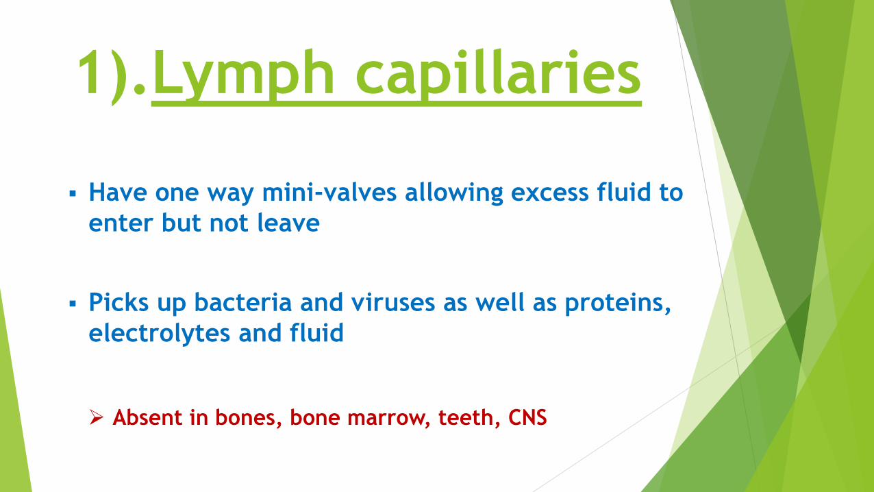
1).Lymph capillaries
Have one way mini-valves allowing excess fluid to
enter but not leave
Picks up bacteria and viruses as well as proteins,
electrolytes and fluid
Absent in bones, bone marrow, teeth, CNS


2).Lymphatic collecting vessels
Similar to blood vessels (3 layers), but thin &
delicate
Superficial ones in skin travel with superficial veins.
Deep ones ( in trunk and digestive viscera) travel
with deep arteries.
Drain into superficial and deep lymph nodes.

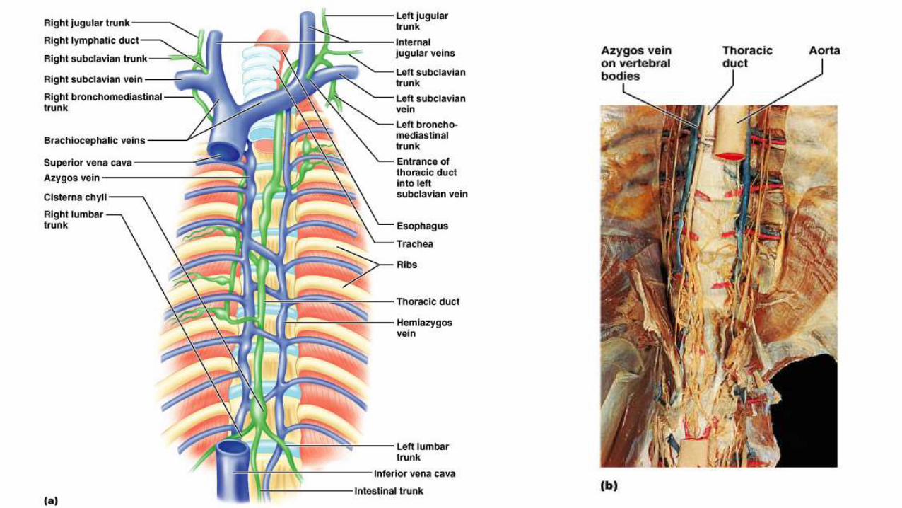
3).Lymphatic Trunks
Lumbar
Intestinal
Receives fatty lymph (chyle) absorbed through lacteals of intestines
Broncho-mediastinal
Subclavian
Jugular
(Paired, except intestinal)
Drain into cisterna chyli

Lymphatic Trunks
Right
Broncho-
Mediastinal
Trunk
Right subclavian trunk
Left
Broncho-
Mediastinal
Trunk
Left subclavian
trunk
Right lumber Trunk Left lumber Trunk
Thoracic Duct
Cisterna Chyli
Right jugular trunk Left jugular trunk
Intestinal Trunk

4).Lymphatic ducts:
Thoracic duct (always present)
(drain into left subclavian vein)
20% also have a right lymphatic duct
(drain into right subclavian vein)

Lymphoid
Organs

Lymph nodes
Spleen
Thymus
Tonsils
Small intestine &
appendix (lymphoid nodules)

Lymphatic system
L
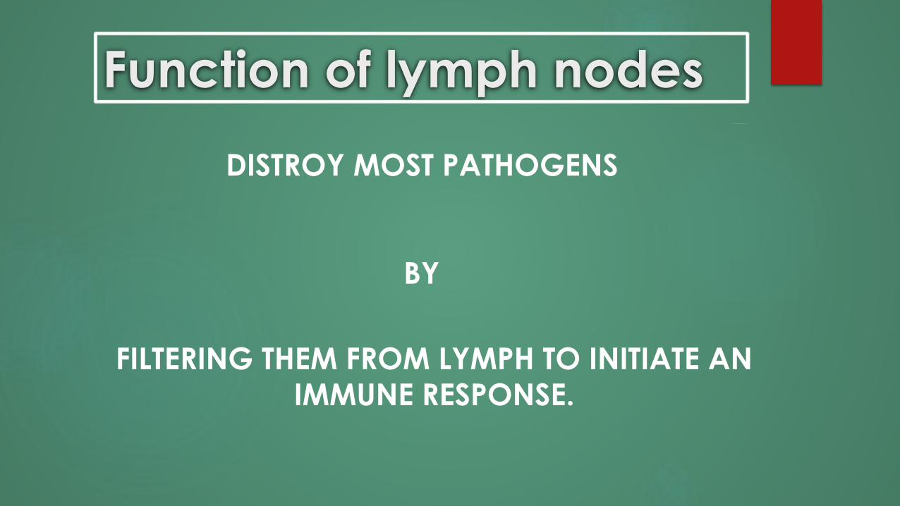
Function of lymph nodes
FILTERING THEM FROM LYMPH TO INITIATE AN
IMMUNE RESPONSE.
DISTROY MOST PATHOGENS
BY

LYMPH NODES ARE FOUND IN
GROUPS
Superficial groups:-Cervical
-Axillary
-Inguinal
Deep groups:-Tracheobronchial
-Aortic
-Iliac


Lymph Nodes Structure
Bean shaped masses of lymphoid tissue( Conn. Tissue & lymphocytes.
Covered by Capsule, gives off trabiculae.
In between trabiculae, Cortical and Medullary sinuses.
Sinuses contain lymph. follicles (nodules)
Follicles are masses of B-Lymphocytes.

Afferent
Lymphatic
vessels
Efferent
Lymphatic
vessel


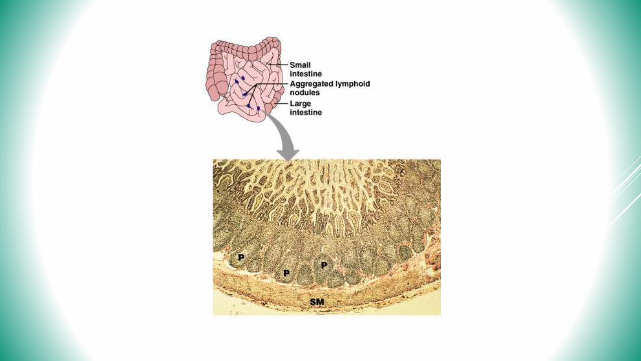
Nodules (also called follicles)
(Spherical masses of mostly B-lymphocytes).
1. Scattered Lymphatic nodules. (in submucosa of GIT).
2. Aggregated lymphatic nodules. (“Payer's Patches”)
About 40 follicles / 1 cm area.
Distal ileum and Appendix


The thymus
Secrete hormones (thymopoietin, thymulin and
thymosin) for the development of T-lymphocytes
Thymus is the organ where T-cells become
mature and differentiate.

Very large in fetus, after age 14 begins involution
(in elderly mostly fatty and fibrous tissue)

Structure of the thymus
Capsule gives off trabeculae, divides parenchyma into lobules of cortex and medulla
Cortex is darker than the medulla.
Medulla has more mature T-lymphocytes.
Medulla contains Hassall's corpuscles.(Masses of aged, degenerated cells)

Thymus Gland

Structure of the thymus

SPLEEN
This is the largest lymphoid organ,
posterior to the stomach.


SPLEEN STRUCTURE
(spleen is about the size of a fist).
a. Capsule
b. Trabeculae
c. Red pulp
d. White pulp

RED PULP:
(made of sinuosoid capillaries)
WHITE PULP:
(made of masses of lymphocytes)
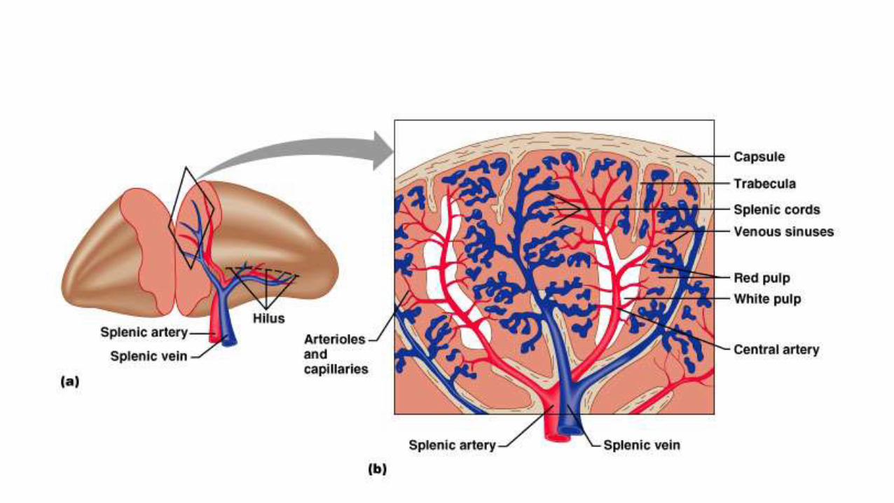

FUNCTIONS OF THE SPLEEN
Phagocytizes bacteria and other foreign materials : “white pulp”
Removal & destruction of aged or defective blood cells: “red pulp”
Stores platelets
In fetus: hematopoiesis
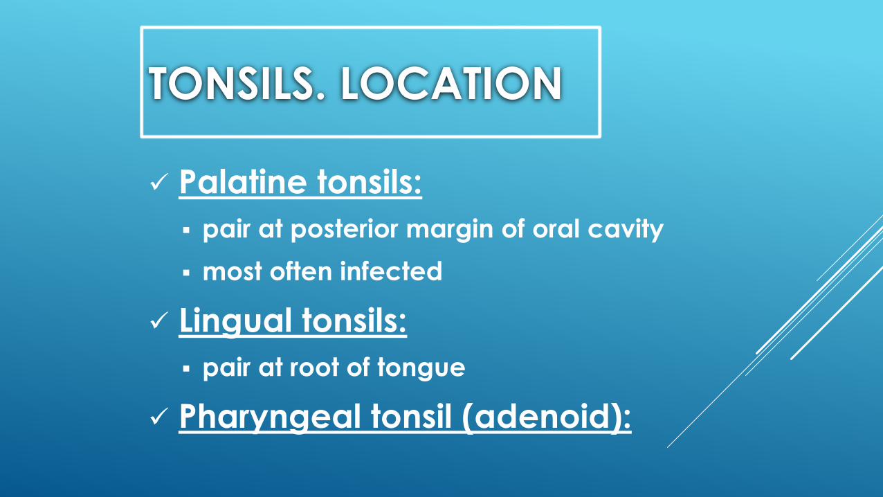
TONSILS. LOCATION
Palatine tonsils:
pair at posterior margin of oral cavity
most often infected
Lingual tonsils:
pair at root of tongue
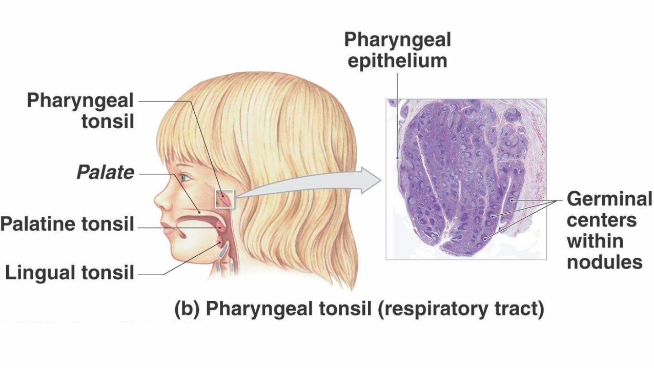
Pharyngeal tonsil (adenoid):

STRUCTURE OF PHARYNGEAL TONSILS
Covered by epithelium
Tonsillar crypts
Lymph Nodules (Follicles)
Lymphocytes

Tonsils
Hematopoiesis

thank you for attention.THE END!


