72
Lymphatic System KAMAL SALIH
| Date post: | 04-Jan-2016 |
| Category: |
Documents |
| Upload: | lorena-powers |
| View: | 224 times |
| Download: | 1 times |

Lymphatic System
KAMAL SALIH

• The lymphatic system is vital to the defense against illness.
• If infectious agents manage to breach the mechanical barriers and gain entry to the milieu interieur, the cells which deal with the invasion are those which have arisen, developed, matured, and/or been stored in lymphatic organs.


The Lymphatic System
• It is intimately related, both structurally and functionally, to the blood vascular system.
• Lymph itself is a clear, slightly yellowish and opalescent fluid derived from blood.
• It contains white blood cells, specifically lymphocytes.

What is the lymph?
• Lymph is a clear, slightly yellowish and opalescent fluid derived from blood.
• What is its content?• It contains white blood cells, specifically
lymphocytes.
Specialized Lymphatic Organs
• Certain specialized organs devoted to processing and modifying lymph and lymphoid cells are present in all normal mammals.
How it circulate?
• Lymph starts as blood fluid that is "strained" into the tissue spaces under the hydrostatic pressure of the pumping blood.
• It's drained from the intercellular spaces by thin vein-like lymphatic vessels, re-entering the venous circulation carrying lymphocytes that enter the stream at certain specific points.

One way to the……
•The lymphatic system forms a one way flow system towards the heart.

Lymphatics……Blind end
• Through this system flows lymph, which starts from blind ended capillaries.

Lymphatics……Blind end
• The capillaries are very permeable with the aid of small anchoring filaments the vessel can take large particles and stay open where the external pressure is greater.
This causes problems, as the vessel also carries large particles, such as:-
• Viruses • Pathogens• Cell debris
Hence can carry infection through the body.


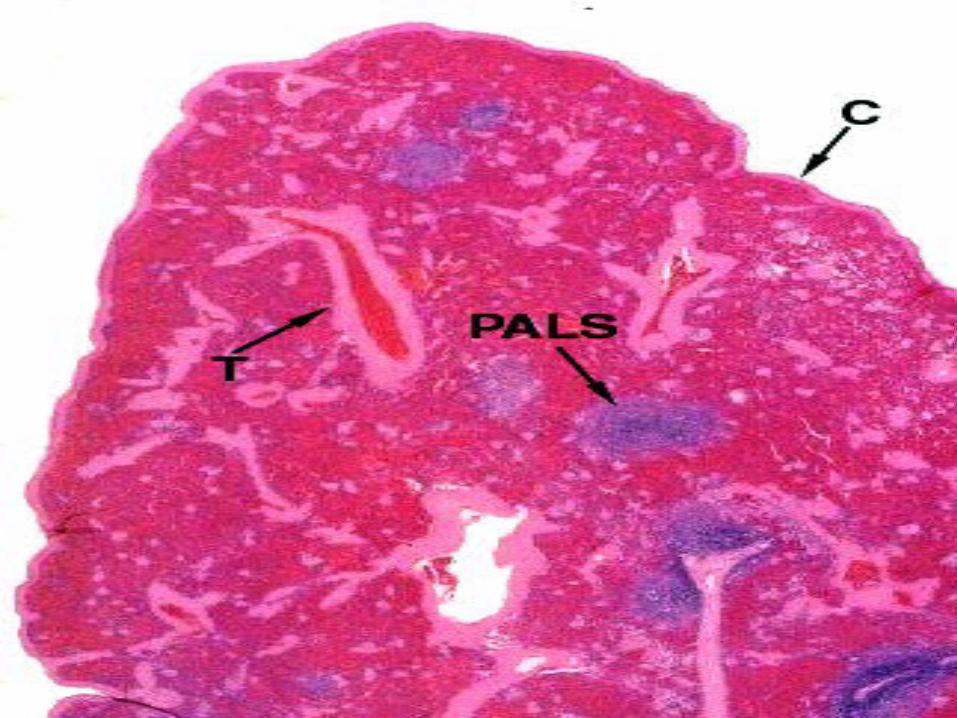
The SpleenThe Spleen• The spleen serves two major functions in the body: • 1. It is responsible for the destruction of old red blood cells (RBC), • 2. It is a major site for mounting the immune response. • The spleen behaves similar to a lymph node but instead of filtering
the lymphatic fluid it filters the blood. • Blood entering the spleen travels through progressively smaller
arterioles until it is deposited in an area known as the red pulp. • This is where the RBCs are processed. • Surrounding each of the arterioles is a sheath of lymphoid cells
which make up the periarteriolar lymphoid sheath (PALS). • The interface between the PALS and the blood is a region of intense
phagocytic activity and sets the stage for an immune response. • The immune reactivity of the spleen is especially effective for
dealing with blood-borne antigens such as bacteria that reach the blood.

Red &White Pulps
• These are two of those troublesome terms that really applicable in gross anatomy, but with which the histologist has to deal as well. Oh, well...here goes:
• Cut a freshly-removed spleen across and it looks like a field of dark red material with white spots in it.
• On the basis of its gross appearance in fresh sections, the spleen is traditionally said to have the bulk of its parenchyma as red pulp, with isolated areas of white pulp interspersed through it.


Red &White Pulps
• The red pulp gets its appearance from the formed elements of the blood (mostly erythrocytes) it contains.
• The white pulp consists almost entirely of lymphocytes, in a peculiar association with the arterial blood supply.

• "white pulp" is equivalent to the lymphocyte population of the spleen, in the form of the periarteriolar lymphocyte sheath or PALS .
• "Red pulp" is everything else, which means the splenic cords and the sinuses between them.

• "Red pulp" fills the bulk of the spleen's volume. White pulp is the blue stained areas visible within it.

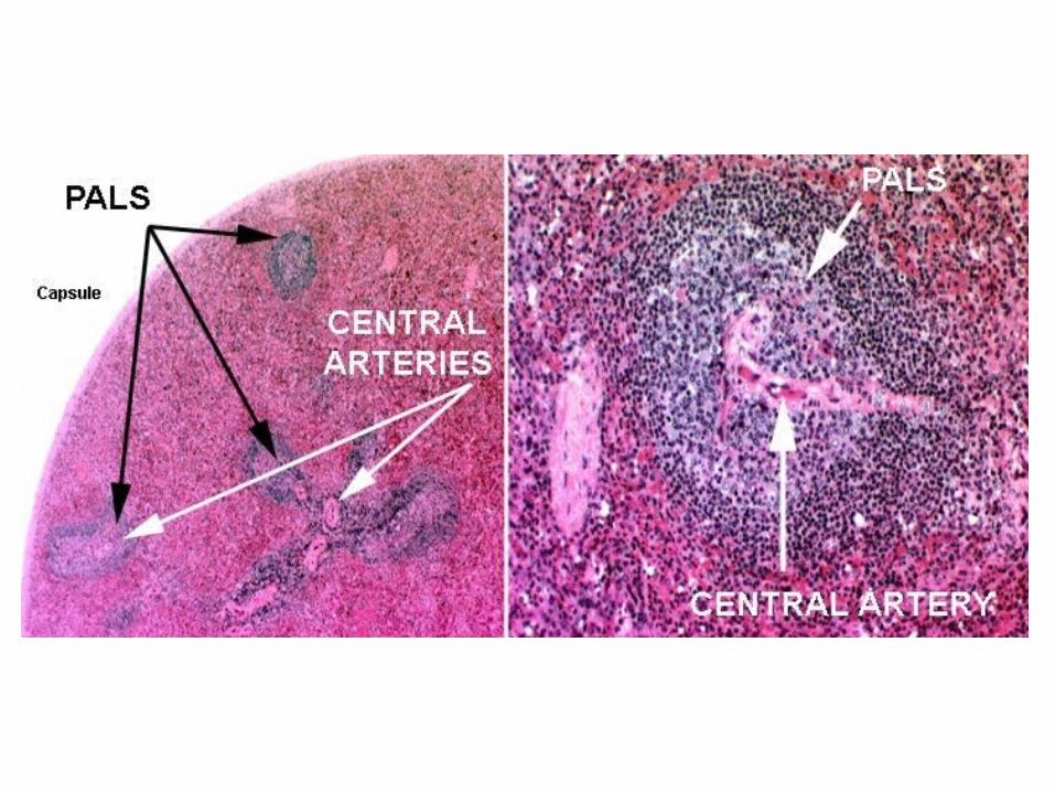
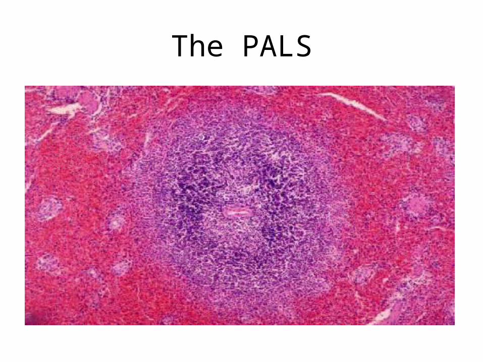
The "PALS”
• Is the periarteriolar lymphocyte sheath, the characteristic association of lymphocytes and blood vessels in the spleen.
• The lymphocytes are arranged along the arteries forming a sleeve or sheath.
• Collectively, the PALS is the "white pulp." • The rest of the interior volume of the spleen
is the splenic cords, collectively constituting the "red pulp," and the blood sinuses between them.

Splenic Circulation

Splenic Circulation
• Blood enters from the splenic arterial supply, via the splenic capsule, and the breakup of arteries into capsular and trabecular segments begins almost immediately.
• As is true in other organs, the angiogenic properties of the CT capsule are are needed to create the routes for the blood supply entering and leaving the spleen.

Splenic Circulation
• The main input, the splenic artery, ramifies in the capsule and sends branches deeper and deeper.
• Blood leaving the organ is drained back through a series of veins in the septa and the capsule, eventually exiting via the splenic vein.
• Both the artery and vein are grossly located at the hilus.

Splenic Circulation
• As soon as a branch of the arterial input reaches the interior space of the spleen, it acquires the periarteriolar lymphocyte sheath, or PALS, which it retains almost to the smallest subdivisions.
• The PALS is a place in which the special conditions required for proliferation of B lymphocytes can be met, and at intervals there will be germinal centers along it.

Splenic Circulation
• Germinal centersGerminal centers are individually transient and depend on the immune state of the animal, as they do in other parts of the immune system; but their presence is a feature of the spleen in all species to some extent.
• The arterial vessels that have been covered with lymphocytes are the central arteries.

Penicillar Arteries
• Eventually the arteriolar supply subdivides to the point where the PALS becomes attenuated to a few cells, or even lost altogether.
• These small arteries tend to run in bundles and hence are termed penicillar arteries, from their resemblance to the hairs of a paintbrush (the Latin word for a paintbrush is penicillum).

The role of the penicillar arteries: "arteries of the red pulp ".
• The role of the penicillar arteries is to deliver the incoming blood to the red pulp (which is not shown in this sketch) i.e., the splenic cords and sinuses.
• Some segments of the penicillar arteries acquire a peculiar "sheath" of phagocytic cells in their walls, and have been termed sheathed arterioles in the short regions where this occurs.
• The presence of a sheath is inconstant and there are species variations; its role is unclear, as well.

The PALS
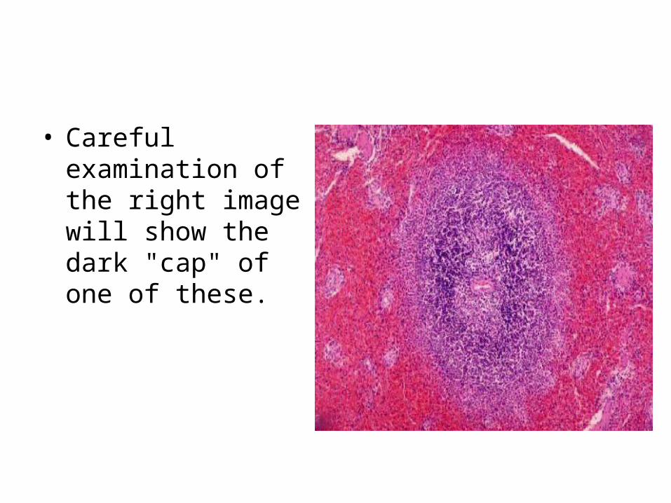
• Careful examination of the right image will show the dark "cap" of one of these.

• The PALS is diffuse lymphatic tissue, but within it germinal centers frequently develop.

• Now, the PALS, being diffuse, has a loose stromal framework of reticular fibers made by a variant for of fibroblast.
• The resident lymphocytes of the diffuse region of the PALS are supported by it, like birds sitting on telephone wires: this sort of arrangement is typical of all diffuse lymphatic tissue.

• Due to the dense packing of the lymphocytes in the PALS you will not be able to make out the fibers in an H&E preparation, even in the diffuse parts.

• Within the germinal centers, however, things are different.
• The germinal center is a special place: it's a clone of B-lymphocytes, and it has to be isolated so that these cells can develop properly.

• Consequently the stroma in germinal centers isn't fibrillar in nature.
• Instead, special cells of a different cell line than the fibroblasts form a stroma within the germinal center and support the B cells in it.
• A similar non-fibrillar stroma is characteristic of the thymus, and will be discussed below.
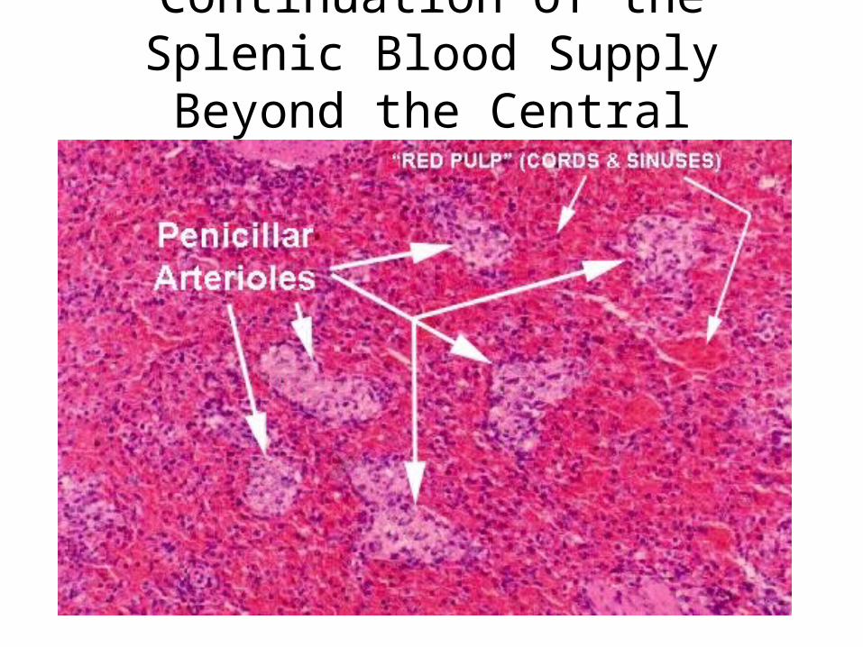
Continuation of the Splenic Blood Supply Beyond the Central Arteries

• The central arteries, like any self-respecting artery, break up into smaller ones eventually. Each one gives rise to a tuft of small vessels, the penicillar arteries.
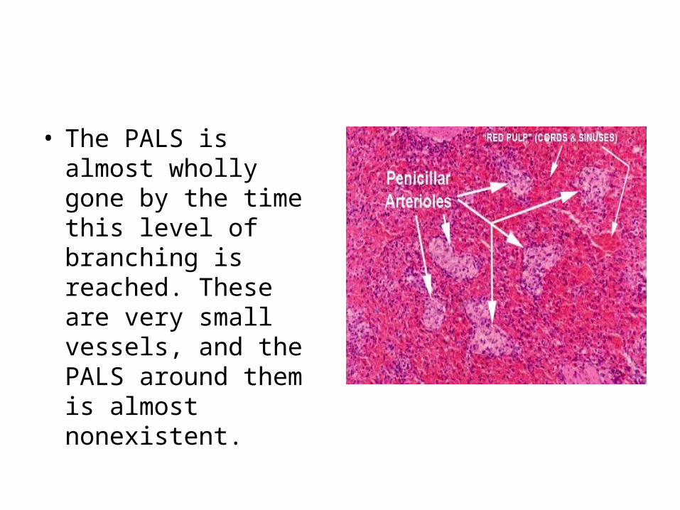
• The PALS is almost wholly gone by the time this level of branching is reached. These are very small vessels, and the PALS around them is almost nonexistent.

Splenic Cords and Sinuses

The Thymus
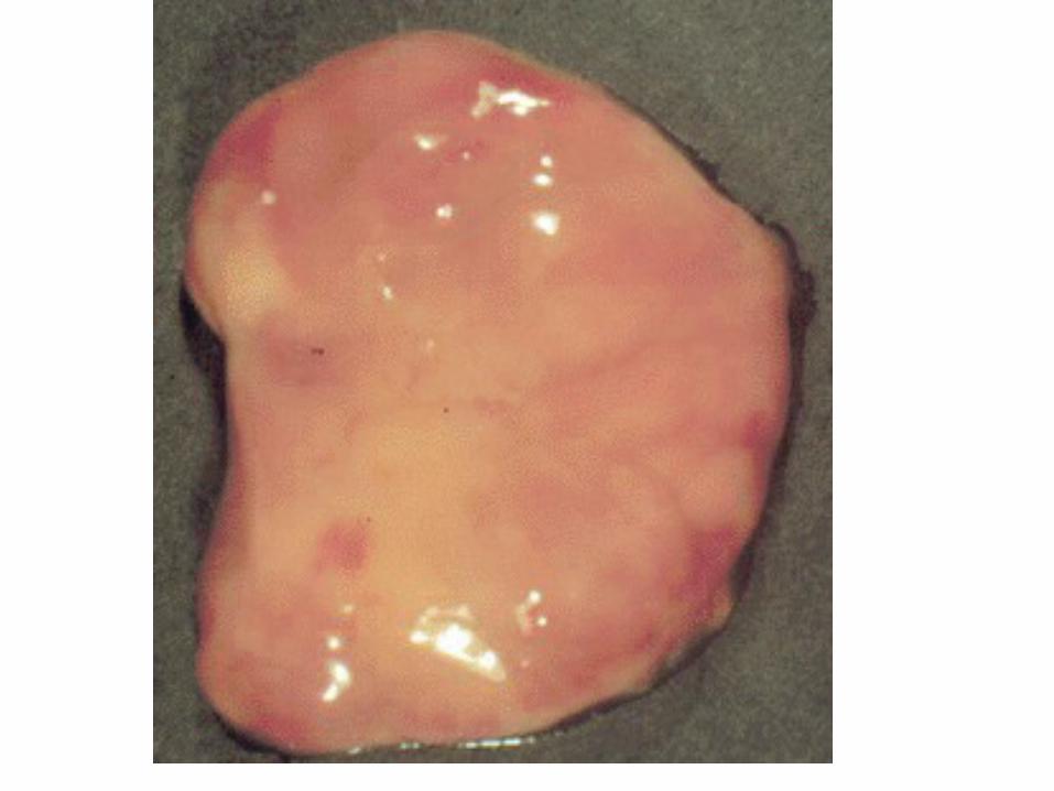
• The thymus is a bilobed, greyish organ located in the thoracic cavity just below the neck.
• Curiously, when the thymus is removed from adult mammals, few effects are seen.
• However, when the thymus is removed at birth, dramatic effects are witnessed as will be explained later.
• The thymus develops from the endoderm. • During its development many cells migrate
towards it, most of which are lymphocytes.


The Thymus
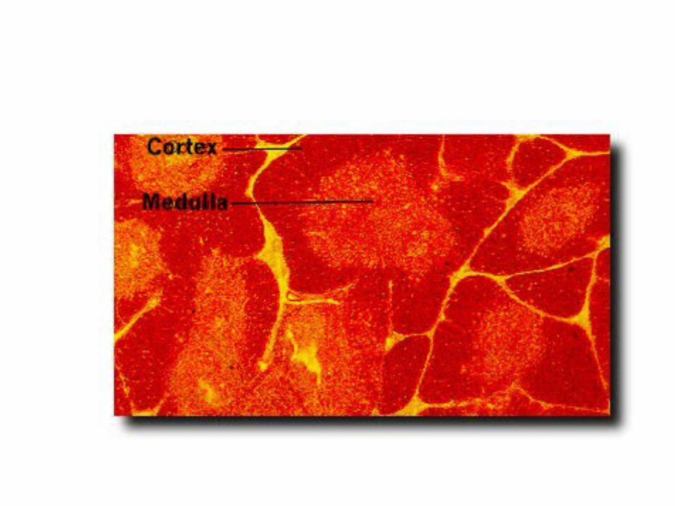
• The thymus is divided into two distinct compartments, the outer cortex and the inner medulla.
• Both regions are densly populated with lymphocytes (or thymocytes while in the thymus).
• Most of the cortical lymphocytes are immature and unable to carry out immune functions.

The Thymus
• Mature immunocompetent cells are found in the medulla in greater numbers.
• The main function of the thymus is to develop immature T-cells into immunocompetent T-cells.
• This process begins with the production of pre-T cells in the bone marrow and their subsequent transport to the thymus via the blood.
• The pre-T cells are then taken into the cortex of the thymus.
• Here, a series of molecular events take place allowing the cells to recognize certain antigens.

The Thymus
• Some of the cells recognize self-components, and these are elmiminated by a process of negative selection.
• Those that fail the selection die and those that live proceed to the medulla and eventually into the blood stream where they act upon foreign agents in the body.


Lymph Nodes
• Lymph nodes can take on many different sizes and shapes, but most are bean-shaped and are around 1 inch in length.

Lymph Nodes
• The node is covered thickly with the fibrous capsule and is subdivided into different compartments by inward pointing trabeculae.
• As with many organs, the lymph node has two basic parts, the cortex and the medulla.
• The cortex is populated mainly with lymphocytes (follicles).
• The germinal centers are the primary resting place for B Cell Lymphocytes (the cells responsible for production of circulating antibodies).

Lymph Nodes
• In the event of an infecting antigen, these B Lymphocytes will rapidly undergo mitosis and divide.
• Each unique kind of B cell produces only one type of antibody.
• Thus, by dividing, they can produce large quantities of a specific antibody to seek out and help destroy the antigen.

Lymph Nodes
• The rest of the cortex contains T lymphocytes- cells that circulate through the lymph nodes, blood stream, and lymphatic ducts to seek out any infection.
• The medulla of the lymph nodes is primarily made up of macrophages attached to reticular fibers.


The lymphatics • They tend to run within the deep or
superficial fascia.
• The fascia tends to contain disease processes until it is eroded.

The lymphatics of the neck
• are arranged into deep and superficial chains.
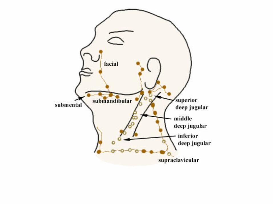

• The deep jugular chain extends from the base of the skull to the clavicle and is formed into
• Superior group of lymph nodes
• Middle group of lymph nodes
• Inferior group of lymph nodes.

The superior deep jugular nodes
• Receive primary drainage from the • Soft palate, • Tonsils, • Palatoglossal and palatopharyngeal arches, • Posterior tongue,• Base of the tongue,• Pyriform sinus and the larynx above the vocal
folds.
The superior deep jugular nodes
• Receive primary drainage from the soft palate, tonsils, palatoglossal and palatopharyngeal arches, posterior tongue, base of the tongue, pyriform sinus and the larynx above the vocal folds.
• This group of lymph nodes also receives lymphatic drainage from more superficial nodes in the upper head and neck (retropharyngeal, spinal accessory, parotid, superficial cervical and submandibular nodes).

• The middle deep jugular nodes receive primary drainage from the larynx above the vocal folds, lower pyriform sinus and posterior cricoid.
• They receive secondary drainage from the deep jugular nodes above them and the lower retropharyngeal nodes.

The inferior deep jugular nodes
• receive primary drainage from the thyroid, trachea and cervical esophagus.
• They receive secondary drainage from the deep jugular nodes above them and the paratracheal nodes.
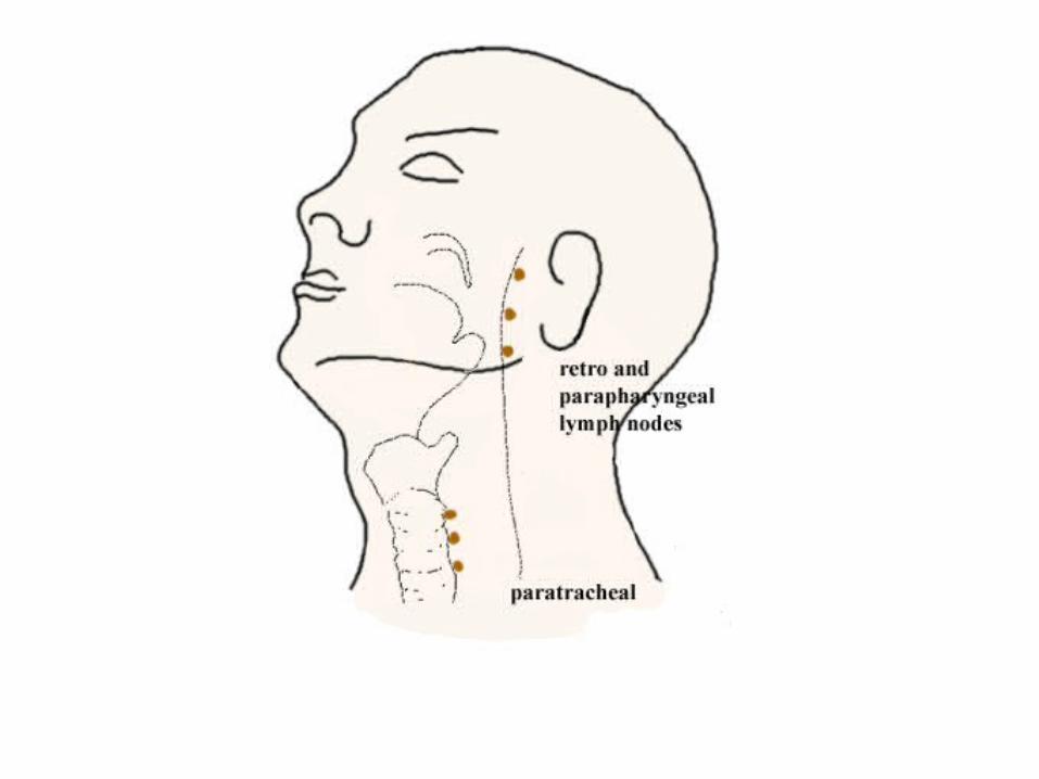

The Retropharyngeal & Paratracheal nodes
• lie posteriorly around the midline viscera.
• They receive drainage from these viscera and from the deep structures in the midline of the head, i.e. the nasopharynx, posterior nasal cavity, paranasal sinuses, posterior oropharynx.
• They drain towards the deep jugular chain.

The superficial nodes
• The superficial nodes tend to drain secondarily as mentioned to the deep nodes.
• The superficial nodes are the submental, superficial cervical, submandibular, spinal accessory and anterior scalene.

The Submental &Submandibular Lymph Nodes
• The submental nodes drain the chin, the middle of the lower lip, tip of the tongue and anterior mouth.
• These nodes in turn drain to the submandibular nodes.
• The submandibular nodes drain the upper lip, lateral lower lip, lower nasal cavity, anterior mouth and the skin of the cheek.
• The submandibular nodes in turn drain to the superior deep jugular nodes.


The superficial cervical nodes
• The superficial cervical nodes located along the external jugular vein receive drainage from the cutaneous lymphatics of the face, especially from around the parotid gland, behind the ear, parotid and occipital nodes.
• The superficial cervical then drain into the superior deep jugular nodes.

The nodes in the posterior triangle
• lie along the spinal accessory nerve.
• They drain the parietal and occipital regions of the scalp.
• The upper nodes drain to the superior deep jugular nodes while the lower nodes drain down to the supraclavicular nodes.

The anterior scalene (Virchow's) nodes
• The anterior scalene (Virchow's) nodes receive drainage from the thoracic duct and are located at the junction of the thoracic duct and left subclavian vein.
• They are usually the site of metastases from lower down in the body (e.g. stomach).

The supraclavicular nodes
• The supraclavicular nodes receive drainage from the spinal accessory nodes and from infraclavicular sources.
• All of these lymphatics eventually drain into the venous system either through or together with the thoracic duct on the left, or the right lymphatic duct


