Page 1
REVIEW
Macrophage–tumor crosstalk: role of TAMR tyrosine kinasereceptors and of their ligands
Thomas Schmidt • Isabel Ben-Batalla •
Alexander Schultze • Sonja Loges
Received: 14 August 2011 / Revised: 14 October 2011 / Accepted: 14 October 2011 / Published online: 11 November 2011
� Springer Basel AG 2011
Abstract Ample clinical and preclinical evidence indi-
cates that macrophages interact with tumor cells as well as
with virtually all populations of host cells present in the
tumor microenvironment. This crosstalk can strongly pro-
mote malignancy, but also has in principle the potential to
inhibit tumor growth. Thus, it is of the utmost importance
to improve our understanding of the mechanisms driving
the pro- and antimalignant behavior of tumor-associated
macrophages (TAMs) in order to develop better anticancer
therapies. In this review, we discuss the biological conse-
quences of reciprocal interactions between TAMs, cancer
cells, endothelial cells, fibroblasts and other leukocyte
subfractions within tumors. It was recently elucidated that
tumors specifically educate macrophages to secrete growth
arrest-specific gene 6 (Gas6), the common ligand of the
Tyro3, Axl, Mer receptor (TAMR) family. In turn, Gas6
fosters tumor growth by promoting cancer cell prolifera-
tion. Therefore, the Gas6–TAMR axis might represent a
novel target for disrupting tumor–macrophage crosstalk.
We summarize here what is known about TAMR and their
ligands in (human) cancer biology. In order to shed more
light on the role of macrophages in human cancer, we
additionally provide an overview of what is currently
known about the prognostic impact of TAMs in human
cancer.
Keywords TAM (Tumor-associated macrophages) �Gas6 � TAMR (Tyro3, Axl, Mer receptors) �Tumor-macrophage crosstalk � Cancer � Inflammation
Introduction
Cancer represents a heterogeneous class of diseases origi-
nating from neoplastic cells capable of uncontrolled
growth. More than 10 years ago, six essential ‘‘hallmarks
of cancer’’ were extracted from several decades of research
in order to define malignancy [1]: (1) self-sufficiency in
growth signals, (2) insensitivity to antigrowth signals,
(3) evasion of apoptosis, (4) limitless replicative potential,
(5) sustained angiogenesis, and (6) tissue invasion and
metastasis. Today, however, this tumor cell-centered pic-
ture of cancer represents a rather simplistic view that
neglects the complex microenvironment of the host. This
microenvironment forms an integral part of every tumor
and it is crucially involved in every single step of carci-
nogenesis, ranging from cancer initiation to metastasis
[2, 3]. Cancer cells are surrounded by numerous different
stromal cell types, including vascular and lymphatic
endothelial cells, pericytes, vascular smooth muscle cells,
Thomas Schmidt and Isabel Ben-Batalla contributed equally to this
work.
Electronic supplementary material The online version of thisarticle (doi:10.1007/s00018-011-0863-7) contains supplementarymaterial, which is available to authorized users.
T. Schmidt
Department of General, Visceral and Transplantation Surgery,
University of Heidelberg, Heidelberg, Germany
I. Ben-Batalla � A. Schultze � S. Loges (&)
Department of Hematology and Oncology with Sections BMT
and Pneumology, Hubertus Wald Tumorzentrum,
University Comprehensive Cancer Center Hamburg,
University Medical Center Hamburg-Eppendorf,
Martinistrasse 52, 20246 Hamburg, Germany
e-mail: [email protected]
I. Ben-Batalla � A. Schultze � S. Loges
Institute of Tumor Biology, Center of Experimental Medicine,
University Medical Center Hamburg-Eppendorf,
Hamburg, Germany
Cell. Mol. Life Sci. (2012) 69:1391–1414
DOI 10.1007/s00018-011-0863-7 Cellular and Molecular Life Sciences
123
Page 2
mesenchymal cells, adipocytes, cancer-associated fibro-
blasts (CAFs) as well as a large variety of bone marrow-
derived cells (BMDCs) [2–5]. Tumor-infiltrating BMDCs
comprise a heterogeneous population of leukocytes with
immunological properties such as B- and T-lymphocytes,
NK cells, macrophages and related myeloid cells, dendritic
cells, granulocytes and mast cells [4, 6]. This leukocyte
infiltrate varies in size, composition and distribution
between different tumor types and stages of progression
and is often termed ‘‘tumor inflammation’’ [4, 6]. However,
we need to keep in mind that this terminology is misleading
because tumor inflammation lacks many of the cardinal
features of inflammation sensu strictu including fever,
swelling and edema [7]. In this review, we also use ‘‘tumor
inflammation’’, but we refer to the smoldering, subacute
and chronic inflammation typically found in tumors [8].
Tumor and stroma cells are embedded in an extracellular
matrix consisting of integrins, collagens, hyaluron, laminins
and proteoglycans amongst others, with which tumor cells
intensively communicate via junctions, receptors, growth
factors, hormones and other soluble molecules [3, 9]. Thus,
cancers represent complex mixtures of malignant and non-
malignant (host-derived) cells and components interacting
with one another in a reciprocal manner throughout tumor
development and progression [2–4, 6]. Unfortunately, in
many cases, tumor cells succeed in exploiting the microen-
vironment for their benefit by creating a supportive
environment that promotes cancer initiation and growth, and
eventually its progression to fatal disease [2]. For instance,
induction of angiogenesis is an extensively studied example
of how cancers exploit their host [10–12]. Tumor-infiltrating
inflammatory cells were once assumed to inhibit tumor
growth or to be a consequence of failed cancer cell destruc-
tion. However, in the light of recent data, it is becoming
increasingly clear that these cells can play key roles in pro-
moting tumors by multiple mechanisms [2, 4, 6, 7]. Even
immunological cell types with potential tumoricidal activity
such as macrophages and neutrophils often are converted
under the influence of cancer cells into tumor-promoting
subpopulations [7, 13]. Macrophages and closely related cell
types can even mediate resistance to conventional chemo-
therapy or targeted antiangiogenic treatment [14, 15].
Thus, cells of the immune system act as ‘‘double-edged
swords’’ in the context of tumor biology because they are
in principle capable of destroying and promoting cancers.
However, in many cases immune cells seem to show pro-
tumoral activity [6]. However, because many patients who
died of non-malignant causes have been found to host
occult carcinomas, for instance in their breast or prostate,
which failed to progress to advanced cancer [16], we also
need to consider that the microenvironment can and does
constrain malignant cells. Obviously, it would be desirable
to tilt the microenvironment more towards destruction of
tumors. As a consequence, development of anticancer
drugs has moved from a traditional cancer cell-centered
approach towards increased targeting of the microenvi-
ronment, as reflected by development of numerous
compounds acting primarily on host-derived cells or
structures [16]. Unfortunately, despite tremendous efforts
in the field of tumor immunology and immunotherapy,
attempts to instruct immune cells to fight the progression of
established tumors has had only limited success [17].
Therefore, it is of the utmost importance to better dissect
the molecular and cellular basis of these fatal interactions
between tumor cells and immune cells in order to improve
our knowledge of cancer biology and to develop more
effective therapies for cancer patients.
The importance and high priority of this issue are also
reflected in the recent appreciation of tumor inflammation
as seventh hallmark of cancer [18–20]. Different cell types
and mechanisms related to this topic have recently been
extensively reviewed elsewhere [4, 6, 7]. In this review, we
specifically highlight the bidirectional crosstalk between
macrophages and tumors (i.e. tumor cells and microenvi-
ronmental host cells) with a special focus on the role of the
Tyro3, Axl and Mer (TAM) receptors and of their ligands
growth arrest-specific gene 6 (Gas6) and protein S.
In the following section we first summarize the role of
macrophages in cancer and then focus on their interaction
with different cell types. We mainly discuss preclinical
insights, but also provide links to human (clinical) data
where appropriate.
Role of macrophages in cancer
Macrophages are differentiated cells of the myelomono-
cytic lineage capable of phagocytosis. They are important
components of the innate immune system. In mice, mac-
rophages express the cell surface markers CD11b, F4/80
and colony-stimulating factor-1 receptor (CSF-1R;
CD115), but they do not display the Ly6G epitope of Gr1.
In humans, macrophages are characterized by the presence
of CD16, CD68, CD163 and CD312 [7]. By combining
these cell surface profiles with morphological parameters
macrophages can be differentiated from other closely
related myeloid cell types with partially overlapping phe-
notypes such as polynuclear neutrophils and eosinophils
[7]. In general, macrophages originate from monocytes,
which are recruited from the peripheral blood into tissues,
where they differentiate into macrophages. Tissue macro-
phages adopt various organ-specific phenotypes such as
Kupffer cells in the liver, Langerhans cells in the skin,
osteoclasts in the bone and microglia in the brain [7]. The
details of this process including monocytic lineage differ-
entiation from CD34? hematopoietic stem cells and the
1392 T. Schmidt et al.
123
Page 3
regulation by growth factors have been reviewed elsewhere
[21].
TAMs and T cells are the most abundant immune cells
in the tumor microenvironment [4, 22]. It is important to
note that mononuclear phagocytes exhibit remarkable
plasticity and diversity. Besides prototypic macrophages
(as described above), subpopulations including a TIE2-
expressing monocyte subset (TEMs), myeloid-derived
suppressor cells and myeloid dendritic cells occur within
the population of myelomonocytes in tumors [4]. These
cells share certain phenotypic and functional properties
with macrophages such as the cell surface marker CD11b
and the ability to promote tumor progression. The precise
role of each of these players in cancer biology remains to
be determined.
Due to the diversity of macrophage function several
attempts were initiated to categorize them, resulting in one
commonly used classification, which is based on their
immunogenic function [7, 23]. ‘‘Classically activated’’
macrophages are involved in the type I immune response
mediated by T helper 1 (Th1) cells and they were therefore
coined M1 macrophages. M1 macrophages are activated by
microbial products, interferon gamma and by Toll-like
receptor signaling [7, 24]. They release high levels of
proinflammatory cytokines including IL-1, IL-6, IL-12, IL-
23 and tumor necrosis factor alpha (TNFa), and express
high levels of major histocompatibility complex molecules
[6, 7, 24]. Moreover, M1 macrophages secrete Th1 cell-
attracting chemokines including CXCL9 and CXCL10.
They also generate reactive oxygen species and nitric oxide
[25]. Thus, M1 macrophages exhibit a proinflammatory
phenotype able to support antitumoral immune responses
by activating other immune cells and by engulfing tumor
cells.
In contrast, another subtype of macrophages is termed
‘‘alternatively activated’’ M2 in response to cytokines of T
helper 2 (Th2) type, such as IL-4, IL-10 and IL-13. Also
alternative mediators, which are abundantly present within
the tumor microenvironment, including IL-6, LIF and
prostaglandin E2 can induce M2 polarization of macro-
phages [26, 27]. M2 macrophages express IL-1RA,
IL-1decoy receptor and the chemokines CCL17, CCL22
and CCL24. Furthermore, they down-modulate MHC II and
IL-12 expression, and thus have a less inflammatory and
immunoactivating phenotype than M1 macrophages. IL-10
activates STAT3 signaling and IL-4 activates STAT6, both
of which further downstream induce transcription of
M2-specific genes such as arginase-1 and arginase-2, and
inhibit NFjB signaling [28–31]. M2 macrophages show
increased expression of immunosuppressive modulators
including IL-10, scavenger receptor A, ornithine and argi-
nase. In addition, they express different proangiogenic
cytokines including vascular endothelial growth factor
(VEGF), epidermal growth factor (EGF) and semaphorin
4D (see below). Besides their proangiogenic activity, M2
macrophages are potent inducers of lymphangiogenesis.
Transcriptional profiling has revealed further significant
differences in the transcriptome of M2 macrophages as
compared to that of M1 macrophages, including expression
of cyclooxygenase 1, mannose receptor 1, macrophage
scavenger receptor 1 and the C-type lectin receptor Dectin-1
[32]. In general, M2 macrophages dampen inflammation
and promote tissue remodeling and tumor progression [23].
As a note of caution, we need to keep in mind that
classifying TAMs into M1 and M2 is a (useful) over-sim-
plification, because macrophages are more plastic and less
determined than Th1 and Th2 cells. Most likely TAMs
rather form a continuum with partially overlapping func-
tions than represent strictly binary M1 or M2 macrophages
[7, 33]. This notion is supported by recent data demon-
strating coexpression of M1 and M2 markers in subsets of
TAMs in murine skin cancer [34]. Similarly, coexpression
of CD163 (M2) and CXCL10 (M1) was observed in liver
metastases of human colon cancer [35]. Interestingly, M1
and M2 macrophages exhibited different spatial distribu-
tions in a model of mammary carcinoma, where M1
macrophages resided more in normoxic tumor tissues,
while M2 macrophages rather accumulated in hypoxic
tumor regions [36]. To add further to the complexity of
TAMs, certain macrophages, which resemble macrophages
involved in tissue development during embryogenesis and
in tissue-shaping during adulthood, coexist within the
tumor microenvironment [21]. These ‘‘trophic’’ macro-
phages fail to fit into the immunological classification of
M1 and M2 macrophages because they mainly develop in
response to CSF-1 and show lower levels of expression of
M1- and M2-related factors [37]. Altogether, TAMs rep-
resent different macrophage phenotypes; thus a dynamic
model probably more accurately reflects their phenotype
than the rather static M1/M2 classification. In any case,
more work is needed to define better the specific fractional
and functional contribution of different TAM phenotypes
to tumor inflammation. However, the majority of TAMs
isolated from established murine and human tumors exhibit
immunosuppressive and tumor-fostering M2 properties,
and thus promote rather than inhibit tumor progression
[23].
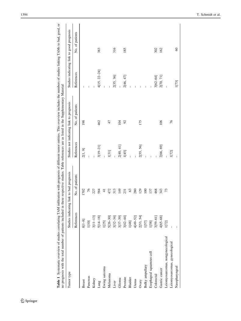
This notion is supported by some clinical data indicating
an adverse prognostic effect of macrophage infiltration in
different cancers (Table 1; see also Supplementary Mate-
rial Table 1 for detailed information). In breast cancer,
uterine cancer, kidney cancer and melanoma the majority
of published studies (three or more independent studies
each) show a negative clinical impact of high TAM num-
bers, and in these cancers no data exist so far supporting
a positive association between TAMs and prognosis.
Macrophage-tumor cross-talk 1393
123
Page 4
Ta
ble
1S
yst
emat
ico
ver
vie
wo
fst
ud
ies
corr
elat
ing
TA
Min
filt
rati
on
wit
hp
rog
no
sis
of
dif
fere
nt
tum
or
enti
ties
.T
his
ov
erv
iew
incl
ud
esth
en
um
ber
so
fst
ud
ies
lin
kin
gT
AM
sto
bad
,g
oo
d,
or
no
pro
gn
osi
sw
ith
the
tota
ln
um
ber
of
pat
ien
tsin
clu
ded
inth
ese
resp
ecti
ve
stu
die
s.T
able
refe
ren
ces
are
asli
sted
inth
eS
up
ple
men
tary
Mat
eria
l
Tu
mo
rty
pe
Stu
die
sin
dic
atin
gli
nk
tob
adp
rog
no
sis
Stu
die
sn
ot
ind
icat
ing
lin
kto
pro
gn
osi
sS
tud
ies
ind
icat
ing
lin
kto
go
od
pro
gn
osi
s
Ref
eren
ces
No
.o
fp
atie
nts
Ref
eren
ces
No
.o
fp
atie
nts
Ref
eren
ces
No
.o
fp
atie
nts
Bre
ast
8[1
–8
]1
70
22
[1,
9]
19
8–
Pan
crea
s1
[10
]7
6–
–
Kid
ney
3[1
1–
13
]2
27
––
Lu
ng
5[1
4–
18
]5
84
3[1
9–
21
]4
62
4[1
5,
22
–2
4]
38
3
Ew
ing
sarc
om
a1
[25
]4
1–
–
Mel
ano
ma
5[2
6–
30
]4
72
1[3
1]
47
–
Liv
er3
[32
–3
4]
31
3–
2[3
5,
36
]3
16
Gli
om
a3
[37
–3
9]
14
92
[40
,4
1]
10
4–
Pro
stat
e3
[42
–4
4]
23
11
[45
]9
22
[46
,4
7]
18
5
Bla
dd
er1
[48
]6
3–
–
Ute
rus
4[4
9–
52
]2
80
––
Ov
ary
2[5
3,
54
]1
29
2[5
5,
56
]1
75
–
Bu
lky
amp
ull
ary
1[5
7]
10
0–
–
Eso
ph
agea
lsq
uam
ou
s-ce
ll1
[58
]1
37
––
Co
lore
ctal
3[5
9–
61
]4
68
–3
[62
–6
4]
30
2
Gas
tric
can
cer
4[6
5–
68
]3
43
2[6
6,
69
]1
06
2[7
0,
71
]1
62
Lei
om
yo
sarc
om
as,
no
ng
yn
eco
log
ical
1[7
2]
73
––
Lei
om
yo
sarc
om
as,
gy
nec
olo
gic
al–
1[7
2]
76
Nas
op
har
yn
gea
l–
–1
[73
]6
0
1394 T. Schmidt et al.
123
Page 5
In contrast, in lung cancer, more studies indicate no associ-
ation (three studies) or a favorable association (four studies)
between TAM numbers and prognosis than a negative
prognostic impact. In other cancer types, including glioma,
prostate cancer, gastric cancer, colorectal cancer and ovarian
cancer, the numbers of studies indicating negative or
no/positive correlations are almost equal. In some cancers
only one or two studies have yet been reported. Hence, it is
too early to draw conclusions about the prognostic impact of
TAMs. Interestingly, in different histological subtypes of
breast cancer (intraductal carcinoma vs. infiltrative lobular
carcinoma) [38] and leiomyosarcoma (gynecological vs.
non-gynecological) [39], TAMs have a negative or no
prognostic impact, respectively (Supplementary Material
Table 1). Thus, it cannot be excluded that interaction of
TAMs with different types of cancer cells influences the
tumor-promoting capacity of macrophages.
The spatial localization of TAMs also seems to matter,
because in lung cancer TAM density in the surrounding
stroma has been shown to have a negative association with
prognosis, while the opposite is true when TAMs are present
within tumor cell nests [40, 41]. Similarly, in melanoma [42]
and uterine cancer [43] macrophages, which were located at
the invasive front of tumors, have a negative impact on
prognosis. Thus, macrophages at the invasive front might
promote malignancy, while macrophages within tumors
might be tumoricidal. Taken together, the picture is not yet
crystal clear, and this might also be due to methodology.
Possible reasons for incoherent data include small sample
sizes, different markers used for macrophage identification
and interobserver variability in immunohistochemical
analysis. Clearly, in order to elucidate the prognostic impact
of TAMs, larger studies are warranted, in which tissues are
simultaneously stained, ideally on tissue microarrays. Also,
it might be more informative to analyze phenotypes and the
activation status of TAMs, rather than only determining their
number, because function is most likely more important. In
line with this, osteopontin-positive macrophages have a
negative impact on prognosis in bulky ampullary cancer,
which might be explained by the well-described promigra-
tory action of this cytokine [44]. In another study, the
presence of TAMs expressing high levels of thymidine
phosphorylase, which promotes tumor growth and metas-
tasis by enhancing angiogenesis, was independently
associated with shorter survival, while thymidine phos-
phorylase expression by tumor cells was not significantly
associated with prognosis [45]. Of note, TAMs isolated from
patients with advanced clinical lung cancer produce higher
levels of IL-10 when isolated and cultured in vitro than
TAMs from patients with earlier disease stages [46].
However, the majority of published studies did not dis-
tinguish between different macrophage phenotypes, because
CD68 expression was mainly used to identify TAMs. Few
studies in humans have analyzed M1 and M2 macrophages,
with CD68?CD163- macrophages being considered M1-
polarized, while M2-polarized macrophages are considered
CD68?CD163? [23]. Some of these analyses are in line with
the concept that M2 macrophages foster tumor progression,
whereas M1 macrophages inhibit tumor development. For
instance, in intrahepatic cholangiocarcinomas and in uveal
melanoma, high infiltration with CD163? M2 macrophages
is correlated with shorter disease-free survival and overall
survival, respectively, compared to low M2 infiltration
[47, 48]. In contrast, the density of M1-polarized macro-
phages is positively associated with survival in lung cancer
[49, 50]. Hence, therapeutic approaches aimed at skewing
macrophage polarization towards an M1 phenotype might
open up novel therapeutic avenues. In this respect, recent
studies indicate that M2 macrophages can be induced to
acquire an M1-like phenotype by inhibitors targeting cyclo-
oxygenase 2 and placental growth factor (PlGF) [51, 52].
Moreover, CD4? Th1 cells are also capable of skewing
macrophages from M2 to M1 polarization [27]. A recent
study has shown that autocrine CXCL12 production by
macrophages enhances their proangiogenic and immuno-
suppressive phenotype in vitro [53]. Interestingly, 60–90% of
TAMs in primary metastatic melanoma coexpress CD68,
CD163 and CXCL12; thus this mechanism might be of rel-
evance in human cancer. Consequently, CXCL12 could be an
additional target with the potential to decrease M2 properties
of TAMs [53]. However, as the majority of human cancer
studies did not distinguish between M1 and M2 (Supple-
mentary Material Table 1), more work is needed to get a
better view of the prognostic impact and target potential of
M2-polarized macrophages. However, such correlative
studies obviously need to be interpreted with caution, and
more mechanistic studies are warranted to answer the ques-
tion when and why TAMs show pro- and antitumoral activity.
Taken together, emerging data suggest an important role
of TAMs in tumor biology. In the following section we
describe how macrophages and closely related myeloid cell
types interact with various cellular players present in
tumors and vice versa (see Fig. 1 for an overview). For
information about how other leukocyte populations interact
with tumors, we refer the readers to recent comprehensive
reviews on this topic [3, 4, 6].
Crosstalk between macrophages and different tumor
components
Crosstalk between macrophages and tumor cells
from cancer initiation until metastasis
There is a tight link between cancer initiation and inflam-
mation, because an inflammatory microenvironment can
Macrophage-tumor cross-talk 1395
123
Page 6
directly increase the mutation rate of cancer cells. Inter-
estingly, during tumor initiation, macrophages obtain a
more proinflammatory profile as opposed to the immuno-
suppressive M2 phenotype observed in established tumors
(see previous section). These proinflammatory macro-
phages are characterized by strong activity of the NFjB
pathway, which is induced by pathogen associated
molecular patterns, by Toll-like receptor ligands and by
cytokines including TNFa and IL-1b. As a result, they
secrete abundant proinflammatory and protumoral cyto-
kines such as IL-12, inducible nitric oxide synthase
(iNOS), TNFa and IL-6 [28, 54, 55].
The importance of the NFjB pathway for tumor initia-
tion is emphasized by data indicating reduced expression of
several proinflammatory cytokines after ablation of IjB
kinase (IKKb) in myeloid cells. This dampened inflam-
matory reaction substantially reduced tumor incidence and
progression in mouse models of intestinal cancer [56].
Furthermore, deletion of IKKb in Kupffer cells (the mac-
rophages of the liver) or inhibition of TNFa reduces
hepatocellular carcinogenesis [57, 58]. The NFjB pathway
also plays an important role in spontaneous mouse models
of skin cancer, because tumorigenesis after application of
carcinogens only occurs after additional induction of a
pronounced inflammation by TNFa through tumor necrosis
factor receptor 1 (TNFR1), a strong inducer of NFjB
activity [59, 60]. Amongst other cell types, macrophages
play a particularly important role in this NFjB-mediated
inflammation, because they are induced to enter tumors by
TNFa and at the same time secrete large amounts of this
cytokine. TNFa can induce the Wnt/b-catenin signaling
pathway in tumor cells, which is a strong promotor of
tumorigenesis [61–63]. As TNFa is one of the main
inducers of NFjB signaling, its secretion creates a feed-
forward autocrine loop fueling further proinflammatory
activity in the macrophages, but also activates other
inflammatory cells present in the microenvironment of the
developing tumor by acting in a paracrine manner [61].
Furthermore, TNFa promotes the formation of reactive
oxygen species in macrophages, which directly induce
DNA damage and genomic instability [25]. Reactive oxy-
gen species and TNFa can cause inactivation of enzymes or
regulators involved in DNA mismatch repair including p53
and poly-adenosine ribose polymerase [18, 25]. This sup-
pression of DNA repair generates a promutagenic
environment, which is further promoted by nitric oxide
synthesized in macrophages by iNOS. Nitric oxide is a
highly reactive substance that gives rise to intermediates
directly causing mutations in epithelial cells [18].
Successful initiation of cancer depends on increased cell
proliferation and reduced cell death, both of which are
fostered by TAMs [7]. To achieve this, they produce a
plethora of cytokines including IL-1, IL-6 and TNFa. After
binding to their receptors at the tumor cell surface, these
mediators activate the transcription factors NFjb, STAT3
and AP1. STAT3 elicits tumor cell proliferation by acti-
vating cyclin D1, cyclin D2, cyclin B and c-myc [64, 65].
NFjb and STAT3 promote cell survival by inducing
expression of the antiapoptotic proteins Bcl-2 and Bcl-xl
[65, 66]. The importance of IL-6 for tumor initiation has
been underscored by the reduced risk of hepatocellular
carcinoma in female mice, which produce lower levels of
Fig. 1 Pleiotropic roles of
macrophages in the tumor
microenvironment. In the tumor
microenvironment different
types of ‘‘tumor-promoting’’
macrophages (red) can display
protumorigenic roles during
virtually every step of tumor
development and progression
(top panel). In contrast, ‘‘tumor-
suppressive’’ macrophages
(green) can fight tumor growth
mainly by stimulating the
immune system to kill cancer
cells (bottom panel)
1396 T. Schmidt et al.
123
Page 7
IL-6 after treatment with a chemical carcinogen, whereas
ablation of this growth factor leads to equal hepatocarci-
nogenesis in both genders [67]. Furthermore, macrophages
can secrete heparin-binding EGF, which supports survival
and proliferation of colon cancer cells via their Her1
receptor. Interestingly, in response, cancer cells produce
GM-CSF, which acts on macrophages and induces them to
further upregulate heparin-binding EGF expression [35].
Thus, macrophages are a rich source of cytokines directly
promoting tumor initiation via autocrine and paracrine
feed-back loops. Interestingly, recent data indicate a posi-
tive correlation between TAM density and the density of
glioma-initiating cells in primary glioma [68]. Thus, TAMs
might support cancer stem cells, which are regarded as key
cellular components sustaining malignancy.
Interestingly, in established tumors, NFjB signaling in
macrophages is suppressed due to constitutive expression
of p50 homodimers. These homodimers are unable to
induce transcription, but possess higher DNA-binding
affinity than the bioactive p50/p65 heterodimer. Conse-
quently, in the presence of p50 homodimers, macrophages
express fewer NFjB target genes including IL-12, iNOS
and TNFa, while M2-specific genes including arginase-1
and Fizz1 become upregulated [28, 69]. Therefore, TAMs
in established human and murine tumors often acquire an
M2 phenotype [70–73] (see above). This phenotypic M1 to
M2 shift can be mimicked in vitro by blocking the NFjB
pathway in macrophages by inhibition of IKKa. In addition
to suppression of NFjB signaling in macrophages, cancer
cells also secrete IL-4, IL-10 and IL-13, mediators capable
of polarizing macrophages towards an M2 phenotype
[4, 20, 74]. Thus, via different pathways, tumor cells can
induce M2 polarization of macrophages, which in turn
promote tumor progression.
In order to recruit more cancer-promoting macrophages,
tumor cells produce different cytokines including CSF-1,
monocyte chemotactic protein-1 (MCP-1/CCL2) and PlGF.
The importance of CSF-1 has been amply demonstrated by
a significant reduction in infiltrating macrophages after
deletion of CSF-1 from transgene models of breast cancer,
colon cancer and osteosarcoma, while their numbers were
increased after overexpressing CSF-1 [62, 75, 76]. As a
consequence, tumor progression was reduced or enhanced,
respectively [62, 75, 76]. Consistent with these findings,
therapeutic inhibition of CSF-1 by neutralizing antibodies
or antisense strategies blocks tumor growth and metastasis
in murine xenograft models [77, 78]. PlGF induces mac-
rophage recruitment via VEGF receptor 1 (VEGFR-1).
Consequently, inhibition and genetic ablation of PlGF
inhibits tumor progression by reducing macrophage
recruitment [14, 79, 80].
Macrophages promote additional hallmarks of malig-
nancy including cancer cell migration, invasion and
metastasis. In murine xenograft breast cancer models and
spontaneous breast tumors, cancer cells produce CSF-1,
thereby stimulating and attracting macrophages, which in
response produce EGF. EGF subsequently activates tumor
cells in a vicious circle to migrate. Interestingly, inhibition
of either CSF-1 or EGF signaling is sufficient to impair
migration and chemotaxis of both cell types, which under-
lines the central importance of this reciprocal paracrine
interaction [81–84]. Of note, this crosstalk is further
amplified in hypoxic conditions, because hypoxia activates
hypoxia-inducible factor 2a (HIF-2a), which upregulates
expression of EGFR in tumor cells and of CSFR in macro-
phages [85, 86]. In this way, via interaction with
macrophages, tumor cells can better escape hostile hypoxic
tumor environments. Interestingly, the presence of IL-4 is
required, because in its absence TAMs are not able to induce
invasion and migration of breast and pancreatic tumor cells,
which strongly reduces the metastatic capacity of cancer
cells [74, 84]. Besides IL-4 and EGF, other macrophage-
derived mediators including Wnt5a and TNFa promote
tumor cell invasiveness [7]. Interestingly, macrophages
even physically promote extravasation of tumor cells by
forming clusters on the abluminal side of blood vessels,
through which tumor cells enter the circulation [82]. Of note,
the transcriptome of these ‘‘tumor cell-bridging’’ macro-
phages closely resembles that of ‘‘trophic’’ macrophages,
but has little similarity to M1 or M2 macrophages [7].
Crosstalk between macrophages and vascular
endothelial cells
TAMs and closely related TEMs are involved in regulation
and remodeling of blood vessels. Both TAMs and TEMs
express higher levels of proangiogenic molecules than
circulating monocytes [87, 88]. TAMs are required for the
angiogenic switch and for vascular remodeling in sponta-
neous mammary tumors, because angiogenesis is impaired
after inhibition of macrophage recruitment due to inacti-
vation of CSF-1 [89]. Similarly, upon macrophage
depletion by different approaches, such as clodronate
liposomes, angiogenesis is reduced in different tumor
models [7]. Conversely, CSF-1 overexpression, and sub-
sequently enhanced TAM infiltration, substantially
increases angiogenesis [89]. The proangiogenic function of
M2-polarized TAMs is at least partly mediated by the
transcription factor Fra-1, because its down-modulation in
macrophages greatly decreases their ability to induce
angiogenesis in experimental breast cancer [90]. The con-
cept of potent induction of angiogenesis by TAMs is
further substantiated by clinical data revealing a correlation
between a high density of TAMs and increased microvessel
density in different cancers, including lung cancer [91] and
breast cancer [92].
Macrophage-tumor cross-talk 1397
123
Page 8
TAMs are recruited specifically to hypoxic tumor
regions, because important macrophage chemoattractors,
including VEGF, endothelins and stromal cell-derived fac-
tor-1, are upregulated in hypoxia [93]. This process is further
amplified in hypoxic conditions, where macrophages acti-
vate HIF-2a. HIF-2a then induces upregulation of M-CSFR
and CXCR4, which potentiate chemotaxis of macrophages
towards hypoxic tumor regions [86]. Once arrived, TAMs
secrete VEGF, PlGF and other proangiogenic cytokines
[94]. These potent mediators increase angiogenesis and at
the same time, by acting on endothelial cells and pericytes,
induce blood vessel abnormalities such as increased leaki-
ness, multilayered endothelium and immaturity, together
termed ‘‘vessel abnormalization’’. As the end result, vessel
abnormalization leads to dysfunctional, hypoperfused
vessels, which fail to adequately supply the tumor with
oxygen and nutrients [95, 96]. Consequently, hypoxia
increases, which leads to further fueling of macrophage
infiltration. Therefore, vessel abnormalization increases
even more, and as consequence, tumor cell intravasation
through a leaky endothelial cell layer is enhanced [7, 97].
VEGF and PlGF not only act on endothelial cells, but can
also stimulate tumor cell motility by activating VEGF
receptors [14, 98]. These multitasking cytokines are addi-
tionally involved in TAM polarization towards an M2
phenotype [52, 79]. Consistently, in PlGF-deficient mice
TAMs are skewed from the proangiogenic M2-like pheno-
type towards a more proinflammatory phenotype [52]. As a
consequence, vessel abnormalization is decreased and tumor
cells elicit less invasiveness and metastasis [52].
The TEM subset of monocytes predominantly resides
close to tumor blood vessels, where they can potently induce
angiogenesis [99]. This close association depends on endo-
thelial secretion of angiopoietin-2 (Ang-2). Consequently,
blockade of Ang-2 reduces tumor growth and angiogenesis
partly by disrupting the close physical interaction of TEMs
and endothelial cells [100]. Recent data indicate a similar
localization of tissue-resident macrophages close to the tips
of branching blood vessels, where they facilitate fusion of
two adjacent vessel sprouts. Implications for tumor biology
as well as molecular mechanisms are still not fully explored,
but the Notch/Dll4 or Tie2/Ang-2 systems might mediate
this interaction between macrophages and blood vessels
[101, 102]. Altogether, TAMs represent potent inducers of
angiogenesis and vessel abnormalization, and hence
approaches aimed at inhibiting these important protumoral
actions of TAMs may lead to novel cancer treatments.
Crosstalk between macrophages and lymphatic
endothelial cells
Besides their role in angiogenesis, macrophages are
involved in lymphangiogenesis during development and
disease. Indeed, lymphatic vessel development was
impaired in op/op mice exhibiting reduced macrophage
numbers due to an inactivating mutation in the Csf1 gene
[75]. Macrophages are an important source of the lymph-
angiogenic cytokines VEGF-C and VEGF-D in different
disease conditions including cancer [14, 103]. Consistent
with this, depletion of macrophages strongly impairs
lymphangiogenesis in different experimental cancer mod-
els, mainly because of reduced intratumoral levels of
prolymphangiogenic cytokines. As a consequence, lym-
phatic metastasis is reduced [14, 104]. These findings may
have implications for human cancer, because TAMs in
primary human cutaneous squamous cell carcinomas are
important producers of VEGF-C [105]. A novel concept of
macrophage and lymphatic interaction has been found in
the RipTag2 pancreatic tumor model and TRAMP-C1
prostate cancer model, in which BMDCs of the myelo-
monocytic lineage become integrated into tumor-
associated lymphatic vessels. This effect is not based on
cell fusion, but rather on phenotypical conversion of
myeloid cells into lymphatic endothelial cells. Depletion of
macrophages consequently reduces the lymphatic vessel
density [106]. In line with the role of macrophages in
lymphangiogenesis, TAM infiltration correlates with tumor
lymphatic vessel density in lung cancer [107] and in pan-
creatic cancer [108].
Crosstalk between macrophages and fibroblasts
The development of cancer is often associated with an
increase in fibroblast proliferation leading to extensive
fibrosis. However, relatively little is known about the
interaction of macrophages and fibroblasts. Recent data in a
chemically induced skin cancer model indicate that FSP1?
CAFs secrete MCP-1 (CCL2), IL-6 and TNFa, by which
they recruit and polarize macrophages towards the M2
phenotype. Depletion of these CAFs consequently reduces
macrophage infiltration and thereby inhibits tumor devel-
opment [109]. These findings were further supported in
experimental breast cancer, because recruitment of TAMs
was enhanced after upregulation of CCL2 in CAFs, pro-
moting tumor progression and metastasis [110]. The
importance of fibroblasts for macrophage recruitment was
also corroborated by in vitro data showing extensive
CCL2-mediated infiltration of tumor-derived 3D fibroblast
spheroids with monocytes. Normal fibroblasts fail to attract
monocytes; hence this fibroblast–macrophage interaction
appears tumor-specific [111]. Of note, CAFs in human
cancer might show similar functions, because in pancreatic
ductal adenocarcinoma and squamous cell carcinoma
models, CAFs attract macrophages by NFjB-mediated
upregulation of CXCL1, CXCL2 and CXCL5 [110]. Sim-
ilarly, in comparison to fibroblasts present in healthy tissue,
1398 T. Schmidt et al.
123
Page 9
CAFs in primary human prostate cancer tissue upregulate
CCL14, which chemoattracts macrophages. Interestingly,
in murine 4T1 breast tumors, CAFs can additionally act as
immunomodulators, because inhibiting their activation
shifts the immune microenvironment from Th2 towards
Th1 polarization. Subsequently, tumor growth and metas-
tasis are strongly reduced, which might be explained by
decreased recruitment and M2 polarization of macrophages
[112]. However, not all CAFs show proinflammatory
functions; for instance CAFs in murine cervical cancer do
not show upregulation of IL-1, IL-6, CXCL1 or CXCL-2
[110]. In addition to recruiting macrophages, CAFs can
also support macrophage maturation. Indeed, coculture of
CAFs with a monocytic cell line induces upregulation of
the macrophage maturation marker F4/80 and induces
morphological changes typical of mature macrophages. At
the functional level, these mature macrophages secrete
higher levels of proinflammatory and protumoral cytokines
such as IL-1b and TNFa upon coculture with tumor cells
when than immature monocytes [113]. In summary, the
crosstalk between CAFs and macrophages plays an
important role in tumor progression; hence it would be of
interest to decipher further interactions at the molecular
and functional levels.
Crosstalk between macrophages and other leukocytes
TAMs are essential players in the suppression of antitu-
moral immune responses. They express high levels of
mediators interfering with T-cell activation and prolifer-
ation such as IL-10, TGFb, prostaglandins and arginase-1
[73, 114]. This might be a reason for the rather weak
antitumoral immune responses observed in most tumors.
The immunosuppressive function of macrophages has
already been extensively reviewed elsewhere [7, 19].
Interestingly, in hypoxic tumor regions, the immune-
suppressive properties of macrophages are even more
enhanced, because activation of HIF-1a leads to upregu-
lation of arginase-1 and iNOS, both of which dampen
T-cell function [115]. Indeed, macrophage-specific
depletion of HIF-1a reduces breast cancer growth by
activating the cytotoxic T-cell response, but without
changing expression levels of the prototypic HIF-1a target
gene VEGF [115]. Alternatively, in response to autocrine
production of IL-10 and TNFa, macrophages upregulate
the cosignaling molecule PD-L1 (also called B7-H1),
which suppresses T-cell function by a yet-undiscovered
mechanism. Therapeutic blockade of PD-L1 reduces
tumor growth by enhancing intratumoral cytotoxic T-cell
function [116]. Of note, in contrast, tumor-infiltrating T
cells can polarize TAMs towards M2 macrophages,
thereby creating another vicious circle promoting tumor
development. Indeed, in a spontaneous breast cancer
model, CD4? cells expressed IL-4, a strong inducer of
M2 polarization of macrophages. Consequently, after
therapeutic or genetic targeting of IL-4, the macrophage
phenotype is skewed towards M1. Furthermore, IL-4
derived from tumor cells and from T cells induces high
levels of cathepsin B and S protease activity in TAMs of
murine breast and pancreatic tumors. As a consequence,
tumor growth, angiogenesis and invasion are induced,
because cathepsin B and S cleave extracellular matrix
proteins, thereby liberating matrix-bound proangiogenic
molecules [74, 84].
Macrophages not only interact with cytotoxic T cells,
but they also cooperate in a complex manner with several
other immune cell populations in tumors. Within the
immunological tumor microenvironment, regulatory T
cells (Tregs) play an important immunosuppressive role.
Their importance has, for example, been proven in pre-
clinical lymphoma models, where Treg depletion led to
rapid tumor rejection by T cells and NK cells. Consistent
with a protumoral role of Tregs, their density correlates
with a poor prognosis in patients with different cancers,
including hepatocellular cancer [117]. TAMs can augment
the recruitment of Tregs by secreting CCL20, which
chemoattracts them via chemokine receptor 6 (CCR6).
Accordingly, tumor growth is reduced after macrophage
depletion, because CCL20 levels are lowered and conse-
quently Treg recruitment is reduced [117, 118]. Similarly,
in colorectal cancer patients, macrophages produce high
levels of CXCL11, which is another strong chemoattractant
for Tregs. Tregs, besides their immunosuppressive action,
produce IL-17 in the tumor microenvironment, which
supports survival of colorectal cancer-initiating cells [119].
Interestingly, the crosstalk between TAMs and Tregs is
bidirectional, because upon Treg depletion, TAM numbers
decrease. Additionally, without the influence of Tregs,
TAMs augment their proinflammatory properties by
increasing MHC class II and immunoactivating chemokine
expression (MIP-1b, MIP-2 and TNFa) [120]. However,
similar to TAMs (see above), the role of Tregs may be
different in different cancers. For example, in patients with
gastric cancer, a high CD68?/FoxP3? cell ratio (macro-
phage/Tregs) is associated with shorter survival, indicating
that inhibition of Tregs might not be useful in some cancers
[121].
Besides interacting with different T-cell subpopulations,
macrophages also cooperate with B cells, and vice versa.
For instance, B cells can skew the macrophage phenotype
towards M2 polarization. This ability has been shown in
the B16 melanoma model, because so-called B1 cells were
able to drive macrophages to acquire an M2-biased phe-
notype mainly by secreting IL-10 [122].
Macrophage-tumor cross-talk 1399
123
Page 10
Macrophages and cancer therapy resistance
Macrophages are implicated in resistance towards chemo-
therapy and biological therapies. Recent data indicate that
breast cancer patients with a high number of TAMs and a
low number of cytotoxic T cells within their tumor tissue
have a poor response to neoadjuvant chemotherapy with
taxanes, antimetabolites and anthracyclines. Interestingly,
both in cancer patients and in mice, elevated CSF-1 levels
and increased numbers of macrophages have been detected
in tumor tissue after chemotherapy. Blockage of CSF-1
signaling or macrophage depletion enhances antitumor
immunity and response to chemotherapy in murine cancer
models, indicating functional involvement of macrophages
in mediating resistance to chemotherapy [123]. Interest-
ingly, TAMs are abundantly present in the bone marrow of
patients with multiple myeloma, where they protect mye-
loma cells from chemotherapy-induced cell death. This
protection depends on direct cell–cell contact and on
ICAM-1, because it does not occur after physical separa-
tion of myeloma cells from TAMs or after antibody-
mediated blockade of ICAM-1. TAMs achieve this
protective effect partly by attenuating the activation and
cleavage of caspase-dependent apoptotic signaling [124].
Interestingly, TAMs can render cancer stem cells, intrin-
sically relatively resistant to chemotherapy, even more
resistant by producing milk-fat globule-epidermal growth
factor VIII (MFG-E8) and IL-6. MFG-E8 is a potent acti-
vator of STAT3 signaling and of the hedgehog pathway,
while IL-6 further fuels STAT3 activation. Both pathways
have been shown to mediate the resistance of colorectal
cancer stem cells by promoting their survival in the pres-
ence of cisplatin [125]. Moreover, in prostate cancer TAMs
can confer resistance to androgen receptor antagonists. In
this process, macrophages adhere to prostate cancer cells
by VCAM-1 and subsequently produce IL-1b, which in
turn blocks the function of nuclear receptor corepressors
N-CoR. N-CoR normally associates with antiandrogens,
and subsequently suppresses androgen-induced gene tran-
scription. However, without binding to N-CoR, androgen
receptor antagonists activate, instead of suppress, andro-
gen-induced gene expression [126].
Via CSF-1, cells of the myeloid lineage are also
recruited into tumors treated with VEGF-targeted antian-
giogenic therapies, where they can directly confer
resistance by producing alternative proangiogenic factors
besides VEGF [15, 127]. Consequently, blockade of the
CSF-1 pathway inhibits tumor angiogenesis and acts syn-
ergistically with anti-VEGFR-2-targeted therapy by
reversing myeloid cell-mediated antiangiogenic therapy
resistance [128]. The importance of pathways mediating
myeloid cell recruitment in antiangiogenic escape is further
supported by detection of increased levels of PlGF, stromal
cell-derived factor-1 and MCP-3 in colorectal cancer
patients treated with anti-VEGF antibodies and chemo-
therapy immediately before disease progression [129].
Overall, TAMs can facilitate a large variety of mechanisms
to render tumors resistant to different therapeutic strategies
[130]. Hence, therapeutic approaches aimed at inhibiting
these TAM properties might be more efficient than current
anticancer therapies.
In summary, TAMs interact with cancer cells and with
different cellular components of the tumor microenviron-
ment. This crosstalk can promote malignancy and therapy
resistance via a plethora of complex mechanisms, but in
principle macrophages can also show antitumoral activity.
In experimental cancer models, some progress has been
made recently in skewing the macrophage phenotype
towards tumoricidal activity and in overcoming therapy
resistance by targeting macrophages, but considerably
more work is necessary to elucidate whether this approach
has therapeutic potential in human cancer.
In the next section we introduce the Tyro3, Axl and Mer
receptor (TAMR) family with their ligands, that are
expressed by tumor cells and macrophages and have
recently been shown to be involved in the tumor–macro-
phage crosstalk (Fig. 2). We also describe their role in
(human) solid cancer and in hematological malignancies.
Fig. 2 Ligand–receptor specificity in TAMR. Gas6 binds to all three
TAMR (Sky, Axl, Mer) with different affinities (Axl � Mer [ Sky)
and signals through them. Current knowledge indicates that protein
S(ProtS) binds to Sky and Axl. Tubby-like protein 1 (tulp1) can bind
and signal through all three TAMR, whereas tubby is only found to
signal through Mer. Each receptor induces certain biological
responses indicated below the respective receptor
1400 T. Schmidt et al.
123
Page 11
TAMR tyrosine kinase receptors and their ligands
Receptor tyrosine kinases (RTKs) are key players in cancer
cell biology. They regulate cell survival, proliferation,
migration and differentiation, cell cycle control and apop-
tosis [131]. The prototypical RTKs are activated by ligands
such as growth factors, which induce receptor dimerization
and subsequent autophosphorylation of tyrosine residues
on the intracellular cytoplasmic domain with further
downstream signaling [132]. Currently 58 RTKs, divided
into 20 subfamilies, are known. The TAMR, named after
Tyro3, Axl and Mer or their homologues, are present in
chordates including urochordates and vertebrates [133, 134]
(see below for alternative nomenclature). This RTK
subfamily was identified only in 1991 and TAMR
were initially considered as orphan receptors [135, 136].
Structurally, the TAMR family is characterized by an
extracellular domain consisting of two immunoglobulin-
like domains followed by two fibronectin type 3-like
domains. These extracellular domains are followed by a
transmembrane domain and a cytoplasmic tyrosine kinase
domain [137, 138]. TAMR can be activated by (1) ligand-
independent dimerization, (2) ligand-dependent dimeriza-
tion, (3) heteromeric dimerization of two different TAMR,
(4) heterotypic dimerization with a non-TAMR, and (5)
trans-cellular binding of extracellular domains [139, 140].
Axl was initially identified as a transforming gene
derived from chronic myeloid leukemia cells [136].
Overexpression of this gene in NIH 3T3 cells led to their
transformation and the gene was therefore named axl from
the Greek word ‘‘anexelekto’’, which means uncontrolled
[136]. The oncogenic potential of Axl depends on its
intracellular tyrosine kinase domain, because 33 amino
acids of the intracellular domain are able to transform NIH
3T3 cells [141]. Axl is ubiquitously expressed and detect-
able in most organs as well as in different cell lines of
mesenchymal, epithelial and hematopoietic origin. Axl
expression becomes detectable in many tissues during
embryonic development from day E12.5 onwards [142].
Due to independent cloning, Axl was also designated Ufo,
Jkt11, Ark, Tyro7; however, Axl is the official NCBI
designation. Axl promotes a large variety of biological
functions including platelet aggregation [143, 144], regu-
lation of proinflammatory cytokine production and control
of the actin cytoskeleton [145]. Moreover, Axl mediates
cell survival, proliferation and migration [139]. The
important function of Axl in regulating survival was
demonstrated in fibroblasts isolated from Axl-/- mice.
These fibroblasts display enhanced serum deprivation-
induced apoptosis when compared to fibroblasts derived
from wild-type mice [146]. Axl controls survival mainly
via the PI3K, AKT, and NFjB pathways, while Axl-
induced proliferation depends mainly on ERK1/2 signaling
[139, 140]. TAMR share structural features with cell
adhesion proteins, in particular the ectodomain of Axl
elicits adhesive properties. This ‘‘stickiness’’ can mediate
cell–cell contact leading to aggregation of cells, which
might facilitate metastasis. In line with this concept, Axl
expression correlates with adherence of human lung cancer
cell lines [147, 148].
Ample evidence in the literature points to an important
functional role of Axl in tumor biology (Fig. 3). Activation
of Axl induces proliferation [149, 150], survival [151–
154], resistance against apoptosis [151, 155, 156], migra-
tion and invasiveness of cancer cells [149–154].
Furthermore, Axl mediates resistance towards chemo- and
targeted therapy including anti-VEGF or anti-EGFR ther-
apy in part by inducing secretion of proinflammatory and
protumoral cytokines such as IL-6, TNFa and G-CSF in
TAMs, where it is highly expressed [157]. Hence, treat-
ment of breast cancer xenografts with anti-Axl antibodies
inhibits the secretion of protumoral inflammatory cytokines
and chemokines from TAMs, which have an inhibitory
effect on tumor growth [157]. The precise mechanism
involved in the inhibition of the production of inflamma-
tory cytokines in TAMs by anti-Axl antibodies remains to
be elucidated. Furthermore, recent data in thyroid cancer
cell lines indicate constitutive Axl and Sky phosphoryla-
tion induced by autocrine production of Gas6. This
autocrine loop, which is not present in normal thyroid cells,
specifically mediates proliferation and apoptosis resistance
of thyroid cancer cells in vitro and in vivo [151]. Alto-
gether, Axl plays a prominent role in cancer biology by
promoting malignancy at several levels.
In 1993 the Sky receptor was identified, which was also
termed Tyro3, Brt (brain tyrosine kinase), Tif, Dtk, BYK
and Etk-2 [158–160]. In a similar manner to Axl, Sky is
also expressed in several embryonic tissues during devel-
opment [161, 162], but shows a more restricted expression
pattern in adulthood with the predominant expression in the
brain [163–165], in hematopoietic cells [161], in pulmon-
ary endothelial cells [166], in osteoclasts [167], and in the
kidney, testis and ovary [163, 168]. In a similar manner to
Axl and Mer, Sky is involved in platelet function, but
single Sky-/- mice display mild platelet dysfunction
without spontaneous bleeding. In contrast, double or triple
TAMR-deficient mice suffer from pronounced bleeding
diathesis [169]. Additionally, Sky plays an important role
in osteoclastic bone resorption [167]. Recently, in exito-
toxic brain injury, protein S has been shown to activate Sky
leading to the suppression of proapoptotic Fas ligand pro-
duction. Thus, by suppressing apoptosis, Sky acts in a
neuroprotective manner [170]. Furthermore, together with
Axl, Sky mediates survival and targeting of GnRH neurons
to the ventral forebrain, which is important for reproductive
function in female mice [152].
Macrophage-tumor cross-talk 1401
123
Page 12
Sky can transform cells in vitro, but its role in cancer is
less well-defined than that of Axl (Fig. 3) [171, 172]. Sky
can promote malignancy by inducing proliferation, which
is at least partially mediated via PI3K signaling [171]. In
malignant melanoma, activation of Sky induces the tran-
scription factor microphthalmia-associated transcription
factor, which strongly promotes malignancy in melanoma.
Indeed, knock-down of Sky in melanoma cells suppresses
proliferation and sensitizes them to chemotherapy. Con-
versely, Sky overexpression promotes cancer cell survival
by overcoming senescence [173]. In addition, in a similar
manner to Axl, Sky can also exert adhesive functions by
homophilic interaction, which potentially enhances
metastasis [174].
The Mer receptor was initially cloned as a human pro-
tooncogene from a leukemic cell line [175]. Sequence
comparison indicates that this human kinase is 83% similar
to the previously isolated chicken retroviral oncogene
v-ryk (v-eyk) [175, 176]. Mer was named after its unique
expression pattern in monocytes, epithelium and repro-
ductive tissue. Mer was also designated mertk, eyk, nyk
and rdy. Mer is expressed during most stages of embryonic
development of the mouse [177]. Like the other members
of the TAMR family, Mer is involved in platelet function
[178], exerts mitogenic signals and has transforming ability
[179]. Furthermore, Mer is crucially involved in apoptotic
cell clearance by phagocytic cells including dendritic cells,
macrophages, monocytes and retinal pigment epithelial
(RPE) cells [180]. In order to mediate the cytoskeletal
reorganization crucial for phagocytosis, Mer activates focal
adhesion kinase via an alpha(v) beta(5) integrin-dependent
pathway. Alternatively, after binding its ligand Gas6, Mer
signaling leads to phosphorylation of Vav1 [181]. Both
pathways finally activate Ras-related C3 botulinum toxin
substrate (Rac), which induces cytoskeletal reorganization
with subsequent phagocytosis [181]. In line with this
mechanism, Mer-deficient mice exhibit delayed phagocy-
tosis of apoptotic cells by macrophages. This defect leads
to uncontrolled disposal of dying cells, thereby activating
autoimmune responses, which are normally prevented due
to ‘‘controlled’’ apoptotic cell clearance. As a consequence,
intracellular antigens become exposed to the immune sys-
tem, which fosters development of diseases such as lupus-
like autoimmune disorders [182, 183]. Furthermore,
clearance of degenerated photoreceptor fragments by RPE
cells is impaired, leading to blindness of Mer-/- mice in
adult life [184, 185]. Of note, a 91-kb deletion in exons 1–7
of the Mer gene was found to be present in 30% of patients
with retinitis pigmentosa in an isolated population on the
Faroe Islands. Interestingly, computed tomography
revealed similar morphological changes such as abundant
photoreceptor debris to those observed in Mer-/- mice.
Thus Mer seems to exert similar functions in humans [186].
Mer is also involved in tumor biology, but data on this
topic is still scarce (Fig. 3). Knocking down Mer in astro-
cytoma cell lines increases apoptosis, but the proapoptotic
effect of knocking down Axl is more pronounced in com-
parison. In a similar manner to Axl, Mer signals through
p-Akt and through p-Erk1/2, thereby enhancing survival
and proliferation. Thus, inhibition of Mer cells leads to
increased chemosensitivity of astrocytoma cells to tem-
ozolomide, carboplatin and vincristine [155]. Interestingly,
Fig. 3 Role of Gas6 and
TAMR in tumor–macrophage
interaction. Tumor cells
stimulate macrophages to
upregulate Gas6 by expressing
IL-10 and M-CSF, thereby
inducing a vicious circle,
because Gas6 stimulates tumor
cells to proliferate. Furthermore,
all three TAMR can foster
cancer by promoting different
hallmarks of malignancy
1402 T. Schmidt et al.
123
Page 13
in prostate cancer cell lines, activation of Mer does not
induce proliferation, but instead mediates differentiation of
the cancer cells [187]. In addition, Mer can induce IL-8
secretion by tumor cells via Erk1/2-mediated signaling.
IL-8 can foster angiogenesis and metastasis in murine
prostate cancer. Therefore it is possible that Mer can
enhance malignancy via this mechanism [187]. Interest-
ingly, in a bioinformatic screening of public databases
aimed at identifying differentially regulated genes in mel-
anoma, Mer was among six genes found to be dysregulated
in several independent studies [188]. This finding implies
that Mer might play a more pronounced role in tumor
biology than currently appreciated.
Initially, TAMR were considered to be orphan recep-
tors until the vitamin K-dependent ligands Gas6 and
protein S were discovered [189, 190]. Gas6 is a common
ligand for all three TAMR with different affinities
(Axl [ Tyro3 [ Mer), whereas protein S activates Tyro3
and Mer, but not Axl [191–194]. Gas6 is upregulated in
NIH 3T3 fibroblasts under starvation conditions and
protein S is a well-known negative regulator of coagula-
tion [195–197]. Gas6 and protein S are structurally related
secreted proteins sharing about 42% amino acid identity.
They consist of a vitamin K-dependent post-translation-
ally modified N-terminal gamma-carboxylated glutamic
acid (Gla) domain, followed by four EGF-like domains
and a C-terminal sex hormone binding globulin that
consists of two laminin G-like domains [139]. Protein S
exhibits a thrombin-sensitive cleavage site, which is not
present in Gas6.
Gas6 shows pleiotropic functions in health and disease.
Gas6-deficient mice are viable, fertile and born at a
Mendelian frequency [144]. Gas6 induces cell prolifera-
tion, survival and migration [139]. Additionally, Gas6
plays a role in cell–cell adhesion, because Axl overex-
pression leads to aggregation of 32D cells only in the
presence of Gas6. Interestingly, this aggregation does not
induce Axl receptor downstream signaling but rather
depends on extracellular calcium [153]. Gas6 and protein S
play important roles in the immune system mainly by
regulating phagocytosis and inflammatory reactions of
antigen-presenting cells [180, 183, 198]. Gas6 amplifies
platelet aggregation during thrombus formation, and as a
consequence, Gas6-/- mice are protected against collagen/
epinephrine-induced thromboembolism, but without
suffering from spontaneous bleeding disorders [143, 144,
198]. Gas6 supports erythropoiesis by enhancing Epo
receptor signaling [199]. Furthermore, it increases leuko-
cyte extravasation by amplifying the response of
endothelial cells in response to inflammatory stimuli [200],
and induces plaque stabilization in atherosclerosis by
enhancing plaque fibrosis [201]. Recent data indicate that
Gas6 deficiency alleviates hepatic graft-versus-host disease
in allogenic liver transplantation and that Gas6 is hepato-
protective against ischemia reperfusion injury, whereas
Gas6 has been found to be upregulated in allograft rejec-
tion in murine kidney transplantation models and in human
graft dysfunction [202–205].
Recently new light has been shed on the role of mac-
rophage-derived Gas6 in experimental models of solid
tumors, including colorectal cancer and breast cancer
[206]. In this study, tumor cells did not express Gas6, while
CD45? tumor-infiltrating leukocytes showed abundant
expression of this protein. These leukocytes specifically
upregulated Gas6 after entering the tumor, because they do
not secrete Gas6 while circulating in the blood or while
residing in the bone marrow. Further analysis revealed that
TAMs are the main source of Gas6 within the tumor
microenvironment. In contrast, tissue-resident macro-
phages isolated from lungs or from the peritoneum express
much lower levels of Gas6 than TAMs. Thus crosstalk
between tumors and macrophages leads to specific upreg-
ulation of Gas6. Interestingly, Gas6 production in
macrophages can be induced by the cytokines IL-10 and
M-CSF, which are also known to polarize macrophages
more towards an M2-like phenotype [206].
Tumor growth was inhibited by 35–55% in mice with
genetic deletions of Gas6 when compared to wild-type
mice indicating that expression of Gas6 within the (host-
derived) tumor microenvironment promotes tumor pro-
gression. This growth inhibition was due to decreased
proliferation in the absence of Gas6, which is in line with
published literature [207–209]. However, angiogenesis or
tumor infiltration with inflammatory cells remained
unchanged. Functionally, Gas6 is delivered into tumors by
BMDCs, because the reduced tumor growth was abrogated
in Gas6-/- mice transplanted with wild-type bone marrow
prior to tumor implantation. Conversely, tumor growth
reduction was phenocopied after transplantation of
Gas6-/- bone marrow into wild-type mice. The importance
of macrophage-derived Gas6 in promoting tumor cell
proliferation has been further underscored by coculture
experiments, in which Gas6-/- macrophages exhibited a
significant reduction in their capacity to stimulate cancer
cell proliferation when compared to wild-type macro-
phages [206]. Altogether, via the crosstalk with tumors,
TAMs become educated to secrete Gas6 which then fuels
proliferation of tumor cells and thereby promotes malig-
nant progression. However, the relevance of this preclinical
study for human disease still needs to be determined. The
situation in human cancer might be different, because some
studies in different primary cancer tissues have indicated
that cancer cells express Gas6, which was not the case in
the preclinical models. Furthermore, in lung cancer Gas6 is
exclusively detected in TAMs, but its expression level as
determined by immunohistochemistry is correlated with
Macrophage-tumor cross-talk 1403
123
Page 14
Ta
ble
2O
ver
vie
wo
fst
ud
ies
inv
esti
gat
ing
the
pro
gn
ost
icim
pac
to
fG
as6
or
Ax
lex
pre
ssio
nw
ith
clin
ical
ou
tco
me
for
dif
fere
nt
can
cers
.T
able
refe
ren
ces
are
asli
sted
inth
eS
up
ple
men
tary
Mat
eria
l
Can
cer
Gas
6A
xl
Ref
eren
ceN
o.
of
pat
ien
tsP
rog
no
stic
imp
act
Ref
eren
ceN
o.
of
pat
ien
tsP
rog
no
stic
imp
act
Gas
tric
[74
]3
3P
oo
rer
ou
tco
me
[75
]9
7N
oco
rrel
atio
n
[76
]9
6P
oo
rer
ou
tco
me
Lu
ng
[77
]6
3B
ette
ro
utc
om
e[7
8]
58
Po
ore
ro
utc
om
e
Pan
crea
tic
[79
]5
3P
oo
rer
ou
tco
me
Bre
ast
[80
]4
9B
ette
ro
utc
om
e[8
2]
19
0P
oo
rer
ou
tco
me
[81
]7
4N
oco
rrel
atio
n
Ren
alce
llca
rcin
om
a[8
3],
pro
tein
(ser
um
EL
ISA
)2
21
Bet
ter
ou
tco
me
[83
]3
08
Po
ore
ro
utc
om
e
[83
],m
RN
A(t
um
or)
28
2P
oo
rer
ou
tco
me
Gli
ob
last
om
am
ult
ifo
rme
[84
]7
6P
oo
rer
ou
tco
me
[84
]7
6P
oo
rer
ou
tco
me
Ov
aria
n[8
5]
90
Po
ore
ro
utc
om
e[8
5]
90
No
corr
elat
ion
[86
]2
97
Po
ore
ro
utc
om
e
En
do
met
rial
[87
]6
0N
oco
rrel
atio
n[8
7]
60
No
corr
elat
ion
Th
yro
id[8
8]
11
2N
oco
rrel
atio
n
Bla
dd
er[8
9]
65
Po
ore
ro
utc
om
e
Eso
ph
agea
lad
eno
carc
ino
ma
[90
]9
2P
oo
rer
ou
tco
me
1404 T. Schmidt et al.
123
Page 15
prolonged survival after tumor resection [210] (see section
below and Supplementary Material Table 2). It remains to
be determined how these findings can be reconciled with
the preclinical data.
The biological consequences of protein S binding to
TAMR are much less well-described than those mediated
by Gas6. Interestingly, protein S reaches plasma concen-
trations around 300 nM, while plasma Gas6 is present only
in subnanomolar concentrations [139]. However, whether
this difference might indicate that Gas6 acts mainly in an
autocrine or paracrine manner over short distances, while
protein S acts as an endocrine factor is currently unknown.
Protein S can exert pro- and antiproliferative effects in
vascular smooth muscle cells and astrocytes, respectively.
Furthermore, protein S promotes phagocytic activity
including bone-resorbing activity of osteoclasts and
phagocytosis of photoreceptor fragments via Sky and Mer
[139]. Additionally, protein S might be implicated in sup-
pressing cell-mediated immune responses [211].
Recently, tubby and tubby-like protein 1 (Tulp1) have
been identified as novel TAMR ligands (Fig. 2). They
facilitate phagocytosis by RPE cells and by macrophages
[212]. Tulp1 interacts with all three TAMR, whereas tubby
exclusively binds to Mer. After binding to their receptors,
both ligands serve as ‘‘eat-me’’ signals, thereby marking
cells for phagocytosis. Mainly via Mer, phagocytes then
bind to the C-terminal ‘‘prey-binding’’ domain of Tulp1 or
tubby, which together with the N-terminal MerTK-bridging
domain induces Mer receptor phosphorylation and sub-
sequent phagocytic activity. Of note, tubby and Tulp1 are
predominantly expressed intracellularly in photoreceptors
and neural tissue, but also occur as soluble ‘‘eat-me’’ sig-
nals, whose role outside their tissues of origin still needs to
be determined [213].
The roles of TAMR and of their ligands in solid tumors
and in leukemia are discussed in the following sections.
TAMR, Gas6 and protein S in human cancer
Ample preclinical evidence indicates prominent involve-
ment of Gas6 and TAMR in the pathobiology of cancer
(see previous section). Therefore, it is not unexpected that
different TAMR and Gas6 are overexpressed in different
human tumor cell lines as well as in primary cancer tissues
[139, 140]. Several studies link expression levels of Gas6
and Axl to prognosis of cancer patients, while only scarce
data exist on the prognostic impact of Sky, Mer and protein
S (Table 2; for more detailed information see Supple-
mentary Material Table 2).
High Axl expression levels as determined by quantita-
tive PCR and/or immunohistochemistry have a negative
prognostic impact in the majority of cancers, while in some
neoplasms, including thyroid and uterine cancer, Axl levels
have no prognostic impact. In ovarian and gastric cancer,
conflicting evidence about the prognostic impact of Axl
exists (Table 2). Of note, no study has thus far found a
positive prognostic impact of Axl expression. Thus,
although more work is necessary to fully elucidate the
prognostic impact of Axl, this receptor represents a
promising novel target for cancer treatment based on
functional and clinical correlative data. Interestingly,
R428, a small-molecule Axl inhibitor, blocked metastasis
in different preclinical cancer models, but surprisingly
treatment did not influence primary tumor growth. In any
case, it will be worthwhile to evaluate the therapeutic
efficacy of R428 in the clinic [214].
The picture concerning the prognostic implications of
Gas6 expression levels in cancer patients is considerably
more heterogeneous compared with that of Axl, because
positive, negative and absence of correlation with clinical
outcome have been reported (Table 2). Interestingly, one
study in renal cancer found prolonged survival of patients
expressing high levels of Gas6 mRNA in their tumor tissue,
while the opposite was true for high serum Gas6 levels
[215]. This divergence might reflect differences in the
function of Gas6 depending on its location. However, as
mentioned above, in general caution is warranted when
interpreting descriptive data generated with human tumor
tissues using diverse methodology (Supplementary Mate-
rial Table 2). Overall, large studies on uniform sample
types, ideally on tissue microarrays, are warranted to more
accurately determine the prognostic impact of Gas6 and
Axl in different malignancies. Additionally, it would be of
special interest to analyze the impact of Sky, Mer and
protein S in more detail, because almost no data exist on
their roles in human cancer (Supplementary Material
Table 2).
TAMR, Gas6 and protein S in leukemia
Gas6 and TAMR are present in normal and malignant
hematopoiesis as well as in different populations of tumor-
infiltrating leukocytes [138, 206, 216]. Axl is currently the
most well-studied member of this ligand–receptor family in
hematology. Axl is expressed in most hematopoietic
lineages with predominant expression in myeloid precursors
[138, 206, 216]. In addition, different myeloid and lymphoid
leukemia cell lines exhibit Axl on their surface [149, 216,
217]. Axl has also been detected in primary leukemia cells
isolated from AML patients and, interestingly, became
upregulated upon treatment with chemotherapy, which
indicates a link to drug resistance [218, 219]. Further data
supporting a role for Axl in chemoresistance in AML cells
comes from a small clinical study indicating a worse
Macrophage-tumor cross-talk 1405
123
Page 16
prognosis for patients with detectable Axl expression
(n = 19) as compared to those who did not express Axl
[219]. However, as a note of caution, the sample size in this
study was small, patients were treated with different
chemotherapeutic regimens and the link to prognosis was
shown only by multivariate analysis, and not by univariate
analysis. In this multivariate analysis, several important risk
factors including cytogenetics were not analyzed [219].
Thus these data need to be corroborated in larger, well-
defined patient cohorts. In addition to its potential role in
AML, Axl was upregulated upon development of imatinib
resistance in a CML cell line [220], and has very recently
been linked to resistance to nilotinib [221]. Axl also plays a
role in B-cell chronic lymphocytic leukemia, because it is
constitutively activated in these leukemia cells and acts as a
docking site for nonreceptor kinases [222]. Hence, Axl plays
a role in myeloid and lymphoid leukemia, but further
research is necessary to better define its functional and
prognostic implications.
Only scarce data exist about the role of Mer in leu-
kemia. Mer is abundantly expressed in pediatric B-cell
and T-cell acute lymphoblastic leukemia (ALL), whereas
healthy B and T cells are negative for Mer expression
[138, 223, 224]. The Gas6–Mer axis has been shown to
mediate homing and survival of B-cell ALL cells in the
bone marrow niche, because Mer-expressing leukemia
cells migrated towards Gas6, which is secreted by bone
marrow stromal cells. Furthermore, Gas6–Mer interaction
mediates survival and prevents chemotherapy-mediated
apoptosis of B-cell ALL cells [225]. Almost nothing is
known about the impact of Tyro3 in leukemia besides its
expression in the chronic myeloid leukemia cell line
K562 [226].
Summary
Crosstalk between tumors and macrophages is emerging
as one of the key mechanisms in the promotion of all the
essential hallmarks of malignancy. However, in principle
macrophages can also support the host in suppressing
cancer growth. This ‘‘ying and yang’’ of TAMs is
reflected in heterogeneous clinical datasets reporting both
positive and negative impacts of macrophage density on
clinical outcome. Thus, it is of high relevance to improve
our understanding of the essential cues driving macro-
phages into a protumoral or an antitumoral mode of
action. Amongst other molecular mechanisms, the
importance of the Gas6–TAMR axis in fostering a pro-
tumoral action of TAMs has recently been recognized.
Disrupting this axis, for example by small molecule Axl
inhibitors, might therefore open up novel avenues in
cancer treatment.
Acknowledgments S.L. is funded by the Max-Eder group leader
program of the Deutsche Krebshilfe, by the Hamburger Krebsge-
sellschaft and by the Roggenbuck Stiftung. A.S. receives funding
from the Roggenbuck Stiftung.
References
1. Hanahan D, Weinberg RA (2000) The hallmarks of cancer. Cell
100(1):57–70 S0092-8674(00)81683-9[pii]
2. Allen M, Louise Jones J (2011) Jekyll and Hyde: the role of the
microenvironment on the progression of cancer. J Pathol 223(2):
162–176. doi:10.1002/path.2803
3. Joyce JA, Pollard JW (2009) Microenvironmental regulation of
metastasis. Nat Rev Cancer 9(4):239–252. doi:10.1038/nrc2618
4. Mantovani A, Allavena P, Sica A, Balkwill F (2008) Cancer-
related inflammation. Nature 454(7203):436–444. doi:10.1038/
nature07205
5. Tlsty TD, Coussens LM (2006) Tumor stroma and regulation of
cancer development. Annu Rev Pathol 1:119–150. doi:10.1146/
annurev.pathol.1.110304.100224
6. Grivennikov SI, Greten FR, Karin M (2010) Immunity,
inflammation, and cancer. Cell 140(6):883–899. doi:10.1016/
j.cell.2010.01.025
7. Qian BZ, Pollard JW (2010) Macrophage diversity enhances
tumor progression and metastasis. Cell 141(1):39–51. doi:
10.1016/j.cell.2010.03.014
8. Balkwill F, Charles KA, Mantovani A (2005) Smoldering and
polarized inflammation in the initiation and promotion of
malignant disease. Cancer Cell 7(3):211–217. doi:10.1016/j.ccr.
2005.02.013
9. DeClerck YA, Mercurio AM, Stack MS, Chapman HA, Zutter
MM, Muschel RJ, Raz A, Matrisian LM, Sloane BF, Noel A,
Hendrix MJ, Coussens L, Padarathsingh M (2004) Proteases,
extracellular matrix, and cancer: a workshop of the path B study
section. Am J Pathol 164(4):1131–1139. doi:10.1016/S0002-
9440(10)63200-2
10. Carmeliet P, Jain RK (2011) Molecular mechanisms and clinical
applications of angiogenesis. Nature 473(7347):298–307. doi:
10.1038/nature10144
11. Folkman J (1971) Tumor angiogenesis: therapeutic implications.
N Engl J Med 285(21):1182–1186. doi:10.1056/NEJM1971111
82852108
12. Kerbel RS (2008) Tumor angiogenesis. N Engl J Med
358(19):2039–2049. doi:10.1056/NEJMra0706596
13. Fridlender ZG, Sun J, Kim S, Kapoor V, Cheng G, Ling L,
Worthen GS, Albelda SM (2009) Polarization of tumor-associ-
ated neutrophil phenotype by TGF-beta: ‘‘N1’’ versus ‘‘N2’’
TAN. Cancer Cell 16(3):183–194. doi:10.1016/j.ccr.2009.06.
017
14. Fischer C, Jonckx B, Mazzone M, Zacchigna S, Loges S,
Pattarini L, Chorianopoulos E, Liesenborghs L, Koch M, De
Mol M, Autiero M, Wyns S, Plaisance S, Moons L, van Rooijen
N, Giacca M, Stassen JM, Dewerchin M, Collen D, Carmeliet P
(2007) Anti-PlGF inhibits growth of VEGF(R)-inhibitor-resis-
tant tumors without affecting healthy vessels. Cell 131(3):
463–475
15. Shiai F, Wu X, Malik AK, Zhong C, Baldwin ME, Schanz S,
Fuh G, Gerber HP, Ferrara N (2007) Tumor refractoriness to
anti-VEGF treatment is mediated by CD11b? Gr1? myeloid
cells. Nat Biotechnol 25(8):911–920. doi:10.1038/nbt1323
16. Bissell MJ, Hines WC (2011) Why don’t we get more cancer? A
proposed role of the microenvironment in restraining cancer
progression. Nat Med 17(3):320–329. doi:10.1038/nm.2328
1406 T. Schmidt et al.
123
Page 17
17. Tiwari M (2010) From tumor immunology to cancer immuno-
therapy: miles to go. J Cancer Res Ther 6(4):427–431. doi:
10.4103/0973-1482.77071
18. Colotta F, Allavena P, Sica A, Garlanda C, Mantovani A (2009)
Cancer-related inflammation, the seventh hallmark of cancer:
links to genetic instability. Carcinogenesis 30(7):1073–1081.
doi:10.1093/carcin/bgp127
19. de Visser KE, Eichten A, Coussens LM (2006) Paradoxical roles
of the immune system during cancer development. Nat Rev
Cancer 6(1):24–37. doi:10.1038/nrc1782
20. Mantovani A, Sica A (2010) Macrophages, innate immunity and
cancer: balance, tolerance, and diversity. Curr Opin Immunol
22(2):231–237. doi:10.1016/j.coi.2010.01.009
21. Pollard JW (2009) Trophic macrophages in development and
disease. Nat Rev Immunol 9(4):259–270. doi:10.1038/nri2528
22. Condeelis J, Pollard JW (2006) Macrophages: obligate partners
for tumor cell migration, invasion, and metastasis. Cell
124(2):263–266. doi:10.1016/j.cell.2006.01.007
23. Biswas SK, Mantovani A (2010) Macrophage plasticity and
interaction with lymphocyte subsets: cancer as a paradigm. Nat
Immunol 11(10):889–896. doi:10.1038/ni.1937
24. Gordon S, Taylor PR (2005) Monocyte and macrophage heter-
ogeneity. Nat Rev Immunol 5(12):953–964. doi:10.1038/nri
1733
25. Hussain SP, Hofseth LJ, Harris CC (2003) Radical causes of
cancer. Nat Rev Cancer 3(4):276–285. doi:10.1038/nrc1046
26. Jeannin P, Duluc D, Delneste Y (2011) IL-6 and leukemia-
inhibitory factor are involved in the generation of tumor-asso-
ciated macrophage: regulation by IFN-gamma. Immunotherapy
3(4 Suppl):23–26. doi:10.2217/imt.11.30
27. Heusinkveld M, van Steenwijk PJ, Goedemans R, Ram-
wadhdoebe TH, Gorter A, Welters MJ, van Hall T, van der Burg
SH (2011) M2 macrophages induced by prostaglandin E2 and
IL-6 from cervical carcinoma are switched to activated M1
macrophages by CD4? Th1 cells. J Immunol 187(3):1157–
1165. doi:10.4049/jimmunol.1100889
28. Hagemann T, Biswas SK, Lawrence T, Sica A, Lewis CE (2009)
Regulation of macrophage function in tumors: the multifaceted
role of NF-kappaB. Blood 113(14):3139–3146. doi:10.1182/
blood-2008-12-172825
29. Sica A, Bronte V (2007) Altered macrophage differentiation and
immune dysfunction in tumor development. J Clin Invest
117(5):1155–1166. doi:10.1172/JCI31422
30. Jiang H, Harris MB, Rothman P (2000) IL-4/IL-13 signaling
beyond JAK/STAT. J Allergy Clin Immunol 105(6 Pt 1):
1063–1070 S0091674900452255[pii]
31. Donnelly RP, Dickensheets H, Finbloom DS (1999) The inter-
leukin-10 signal transduction pathway and regulation of gene
expression in mononuclear phagocytes. J Interferon Cytokine
Res 19(6):563–573. doi:10.1089/107999099313695
32. Martinez FO, Gordon S, Locati M, Mantovani A (2006) Tran-
scriptional profiling of the human monocyte-to-macrophage
differentiation and polarization: new molecules and patterns
of gene expression. J Immunol 177(10):7303–7311 177/10/
7303[pii]
33. Mosser DM, Edwards JP (2008) Exploring the full spectrum of
macrophage activation. Nat Rev Immunol 8(12):958–969. doi:
10.1038/nri2448
34. Pettersen JS, Fuentes-Duculan J, Suarez-Farinas M, Pierson KC,
Pitts-Kiefer A, Fan L, Belkin DA, Wang CQ, Bhuvanendran S,
Johnson-Huang LM, Bluth MJ, Krueger JG, Lowes MA, Carucci
JA (2011) Tumor-associated macrophages in the cutaneous SCC
microenvironment are heterogeneously activated. J Invest Der-
matol 131(6):1322–1330. doi:10.103/jid.2011.9
35. Rigo A, Gottardi M, Zamo A, Mauri P, Bonifacio M, Krampera
M, Damiani E, Pizzolo G, Vinante F (2010) Macrophages may
promote cancer growth via a GM-CSF/HB-EGF paracrine loop
that is enhanced by CXCL12. Mol Cancer 9:273. doi:10.1186/
1476-4598-9-273
36. Movahedi K, Laoui D, Gysemans C, Baeten M, Stange G, Van
den Bossche J, Mack M, Pipeleers D, In’t Veld P, De Baetselier
P, Van Ginderachter JA (2010) Different tumor microenviron-
ments contain functionally distinct subsets of macrophages
derived from Ly6C(high) monocytes. Cancer Res 70(14):
5728–5739. doi:10.1158/0008-5472.CAN-09-4672
37. Hamilton JA (2008) Colony-stimulating factors in inflammation
and autoimmunity. Nat Rev Immunol 8(7):533–544. doi:
10.1038/nri2356
38. Bolat F, Kayaselcuk F, Nursal TZ, Yagmurdur MC, Bal N,
Demirhan B (2006) Microvessel density, VEGF expression, and
tumor-associated macrophages in breast tumors: correlations
with prognostic parameters. J Exp Clin Cancer Res 25(3):
365–372
39. Lee CH, Espinosa I, Vrijaldenhoven S, Subramanian S, Mont-
gomery KD, Zhu S, Marinelli RJ, Peterse JL, Poulin N, Nielsen
TO, West RB, Gilks CB, van de Rijn M (2008) Prognostic
significance of macrophage infiltration in leiomyosarcomas.
Clin Cancer Res 14(5):1423–1430. doi:10.1158/1078-0432.CCR-
07-1712
40. Ohno S, Inagawa H, Dhar DK, Fujii T, Ueda S, Tachibana M,
Ohno Y, Suzuki N, Inoue M, Soma G, Nagasue N (2005) Role
of tumor-associated macrophages (TAM) in advanced gastric
carcinoma: the impact on FasL-mediated counterattack. Anti-
cancer Res 25(1B):463–470
41. Ohno S, Inagawa H, Dhar DK, Fujii T, Ueda S, Tachibana M,
Suzuki N, Inoue M, Soma G, Nagasue N (2003) The degree of
macrophage infiltration into the cancer cell nest is a significant
predictor of survival in gastric cancer patients. Anticancer Res
23(6D):5015–5022
42. Jensen TO, Schmidt H, Moller HJ, Hoyer M, Maniecki MB,
Sjoegren P, Christensen IJ, Steiniche T (2009) Macrophage
markers in serum and tumor have prognostic impact in Ameri-
can Joint Committee on Cancer stage I/II melanoma. J Clin
Oncol 27(20):3330–3337. doi:10.1200/JCO.2008.19.9919
43. Soeda S, Nakamura N, Ozeki T, Nishiyama H, Hojo H, Yamada
H, Abe M, Sato A (2008) Tumor-associated macrophages cor-
relate with vascular space invasion and myometrial invasion in
endometrial carcinoma. Gynecol Oncol 109(1):122–128. doi:
10.1016/j.ygyno.2007.12.033
44. Hsu HP, Shan YS, Lai MD, Lin PW (2010) Osteopontin-positive
infiltrating tumor-associated macrophages in bulky ampullary
cancer predict survival. Cancer Biol Ther 10(2):144–154
45. Tanaka Y, Kobayashi H, Suzuki M, Kanayama N, Terao T
(2002) Thymidine phosphorylase expression in tumor-infiltrat-
ing macrophages may be correlated with poor prognosis in
uterine endometrial cancer. Hum Pathol 33(11):1105–1113. doi:
10.1053/hupa.2002.129203S0046817702001636
46. Wang R, Lu M, Zhang J, Chen S, Luo X, Qin Y, Chen H (2011)
Increased IL-10 mRNA expression in tumor-associated macro-
phage correlated with late stage of lung cancer. J Exp Clin
Cancer Res 30:62. doi:10.1186/1756-9966-30-62
47. Bronkhorst IH, Ly LV, Jordanova ES, Vrolijk J, Versluis M,
Luyten GP, Jager MJ (2011) Detection of M2-macrophages in
uveal melanoma and relation with survival. Invest Ophthalmol
Vis Sci 52(2):643–650. doi:10.1167/iovs.10-5979
48. Hasita H, Komohara Y, Okabe H, Masuda T, Ohnishi K, Lei XF,
Beppu T, Baba H, Takeya M (2010) Significance of alterna-
tively activated macrophages in patients with intrahepatic
cholangiocarcinoma. Cancer Sci 101(8):1913–1919. doi:
10.1111/j.1349-7006.2010.01614.x
49. Ma J, Liu L, Che G, Yu N, Dai F, You Z (2010) The M1 form of
tumor-associated macrophages in non-small cell lung cancer is
Macrophage-tumor cross-talk 1407
123
Page 18
positively associated with survival time. BMC Cancer 10:112.
doi:10.1186/1471-2407-10-112
50. Ohri CM, Shikotra A, Green RH, Waller DA, Bradding P (2009)
Macrophages within NSCLC tumour islets are predominantly of
a cytotoxic M1 phenotype associated with extended survival.
Eur Respir J 33(1):118–126. doi:10.1183/09031936.00065708
51. Markosyan N, Chen E, Ndong V, Yao Y, Sterner CJ, Chodosh
LA, Lawson JA, Fitzgerald GA, Smyth EM (2011) Deletion of
cyclooxygenase 2 in mouse mammary epithelial cells delays
breast cancer onset through augmentation of type 1 immune
responses in tumors. Carcinogenesis 32(10):1441–1449. doi:
10.1093/carcin/bgr134
52. Rolny C, Mazzone M, Tugues S, Laoui D, Johansson I, Coulon
C, Squadrito ML, Segura I, Li X, Knevels E, Costa S, Vinckier
S, Dresselaer T, Akerud P, De Mol M, Salomaki H, Phillipson
M, Wyns S, Larsson E, Buysschaert I, Botling J, Himmelreich
U, Van Ginderachter JA, De Palma M, Dewerchin M, Claesson-
Welsh L, Carmeliet P (2011) HRG inhibits tumor growth and
metastasis by inducing macrophage polarization and vessel
normalization through downregulation of PlGF. Cancer Cell
19(1):31–44. doi:10.1016/j.ccr.2010.11.009
53. Sanchez-Martin L, Estecha A, Samaniego R, Sanchez-Ramon S,
Vega MA, Sanchez-Mateos P (2011) The chemokine CXCL12
regulates monocyte-macrophage differentiation and RUNX3
expression. Blood 117(1):88–97. doi:10.1182/blood-2009-12-
258186
54. Deng L, Zhou JF, Sellers RS, Li JF, Nguyen AV, Wang Y,
Orlofsky A, Liu Q, Hume DA, Pollard JW, Augenlicht L,
Lin EY (2010) A novel mouse model of inflammatory bowel
disease links mammalian target of rapamycin-dependent
hyperproliferation of colonic epithelium to inflammation-asso-
ciated tumorigenesis. Am J Pathol 176(2):952–967. doi:10.2353/
ajpath.2010.090622
55. Karin M, Greten FR (2005) NF-kappaB: linking inflammation
and immunity to cancer development and progression. Nat Rev
Immunol 5(10):749–759. doi:10.1038/nri1703
56. Greten FR, Eckmann L, Greten TF, Park JM, Li ZW, Egan LJ,
Kagnoff MF, Karin M (2004) IKKbeta links inflammation and
tumorigenesis in a mouse model of colitis-associated cancer.
Cell 118(3):285–296. doi:10.1016/j.cell.2004.07.013S0092867
404006713
57. Maeda S, Kamata H, Luo JL, Leffert H, Karin M (2005) IKKbeta
couples hepatocyte death to cytokine-driven compensatory pro-
liferation that promotes chemical hepatocarcinogenesis. Cell
121(7):977–990. doi:10.1016/j.cell.2005.04.014
58. Pikarsky E, Porat RM, Stein I, Abramovitch R, Amit S, Kasem
S, Gutkovich-Pyest E, Urieli-Shoval S, Galun E, Ben-Neriah Y
(2004) NF-kappaB functions as a tumour promoter in inflam-
mation-associated cancer. Nature 431(7007):461–466. doi:
10.1038/nature02924
59. Arnott CH, Scott KA, Moore RJ, Robinson SC, Thompson RG,
Balkwill FR (2004) Expression of both TNF-alpha receptor sub-
types is essential for optimal skin tumour development. Oncogene
23(10):1902–1910. doi:10.1038/sj.onc.12073171207317
60. Moore RJ, Owens DM, Stamp G, Arnott C, Burke F, East N,
Holdsworth H, Turner L, Rollins B, Pasparakis M, Kollias G,
Balkwill F (1999) Mice deficient in tumor necrosis factor-alpha
are resistant to skin carcinogenesis. Nat Med 5(7):828–831. doi:
10.1038/10552
61. Balkwill F (2009) Tumour necrosis factor and cancer. Nat Rev
Cancer 9(5):361–371. doi:10.1038/nrc2628
62. Oguma K, Oshima H, Aoki M, Uchio R, Naka K, Nakamura S,
Hirao A, Saya H, Taketo MM, Oshima M (2008) Activated
macrophages promote Wnt signalling through tumour necrosis
factor-alpha in gastric tumour cells. EMBO J 27(12):1671–1681.
doi:10.1038/emboj.2008.105
63. Popivanova BK, Kitamura K, Wu Y, Kondo T, Kagaya T,
Kaneko S, Oshima M, Fujii C, Mukaida N (2008) Blocking
TNF-alpha in mice reduces colorectal carcinogenesis associated
with chronic colitis. J Clin Invest 118(2):560–570. doi:10.1172/
JCI32453
64. Bollrath J, Phesse TJ, von Burstin VA, Putoczki T, Bennecke M,
Bateman T, Nebelsiek T, Lundgren-May T, Canli O, Schwitalla
S, Matthews V, Schmid RM, Kirchner T, Arkan MC, Ernst M,
Greten FR (2009) gp130-mediated Stat3 activation in entero-
cytes regulates cell survival and cell-cycle progression during
colitis-associated tumorigenesis. Cancer Cell 15(2):91–102.
doi:10.1016/j.ccr.2009.01.002
65. Yu H, Kortylewski M, Pardoll D (2007) Crosstalk between cancer
and immune cells: role of STAT3 in the tumour microenviron-
ment. Nat Rev Immunol 7(1):41–51. doi:10.1038/nri1995
66. Karin M, Lawrence T, Nizet V (2006) Innate immunity gone
awry: linking microbial infections to chronic inflammation and
cancer. Cell 124(4):823–835. doi:10.1016/j.cell.2006.02.016
67. Naugler WE, Sakurai T, Kim S, Maeda S, Kim K, Elsharkawy
AM, Karin M (2007) Gender disparity in liver cancer due to sex
differences in MyD88-dependent IL-6 production. Science
317(5834):121–124. doi:10.1126/science.1140485
68. Yi L, Xiao H, Xu M, Ye X, Hu J, Li F, Li M, Luo C, Yu S, Bian
X, Feng H (2011) Glioma-initiating cells: a predominant role in
microglia/macrophages tropism to glioma. J Neuroimmunol
232(1–2):75–82. doi:10.1016/j.jneuroim.2010.10.011
69. Mantovani A, Sozzani S, Locati M, Allavena P, Sica A (2002)
Macrophage polarization: tumor-associated macrophages as a
paradigm for polarized M2 mononuclear phagocytes. Trends
Immunol 23(11):549–555 S1471490602023025[pii]
70. Biswas SK, Gangi L, Paul S, Schioppa T, Saccani A, Sironi M,
Bottazzi B, Doni A, Vincenzo B, Pasqualini F, Vago L, Nebu-
loni M, Mantovani A, Sica A (2006) A distinct and unique
transcriptional program expressed by tumor-associated macro-
phages (defective NF-kappaB and enhanced IRF-3/STAT1
activation). Blood 107(5):2112–2122. doi:10.1182/blood-2005-
01-0428
71. Porta C, Rimoldi M, Raes G, Brys L, Ghezzi P, Di Liberto D,
Dieli F, Ghisletti S, Natoli G, De Baetselier P, Mantovani A,
Sica A (2009) Tolerance and M2 (alternative) macrophage
polarization are related processes orchestrated by p50 nuclear
factor kappaB. Proc Natl Acad Sci U S A 106(35):14978–14983.
doi:10.1073/pnas.0809784106
72. Saccani A, Schioppa T, Porta C, Biswas SK, Nebuloni M, Vago
L, Bottazzi B, Colombo MP, Mantovani A, Sica A (2006) p50
nuclear factor-kappaB overexpression in tumor-associated
macrophages inhibits M1 inflammatory responses and antitumor
resistance. Cancer Res 66(23):11432–11440. doi:10.1158/0008-
5472.CAN-06-1867
73. Sica A, Saccani A, Bottazzi B, Polentarutti N, Vecchi A, van
Damme J, Mantovani A (2000) Autocrine production of IL-10
mediates defective IL-12 production and NF-kappa B activation
in tumor-associated macrophages. J Immunol 164(2):762–767
ji_v164n2p762[pii]
74. Gocheva V, Wang HW, Gadea BB, Shree T, Hunter KE, Garfall
AL, Berman T, Joyce JA (2010) IL-4 induces cathepsin protease
activity in tumor-associated macrophages to promote cancer
growth and invasion. Genes Dev 24(3):241–255. doi:10.1101/
gad.1874010
75. Kubota Y, Takubo K, Shimizu T, Ohno H, Kishi K, Shibuya M,
Saya H, Suda T (2009) M-CSF inhibition selectively targets
pathological angiogenesis and lymphangiogenesis. J Exp Med
206(5):1089–1102. doi:10.1084/jem.20081605
76. Lin EY, Nguyen AV, Russell RG, Pollard JW (2001) Colony-
stimulating factor 1 promotes progression of mammary tumors
to malignancy. J Exp Med 193(6):727–740
1408 T. Schmidt et al.
123
Page 19
77. Abraham D, Zins K, Sioud M, Lucas T, Schafer R, Stanley ER,
Aharinejad S (2010) Stromal cell-derived CSF-1 blockade pro-
longs xenograft survival of CSF-1-negative neuroblastoma. Int J
Cancer 126(6):1339–1352. doi:10.1002/ijc.24859
78. Aharinejad S, Sioud M, Lucas T, Abraham D (2009) Targeting
stromal-cancer cell interactions with siRNAs. Methods Mol Biol
487:243–266
79. Loges S, Schmidt T, Carmeliet P (2009) ‘‘Antimyeloangiogen-
ic’’ therapy for cancer by inhibiting PlGF. Clin Cancer Res
15(11):3648–3653. doi:10.1158/1078-0432.CCR-08-2276
80. Van de Veire S, Stalmans I, Heindryckx F, Oura H, Tijeras-
Raballand A, Schmidt T, Loges S, Albrecht I, Jonckx B,
Vinckier S, Van Steenkiste C, Tugues S, Rolny C, De Mol M,
Dettori D, Hainaud P, Coenegrachts L, Contreres JO, Van
Bergen T, Cuervo H, Xiao WH, Le Henaff C, Buysschaert I,
Kharabi Masouleh B, Geerts A, Schomber T, Bonnin P, Lambert
V, Haustraete J, Zacchigna S, Rakic JM, Jimenez W, Noel A,
Giacca M, Colle I, Foidart JM, Tobelem G, Morales-Ruiz M,
Vilar J, Maxwell P, Vinores SA, Carmeliet G, Dewerchin M,
Claesson-Welsh L, Dupuy E, Van Vlierberghe H, Christofori G,
Mazzone M, Detmar M, Collen D, Carmeliet P (2010) Further
pharmacological and genetic evidence for the efficacy of PlGF
inhibition in cancer and eye disease. Cell 141(1):178–190. doi:
10.1016/j.cell.2010.02.039
81. Wyckoff J, Wang W, Lin EY, Wang Y, Pixley F, Stanley ER,
Graf T, Pollard JW, Segall J, Condeelis J (2004) A paracrine
loop between tumor cells and macrophages is required for tumor
cell migration in mammary tumors. Cancer Res 64(19):7022–
7029. doi:10.1158/0008-5472.CAN-04-1449
82. Wyckoff JB, Wang Y, Lin EY, Li JF, Goswami S, Stanley ER,
Segall JE, Pollard JW, Condeelis J (2007) Direct visualization
of macrophage-assisted tumor cell intravasation in mammary
tumors. Cancer Res 67(6):2649–2656. doi:10.1158/0008-5472.
CAN-06-1823
83. Green CE, Liu T, Montel V, Lester RD, Subramaniam S, Gonias
SL, Klemke RL (2009) Chemoattractant signaling between
tumor cells and macrophages regulates cancer cell migration,
metastasis and neovascularization. PLoS One 4(8):e6713. doi:
10.1371/journal.pone.0006713
84. DeNardo DG, Barreto JB, Andreu P, Vasquez L, Tawfik D,
Kolhatkar N, Coussens LM (2009) CD4(?) T cells regulate
pulmonary metastasis of mammary carcinomas by enhancing
protumor properties of macrophages. Cancer Cell 16(2):91–102.
doi:10.1016/j.ccr.2009.06.018
85. Franovic A, Gunaratnam L, Smith K, Robert I, Patten D, Lee S
(2007) Translational up-regulation of the EGFR by tumor
hypoxia provides a nonmutational explanation for its overex-
pression in human cancer. Proc Natl Acad Sci U S A 104(32):
13092–13097. doi:10.1073/pnas.0702387104
86. Imtiyaz HZ, Williams EP, Hickey MM, Patel SA, Durham AC,
Yuan LJ, Hammond R, Gimotty PA, Keith B, Simon MC (2010)
Hypoxia-inducible factor 2alpha regulates macrophage function
in mouse models of acute and tumor inflammation. J Clin Invest
120(8):2699–2714. doi:10.1172/JCI39506
87. Ojalvo LS, Whittaker CA, Condeelis JS, Pollard JW (2010)
Gene expression analysis of macrophages that facilitate tumor
invasion supports a role for Wnt-signaling in mediating their
activity in primary mammary tumors. J Immunol 184(2):
702–712. doi:10.4049/jimmunol.0902360
88. Pucci F, Venneri MA, Biziato D, Nonis A, Moi D, Sica A, Di
Serio C, Naldini L, De Palma M (2009) A distinguishing gene
signature shared by tumor-infiltrating Tie2-expressing mono-
cytes, blood ‘‘resident’’ monocytes, and embryonic macrophages
suggests common functions and developmental relationships.
Blood 114(4):901–914. doi:10.1182/blood-2009-01-200931
89. Lin EY, Li JF, Gnatovskiy L, Deng Y, Zhu L, Grzesik DA, Qian
H, Xue XN, Pollard JW (2006) Macrophages regulate the
angiogenic switch in a mouse model of breast cancer. Cancer Res
66(23):11238–11246. doi:10.1158/0008-5472.CAN-06-1278
90. Luo YP, Zhou H, Krueger J, Kaplan C, Liao D, Markowitz D,
Liu C, Chen T, Chuang TH, Xiang R, Reisfeld RA (2010) The
role of proto-oncogene Fra-1 in remodeling the tumor micro-
environment in support of breast tumor cell invasion and
progression. Oncogene 29(5):662–673. doi:10.1038/onc.2009.
308
91. Takanami I, Takeuchi K, Kodaira S (1999) Tumor-associated
macrophage infiltration in pulmonary adenocarcinoma: associ-
ation with angiogenesis and poor prognosis. Oncology 57(2):
138–142
92. Murri AM, Hilmy M, Bell J, Wilson C, McNicol AM, Lannigan A,
Doughty JC, McMillan DC (2008) The relationship between the
systemic inflammatory response, tumour proliferative activity,
T-lymphocytic and macrophage infiltration, microvessel density
and survival in patients with primary operable breast cancer. Br J
Cancer 99(7):1013–1019. doi:10.1038/sj.bjc.6604667
93. Fraisl P, Mazzone M, Schmidt T, Carmeliet P (2009) Regulation
of angiogenesis by oxygen and metabolism. Dev Cell 16(2):
167–179. doi:10.1016/j.devcel.2009.01.003
94. Murdoch C, Muthana M, Coffelt SB, Lewis CE (2008) The role
of myeloid cells in the promotion of tumour angiogenesis. Nat
Rev Cancer 8(8):618–631
95. Carmeliet P, Jain RK (2011) Principles and mechanisms of
vessel normalization for cancer and other angiogenic diseases.
Nat Rev Drug Discov 10(6):417–427. doi:10.1038/nrd3455
96. Jain RK (2005) Normalization of tumor vasculature: an
emerging concept in antiangiogenic therapy. Science 307(5706):
58–62
97. De Bock K, Cauwenberghs S, Carmeliet P (2011) Vessel ab-
normalization: another hallmark of cancer? Molecular
mechanisms and therapeutic implications. Curr Opin Genet Dev
21(1):73–79. doi:10.1016/j.gde.2010.10.008
98. Du R, Lu KV, Petritsch C, Liu P, Ganss R, Passegue E, Song H,
Vandenberg S, Johnson RS, Werb Z, Bergers G (2008)
HIF1alpha induces the recruitment of bone marrow-derived
vascular modulatory cells to regulate tumor angiogenesis and
invasion. Cancer Cell 13(3):206–220. doi:10.1016/j.ccr.2008.01.
034
99. De Palma M, Venneri MA, Galli R, Sergi Sergi L, Politi LS,
Sampaolesi M, Naldini L (2005) Tie2 identifies a hematopoietic
lineage of proangiogenic monocytes required for tumor vessel
formation and a mesenchymal population of pericyte progeni-
tors. Cancer Cell 8(3):211–226
100. Mazzieri R, Pucci F, Moi D, Zonari E, Ranghetti A, Berti A,
Politi LS, Gentner B, Brown JL, Naldini L, De Palma M (2011)
Targeting the ANG2/TIE2 axis inhibits tumor growth and
metastasis by impairing angiogenesis and disabling rebounds of
proangiogenic myeloid cells. Cancer Cell 19(4):512–526. doi:
10.1016/j.ccr.2011.02.005
101. Fantin A, Vieira JM, Gestri G, Denti L, Schwarz Q, Prykhozhij
S, Peri F, Wilson SW, Ruhrberg C (2010) Tissue macrophages
act as cellular chaperones for vascular anastomosis downstream
of VEGF-mediated endothelial tip cell induction. Blood
116(5):829–840. doi:10.1182/blood-2009-12-257832
102. Schmidt T, Carmeliet P (2010) Blood-vessel formation: bridges
that guide and unite. Nature 465(7299):697–699. doi:10.1038/
465697a
103. Baluk P, Tammela T, Ator E, Lyubynska N, Achen MG, Hicklin
DJ, Jeltsch M, Petrova TV, Pytowski B, Stacker SA, Yla-Hert-
tuala S, Jackson DG, Alitalo K, McDonald DM (2005)
Pathogenesis of persistent lymphatic vessel hyperplasia in
Macrophage-tumor cross-talk 1409
123
Page 20
chronic airway inflammation. J Clin Invest 115(2):247–257. doi:
10.1172/JCI22037
104. Jeon BH, Jang C, Han J, Kataru RP, Piao L, Jung K, Cha HJ,
Schwendener RA, Jang KY, Kim KS, Alitalo K, Koh GY (2008)
Profound but dysfunctional lymphangiogenesis via vascular
endothelial growth factor ligands from CD11b? macrophages
in advanced ovarian cancer. Cancer Res 68(4):1100–1109. doi:
10.1158/0008-5472.CAN-07-2572
105. Moussai D, Mitsui H, Pettersen JS, Pierson KC, Shah KR, Su-
arez-Farinas M, Cardinale IR, Bluth MJ, Krueger JG, Carucci
JA (2011) The human cutaneous squamous cell carcinoma
microenvironment is characterized by increased lymphatic
density and enhanced expression of macrophage-derived VEGF-
C. J Invest Dermatol 131(1):229–236. doi:10.1038/jid.2010.266
106. Zumsteg A, Baeriswyl V, Imaizumi N, Schwendener R, Ruegg
C, Christofori G (2009) Myeloid cells contribute to tumor
lymphangiogenesis. PLoS One 4(9):e7067. doi:10.1371/journal.
pone.0007067
107. Zhang BC, Gao J, Wang J, Rao ZG, Wang BC, Gao JF (2010)
Tumor-associated macrophages infiltration is associated with
peritumoral lymphangiogenesis and poor prognosis in lung
adenocarcinoma. Med Oncol. doi:10.1007/s12032-010-9638-5
108. Kurahara H, Shinchi H, Mataki Y, Maemura K, Noma H, Kubo
F, Sakoda M, Ueno S, Natsugoe S, Takao S (2011) Significance
of M2-polarized tumor-associated macrophage in pancreatic
cancer. J Surg Res 167(2):e211–e219. doi:10.1016/j.jss.2009.
05.026
109. Zhang J, Chen L, Xiao M, Wang C, Qin Z (2010)
FSP1? fibroblasts promote skin carcinogenesis by maintaining
MCP-1-mediated macrophage infiltration and chronic inflam-
mation. Am J Pathol 178(1):382–390. doi:10.1016/j.ajpath.2010.
11.017
110. Erez N, Truitt M, Olson P, Arron ST, Hanahan D (2010) Cancer-
associated fibroblasts are activated in incipient neoplasia to
orchestrate tumor-promoting inflammation in an NF-kappaB-
dependent manner. Cancer Cell 17(2):135–147. doi:10.1016/
j.ccr.2009.12.041
111. Silzle T, Kreutz M, Dobler MA, Brockhoff G, Knuechel R,
Kunz-Schughart LA (2003) Tumor-associated fibroblasts recruit
blood monocytes into tumor tissue. Eur J Immunol 33(5):
1311–1320. doi:10.1002/eji.200323057
112. Liao D, Luo Y, Markowitz D, Xiang R, Reisfeld RA (2009)
Cancer associated fibroblasts promote tumor growth and
metastasis by modulating the tumor immune microenvironment
in a 4T1 murine breast cancer model. PLoS One 4(11):e7965.
doi:10.1371/journal.pone.0007965
113. Rama D, Esendagli G, Guc D (2011) Expression of chemokine-
like receptor 1 (CMKLR1) on J744A.1 macrophages co-cultured
with fibroblast and/or tumor cells: modeling the influence of
microenvironment. Cell Immunol 271(1):134–140. doi:10.1016/
j.cellimm.2011.06.016
114. Sinha P, Clements VK, Ostrand-Rosenberg S (2005) Interleukin-
13-regulated M2 macrophages in combination with myeloid
suppressor cells block immune surveillance against metastasis.
Cancer Res 65(24):11743–11751. doi:10.1158/0008-5472.
CAN-05-0045
115. Doedens AL, Stockmann C, Rubinstein MP, Liao D, Zhang N,
DeNardo DG, Coussens LM, Karin M, Goldrath AW, Johnson
RS (2010) Macrophage expression of hypoxia-inducible factor-1
alpha suppresses T-cell function and promotes tumor progres-
sion. Cancer Res 70(19):7465–7475. doi:10.1158/0008-5472.
CAN-10-1439
116. Kuang DM, Zhao Q, Peng C, Xu J, Zhang JP, Wu C, Zheng L
(2009) Activated monocytes in peritumoral stroma of hepato-
cellular carcinoma foster immune privilege and disease
progression through PD-L1. J Exp Med 206(6):1327–1337. doi:
10.1084/jem.20082173
117. Zhou J, Ding T, Pan W, Zhu LY, Li L, Zheng L (2009)
Increased intratumoral regulatory T cells are related to intratu-
moral macrophages and poor prognosis in hepatocellular
carcinoma patients. Int J Cancer 125(7):1640–1648. doi:
10.1002/ijc.24556
118. Liu J, Zhang N, Li Q, Zhang W, Ke F, Leng Q, Wang H, Chen J
(2011) Tumor-associated macrophages recruit CCR6? regula-
tory T cells and promote the development of colorectal cancer
via enhancing CCL20 production in mice. PLoS One
6(4):e19495. doi:10.1371/journal.pone
119. Yang S, Wang B, Guan C, Wu B, Cai C, Wang M, Zhang B, Liu
T, Yang P (2011) Foxp3? IL-17? T cells promote development
of cancer-initiating cells in colorectal cancer. J Leukoc Biol
89(1):85–91. doi:10.1189/jlb.0910506
120. Galani IE, Wendel M, Stojanovic A, Jesiak M, Muller MM,
Schellack C, Suri-Payer E, Cerwenka A (2010) Regulatory T
cells control macrophage accumulation and activation in lym-
phoma. Int J Cancer 127(5):1131–1140. doi:10.1002/ijc.25132
121. Haas M, Dimmler A, Hohenberger W, Grabenbauer GG,
Niedobitek G, Distel LV (2009) Stromal regulatory T-cells are
associated with a favourable prognosis in gastric cancer of the
cardia. BMC Gastroenterol 9:65. doi:10.1186/1471-230X-9-65
122. Wong SC, Puaux AL, Chittezhath M, Shalova I, Kajiji TS,
Wang X, Abastado JP, Lam KP, Biswas SK (2010) Macrophage
polarization to a unique phenotype driven by B cells. Eur J
Immunol 40(8):2296–2307. doi:10.1002/eji.200940288
123. DeNardo DG, Brennan DJ, Rexhapaj E, Ruffell B, Shiao SL,
Madden SF, Gallagher WM, Wadhwani N, Keil SD, Junaid SA,
Rugo HS, Hwang ES, Jirstrom K, West BL, Coussens LM
(2011) Leukocyte complexity predicts breast cancer survival and
functionally regulates response to chemotherapy. Cancer Dis-
covery 1(1):54–67
124. Zheng Y, Cai Z, Wang S, Zhang X, Qian J, Hong S, Li H, Wang
M, Yang J, Yi Q (2009) Macrophages are an abundant com-
ponent of myeloma microenvironment and protect myeloma
cells from chemotherapy drug-induced apoptosis. Blood
114(17):3625–3628. doi:10.1182/blood-2009-05-220285
125. Jinushi M, Chiba S, Yoshiyama H, Masutomi K, Kinoshita I,
Dosaka-Akita H, Yagita H, Takaoka A, Tahara H (2011) Tumor-
associated macrophages regulate tumorigenicity and anticancer
drug responses of cancer stem/initiating cells. Proc Natl Acad Sci
U S A 108(30):12425–12430. doi:10.1073/pnas.1106645108
126. Zhu P, Baek SH, Bourk EM, Ohgi KA, Garcia-Bassets I, Sanjo
H, Akira S, Kotol PF, Glass CK, Rosenfeld MG, Rose DW
(2006) Macrophage/cancer cell interactions mediate hormone
resistance by a nuclear receptor derepression pathway. Cell
124(3):615–629. doi:10.1016/j.cell.2005.12.032
127. Shiai F, Wu X, Zhong C, Yu L, Liang XH, Yao J, Blanchard D,
Bais C, Peale FV, van Bruggen N, Ho C, Ross J, Tan M, Carano
RA, Meng YG, Ferrara N (2007) Bv8 regulates myeloid-cell-
dependent tumour angiogenesis. Nature 450(7171):825–831.
doi:10.1038/nature06348
128. Priceman SJ, Sung JL, Shaposhnik Z, Burton JB, Torres-Collado
AX, Moughon DL, Johnson M, Lusis AJ, Cohen DA, Iruela-
Arispe ML, Wu L (2011) Targeting distinct tumor-infiltrating
myeloid cells by inhibiting CSF-1 receptor: combating tumor
evasion of antiangiogenic therapy. Blood 115(7):1461–1471.
doi:10.1182/blood-2009-08-237412
129. Kopetz S, Hoff PM, Morris JS, Wolff RA, Eng C, Glover KY,
Adinin R, Overman MJ, Valero V, Wen S, Lieu C, Yan S, Tran
HT, Ellis LM, Abbruzzese JL, Heymach JV (2010) Phase II trial
of infusional fluorouracil, irinotecan, and bevacizumab for
metastatic colorectal cancer: efficacy and circulating angiogenic
1410 T. Schmidt et al.
123
Page 21
biomarkers associated with therapeutic resistance. J Clin Oncol
28(3):453–459. doi:10.1200/JCO.2009.24.8252
130. Loges S, Schmidt T, Carmeliet P (2010) Mechanisms of resis-
tance to anti-angiogenic therapy and development of third-
generation anti-angiogenic drug candidates. Genes Cancer
1(1):12–25. doi:10.1177/1947601909356574
131. Lemmon MA, Schlessinger J (2010) Cell signaling by receptor
tyrosine kinases. Cell 141(7):1117–1134. doi:10.1016/j.cell.2010.
06.011
132. Schlessinger J (2000) Cell signaling by receptor tyrosine kina-
ses. Cell 103(2):211–225 S0092-8674(00)00114-8[pii]
133. Lapraz F, Rottinger E, Duboc V, Range R, Duloquin L, Walton
K, Wu SY, Bradham C, Loza MA, Hibino T, Wilson K, Poustka
A, McClay D, Angerer L, Gache C, Lepage T (2006) RTK and
TGF-beta signaling pathways genes in the sea urchin genome.
Dev Biol 300(1):132–152. doi:10.1016/j.ydbio.2006.08.048
134. Satou Y, Imai KS, Levine M, Kohara Y, Rokhsar D, Satoh N
(2003) A genomewide survey of developmentally relevant genes
in Ciona intestinalis. I. Genes for bHLH transcription factors. Dev
Genes Evol 213(5-6):213–221. doi:10.1007/s00427-003-0319-7
135. Lai C, Lemke G (1991) An extended family of protein-tyrosine
kinase genes differentially expressed in the vertebrate nervous
system. Neuron 6(5):691–704 0896-6273(91)90167-X[pii]
136. O’Bryan JP, Frye RA, Cogswell PC, Neubauer A, Kitch B,
Prokop C, Espinosa R 3rd, Le Beau MM, Earp HS, Liu ET
(1991) axl, a transforming gene isolated from primary human
myeloid leukemia cells, encodes a novel receptor tyrosine
kinase. Mol Cell Biol 11(10):5016–5031
137. Sasaki T, Knyazev PG, Clout NJ, Cheburkin Y, Gohring W,
Ullrich A, Timpl R, Hohenester E (2006) Structural basis for
Gas6-Axl signalling. EMBO J 25(1):80–87. doi:10.1038/sj.
emboj.7600912
138. Lemke G, Rothlin CV (2008) Immunobiology of the TAM
receptors. Nat Rev Immunol 8(5):327–336. doi:10.1038/nri2303
139. Hafizi S, Dahlback B (2006) Gas6 and protein S. Vitamin
K-dependent ligands for the Axl receptor tyrosine kinase sub-
family. FEBS J 273(23):5231–5244. doi:10.1111/j.1742-4658.
2006.05529.x
140. Linger RM, Keating AK, Earp HS, Graham DK (2011) Taking
aim at Mer and Axl receptor tyrosine kinases as novel thera-
peutic targets in solid tumors. Expert Opin Ther Targets
14(10):1073–1090. doi:10.1517/14728222.2010.515980
141. Zhang QK, Boast S, de los Santos K, Begemann M, Goff SP
(1996) Transforming activity of retroviral genomes encoding
Gag-Axl fusion proteins. J Virol 70(11):8089–8097
142. Faust M, Ebensperger C, Schulz AS, Schleithoff L, Hameister
H, Bartram CR, Janssen JW (1992) The murine ufo receptor:
molecular cloning, chromosomal localization and in situ
expression analysis. Oncogene 7(7):1287–1293
143. Angelillo-Scherrer A, Burnier L, Flores N, Savi P, DeMol M,
Schaeffer P, Herbert JM, Lemke G, Goff SP, Matsushima GK,
Earp HS, Vesin C, Hoylaerts MF, Plaisance S, Collen D, Con-
way EM, Wehrle-Haller B, Carmeliet P (2005) Role of Gas6
receptors in platelet signaling during thrombus stabilization and
implications for antithrombotic therapy. J Clin Invest
115(2):237–246. doi:10.1172/JCI22079
144. Angelillo-Scherrer A, de Frutos P, Aparicio C, Melis E, Savi P,
Lupu F, Arnout J, Dewerchin M, Hoylaerts M, Herbert J, Collen
D, Dahlback B, Carmeliet P (2001) Deficiency or inhibition of
Gas6 causes platelet dysfunction and protects mice against
thrombosis. Nat Med 7(2):215–221. doi:10.1038/84667
145. Linger RM, Keating AK, Earp HS, Graham DK (2008) TAM
receptor tyrosine kinases: biologic functions, signaling, and
potential therapeutic targeting in human cancer. Adv Cancer Res
100:35–83. doi:10.1016/S0065-230X(08)00002-X
146. Bellosta P, Zhang Q, Goff SP, Basilico C (1997) Signaling
through the ARK tyrosine kinase receptor protects from apop-
tosis in the absence of growth stimulation. Oncogene 15(20):
2387–2397. doi:10.1038/sj.onc.1201419
147. Bellosta P, Costa M, Lin DA, Basilico C (1995) The receptor
tyrosine kinase ARK mediates cell aggregation by homophilic
binding. Mol Cell Biol 15(2):614–625
148. Wimmel A, Glitz D, Kraus A, Roeder J, Schuermann M (2001)
Axl receptor tyrosine kinase expression in human lung cancer
cell lines correlates with cellular adhesion. Eur J Cancer
37(17):2264–2274 S0959-8049(01)00271-4[pii]
149. Neubauer A, Burchert A, Maiwald C, Gruss HJ, Serke S, Huhn
D, Wittig B, Liu E (1997) Recent progress on the role of Axl, a
receptor tyrosine kinase, in malignant transformation of myeloid
leukemias. Leuk Lymphoma 25(1–2):91–96. doi:10.3109/104
28199709042499
150. Ou WB, Corson JM, Flynn DL, Lu WP, Wise SC, Bueno R,
Sugarbaker DJ, Fletcher JA (2011) AXL regulates mesothelioma
proliferation and invasiveness. Oncogene 30(14):1643–1652.
doi:10.1038/onc.2010.555
151. Avilla E, Guarino V, Visciano C, Liotti F, Svelto M, Krishna-
moorthy G, Franco R, Melillo RM (2011) Activation of TYRO3/
AXL tyrosine kinase receptors in thyroid cancer. Cancer Res
71(5):1792–1804. doi:10.1158/0008-5472.CAN-10-2186
152. Pierce A, Bliesner B, Xu M, Nielsen-Preiss S, Lemke G, Tobet
S, Wierman ME (2008) Axl and Tyro3 modulate female
reproduction by influencing gonadotropin-releasing hormone
neuron survival and migration. Mol Endocrinol 22(11):
2481–2495. doi:10.1210/me.2008-0169
153. Scutera S, Fraone T, Musso T, Cappello P, Rossi S, Pierobon D,
Orinska Z, Paus R, Bulfone-Paus S, Giovarelli M (2009) Sur-
vival and migration of human dendritic cells are regulated by an
IFN-alpha-inducible Axl/Gas6 pathway. J Immunol 183(5):
3004–3013. doi:10.4049/jimmunol.0804384
154. Song X, Wang H, Logsdon CD, Rashid A, Fleming JB,
Abbruzzese JL, Gomez HF, Evans DB (2011) Overexpression of
receptor tyrosine kinase Axl promotes tumor cell invasion and
survival in pancreatic ductal adenocarcinoma. Cancer 117(4):
734–743. doi:10.1002/cncr.25483
155. Keating AK, Kim GK, Jones AE, Donson AM, Ware K,
Mulcahy JM, Salzberg DB, Foreman NK, Liang X, Thorburn A,
Graham DK (2010) Inhibition of Mer and Axl receptor tyrosine
kinases in astrocytoma cells leads to increased apoptosis and
improved chemosensitivity. Mol Cancer Ther 9(5):1298–1307.
doi:10.1158/1535-7163.MCT-09-0707
156. Papadakis ES, Cichon MA, Vyas JJ, Patel N, Ghali L, Cerio R,
Storey A, O’Toole EA (2010) Axl promotes cutaneous squa-
mous cell carcinoma survival through negative regulation of
pro-apoptotic Bcl-2 family members. J Invest Dermatol 131(2):
509–517. doi:10.1038/jid.2010.326
157. Ye X, Li Y, Stawicki S, Couto S, Eastham-Anderson J, Kallop
D, Weimer R, Wu Y, Pei L (2010) An anti-Axl monoclonal
antibody attenuates xenograft tumor growth and enhances the
effect of multiple anticancer therapies. Oncogene 29(38):
5254–5264
158. Polvi A, Armstrong E, Lai C, Lemke G, Huebner K, Spritz RA,
Guida LC, Nicholls RD, Alitalo K (1993) The human TYRO3
gene and pseudogene are located in chromosome 15q14-q25.
Gene 134(2):289–293
159. Ohashi K, Mizuno K, Kuma K, Miyata T, Nakamura T (1994)
Cloning of the cDNA for a novel receptor tyrosine kinase, Sky,
predominantly expressed in brain. Oncogene 9(3):699–705
160. Fujimoto J, Yamamoto T (1994) Brt, a mouse gene encoding a
novel receptor-type protein-tyrosine kinase, is preferentially
expressed in the brain. Oncogene 9(3):693–698
Macrophage-tumor cross-talk 1411
123
Page 22
161. Crosier PS, Freeman SA, Orlic D, Bodine DM, Crosier KE
(1996) The Dtk receptor tyrosine kinase, which binds protein S,
is expressed during hematopoiesis. Exp Hematol 24(2):318–323
162. Jansa Perez M, Walshe JA, Crosier KE, Crosier PS (1996)
Expression of the DTK receptor tyrosine kinase during zebrafish
development. Int J Dev Biol Suppl 1:101S–102S
163. Biscardi JS, Denhez F, Buehler GF, Chesnutt DA, Baragona SC,
O’Bryan JP, Der CJ, Fiordalisi JJ, Fults DW, Maness PF (1996)
Rek, a gene expressed in retina and brain, encodes a receptor
tyrosine kinase of the Axl/Tyro3 family. J Biol Chem 271(46):
29049–29059
164. Mark MR, Scadden DT, Wang Z, Gu Q, Goddard A, Godowski
PJ (1994) rse, a novel receptor-type tyrosine kinase with
homology to Axl/Ufo, is expressed at high levels in the brain.
J Biol Chem 269(14):10720–10728
165. Schulz NT, Paulhiac CI, Lee L, Zhou R (1995) Isolation and
expression analysis of tyro3, a murine growth factor receptor
tyrosine kinase preferentially expressed in adult brain. Brain Res
Mol Brain Res 28(2):273–280 0169328X94002162[pii]
166. Healy AM, Schwartz JJ, Zhu X, Herrick BE, Varnum B, Farber
HW (2001) Gas 6 promotes Axl-mediated survival in pulmonary
endothelial cells. Am J Physiol Lung Cell Mol Physiol
280(6):L1273–L1281
167. Nakamura YS, Hakeda Y, Takakura N, Kameda T, Hamaguchi
I, Miyamoto T, Kakudo S, Nakano T, Kumegawa M, Suda T
(1998) Tyro 3 receptor tyrosine kinase and its ligand, Gas6,
stimulate the function of osteoclasts. Stem Cells 16(3):229–238.
doi:10.1002/stem.160229
168. Dai W, Pan H, Hassanain H, Gupta SL, Murphy MJ Jr (1994)
Molecular cloning of a novel receptor tyrosine kinase, tif, highly
expressed in human ovary and testis. Oncogene 9(3):975–979
169. Wang H, Chen S, Chen Y, Wu H, Tang H, Xiong W, Ma J, Ge
Y, Lu Q, Han D (2007) The role of Tyro 3 subfamily receptors
in the regulation of hemostasis and megakaryocytopoiesis.
Haematologica 92(5):643–650
170. Zhong Z, Wang Y, Guo H, Sagare A, Fernandez JA, Bell RD,
Barrett TM, Griffin JH, Freeman RS, Zlokovic BV (2010)
Protein S protects neurons from excitotoxic injury by activating
the TAM receptor Tyro3-phosphatidylinositol 3-kinase-Akt
pathway through its sex hormone-binding globulin-like region.
J Neurosci 30(46):15521–15534. doi:10.1523/JNEUROSCI.
4437-10.2010
171. Lan Z, Wu H, Li W, Wu S, Lu L, Xu M, Dai W (2000)
Transforming activity of receptor tyrosine kinase tyro3 is
mediated, at least in part, by the PI3 kinase-signaling pathway.
Blood 95(2):633–638
172. Taylor IC, Roy S, Varmus HE (1995) Overexpression of the Sky
receptor tyrosine kinase at the cell surface or in the cytoplasm
results in ligand-independent activation. Oncogene 11(12):
2619–2626
173. Zhu S, Wurdak H, Wang Y, Galkin A, Tao H, Li J, Lyssiotis
CA, Yan F, Tu BP, Miraglia L, Walker J, Sun F, Orth A, Schultz
PG, Wu X (2009) A genomic screen identifies TYRO3 as a
MITF regulator in melanoma. Proc Natl Acad Sci U S A
106(40):17025–17030. doi:10.1073/pnas.0909292106
174. Heiring C, Dahlback B, Muller YA (2004) Ligand recognition
and homophilic interactions in Tyro3: structural insights into the
Axl/Tyro3 receptor tyrosine kinase family. J Biol Chem
279(8):6952–6958. doi:10.1074/jbc.M311750200
175. Graham DK, Dawson TL, Mullaney DL, Snodgrass HR, Earp
HS (1994) Cloning and mRNA expression analysis of a novel
human protooncogene, c-mer. Cell Growth Differ 5(6):
647–657
176. Jia R, Hanafusa H (1994) The proto-oncogene of v-eyk (v-ryk)
is a novel receptor-type protein tyrosine kinase with extracel-
lular Ig/GN-III domains. J Biol Chem 269(3):1839–1844
177. Graham DK, Bowman GW, Dawson TL, Stanford WL, Earp
HS, Snodgrass HR (1995) Cloning and developmental expres-
sion analysis of the murine c-mer tyrosine kinase. Oncogene
10(12):2349–2359
178. Chen C, Li Q, Darrow AL, Wang Y, Derian CK, Yang J, de
Garavilla L, Andrade-Gordon P, Damiano BP (2004) Mer
receptor tyrosine kinase signaling participates in platelet func-
tion. Arterioscler Thromb Vasc Biol 24(6):1118–1123. doi:
10.1161/01.ATV.0000130662.30537.0801
179. Ling L, Kung HJ (1995) Mitogenic signals and transforming
potential of Nyk, a newly identified neural cell adhesion mole-
cule-related receptor tyrosine kinase. Mol Cell Biol 15(12):
6582–6592
180. Lemke G, Lu Q (2003) Macrophage regulation by Tyro 3 family
receptors. Curr Opin Immunol 15(1):31–36 S095279150
200016X[pii]
181. Binder MD, Kilpatrick TJ (2009) TAM receptor signalling and
demyelination. Neurosignals 17(4):277–287. doi:10.1159/00023
1894
182. Cohen PL, Caricchio R, Abraham V, Camenisch TD, Jennette
JC, Roubey RA, Earp HS, Matsushima G, Reap EA (2002)
Delayed apoptotic cell clearance and lupus-like autoimmunity in
mice lacking the c-mer membrane tyrosine kinase. J Exp Med
196(1):135–140
183. Rothlin CV, Lemke G (2010) TAM receptor signaling and
autoimmune disease. Curr Opin Immunol 22(6):740–746. doi:
10.1016/j.coi.2010.10.001
184. Scott RS, McMahon EJ, Pop SM, Reap EA, Caricchio R, Cohen
PL, Earp HS, Matsushima GK (2001) Phagocytosis and clear-
ance of apoptotic cells is mediated by MER. Nature
411(6834):207–211. doi:10.1038/35075603
185. Todt JC, Hu B, Curtis JL (2004) The receptor tyrosine kinase
MerTK activates phospholipase C gamma2 during recognition
of apoptotic thymocytes by murine macrophages. J Leukoc Biol
75(4):705–713. doi:10.1189/jlb.0903439
186. Ostergaard E, Duno M, Batbayli M, Vilhelmsen K, Rosenberg T
(2011) A novel MERTK deletion is a common founder mutation
in the Faroe Islands and is responsible for a high proportion of
retinitis pigmentosa cases. Mol Vis 17:1485–1492
187. Wu YM, Robinson DR, Kung HJ (2004) Signal pathways in up-
regulation of chemokines by tyrosine kinase MER/NYK in
prostate cancer cells. Cancer Res 64(20):7311–7320. doi:
10.1158/0008-5472.CAN-04-0972
188. Gyorffy B, Lage H (2007) A web-based data warehouse on gene
expression in human malignant melanoma. J Invest Dermatol
127(2):394–399. doi:10.1038/sj.jid.5700543
189. Stitt TN, Conn G, Gore M, Lai C, Bruno J, Radziejewski C,
Mattsson K, Fisher J, Gies DR, Jones PF et al (1995) The
anticoagulation factor protein S and its relative, Gas6, are
ligands for the Tyro 3/Axl family of receptor tyrosine kinases.
Cell 80(4):661–670 0092-8674(95)90520-0[pii]
190. Varnum BC, Young C, Elliott G, Garcia A, Bartley TD, Fridell
YW, Hunt RW, Trail G, Clogston C, Toso RJ et al (1995) Axl
receptor tyrosine kinase stimulated by the vitamin K-dependent
protein encoded by growth-arrest-specific gene 6. Nature
373(6515):623–626. doi:10.1038/373623a0
191. Nagata K, Ohashi K, Nakano T, Arita H, Zong C, Hanafusa H,
Mizuno K (1996) Identification of the product of growth arrest-
specific gene 6 as a common ligand for Axl, Sky, and Mer
receptor tyrosine kinases. J Biol Chem 271(47):30022–30027
192. Godowski PJ, Mark MR, Chen J, Sadick MD, Raab H, Ham-
monds RG (1995) Reevaluation of the roles of protein S and
Gas6 as ligands for the receptor tyrosine kinase Rse/Tyro 3. Cell
82(3):355–358 0092-8674(95)90424-7[pii]
193. Mark MR, Chen J, Hammonds RG, Sadick M, Godowsk PJ
(1996) Characterization of Gas6, a member of the superfamily
1412 T. Schmidt et al.
123
Page 23
of G domain-containing proteins, as a ligand for Rse and Axl.
J Biol Chem 271(16):9785–9789
194. Prasad D, Rothlin CV, Burrola P, Burstyn-Cohen T, Lu Q,
Garcia de Frutos P, Lemke G (2006) TAM receptor function in
the retinal pigment epithelium. Mol Cell Neurosci 33(1):96–108.
doi:10.1016/j.mcn.2006.06.011
195. Schneider C, King RM, Philipson L (1988) Genes specifically
expressed at growth arrest of mammalian cells. Cell 54(6):
787–793 S0092-8674(88)91065-3[pii]
196. Dahlback B (1991) Protein S and C4b-binding protein: com-
ponents involved in the regulation of the protein C anticoagulant
system. Thromb Haemost 66(1):49–61
197. Manfioletti G, Brancolini C, Avanzi G, Schneider C (1993) The
protein encoded by a growth arrest-specific gene (gas6) is a new
member of the vitamin K-dependent proteins related to protein
S, a negative coregulator in the blood coagulation cascade. Mol
Cell Biol 13(8):4976–4985
198. Sather S, Kenyon KD, Lefkowitz JB, Liang X, Varnum BC,
Henson PM, Graham DK (2007) A soluble form of the Mer
receptor tyrosine kinase inhibits macrophage clearance of
apoptotic cells and platelet aggregation. Blood 109(3):1026–
1033. doi:10.1182/blood-2006-05-021634
199. Angelillo-Scherrer A, Burnier L, Lambrechts D, Fish RJ, Tjwa
M, Plaisance S, Sugamele R, DeMol M, Martinez-Soria E,
Maxwell PH, Lemke G, Goff SP, Matsushima GK, Earp HS,
Chanson M, Collen D, Izui S, Schapira M, Conway EM,
Carmeliet P (2008) Role of Gas6 in erythropoiesis and anemia in
mice. J Clin Invest 118(2):583–596. doi:10.1172/JCI30375
200. Tjwa M, Bellido-Martin L, Lin Y, Lutgens E, Plaisance S, Bono
F, Delesque-Touchard N, Herve C, Moura R, Billiau AD,
Aparicio C, Levi M, Daemen M, Dewerchin M, Lupu F, Arnout
J, Herbert JM, Waer M, Garcia de Frutos P, Dahlback B, Car-
meliet P, Hoylaerts MF, Moons L (2008) Gas6 promotes
inflammation by enhancing interactions between endothelial
cells, platelets, and leukocytes. Blood 111(8):4096–4105. doi:
10.1182/blood-2007-05-089565
201. Lutgens E, Tjwa M, Garcia de Frutos P, Wijnands E, Beckers L,
Dahlback B, Daemen MJ, Carmeliet P, Moons L (2008) Genetic
loss of Gas6 induces plaque stability in experimental athero-
sclerosis. J Pathol 216(1):55–63. doi:10.1002/path.2381
202. Burnier L, Saller F, Kadi L, Brisset AC, Sugamele R, Baudino
L, Bono F, Herbert JM, Carmeliet P, Schapira M, Izui S,
Angelillo-Scherrer A (2011) Gas6 deficiency in recipient mice
of allogeneic transplantation alleviates hepatic graft-versus-host
disease. Blood 115(16):3390–3397. doi:10.1182/blood-2009-02-
206920
203. Llacuna L, Barcena C, Bellido-Martin L, Fernandez L, Stefa-
novic M, Mari M, Garcia-Ruiz C, Fernandez-Checa JC, Garcia
de Frutos P, Morales A (2010) Growth arrest-specific protein 6
is hepatoprotective against murine ischemia/reperfusion injury.
Hepatology 52(4):1371–1379. doi:10.1002/hep.23833
204. Yin JL, Pilmore HL, Yan YQ, McCaughan GW, Bishop GA,
Hambly BD, Eris JM (2002) Expression of growth arrest-spe-
cific gene 6 and its receptors in a rat model of chronic renal
transplant rejection. Transplantation 73(4):657–660
205. Yin JL, Hambly BD, Bao SS, Painter D, Bishop GA, Eris JM
(2003) Expression of growth arrest-specific gene 6 and its
receptors in dysfunctional human renal allografts. Transpl Int
16(9):681–688. doi:10.1007/s00147-003-0593-3
206. Loges S, Schmidt T, Tjwa M, van Geyte K, Lievens D, Lutgens
E, Vanhoutte D, Borgel D, Plaisance S, Hoylaerts M, Luttun A,
Dewerchin M, Jonckx B, Carmeliet P (2010) Malignant cells
fuel tumor growth by educating infiltrating leukocytes to pro-
duce the mitogen Gas6. Blood 115(11):2264–2273. doi:10.1182/
blood-2009-06-228684
207. Goruppi S, Chiaruttini C, Ruaro ME, Varnum B, Schneider C
(2001) Gas6 induces growth, beta-catenin stabilization, and
T-cell factor transcriptional activation in contact-inhibited C57
mammary cells. Mol Cell Biol 21(3):902–915. doi:10.1128/
MCB.21.3.902-915.2001
208. Sainaghi PP, Castello L, Bergamasco L, Galletti M, Bellosta P,
Avanzi GC (2005) Gas6 induces proliferation in prostate car-
cinoma cell lines expressing the Axl receptor. J Cell Physiol
204(1):36–44. doi:10.1002/jcp.20265
209. van Ginkel PR, Gee RL, Shearer RL, Subramanian L, Walker
TM, Albert DM, Meisner LF, Varnum BC, Polans AS (2004)
Expression of the receptor tyrosine kinase Axl promotes ocular
melanoma cell survival. Cancer Res 64(1):128–134
210. Koessler W, Fiebeler A, Willms A, Elaidi T, Klosterhalfen B,
Klinge U (2011) Formation of translational risk score based on
correlation coefficients as an alternative to Cox regression
models for predicting outcome in patients with NSCLC. Theor
Biol Med Model 8(1):28. doi:10.1186/1742-4682-8-28
211. Smiley ST, Boyer SN, Heeb MJ, Griffin JH, Grusby MJ (1997)
Protein S is inducible by interleukin 4 in T cells and inhibits
lymphoid cell procoagulant activity. Proc Natl Acad Sci U S A
94(21):11484–11489
212. Caberoy NB, Zhou Y, Li W (2010) Tubby and tubby-like pro-
tein 1 are new MerTK ligands for phagocytosis. EMBO J
29(23):3898–3910. doi:10.1038/emboj.2010.265
213. Li W (2011) Eat-me signals: keys to molecular phagocyte
biology and ‘‘appetite’’ control. J Cell Physiol. doi:10.1002/jcp.
22815
214. Holland SJ, Pan A, Franci C, Hu Y, Chang B, Li W, Duan M,
Torneros A, Yu J, Heckrodt TJ, Zhang J, Ding P, Apatira A,
Chua J, Brandt R, Pine P, Goff D, Singh R, Payan DG, Hitoshi Y
(2010) R428, a selective small molecule inhibitor of Axl kinase,
blocks tumor spread and prolongs survival in models of meta-
static breast cancer. Cancer Res 70(4):1544–1554. doi:10.1158/
0008-5472.CAN-09-2997
215. Gustafsson A, Martuszewska D, Johansson M, Ekman C, Hafizi
S, Ljungberg B, Dahlback B (2009) Differential expression of
Axl and Gas6 in renal cell carcinoma reflecting tumor
advancement and survival. Clin Cancer Res 15(14):4742–4749.
doi:10.1158/1078-0432.CCR-08-2514
216. Neubauer A, Fiebeler A, Graham DK, O’Bryan JP, Schmidt CA,
Barckow P, Serke S, Siegert W, Snodgrass HR, Huhn D et al(1994) Expression of axl, a transforming receptor tyrosine
kinase, in normal and malignant hematopoiesis. Blood 84(6):
1931–1941
217. Challier C, Uphoff CC, Janssen JW, Drexler HG (1996) Dif-
ferential expression of the ufo/axl oncogene in human leukemia-
lymphoma cell lines. Leukemia 10(5):781–787
218. Hong CC, Lay JD, Huang JS, Cheng AL, Tang JL, Lin MT, Lai
GM, Chuang SE (2008) Receptor tyrosine kinase AXL is
induced by chemotherapy drugs and overexpression of AXL
confers drug resistance in acute myeloid leukemia. Cancer Lett
268(2):314–324. doi:10.1016/j.canlet.2008.04.017
219. Rochlitz C, Lohri A, Bacchi M, Schmidt M, Nagel S, Fopp M,
Fey MF, Herrmann R, Neubauer A (1999) Axl expression is
associated with adverse prognosis and with expression of Bcl-2
and CD34 in de novo acute myeloid leukemia (AML): results
from a multicenter trial of the Swiss Group for Clinical Cancer
Research (SAKK). Leukemia 13(9):1352–1358
220. Grosso S, Puissant A, Dufies M, Colosetti P, Jacquel A, Lebri-
gand K, Barbry P, Deckert M, Cassuto JP, Mari B, Auberger P
(2009) Gene expression profiling of imatinib and PD166326-
resistant CML cell lines identifies Fyn as a gene associated with
resistance to BCR-ABL inhibitors. Mol Cancer Ther 8(7):
1924–1933. doi:10.1158/1535-7163.MCT-09-0168
Macrophage-tumor cross-talk 1413
123
Page 24
221. Gioia R, Leroy C, Drullion C, Lagarde V, Etienne G, Dulucq S,
Lippert E, Roche S, Mahon FX, Pasquet JM (2011) Quantitative
phosphoproteomics revealed interplay between Syk and Lyn in
the resistance to nilotinib in chronic myeloid leukemia cells.
Blood 118(8):2211–2221. doi:10.1182/blood-2010-10-313692
222. Ghosh AK, Secreto C, Boysen J, Sassoon T, Shanafelt TD,
Mukhopadhyay D, Kay NE (2011) The novel receptor tyrosine
kinase Axl is constitutively active in B-cell chronic lymphocytic
leukemia and acts as a docking site of nonreceptor kinases:
implications for therapy. Blood 117(6):1928–1937. doi:
10.1182/blood-2010-09-305649
223. Graham DK, Salzberg DB, Kurtzberg J, Sather S, Matsushima
GK, Keating AK, Liang X, Lovell MA, Williams SA, Dawson
TL, Schell MJ, Anwar AA, Snodgrass HR, Earp HS (2006)
Ectopic expression of the proto-oncogene Mer in pediatric
T-cell acute lymphoblastic leukemia. Clin Cancer Res
12(9):2662–2669. doi:10.1158/1078-0432.CCR-05-2208
224. Linger RM, DeRyckere D, Brandao L, Sawczyn KK, Jacobsen
KM, Liang X, Keating AK, Graham DK (2009) Mer receptor
tyrosine kinase is a novel therapeutic target in pediatric B-cell
acute lymphoblastic leukemia. Blood 114(13):2678–2687. doi:
10.1182/blood-2009-03-209247
225. Shiozawa Y, Pedersen EA, Taichman RS (2010) GAS6/Mer axis
regulates the homing and survival of the E2A/PBX1-positive B-cell
precursor acute lymphoblastic leukemia in the bone marrow niche.
Exp Hematol 38(2):132–140. doi:10.1016/j.exphem.2009.11.002
226. Dai W, Pan HQ, Ouyang B, Greenberg JM, Means RT Jr, Li B,
Cardie J (1996) Expression of receptor protein tyrosine kinase tif
is regulated during leukemia cell differentiation. Leukemia
10(6):978–983
1414 T. Schmidt et al.
123