Major Histocompatibility Complex and Transplantation • Major histocompatibility complex (MHC) proteins were discovered for the first time with the advent of tissue transplantation • The success of tissue and organ transplantation depends upon the donor’s and recipient’s “human leukocyte antigens” (HLA) encoded by HLA genes • These proteins are allo-antigens
Transcript
Major Histocompatibility Complex and Transplantation
• Major histocompatibility complex (MHC) proteins were discovered for the first time with the advent of tissue transplantation
• The success of tissue and organ transplantation depends upon the donor’s and recipient’s “human leukocyte antigens” (HLA) encoded by HLA genes
• These proteins are allo-antigens
Major Histocompatibility Complex and Transplantation
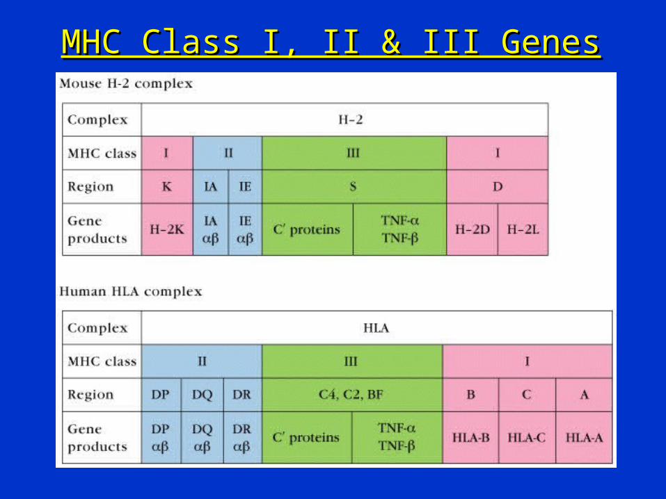
• Genes for HLA proteins are clustered in the MHC complex located on the short arm of chromosome 6
• Three genes HLA-A, HLA-B and HLA-C code for Class I MHC proteins
• HLA-D loci encode for Class II MHC proteins ie, DP, DQ and DR
Major Histocompatibility Complex and Transplantation
• Each individual has two “haplotypes” ie, two sets of these genes one paternal and one maternal
• These genes are very diverse “polymorphic”– 47 HLA-A– 88 HLA-B– 29 HLA-C– More than 300 HLA-D
Major Histocompatibility Complex and Transplantation
• Minor HLA genes – unknown
• They mount a weak immune response
• Play role in chronic rejection of a graft
• There are no laboratory tests to detect minor antigens
• Class III MHC locus – between MHC I & II
• Encode for TNF, lymphotoxin, C2 and C4
MHC Class I, II & III GenesMHC Class I, II & III Genes
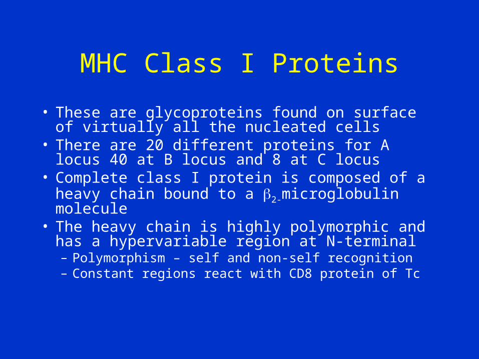
MHC Class I Proteins
• These are glycoproteins found on surface of virtually all the nucleated cells
• There are 20 different proteins for A locus 40 at B locus and 8 at C locus
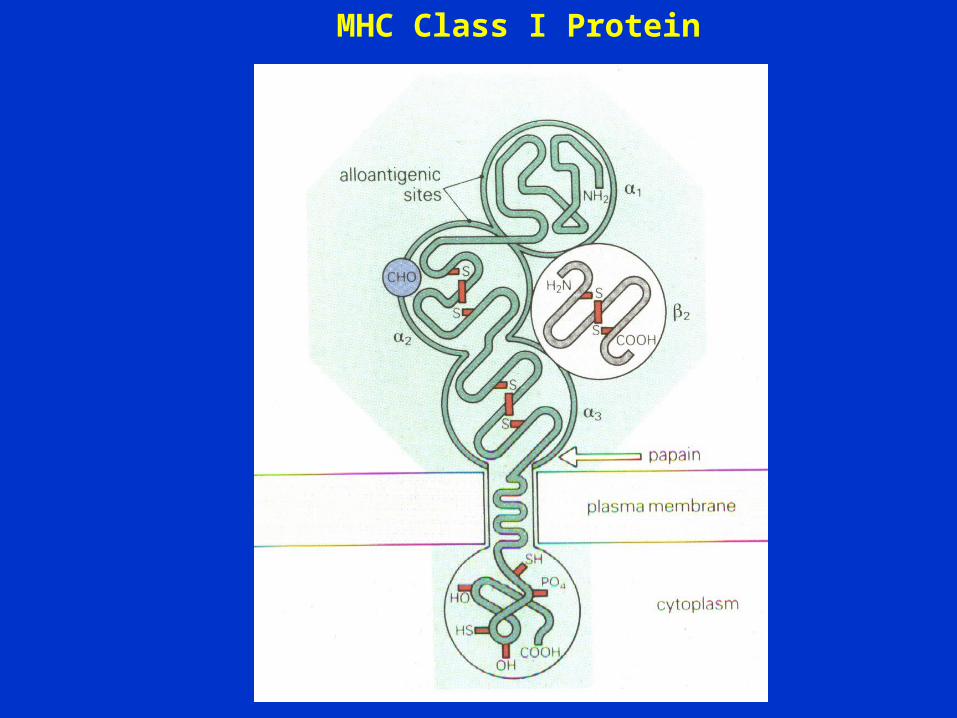
• Complete class I protein is composed of a heavy chain bound to a 2-microglobulin molecule
• The heavy chain is highly polymorphic and has a hypervariable region at N-terminal – Polymorphism – self and non-self recognition– Constant regions react with CD8 protein of Tc
MHC Class I Protein
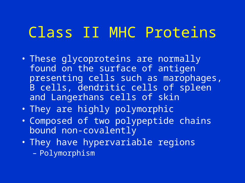
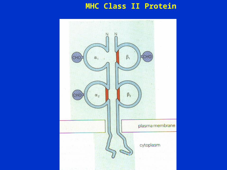
Class II MHC Proteins
• These glycoproteins are normally found on the surface of antigen presenting cells such as marophages, B cells, dendritic cells of spleen and Langerhans cells of skin
• They are highly polymorphic• Composed of two polypeptide chains bound non-
covalently• They have hypervariable regions
– Polymorphism
MHC Class II Protein
Major Histocompatibility Complex and Transplantation
• Both chains of Class II MHC proteins are encoded by the MHC locus
• Constant regions of both the peptides interact with CD4 proteins of helper T cells
Biologic Importance of MHC
• Tc kills virus infected cells in association with class I MHC proteins
• Helper T cell recognize antigen in association with class II MHC proteins
• This is called MHC restriction
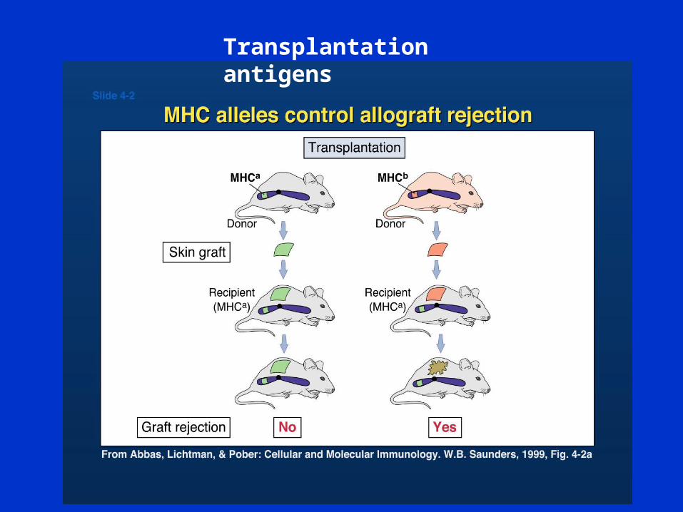
• Success of organ transplant is determined by compatibility of the MHC genes
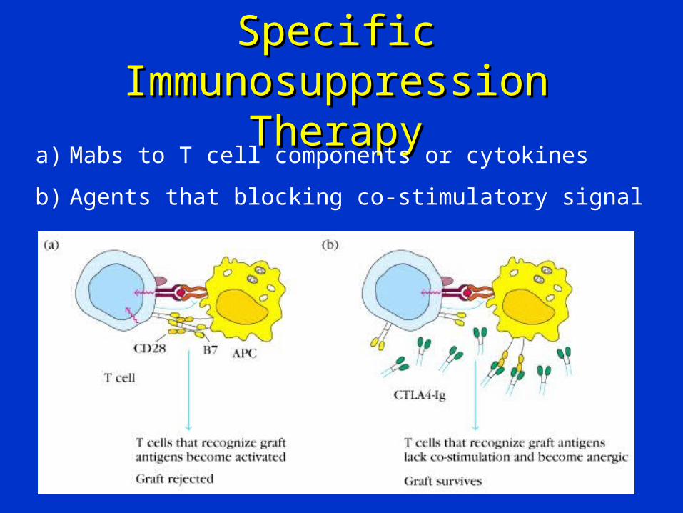
Transplantation antigens
TransplantationTransplantation• Types of transplants:
– Autografts, Autologous grafts• Donor and recipient are same individual• Common in skin grafting; bone marrow
– Syngeneic grafts or (isograft)• Donor and recipient are genetically
identical• Animal models; identical twins
TransplantationTransplantation• Types of transplants:
– Allogeneic grafts• Donor and recipient are same species,
but genetically unrelated• Common heart, lung, kidney, liver graft
– Xenogeneic grafts• Donor and recipient are different species
– Artificial grafts
TransplantationTransplantation• Major Barrier to transplantation is the
immune response– T cells play primary role– B cells can/do play a role– Classic adaptive/acquired immune
response• Memory• Specificity
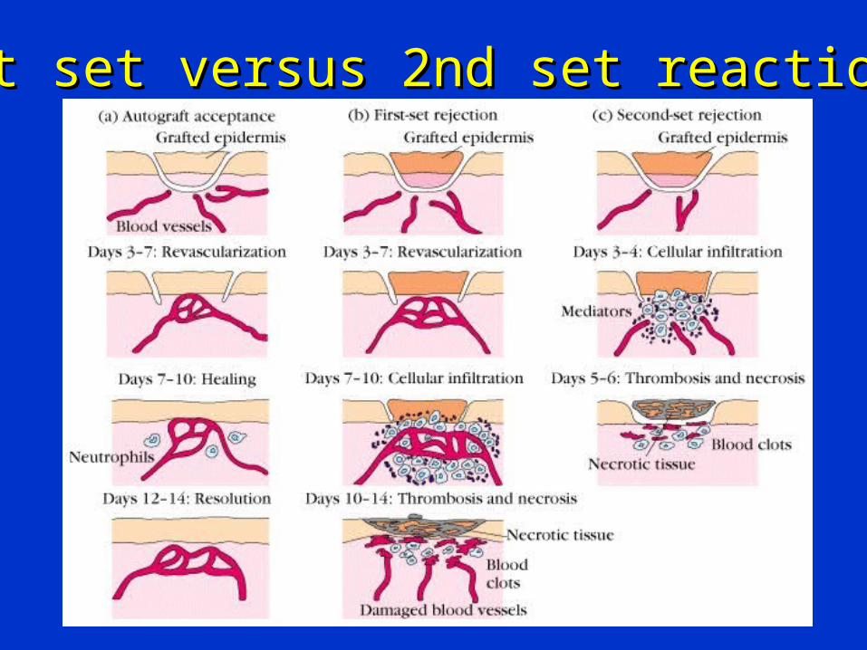
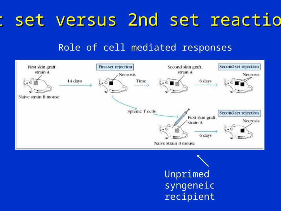
1st set versus 2nd set reactions1st set versus 2nd set reactions
1st set versus 2nd set reactions1st set versus 2nd set reactions
Unprimed syngeneic recipient
Role of cell mediated responses
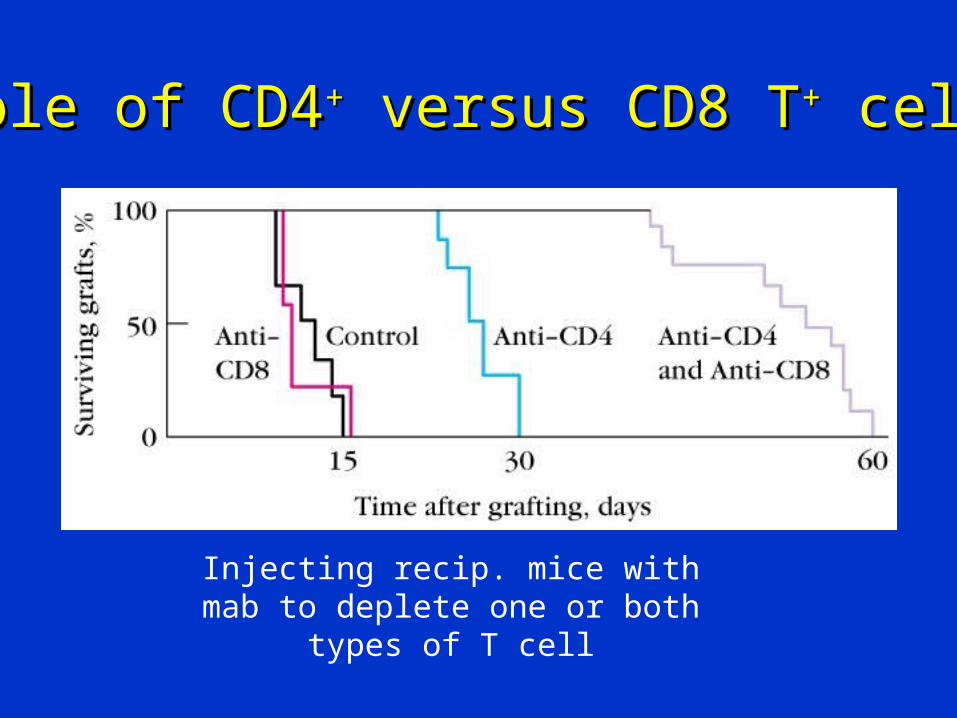
Role of CD4Role of CD4++ versus CD8 T versus CD8 T++ cells cells
Injecting recip. mice with mab to deplete one or both types of T cell
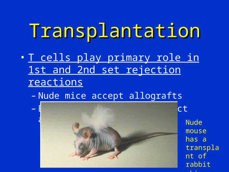
TransplantationTransplantation• T cells play primary role in 1st and 2nd
set rejection reactions– Nude mice accept allografts– B cell deficient mice reject allografts
Nude mouse has a transplant of rabbit skin
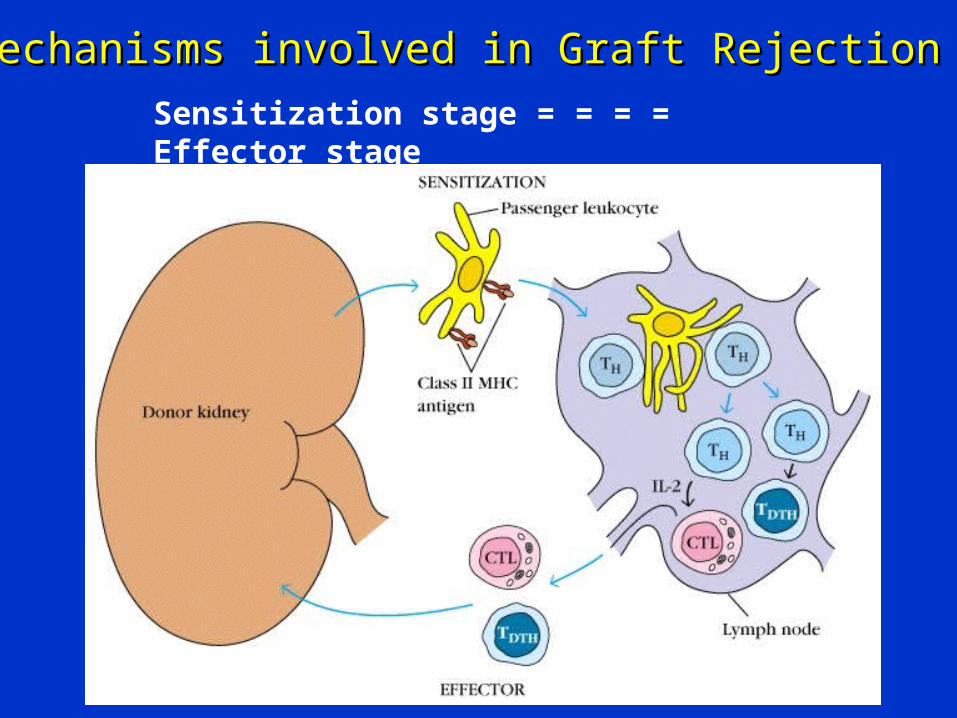
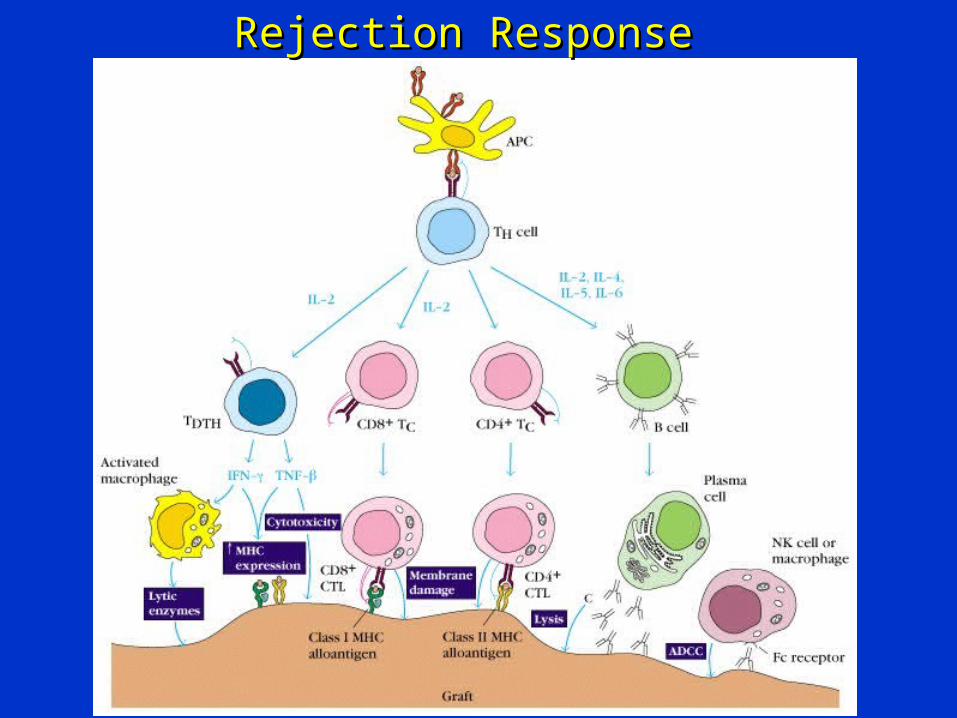
Mechanisms involved in Graft RejectionMechanisms involved in Graft Rejection
Sensitization stage = = = = Effector stage
Rejection ResponseRejection Response
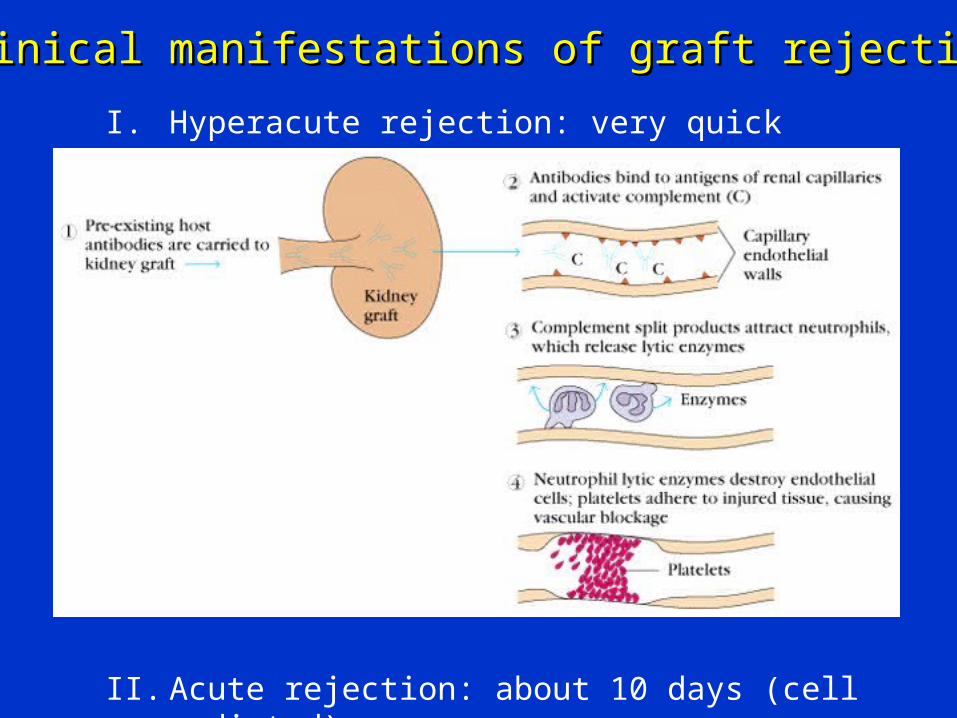
Clinical manifestations of graft rejectionClinical manifestations of graft rejection
I. Hyperacute rejection: very quick
II. Acute rejection: about 10 days (cell mediated)
III. Chronic rejection: months-years (both)
Chronic Rejection
– This occurs months to years after engraftment– Main pathologic finding in chronic rejection is
atherosclerosis of the vascular endothelium– Main cause of chronic rejection is not known
• Minor histocompatibility antigen miss match
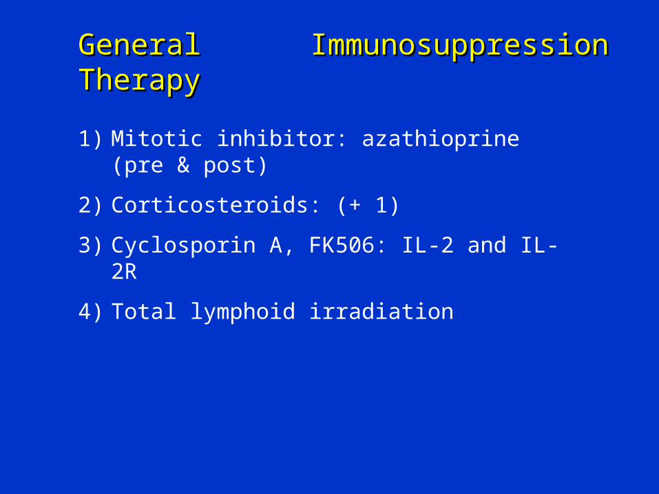
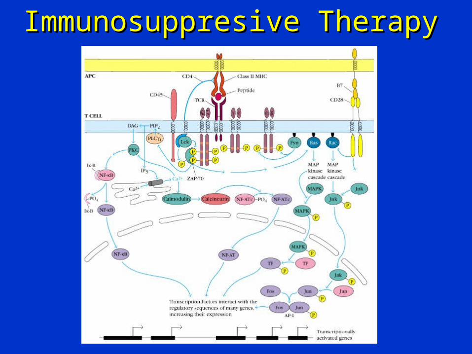
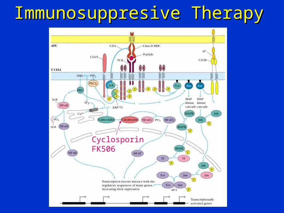
• Side effects of immunosuppressive drugs
Graft-versus-Host (GVH) Reaction
• Occurs in about two thirds of bone marrow transplants• Occurs because grafted immunocompetent T cells
proliferate in the irradiated immunocompromised host and reject cells with foreign proteins resulting in sever organ dysfunction
• Donor’s Tc cells play a major role in destroying the recipient’s cells
• Symptoms are: maculopapular rash, jaundice, hepatosplenomegaly and diarrhea
• GVH reactions usually end in infections and death
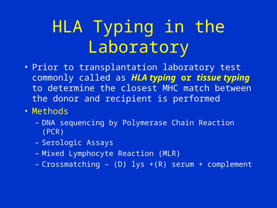
HLA Typing in the Laboratory
• Prior to transplantation laboratory test commonly called as HLA typing or tissue typing to determine the closest MHC match between the donor and recipient is performed
• Methods– DNA sequencing by Polymerase Chain Reaction (PCR)
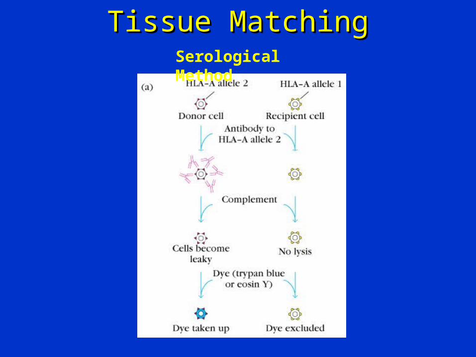
– Serologic Assays
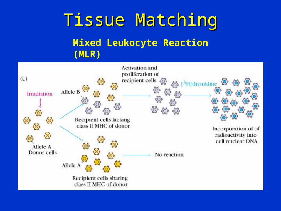
– Mixed Lymphocyte Reaction (MLR)
– Crossmatching – (D) lys +(R) serum + complement
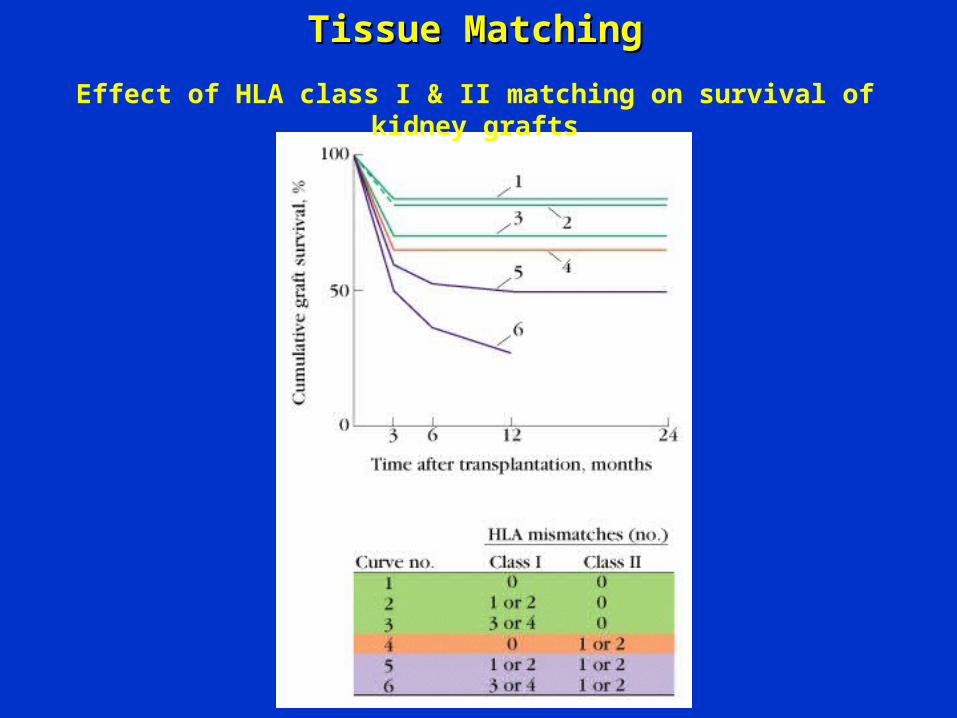
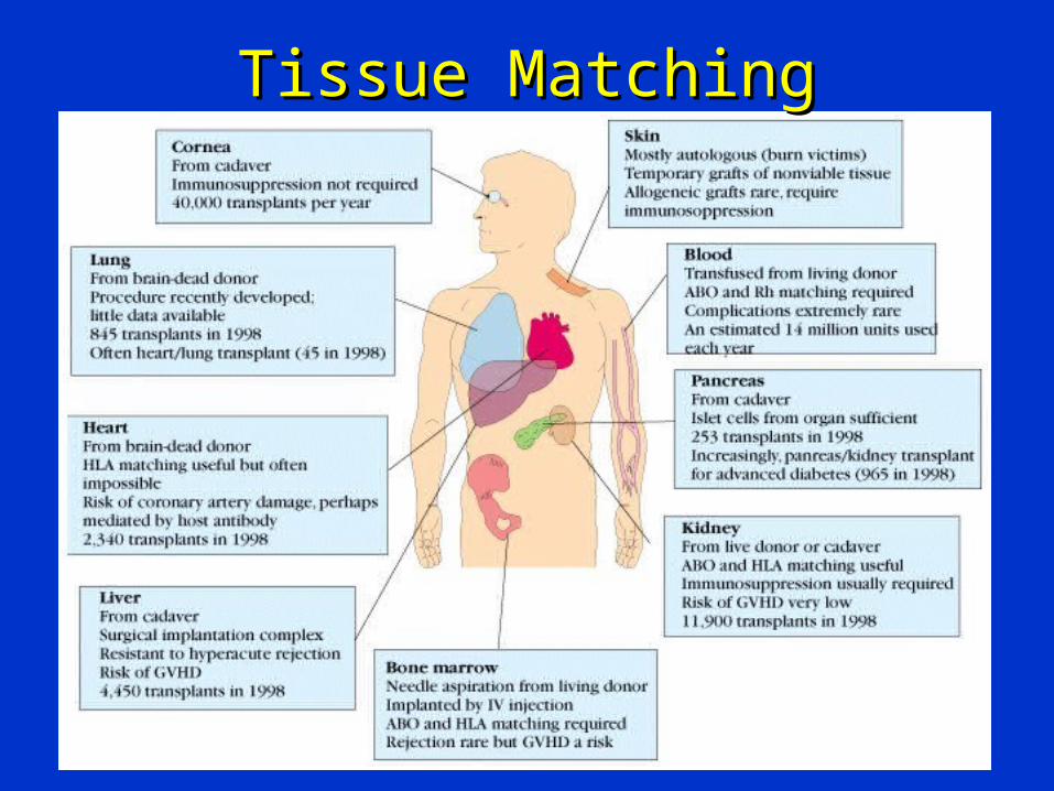
Tissue MatchingTissue Matching
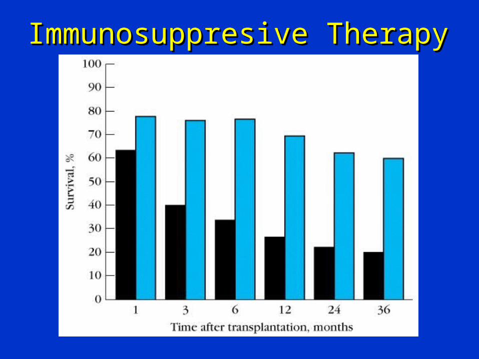
Effect of HLA class I & II matching on survival of kidney grafts