108
Management of the Pediatric Trauma Patient Christopher M. Woleben, M.D. Assistant Professor Emergency Medicine and Pediatrics Associate Dean of Student Affairs VCU School of Medicine

Management of the Pediatric Trauma Patient Christopher M. Woleben, M.D. Assistant Professor Emergency Medicine and Pediatrics Associate Dean of Student Affairs VCU School of Medicine

Objectives • After attending this lecture, you should be able to:
• Discuss developmental stages of infants, toddlers, and children
• Discuss anatomical features of infants and toddlers that may predispose or protect them from certain injuries
• Identify common mechanisms of injury in pediatric patients
• Describe basic management of common injuries seen in pediatric patients
• Identify injuries that may be suspicious for abuse

Epidemiology • Trauma is leading cause of morbidity and mortality in children
• Injuries cause more deaths than all illnesses combined in
children aged 1 to 19: • 40% of deaths age 1 – 4 • 70% of deaths age 5 – 19
• 20% of all pediatric hospitalizations due to injury • 1.5 million injuries per year • 20,000 deaths per year • 500,000 admissions per year • 25 times more ER visits • 30,000 children with injuries experience permanent disabilities
annually
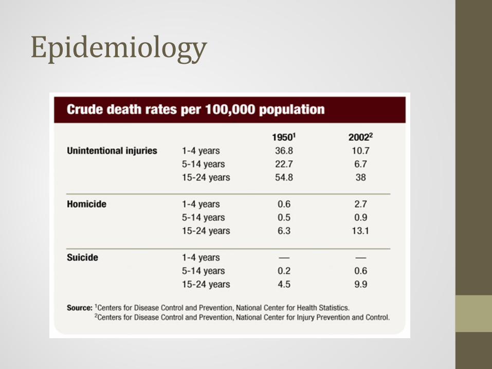
Epidemiology

Developmental Stages • Neonates – birth to one month of age
• May lose 10% body weight in first few days of life
• Should regain birth weight by two weeks of age
• Developmental guidelines based on gestational age at birth
• Key factors to management: • Keep infant warm • Observe skin color, tone, respiratory pattern

Developmental Stages • Infants – 1 to 6 months of age
• Double birth weight by 5 to 6 months of age
• Muscle control develops in cephalo-caudal progression
• Head control at 2 months of age
• Roll over at 4 months of age
• Sit up unassisted by 6 months of age
• Infant – 6 to 12 months of age
• Crawling? • Stand, cruise, walk
• Starting at 9 months of age • Risk for falls
• Stranger anxiety!

Developmental Stages • Toddlers – 1 to 3 years of age
• Great strides in motor development • More curious about their surroundings
• Test their capabilities
• Rapid development of language skills • Can answer simple and specific questions
• Approach slowly and gain confidence • Allow them to hold transitional objects
• Be honest about painful procedures
• Avoid use of dominant hand for IV sticks

Anatomic Considerations • Head –
• Proportionately larger than adult • Occipital region more prominent
• Smaller face, flatter nose
• Greater risk for blunt head trauma
• Fontanelles • Anterior - closes around 12 months of age
• Posterior - closes around 4 months of age
• May become tight and bulging with head trauma or meningitis

Anatomic Considerations • Airway –
• Narrower at all levels • More easily blocked by secretions or obstructions • Cricoid ring is narrowest part of airway
• Infants are obligate nose breathers
• Tongue takes up more proportionate space in children than in adults
• Can more easily obstruct in unconscious patient
• Trachea is softer and more flexible • Can collapse if neck and head are hyper-extended
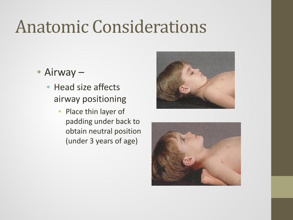
Anatomic Considerations
• Airway – • Head size affects
airway positioning • Place thin layer of
padding under back to obtain neutral position (under 3 years of age)

Anatomic Considerations
• Epiglottis – • Horseshoe shaped
• Extends at 45-degree angle into airway
• Softer cartilage – more floppy

Airway • Formula for ET Tube Choice:
(Age / 4 ) + 4
• Cuffed ET tubes preferred
• Laryngoscope • Straight blade better suited for younger children and infants
• Displaces relatively large tongue
• Better visualization of relatively cephalad and anterior glottis
• Likely to induce a vagal response

Anatomic Considerations • Chest / Lungs -
• Soft, pliable ribs offer less protection to organs • Pulmonary contusions common
• Rib fractures occur less commonly
• Rib fractures raise suspicion of abuse
• Chest muscles tire more easily • Infants are diaphragmatic breathers

Anatomic Considerations • Cardiovascular System –
• Vigorous but limited cardiovascular reserve
• Increase heart rate to maintain cardiac output
• Circulating blood volume is proportionately larger in infants but absolute blood volume is smaller • Can maintain blood pressure longer than an adult but still be
at risk for shock
• Hypotension is a late sign of shock • Tissue perfusion is a better indicator of impending shock in
infants and children
Anatomic Considerations • Abdomen –
• Liver and spleen are proportionately larger than in adults • Vascular organs
• Source of blood loss
• Extremities – • Softer, more porous bones
• Treat sprains as fractures with immobilization
• Injuries to growth plate may disrupt bone growth

Anatomic Considerations • Skin –
• Skin is thinner than adult skin with less subcutaneous fat
• Larger body surface area : weight ratio • Greater risk for injury in extremes of temperature
• Lose fluids and heat more quickly
• Burn more easily and deeply than adults
• Skin may appear mottled due environmental ambient temperature
Anatomic Considerations • Nervous System –
• Continually developing during early childhood
• Brain tissue is less myelinated • Skull is thinner • Subarachnoid is shallow and accumulated fluid causes
damage sooner
• 80% of young children dying from trauma have a significant head injury

Normal Vital Signs - Pulse
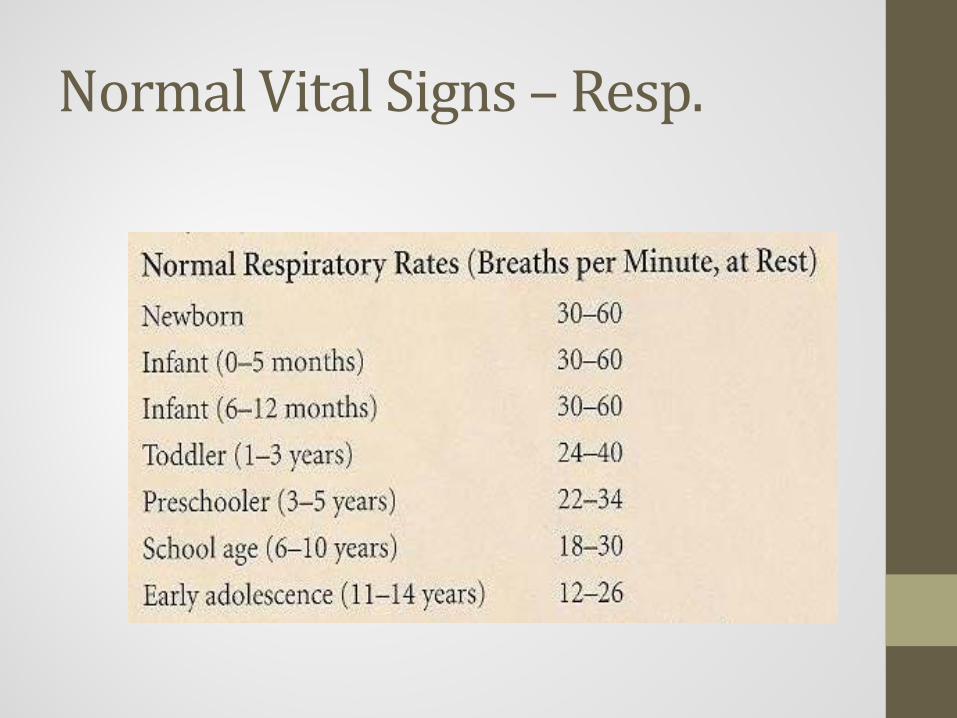
Normal Vital Signs – Resp.

Normal Vital Signs - BP

“Golden Hour of Care”
• Advanced Trauma Life Support
• Also known as Early Management of Severe Trauma (EMST)
• Standard of care for initial assessment and treatment of trauma patients in trauma centers

Pediatric Trauma Score • Developed to reflect the children’s vulnerability to traumatic
injury. • The minimal score is -6 and the maximum score is +12.
• Emphasizes the importance of the child’s weight and airway.
• Several studies have confirmed that the PTS is a valid tool in
predicting mortality of a traumatically injured child. • Mortality is estimated at 9% with a PTS > 8 • Mortality close to 100% with a PTS ≤ 0. • There is a linear relationship between the decrease in PTS and the
mortality risk (i.e. the lower the PTS, the higher the mortality risk)

Pediatric Trauma Score Variable +2 +1 -1
Weight > 20 kg 10-20 kg <10 kg
Airway Patent Maintainable Not Maintainable
Systolic BP > 90 mm Hg 50 – 90 mm Hg < 50 mm Hg
CNS Awake + LOC Unresponsive
Fractures None Closed / Suspected
Multiple Closed / Open
Wounds None Minor Major, Penetrating, or Burns
Primary Survey and Resuscitation • Airway with Cervical Spine Protection
• Assessment • Ascertain patency of airway
• Rapidly assess for airway obstruction
• Management • Chin Lift or Jaw Thrust
• Clear airway of foreign bodies
• Insert oro- or nasopharyngeal airway

Primary Survey and Resuscitation
• Airway with Cervical Spine Protection • Establish definitive airway
• Oro- or nasotracheal airway
• Surgical cricothyroidotomy
• Maintain cervical spine in neutral position with manual immobilization as necessary when establishing airway
• Reinstate immobilization of c-spine with appropriate device after establishing airway

Primary Survey and Resuscitation • Breathing: Ventilation and Oxygenation
• Assessment • Expose neck and chest
• Assure immobilization of head and neck
• Determine depth and rate of respirations
• Inspect and palpate neck and chest for tracheal deviation, unilateral and bilateral chest movement, use of accessory muscles, any signs of injury
• Percuss chest for dullness or hyper-resonance
• Auscultate chest bilaterally

Primary Survey and Resuscitation • Breathing: Ventilation and Oxygenation
• Management • Administer high concentration oxygen
• Ventilate with bag-valve-mask device
• Alleviate tension pneumothorax
• Seal open pneumothorax
• Use end-tidal CO2 monitoring device in conjunction with pulse oximetry

Primary Survey and Resuscitation • Circulation with Hemorrhage Control
• Assessment • Identify source of external, exsanguinating hemorrhage
• Identify potential sources of internal hemorrhage
• Pulse (quality, rate, regularity, paradoxical)
• Skin color
• Decrease in blood pressure is a late finding in pediatric shock
Primary Survey and Resuscitation • Circulation with Hemorrhage Control
• Management • Apply direct pressure to external bleeding site
• Consider presence of internal hemorrhage and potential need for operative management
• Insert 2 large-caliber IV catheters
• IV hydration with warm LR/NS or blood replacement
• Prevent hypothermia
Primary Survey and Resuscitation • Disability: Brief Neurologic Examination
• Determine level of consciousness
• AVPU method
• A = Alert at baseline • V = Verbal stimuli • P = Painful stimuli • U = Unresponsive
• GCS score (score ranges from 3 to 15)
• Severe (GCS ≤ 8) • Moderate (GCS 9 – 12) • Minor (GCS ≥ 13)
• Assess pupils for size, equality, reactivity

Pediatric Glasgow Coma Score: Eye Opening
Age > 2yrs Score Age < 2 yrs
Spontaneous 4 Spontaneous
To Voice 3 To Speech
To Pain 2 To Pain
None 1 None
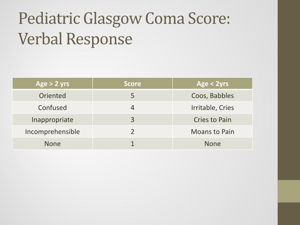
Pediatric Glasgow Coma Score: Verbal Response
Age > 2 yrs Score Age < 2yrs
Oriented 5 Coos, Babbles
Confused 4 Irritable, Cries
Inappropriate 3 Cries to Pain
Incomprehensible 2 Moans to Pain
None 1 None
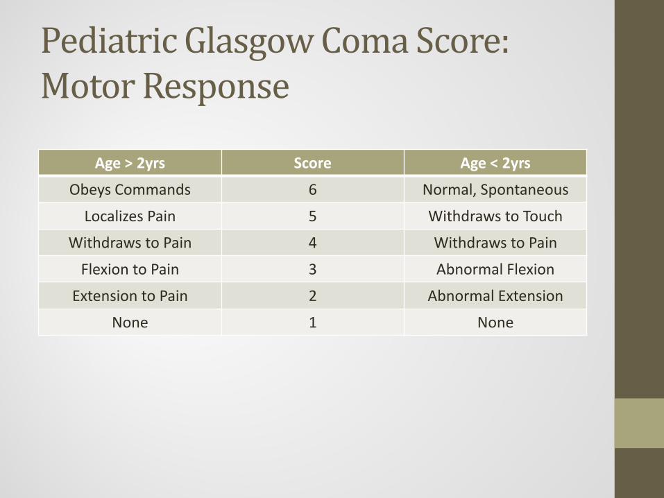
Pediatric Glasgow Coma Score: Motor Response
Age > 2yrs Score Age < 2yrs
Obeys Commands 6 Normal, Spontaneous
Localizes Pain 5 Withdraws to Touch
Withdraws to Pain 4 Withdraws to Pain
Flexion to Pain 3 Abnormal Flexion
Extension to Pain 2 Abnormal Extension
None 1 None

Primary Survey and Resuscitation • Environment / Exposure
• Completely undress patient
• Prevent hypothermia
• Adjuncts to Primary Survey • Urinary and gastric catheters (unless contraindicated)
• Monitor hourly urine output
• Consider need for radiographic studies • Chest x-ray
• Lateral Cross-table Cervical-Spine x-ray
• Pelvis x-ray

Secondary Survey • Obtain AMPLE history
• A = Allergies
• M = Medications
• P = Past Medical History
• L = Last Meal
• E = Events Leading to Injury
• Thorough systematic physical exam

Secondary Survey • Head and Maxillofacial
• Inspect and palpate entire head and face for lacerations, contusions, fractures
• Re-evaluate pupils, level of consciousness • Assess eyes for hemorrhage, penetrating injury, visual
acuity, presence of contact lens • Evaluate cranial nerve function • Inspect ears and nose for CSF leakage • Inspect mouth for evidence of bleeding, soft-tissue
lacerations, dentition

Secondary Survey • Cervical Spine and Neck
• Inspect for signs of blunt trauma or penetrating injury, tracheal deviation, use of accessory muscles
• Palpate for tenderness, deformity, swelling, subcutaneous emphysema, tracheal deviation, symmetry of pulses
• Auscultate carotids for bruits
Management of C-Spine • Any time an infant or child sustains a significant
head injury, assume a neck injury may also be present • Children should remain immobilized in a cervical collar
until cleared by hospital personnel • Multiple devices can be utilized • Try to have family member or caretaker nearby to help
keep the child calm • Maintain supine, neutral, in-line position by placing
padding from shoulders to hips

Utilization of Car Seat for Immobilization

Utilization of Car Seat for Immobilization

Secondary Survey • Chest
• Inspect chest wall for signs of blunt or penetrating trauma, use of accessory muscles
• Auscultate for breath and cardiac sounds
• Palpate entire chest wall for evidence of blunt or penetrating trauma, subcutaneous emphysema, tenderness, crepitus
• Percuss for evidence of hyper-resonance or dullness

Secondary Survey • Abdomen
• Inspect anterior and posterior abdomen for signs of blunt or penetrating trauma or ecchymosis
• Auscultate for presence or absence of bowel sounds
• Percuss to assess for subtle rebound tenderness
• Palpate for tenderness, involuntary guarding, rebound tenderness

Secondary Survey • Genitourinary / Rectal
• Inspect for contusions, hematomas, lacerations, urethral bleeding
• Rectal exam to assess for gross blood, anal sphincter tone, bowel wall integrity, bony fragments
Secondary Survey • Adjuncts to secondary survey:
• Lab tests • Complete Blood Count (CBC) • Coagulopathy Panel (PT, APTT, INR) • Basic Metabolic Panel (BMP) • Liver / Pancreatic Function Tests (AST, ALT, ALP,
Amylase, Lipase) • Venous Blood Gas (VBG and Lactate) • Blood Type and Screen (STBB) • Urinalysis (UA) • Urine Pregnancy (UHcG)

Hepatic Trans-aminase Abnormalities (Hennes et al in Pediatrics, July 1990)
• AST>450 and ALT> 250 predictive of liver injury identifiable
with abdominal imaging • Degree of elevation of levels does not correlate with degree of
injury or outcome
• Hemodynamically stable pediatric patients with blunt
abdominal trauma and AST>450, ALT>250 should undergo abdominal CT

Laboratory Values in Trauma Capraro et al in Pediatric Emergency Care (July 2006)
• Retrospective study of 83 children with CT-confirmed abdominal injuries
• Abnormal lab values: • Glucose (67%) • AST (47%) • Urinalysis (43%) • WBC (43%)
• Glucose (75%) and AST (63%) had highest sensitivity • Lipase (75%) had highest positive predictive value • AST (71%) had lowest negative predictive value
• No single test had excellent sensitivity, specificity, PPV, NPV

Hematuria in Trauma Capraro et al in Pediatric Emergency Care (July 2006)
• Microscopic hematuria common in splenic (11%),
liver (10%), and renal (8%) injury
• Gross hematuria more common in renal injuries (22%) but also seen with splenic (17%) and liver (8%) injuries • Can indicate trauma at any level in the GU system • Can be absent in up to 50% of patients with renal
pedicle injury • May be traumatic from Foley catheter placement

Lab Values: Take Home Message
• Controversy exists over utility of labs to detect abdominal injury: • 1999 – Holmes et al – abdominal abrasions and
abnormal chest exam combined with microscopic hematuria, elevated transaminases, leukocytosis
• 2002 – Holmes et al – six factors associated with intra-abdominal injury: • Low systolic BP, abdominal tenderness, concomitant
femur fracture, elevated AST/ALT, microscopic hematuria and HCT<30
• 2005 – Sokolove et al – seat belt sign associated with 3-fold increase in intra-abdominal injury; 13-fold increase in gastrointestinal tract injury

Secondary Survey • Consider FAST exam • Consider CT of Head, Cervical Spine, Chest,
Abdomen/Pelvis • Consider contrast urography • Consider angiography • Consider extremity, thoracic and lumbar
spine films • Consider bronchoscopy, esophagoscopy

FAST (Focused Assessment with Sonography of Trauma) • Quick ultrasound examination of the
peritoneal and abdominal cavities • This can typically be completed in 3-5 minutes • Performed during the resuscitation or secondary
survey of the trauma patient • The basic FAST examination includes 4 views:
• Right upper quadrant (Peri-hepatic or Morison's pouch) • Left upper quadrant (Peri-splenic) • Pelvic • Cardiac
FAST (Focused Assessment with Sonography of Trauma) • Advantages
• Rapid noninvasive procedure that can be performed at the bedside
• Easily repeatable and involves the use of no radiation or contrast
• Disadvantages • Cannot localize the source of bleeding • Not sensitive for most solid organ injuries during rapid exam • Insensitive for retroperitoneal injuries • Insensitive for injuries to hollow viscera
FAST (Focused Assessment with Sonography of Trauma) • Normal
• Normal sonographic appearance
• Abnormal • Free intraperitoneal or pericardial fluid • Disruption of the normal sonographic appearance of solid organs
• Cause of abnormal result
• Intra-abdominal free fluid • Hemoperitoneum • Perforated viscous • Ascites • Pericardial fluid • Hemopericardium • Pre-existing malignancy or pericarditis
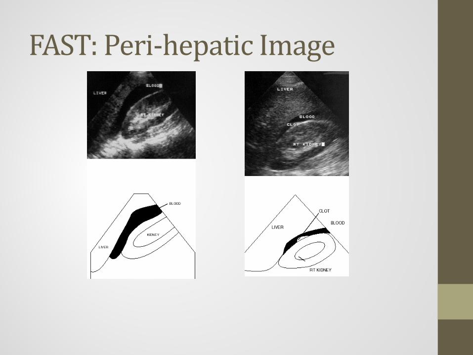
FAST: Peri-hepatic Image

FAST: Peri-Splenic View

FAST: Pelvic View
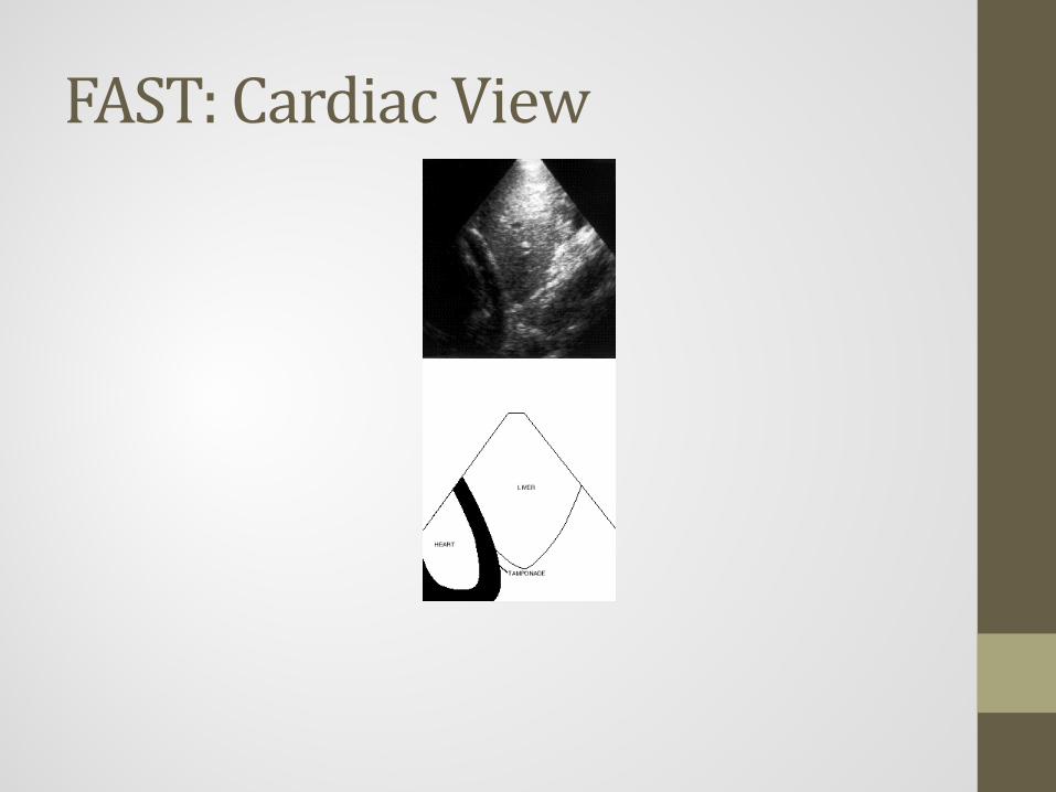
FAST: Cardiac View

Utility of FAST Holmes et al in Journal of Pediatric Surgery (September 2007)
• Recent meta-analysis of 25 articles evaluating use of FAST in 3800 pediatric patients: • Modest sensitivity (66%) for detection of children
with hemoperitoneum • Specificity close to 95%
• Negative FAST has questionable utility as sole diagnostic test to rule out presence of intra-abdominal injury

Blunt Trauma • Thinner body wall allows forces to be readily
transmitted to body contents • Predominant cause of injury in children • Motor vehicle crashes are leading cause of death
and serious injury in children • Leading cause of permanent brain injury and new
onset epilepsy

Motor Vehicle Accidents • 1/3 of childhood deaths results from motor
vehicle accidents • Leading cause of traumatic death
• Leading cause of permanent brain injury and new onset epilepsy
• Improperly seated children at increased risk of injury • EMS prevention strategies can make a difference

Blunt Trauma • Pedestrian Vehicle Crashes
• Particularly lethal cause of trauma
• Initial injury due to impact with vehicle (extremity or trunk)
• Child is thrown from force of impact causing additional injury to head or spine upon impact with other objects

Penetrating Trauma • Becoming an increasing problem in adolescents
• Stab wounds and firearm injuries 10-15% of all trauma admissions
• Higher incidence in inner city (intentional) vs. other areas (unintentional)
• Visual inspection of external injuries cannot evaluate extent of internal injuries

Traumatic Brain Injury (TBI) • Large size of head and weak neck musculature
lead to increased risk of TBI
• Early recognition and aggressive management can reduce mortality and morbidity
• Modified Glasgow Coma Score • Mild = GCS 13-15
• Moderate = GCS 9-12
• Severe = GCS 3-8

Traumatic Brain Injury (TBI) • Diffuse injuries more common in children
• Soft tissues, skull and brain are more compliant
• Due to open fontanelles and sutures, infants up to age 16 months may be more tolerant of increases in ICP • Delayed signs
• Significant blood loss through scalp lacerations • Apply direct pressure

Falls • Single most common cause of injury in children
• Serious injury or death more common with falls
from greater height
• Falls from bicycles account for significant number of injuries • Decreased number of head injuries due to bicycle
safety helmet use
• Often result in facial lacerations or hematomas
• Fractures!!!
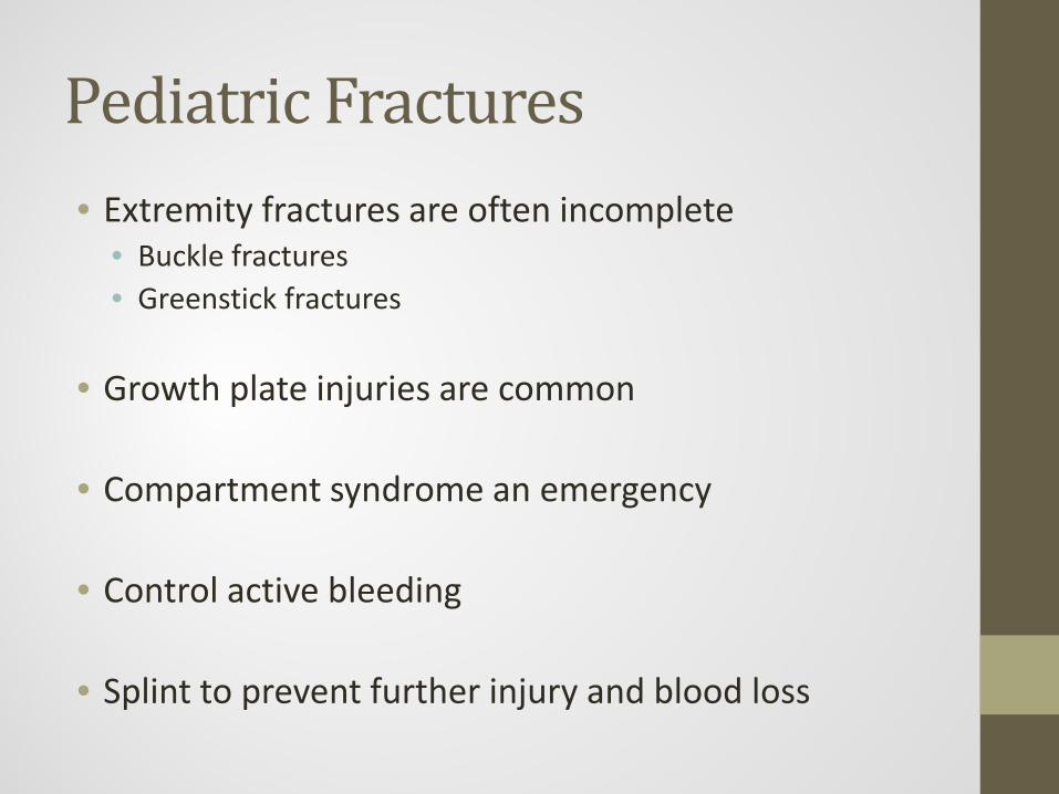
Pediatric Fractures • Extremity fractures are often incomplete
• Buckle fractures • Greenstick fractures
• Growth plate injuries are common
• Compartment syndrome an emergency
• Control active bleeding
• Splint to prevent further injury and blood loss

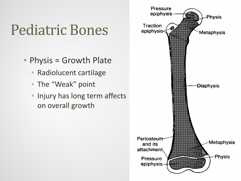
Pediatric Bones
• Epiphysis
• Physis • Cartilage
• Metaphysis
• Diaphysis

• Growth from the ends
• Periosteum very thick and active • Usually stays intact
• Aids in callus formation
• Growth centers • Often multiple
Pediatric Bones
Pediatric Bones
• Physis = Growth Plate • Radiolucent cartilage
• The “Weak” point
• Injury has long term affects on overall growth

Pediatric Fractures • Physical Exam Findings Include:
• Gross deformity
• Bony / joint instability
• Crepitus
• Point tenderness
• Activity not routine
• Painful
• Loss of function

Transverse Fractures • Fracture line perpendicular to long axis of the bone
• Mechanism of injury: • Direct force
• Stretch beyond its limit
• &/or Opposing forces

Transverse Fractures

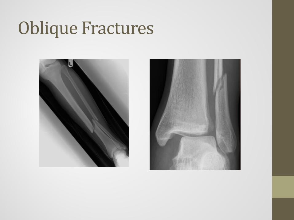
Oblique Fractures • Fracture line < 90° to long axis of bone
• Parallel ⇒ Longitudinal fracture
• Mechanism of injury: • Compression forces
• Tangential forces (?)
• &/or Opposing forces
Oblique Fractures
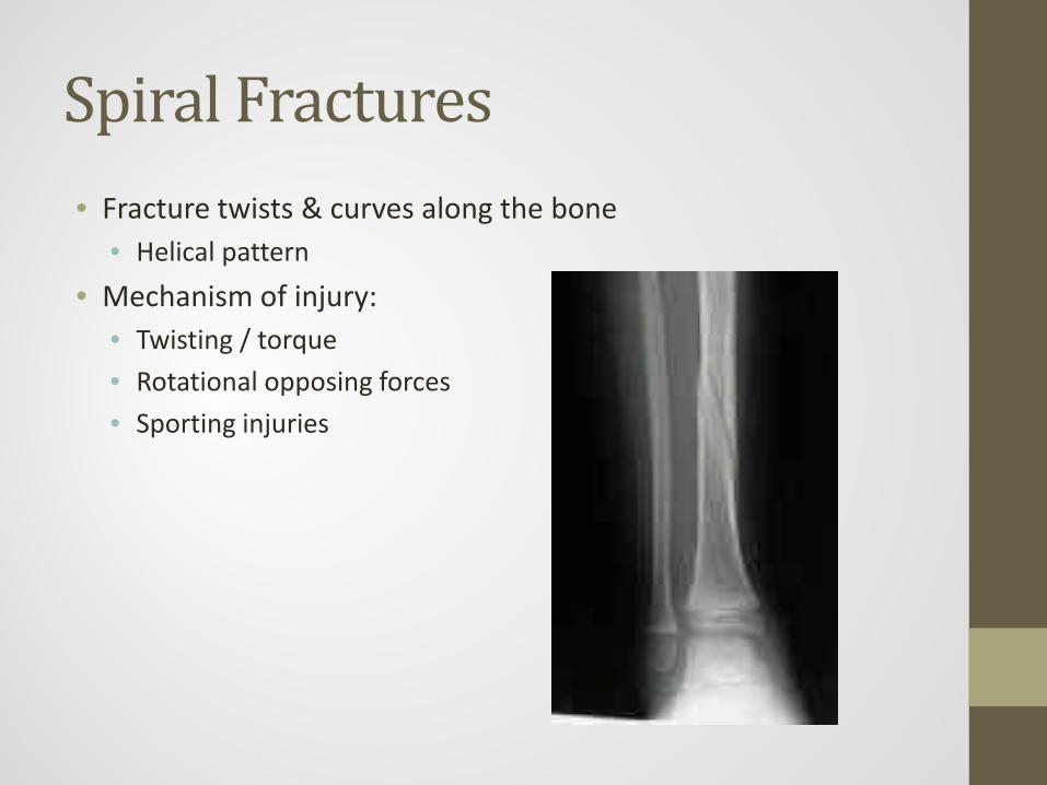
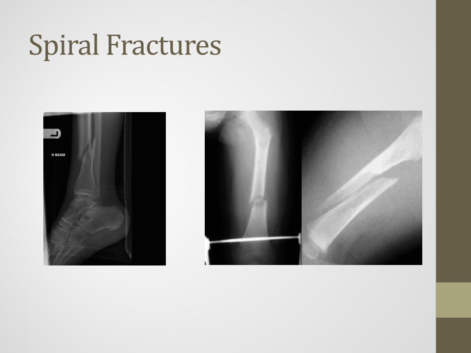
Spiral Fractures • Fracture twists & curves along the bone
• Helical pattern
• Mechanism of injury: • Twisting / torque
• Rotational opposing forces
• Sporting injuries
Spiral Fractures

Comminuted Fractures • > 1 Fracture line
• At least one or more separate bone fragment(s)
• Can lack stability
• Mechanism of injury: • High impact force
• Crush injuries

Torus Fractures
• Very common • Porous bone buckles
• AKA Buckle Fracture • No break in cortex • No angulation
• Stable Fracture • MOI: Axial load force
• FOOSH • Longitudinal compression force

Torus Fracture

Torus Fractures

• Incomplete Fx • Intact periosteum one side
• Very common Pediatric Fx • 50% prior to 12yo
• MOI: Angulated forces • Applied & released force
• FOOSH
• Tx by completing Fx & reduction
Greenstick Fracture

Greenstick Fractures

Bowing Fracture
• Due to porosity of bone • Force not enough to Fx
• Bone bends or bows with no obvious break in cortex
• Common with adjacent Fx • Functional & cosmetic deficits • MOI: Axial load
• FOOSH • Longitudinal compression/stress

Bowing Fracture

Bowing and Greenstick Fractures

Physeal Fractures • Unique pediatric fractures
• Involving physis/growth plate
• More common in pre-adolescents
• Peak at 11–12 years
• Majority involve the radius and ulna
• Salter-Harris Classification

Physeal Fractures

• Fracture thru the physis • Physis may slip
• Difficult to diagnose if non-displaced • Over laying tenderness • +/- findings on x-ray
• Minimal growth disturbance if near anatomic position
Salter-Harris Type I

Salter-Harris Type I

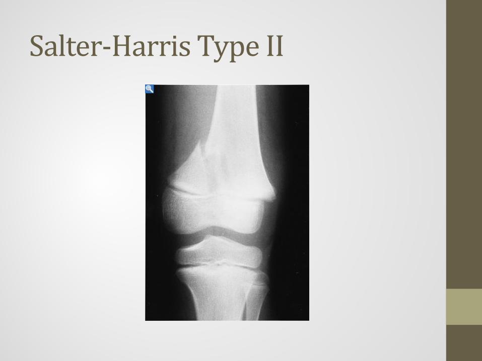
Salter-Harris Type II
• Fracture thru Physis & Metaphysis • Shearing off fragment of
metaphysis • Most commonly
diagnosed physeal fracture
• Conservative management if not displaced
• Normal growth if aligned
Salter-Harris Type II

Salter-Harris Type III
• Fracture thru Physis & Epiphysis
• Intra-articular surface involvement
• May displace along the line of the epiphysis
• Unstable Fracture • Growth disturbance common • Requires orthopedic repair &
follow up

Salter-Harris Type III

Salter-Harris Type IV
• Fracture thru Metaphysis Physis & Epiphysis
• Intra-articular surface involvement
• Surgery to stabilize fragment
• Frequent growth disturbance
• Many long term complications

Salter-Harris Type IV

Salter-Harris Type V
• Compression of growth plate • No obvious Fracture,
angulation or displacement • Retrospective diagnosis • Often missed diagnosis as
SH-I • Significant growth
disturbance or arrest • Long term complications

Salter-Harris Type V

Nursemaid’s Elbow • Subluxation of the radial head
• Usually caused by pulling of arms • Child refuses to move one arm
• Physical exam • Arm is extended and pronated • Child cries with attempts to move arm
• Treatment • Hold the hand, supinate the arm, flex the arm while
applying pressure over radial head with thumb
Child Abuse • Second leading cause of death in infants less
than 6 months of age • About 5000 children die per year as a result of abuse
or neglect
• Forms of Abuse: • Psychological / Emotional
• Physical
• Sexual
• Neglect (physical or emotional)

Child Abuse • Characteristics of Child:
• Premature infants and twins
• Age less than 5 years
• Physical or mental handicaps
• Boys more common than girls
• Unwanted, unexpected children
• Characteristic of Abuser: • Usually a parent or full-time caregiver
• Many were abused themselves as children
• Displays hostile or evasive behavior
• Presence of a crisis (financial stress, marital or relationship problems, illness)

Child Abuse • Red Flags for Abuse:
• Obvious or suspected fracture in child less than 2 years of age
• Multiple injuries in various stages of healing
• Bruises or burns in patterns or locations that suggest intentional infliction
• Injury does not fit with the description of the cause given
• Head injuries or abdominal injuries in infants

Child Abuse • Red Flags in the History:
• History does not match nature or severity of injury • Vague parental accounts or accounts that change
during the interview • Accusations that the child injured himself intentionally • Delay in seeking help • Child dressed inappropriately • Revealing comments by bystanders or siblings

Burns • Leading cause of accidental death in the home
for children under age 14 • Survival a function of burn size and concomitant
injuries • Unsupervised children playing with matches • Stress proper use of smoke detectors and
changing batteries frequently

Burns • Chemical, thermal, or electrical • Smoke Inhalation
• Look for singed nasal hairs, changes in cry or voice
• Scald burns • Pull hot water off table • Immersion in water (abuse?)
• Calculation of Body Surface Area • “Rule of Palms” versus “Rule of Nines”

Modified Rule of Nines

Burns - Management • Prompt management of airway
• Swelling can develop rapidly • Consider use of smaller endotracheal tubes
• Maintain body temperature • Pain control • IV access – good rule of thumb is to start IV hydration at
maintenance • Will calculate fluid needs based on BSA involved once in ED – DO
NOT over-hydrate!
• Suspect C-spine injuries with serious electrical burns
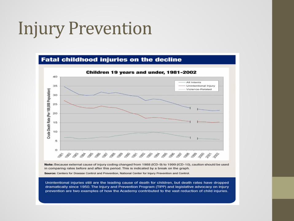
Injury Prevention

Injury Prevention • As EMS providers, you can take an active role in
injury prevention education: • School or community programs
• Specific talks aimed at age-appropriate interventions • Speak at career days
• Anticipatory guidance • Educate care-givers about child-proofing home or
neighborhood
• Offer safety inspections • Bike helmets • Car seats


![Trauma Reach Workshop - Pediatric Trauma.pptx [Read-Only]...Pediatric Trauma Trauma REACH Workshop May 5th, 2015 Tamer A. Ahmed, MD Pediatric Trauma Medical Director Upstate’s GolisanoChildren’s](https://static.documents.pub/doc/80x56/5fe9ec9ba1b3915c9800251e/trauma-reach-workshop-pediatric-read-only-pediatric-trauma-trauma-reach.jpg)


