209 Marginal and internal fits of zirconia single crown and Four-unit Fixed Dental Prostheses Substructures Kulnaree Charukietkajorn, Chuchai Anunmana Kulnaree Charukietkajorn 1 , Chuchai Anunmana 2 1 Neodent Dental Center, Bangkok. 2 Department of Prosthodontics, Faculty of Dentistry, Mahidol University. Marginal and internal fits of zirconia single crown and Four-unit Fixed Dental Prostheses Substructures Corresponding author: Chuchai Anunmana Department of Prosthodontics, Faculty of Dentistry, Mahidol University 6 Yothi st., Rajthevi, Bangkok, 10400 Thailand Tel. (66)-2-2007817 Fax: (66)-2-2007816 e-mail address: [email protected]Received: 21 April 2015 Accepted: 30 September 2015 Abstract Purpose: To compare marginal and internal gaps of zirconia single crown substructures with those of four-unit fixed dental prostheses substructures. Materials and Methods: A standard Co-Cr model of maxillary first premolar and maxillary second molar abutments was made. Twenty polyether impressions of the standard model were taken and subsequently poured with type-IV dental stone. Zirconia substructures were fabricated as premolar and molar single crowns (Cr, n=10) and four-unit bridges (Br, n=10) using LAVA™ Zirconia system. The gaps between substructures and their standard abutments were evaluated using silicone replica technique. Twenty-two different points were measured along the marginal and internal surfaces. The differences of the gap widths between single crown and four-unit bridge substructures were analyzed using Mann Whitney U-test (α=0.05). Results: There was significant difference of marginal gap between Cr and Br groups at mesial marginal gap of molar (30±44 and 112±19 µm for Cr and Br, respectively). For the internal gaps, mid-occlusal point revealed the largest gap. No significant difference of internal gaps between Cr and Br groups was observed except at some points of mesial chamfer area of molar (123±18 and 218±33 µm for Cr and Br, respectively) and at buccal axial wall of premolar (45±15 and 76±47 µm for Cr and Br, respectively). Conclusions: The differences between the gaps of zirconia single crown and those of four-unit bridge substructures were found at the mesial side of molar marginal area. Most of internal gaps of single crown substructures did not significantly differ from those of four-unit bridge substructures. Key words: CAD-CAM, Gap, Zirconia, Fixed dental prostheses, Crown, Bridge, LAVA How to cite: Charukietkajorn K, Anunmana C. Marginal and internal fits of zirconia single crown and four-unit fixed dental prostheses substructures. M Dent J 2015; 35: 209-19. Dental Journal Original Article Mahidol Dental Journal
Transcript
209Marginal and internal fits of zirconia single crown and Four-unit Fixed Dental Prostheses Substructures Kulnaree Charukietkajorn, Chuchai Anunmana
Kulnaree Charukietkajorn1, Chuchai Anunmana2
1 Neodent Dental Center, Bangkok. 2 Department of Prosthodontics, Faculty of Dentistry, Mahidol University.
Marginal and internal fits of zirconia single crown and Four-unit Fixed Dental Prostheses Substructures
Corresponding author: Chuchai Anunmana Department of Prosthodontics, Faculty of Dentistry, Mahidol University 6 Yothi st., Rajthevi, Bangkok, 10400 Thailand Tel. (66)-2-2007817 Fax: (66)-2-2007816 e-mail address: [email protected] Received: 21 April 2015 Accepted: 30 September 2015
AbstractPurpose: To compare marginal and internal gaps of zirconia single crown substructures with those of four-unit fixed dental prostheses substructures.
Materials and Methods: A standard Co-Cr model of maxillary first premolar and maxillary second molar abutments was made. Twenty polyether impressions of the standard model were taken and subsequently poured with type-IV dental stone. Zirconia substructures were fabricated as premolar and molar single crowns (Cr, n=10) and four-unit bridges (Br, n=10) using LAVA™ Zirconia system. The gaps between substructures and their standard abutments were evaluated using silicone replica technique. Twenty-two different points were measured along the marginal and internal surfaces. The differences of the gap widths between single crown and four-unit bridge substructures were analyzed using Mann Whitney U-test (α=0.05).
Results: There was significant difference of marginal gap between Cr and Br groups at mesial marginal gap of molar (30±44 and 112±19 µm for Cr and Br, respectively). For the internal gaps, mid-occlusal point revealed the largest gap. No significant difference of internal gaps between Cr and Br groups was observed except at some points of mesial chamfer area of molar (123±18 and 218±33 µm for Cr and Br, respectively) and at buccal axial wall of premolar (45±15 and 76±47 µm for Cr and Br, respectively).
Conclusions: The differences between the gaps of zirconia single crown and those of four-unit bridge substructures were found at the mesial side of molar marginal area. Most of internal gaps of single crown substructures did not significantly differ from those of four-unit bridge substructures.
How to cite: Charukietkajorn K, Anunmana C. Marginal and internal fits of zirconia single crown and four-unit fixed dental prostheses substructures. M Dent J 2015; 35: 209-19.
Dental Journal Original ArticleMahidol Dental Journal
210 Marginal and internal fits of zirconia single crown and Four-unit Fixed Dental Prostheses Substructures Marginal and internal fits of zirconia single crown and Four-unit Fixed Dental Prostheses Substructures Kulnaree Charukietkajorn, Chuchai Anunmana
M Dent J Volume 35 Number 3 September-December 2015
Marginal and internal fits of zirconia single crown and Four-unit Fixed Dental Prostheses Substructures
Introduction All-ceramic restorations have recently become increasingly popular because of patient’s esthetic expectation. These restorations do not have metals underneath; therefore they look more natural. Nevertheless, only anterior fixed dental prostheses were initially indicated due to its unfavorable mechanical properties. In the past decades, high strength polycrystalline ceramics have been developed as the substructure for posterior fixed dental prostheses (FDPs).1 A variety of ceramic substructures have been used, and zirconia or zirconium dioxide (ZrO2) is one of the most optimal choices for posterior FDPs because of its great mechanical properties.1 Zirconia-based dental restorations can only be fabricated using CAD-CAM technique. Previously, CAD-CAM system was not widely used in dentistry because of its limitation in the accuracy of digitizing tools, quality of materials and efficiency of computer and machine.2 Nowadays, these problems have been continuously solved, and its usage has been dramatically increased. The fit of restorations is challenging for the materials that are fabricated from the CAD-CAM systems. For zirconia-based dental ceramics, partially sintered zirconia block is mostly used because it is cost-effective and easy to be milled; however, it has to be subsequently sintered to full density that may affect its adaptability to abutment tooth from the sintering shrinkage.3 Approximately 25 percent of sintering shrinkage will occur from the firing of the enlarged framework that is initially milled from the pre-sintered zirconia block. To compensate for the large magnitude of sintering shrinkage, the efficiency of the software is crucial to achieve the optimal adaptation and the precision of the prostheses.4 In addition, the composition and homogeneity of the pre-sintered zirconia blank may affect the
integrity of restorations.4, 5
Poor adaptation of restorations during manufacturing process affects the long-term success of FDPs. Poor marginal adaptation increases plaque accumulation that causes marginal caries and periodontal disease6, 7 and also involves esthetic problem from marginal discoloration.8, 9 Poor internal adaptation influences seating ability,10 resistance and retention forms, and it also reduces load-bearing capacity.11 Oneoftheparametersthataffectesthefitof zirconia FDPs is the span length.5 Tinschert et al.12 compared the marginal discrepancies of 3-, 4-, and 5-unit framework of the milled fully sintered zirconia. They found that the mean marginal discrepancies did not significantlydiffer. In contrast, Reich et al.13 compared 3-unit and 4-unit frameworks of pre-sintered zirconia andfoundthattherewassignificantdifferenceinmarginalfitbetween3-unitand4-unitzirconiaframeworks. However, both studies showed tendency for an increasing discrepancy with larger span FDPs.12, 13 Therefore, the dimension oftherestorationsmayaffectthefittingaccuracyof the retainer to the underlying abutment. The aim of this in vitro study was to compare the marginal and internal fit of zirconia substructures between single crown and four-unit fixed dental prostheses which fabricated from a commercially available CAD/CAM system (Lava; 3M ESPE, Seefeld, Germany). Materials and Methods Two Ivorine TM teeth, upper right first premolar and second molar (Columbia Dentoform® Corp, Long Island City, NY, USA), were embedded in type III dental stone platform as abutments of four-unit FDPs. The abutment teeth were prepared as follows: 1 mm-circumferential deep chamfer, 2 mm occlusal reduction, and rounded all edges. The approximate height of each
Marginal and internal fits of zirconia single crown and Four-unit Fixed Dental Prostheses Substructures 211Marginal and internal fits of zirconia single crown and Four-unit Fixed Dental Prostheses Substructures Kulnaree Charukietkajorn, Chuchai Anunmana
M Dent J Volume 35 Number 3 September-December 2015
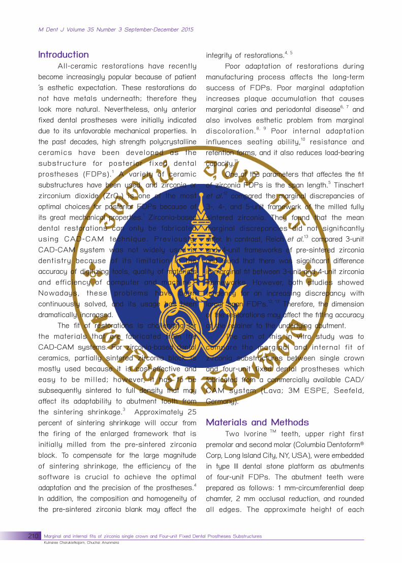
prepared tooth was 4 mm with 8° total occlusal convergence. A standard model of prepared abutments was fabricated using Co-Cr alloy (Fig. 1). Twenty polyether impressions (Impregum, 3M ESPE, St.Paul, MN, USA) of the standard
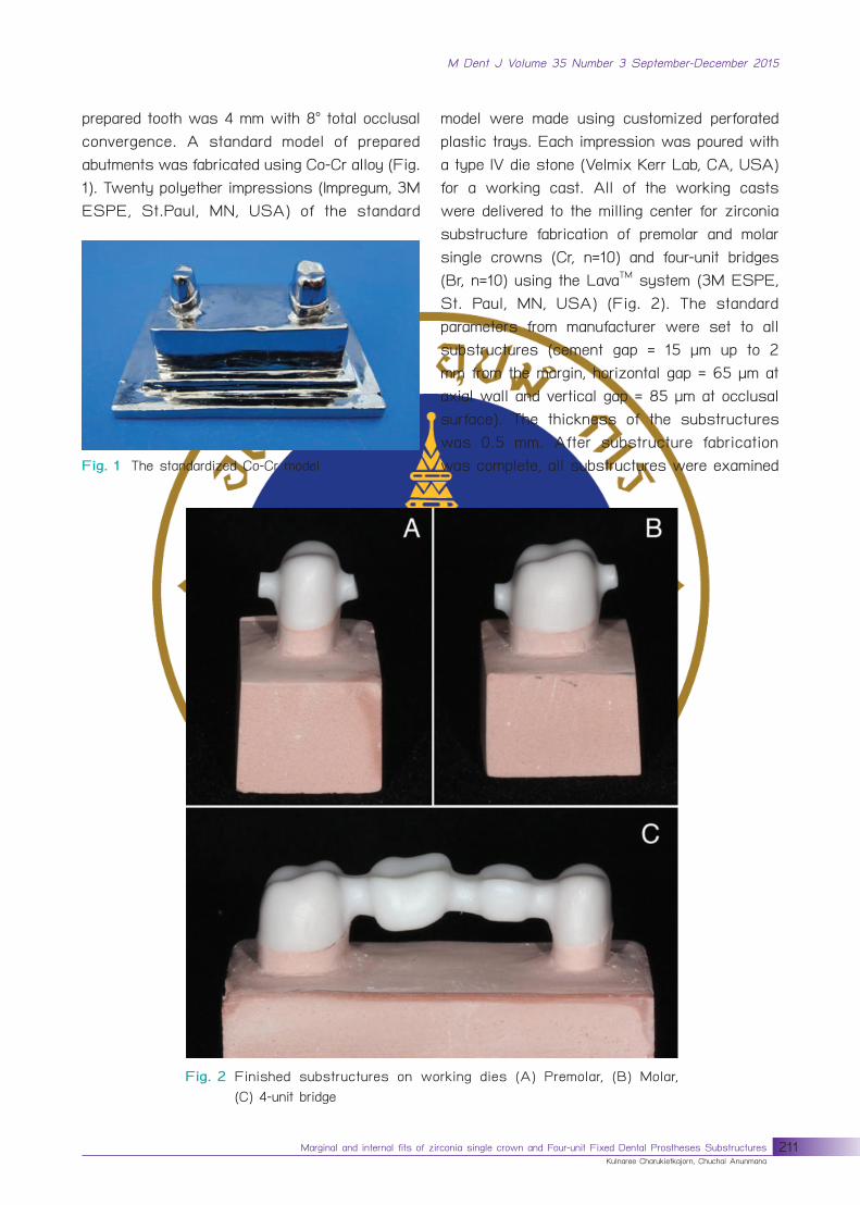
model were made using customized perforated plastic trays. Each impression was poured with a type IV die stone (Velmix Kerr Lab, CA, USA) for a working cast. All of the working casts were delivered to the milling center for zirconia substructure fabrication of premolar and molar single crowns (Cr, n=10) and four-unit bridges (Br, n=10) using the LavaTM system (3M ESPE, St. Paul, MN, USA) (Fig. 2). The standard parameters from manufacturer were set to all substructures (cement gap = 15 µm up to 2 mm from the margin, horizontal gap = 65 µm at axial wall and vertical gap = 85 µm at occlusal surface). The thickness of the substructures was 0.5 mm. After substructure fabrication was complete, all substructures were examined Fig. 1 The standardized Co-Cr model
Fig. 2 Finished substructures on working dies (A) Premolar, (B) Molar, (C) 4-unit bridge
212 Marginal and internal fits of zirconia single crown and Four-unit Fixed Dental Prostheses Substructures Marginal and internal fits of zirconia single crown and Four-unit Fixed Dental Prostheses Substructures Kulnaree Charukietkajorn, Chuchai Anunmana
M Dent J Volume 35 Number 3 September-December 2015
Marginal and internal fits of zirconia single crown and Four-unit Fixed Dental Prostheses Substructures
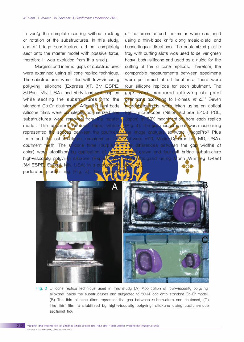
to verify the complete seating without rocking or rotation of the substructures. In this study, one of bridge substructure did not completely seat onto the master model with passive force, therefore it was excluded from this study. Marginal and internal gaps of substructures were examined using silicone replica technique. Thesubstructureswerefilledwithlow-viscositypolyvinyl siloxane (Express XT, 3M ESPE, St.Paul, MN, USA), and 50-N load was applied while seating the substructures onto the standard Co-Cr abutments. After the light-body siliconefilmswerecompletelypolymerized,thesubstructures were removed from the master model. The apparent silicone films, which represented the spaces between the abutment teeth and the substructures, remained on the abutment teeth. The silicone films (purple incolor) were stabilized by application of green high-viscosity polyvinyl siloxane (Express XT, 3M ESPE, St.Paul, MN, USA) in a customized perforated plastic tray (Fig. 3). The replicas
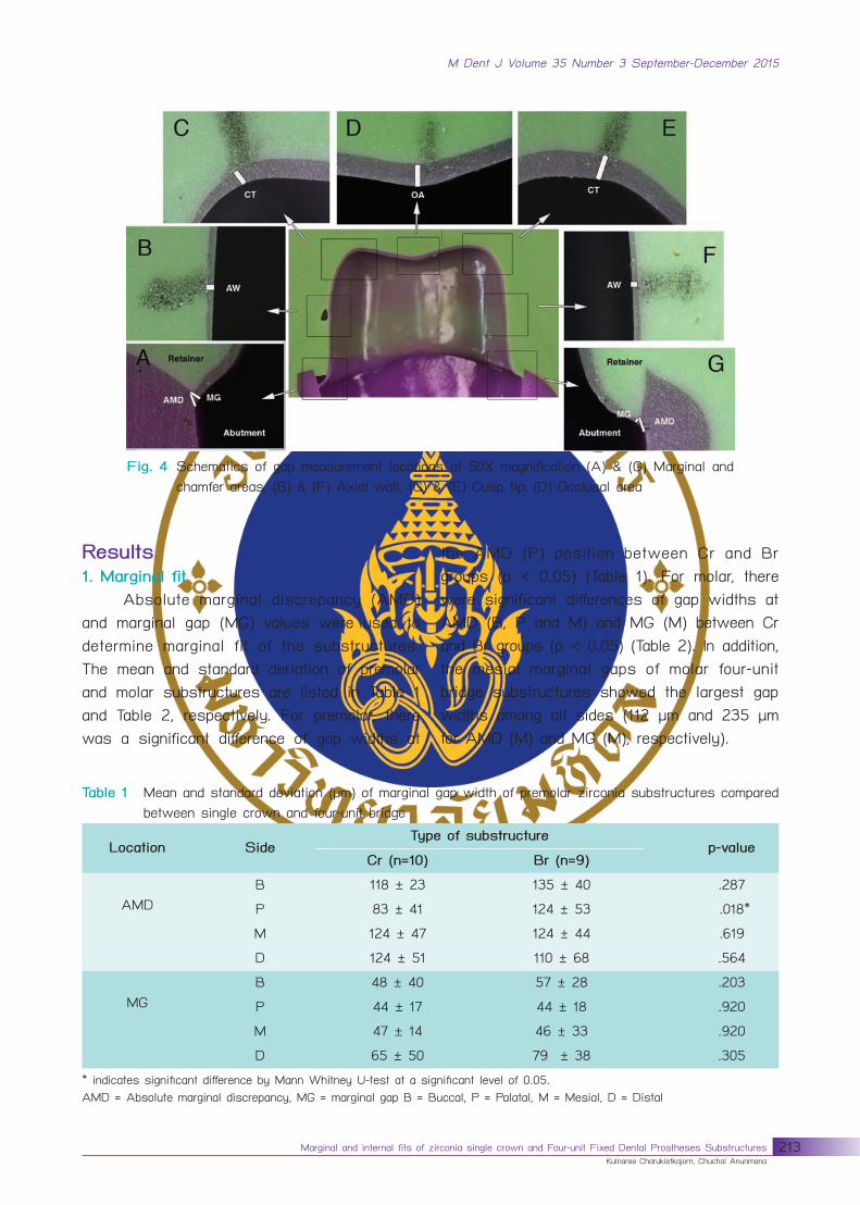
of the premolar and the molar were sectioned using a thin-blade knife along mesio-distal and bucco-lingual directions. The customized plastic tray with cutting slots was used to deliver green heavy body silicone and used as a guide for the cutting of the silicone replicas. Therefore, the comparable measurements between specimens were performed at all locations. There were four silicone replicas for each abutment. The gaps were measured following six point definitionsaccording toHolmeset al.14 Seven microphotographs were taken using an optical light microscope (Nikon eclipse E400 POL, Japan)at50Xmagnificationfromeachreplica(Fig. 4). The gap measurement was made using the image analysis software (ImagePro® Plus software v.7.0, Media Cybernetics, MD, USA). The differences between the gap widths of single crown and four-unit bridge substructure were analyzed using Mann Whitney U-test (α=0.05)
Fig. 3 Silicone replica technique used in this study (A) Application of low-viscosity polyvinyl siloxane inside the substructures and subjected to 50-N load onto standard Co-Cr model, (B) The thin silicone films represent the gap between substructure and abutment, (C) The thin film is stabilized by high-viscosity polyvinyl siloxane using custom-made sectional tray
Marginal and internal fits of zirconia single crown and Four-unit Fixed Dental Prostheses Substructures 213Marginal and internal fits of zirconia single crown and Four-unit Fixed Dental Prostheses Substructures Kulnaree Charukietkajorn, Chuchai Anunmana
M Dent J Volume 35 Number 3 September-December 2015
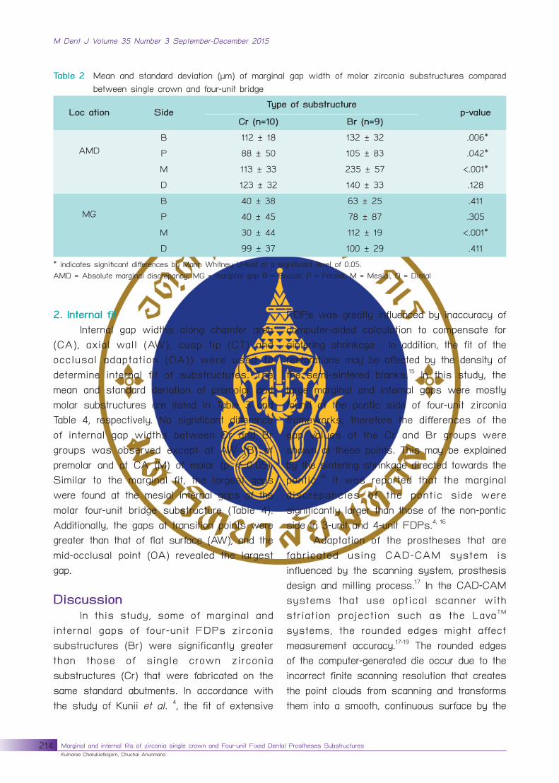
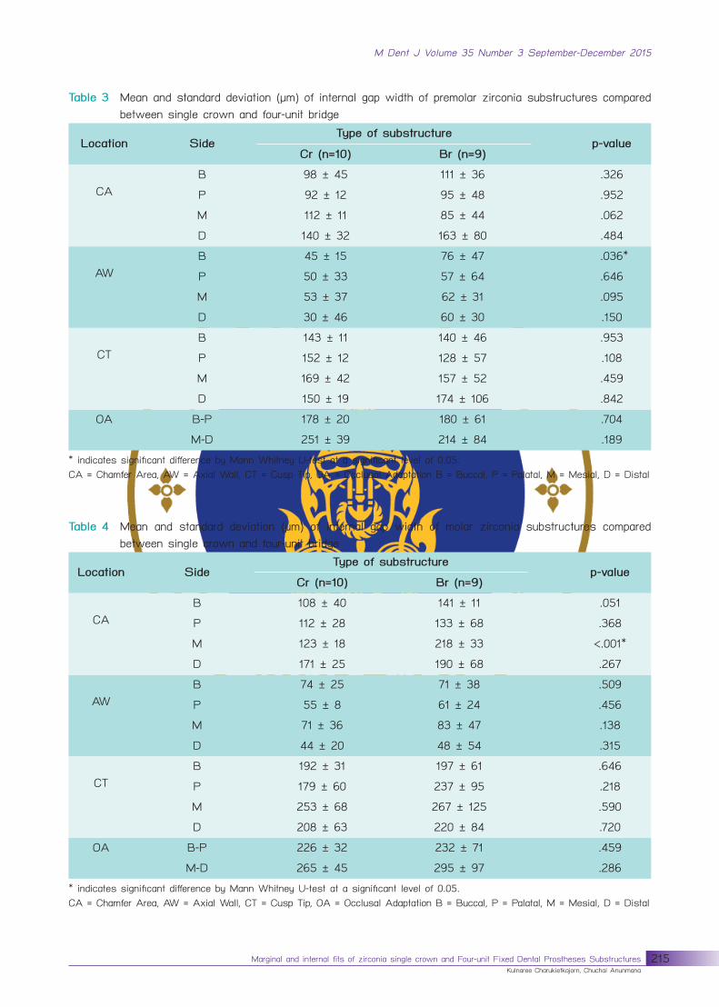
Results 1. Marginal fit Absolute marginal discrepancy (AMD) and marginal gap (MG) values were used to determine marginal fit of the substructures. The mean and standard deriation of premolar and molar substructures are listed in Table 1 and Table 2, respectively. For premolar, there was a significant difference of gap widths at
the AMD (P) position between Cr and Br groups (p < 0.05) (Table 1). For molar, there were significant differences of gap widths at AMD (B, P and M) and MG (M) between Cr and Br groups (p < 0.05) (Table 2). In addition, the mesial marginal gaps of molar four-unit bridge substructures showed the largest gap widths among all sides (112 µm and 235 µm for AMD (M) and MG (M), respectively).
Fig. 4 Schematics of gap measurement locations at 50X magnification (A) & (G) Marginal and chamfer areas, (B) & (F) Axial wall, (C) & (E) Cusp tip, (D) Occlusal area
Table 1 Mean and standard deviation (µm) of marginal gap width of premolar zirconia substructures compared between single crown and four-unit bridge
Location Side Type of substructure
p-value Cr (n=10) Br (n=9)
AMD
B 118 ± 23 135 ± 40 .287
P 83 ± 41 124 ± 53 .018*
M 124 ± 47 124 ± 44 .619
D 124 ± 51 110 ± 68 .564
MG
B 48 ± 40 57 ± 28 .203
P 44 ± 17 44 ± 18 .920
M 47 ± 14 46 ± 33 .920
D 65 ± 50 79 ± 38 .305 *indicatessignificantdifferencebyMannWhitneyU-testatasignificantlevelof0.05.AMD = Absolute marginal discrepancy, MG = marginal gap B = Buccal, P = Palatal, M = Mesial, D = Distal
214 Marginal and internal fits of zirconia single crown and Four-unit Fixed Dental Prostheses Substructures Marginal and internal fits of zirconia single crown and Four-unit Fixed Dental Prostheses Substructures Kulnaree Charukietkajorn, Chuchai Anunmana
M Dent J Volume 35 Number 3 September-December 2015
Marginal and internal fits of zirconia single crown and Four-unit Fixed Dental Prostheses Substructures
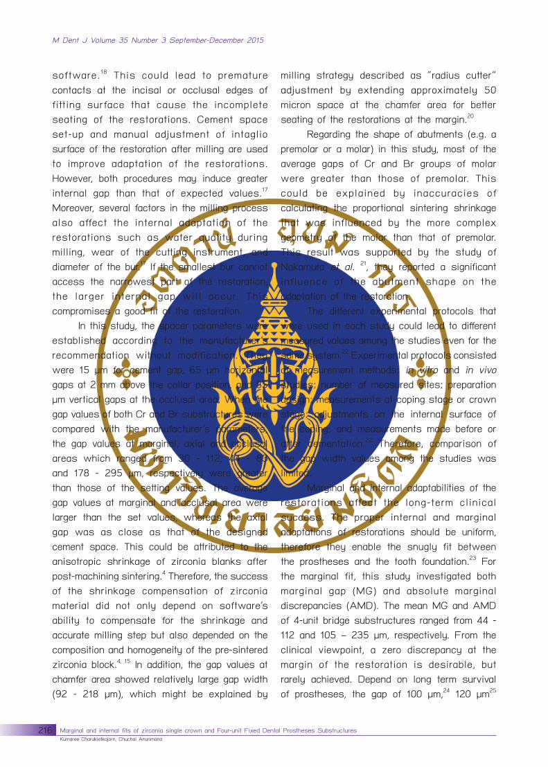
2. Internal fit Internal gap widths along chamfer area (CA), axial wall (AW), cusp tip (CT) and occlusal adaptation (OA)) were used to determine internal fit of substructures. The mean and standard deriation of premolar and molar substructures are listed in Table 3 and Table 4, respectively. No significant difference of internal gap widths between Cr and Br groups was observed except at AW (B) of premolar and at CA (M) of molar (p < 0.05). Similar to the marginal fit, the largest gaps were found at the mesial internal gaps of the molar four-unit bridge substructure (Table 4). Additionally, the gaps at transition points were greater than that of flat surface (AW), and the mid-occlusal point (OA) revealed the largest gap. Discussion In this study, some of marginal and internal gaps of four-unit FDPs zirconia substructures (Br) were significantly greater than those of single crown zirconia substructures (Cr) that were fabricated on the same standard abutments. In accordance with the study of Kunii et al. 4, the fit of extensive
FDPs was greatly influenced by inaccuracy of computer-aided calculation to compensate for sintering shrinkage. In addition, the fit of the restorations may be affected by the density of the semi-sintered blanks.15 In this study, the large marginal and internal gaps were mostly found at the pontic side of four-unit zirconia frameworks, therefore the differences of the gap values of the Cr and Br groups were shown at these points. This may be explained by the sintering shrinkage directed towards the pontic.16 It was reported that the marginal discrepancies of the pontic side were significantly larger than those of the non-pontic side in 3-unit and 4-unit FDPs.4, 16
Adaptation of the prostheses that are fabricated using CAD-CAM system is influenced by the scanning system, prosthesis design and milling process.17 In the CAD-CAM systems that use optical scanner with striation projection such as the LavaTM systems, the rounded edges might affect measurement accuracy.17-19 The rounded edges of the computer-generated die occur due to the incorrect finite scanning resolution that creates the point clouds from scanning and transforms them into a smooth, continuous surface by the
Table 2 Mean and standard deviation (µm) of marginal gap width of molar zirconia substructures compared between single crown and four-unit bridge
Loc ation Side Type of substructure
p-value Cr (n=10) Br (n=9)
AMD
B 112 ± 18 132 ± 32 .006*
P 88 ± 50 105 ± 83 .042*
M 113 ± 33 235 ± 57 <.001*
D 123 ± 32 140 ± 33 .128
MG
B 40 ± 38 63 ± 25 .411
P 40 ± 45 78 ± 87 .305
M 30 ± 44 112 ± 19 <.001*
D 99 ± 37 100 ± 29 .411 *indicatessignificantdifferencesbyMannWhitneyU-testatasignificantlevelof0.05.AMD = Absolute marginal discrepancy, MG = marginal gap B = Buccal, P = Palatal, M = Mesial, D = Distal
Marginal and internal fits of zirconia single crown and Four-unit Fixed Dental Prostheses Substructures 215Marginal and internal fits of zirconia single crown and Four-unit Fixed Dental Prostheses Substructures Kulnaree Charukietkajorn, Chuchai Anunmana
M Dent J Volume 35 Number 3 September-December 2015
Table 3 Mean and standard deviation (µm) of internal gap width of premolar zirconia substructures compared between single crown and four-unit bridge
Location Side Type of substructure
p-value Cr (n=10) Br (n=9)
CA
B 98 ± 45 111 ± 36 .326
P 92 ± 12 95 ± 48 .952
M 112 ± 11 85 ± 44 .062
D 140 ± 32 163 ± 80 .484
AW
B 45 ± 15 76 ± 47 .036*
P 50 ± 33 57 ± 64 .646
M 53 ± 37 62 ± 31 .095
D 30 ± 46 60 ± 30 .150
CT
B 143 ± 11 140 ± 46 .953
P 152 ± 12 128 ± 57 .108
M 169 ± 42 157 ± 52 .459
D 150 ± 19 174 ± 106 .842
OA B-P 178 ± 20 180 ± 61 .704
M-D 251 ± 39 214 ± 84 .189 *indicatessignificantdifferencebyMannWhitneyU-testatasignificantlevelof0.05.CA = Chamfer Area, AW = Axial Wall, CT = Cusp Tip, OA = Occlusal Adaptation B = Buccal, P = Palatal, M = Mesial, D = Distal
Table 4 Mean and standard deviation (µm) of internal gap width of molar zirconia substructures compared between single crown and four-unit bridge
Location Side Type of substructure
p-value Cr (n=10) Br (n=9)
CA
B 108 ± 40 141 ± 11 .051
P 112 ± 28 133 ± 68 .368
M 123 ± 18 218 ± 33 <.001*
D 171 ± 25 190 ± 68 .267
AW
B 74 ± 25 71 ± 38 .509
P 55 ± 8 61 ± 24 .456
M 71 ± 36 83 ± 47 .138
D 44 ± 20 48 ± 54 .315
CT
B 192 ± 31 197 ± 61 .646
P 179 ± 60 237 ± 95 .218
M 253 ± 68 267 ± 125 .590
D 208 ± 63 220 ± 84 .720
OA B-P 226 ± 32 232 ± 71 .459
M-D 265 ± 45 295 ± 97 .286 *indicatessignificantdifferencebyMannWhitneyU-testatasignificantlevelof0.05.CA = Chamfer Area, AW = Axial Wall, CT = Cusp Tip, OA = Occlusal Adaptation B = Buccal, P = Palatal, M = Mesial, D = Distal
216 Marginal and internal fits of zirconia single crown and Four-unit Fixed Dental Prostheses Substructures Marginal and internal fits of zirconia single crown and Four-unit Fixed Dental Prostheses Substructures Kulnaree Charukietkajorn, Chuchai Anunmana
M Dent J Volume 35 Number 3 September-December 2015
Marginal and internal fits of zirconia single crown and Four-unit Fixed Dental Prostheses Substructures
software.18 This could lead to premature contacts at the incisal or occlusal edges of fitting surface that cause the incomplete seating of the restorations. Cement space set-up and manual adjustment of intaglio surface of the restoration after milling are used to improve adaptation of the restorations. However, both procedures may induce greater internal gap than that of expected values.17 Moreover, several factors in the milling process also affect the internal adaptation of the restorations such as water quality during milling, wear of the cutting instrument, and diameter of the bur.17 If the smallest bur cannot access the narrowest part of the restoration, the larger internal gap wil l occur. This compromises a good fit of the restoration. In this study, the spacer parameters were established according to the manufacturer’s recommendation without modification. They were 15 µm for cement gap, 65 µm horizontal gaps at 2 mm above the collar position, and 85 µm vertical gaps at the occlusal area. When the gap values of both Cr and Br substructures were compared with the manufacturer’s parameters, the gap values at marginal, axial and occlusal areas which ranged from 30 - 112, 44 - 83 and 178 - 295 µm, respectively were greater than those of the setting values. The average gap values at marginal and occlusal area were larger than the set values, whereas the axial gap was as close as that of the designed cement space. This could be attributed to the anisotropic shrinkage of zirconia blanks after post-machining sintering.4 Therefore, the success of the shrinkage compensation of zirconia material did not only depend on software’s ability to compensate for the shrinkage and accurate milling step but also depended on the composition and homogeneity of the pre-sintered zirconia block.4, 15 In addition, the gap values at chamfer area showed relatively large gap width (92 - 218 µm), which might be explained by
milling strategy described as “radius cutter” adjustment by extending approximately 50 micron space at the chamfer area for better seating of the restorations at the margin.20 Regarding the shape of abutments (e.g. a premolar or a molar) in this study, most of the average gaps of Cr and Br groups of molar were greater than those of premolar. This could be explained by inaccuracies of calculating the proportional sintering shrinkage that was influenced by the more complex geometry of the molar than that of premolar. This result was supported by the study of Nakamura et al. 21, they reported a significant influence of the abutment shape on the adaptation of the restoration. The different experimental protocols that were used in each study could lead to different measured values among the studies even for the same system.22 Experimental protocols consisted of measurement methods; in vitro and in vivo studies; number of measured sites; preparation design; measurements at coping stage or crown stage; adjustments on the internal surface of the coping; and measurements made before or after cementation.22 Therefore, comparison of the gap width values among the studies was limited. Marginal and internal adaptabilities of the restorations affect the long-term clinical success. The proper internal and marginal adaptations of restorations should be uniform, therefore they enable the snugly fit between the prostheses and the tooth foundation.23 For the marginal fit, this study investigated both marginal gap (MG) and absolute marginal discrepancies (AMD). The mean MG and AMD of 4-unit bridge substructures ranged from 44 - 112 and 105 – 235 µm, respectively. From the clinical viewpoint, a zero discrepancy at the margin of the restoration is desirable, but rarely achieved. Depend on long term survival of prostheses, the gap of 100 µm,24 120 µm25
Marginal and internal fits of zirconia single crown and Four-unit Fixed Dental Prostheses Substructures 217Marginal and internal fits of zirconia single crown and Four-unit Fixed Dental Prostheses Substructures Kulnaree Charukietkajorn, Chuchai Anunmana
M Dent J Volume 35 Number 3 September-December 2015
or up to 200 µm26 was recommended. Most authors agree that marginal opening of less than 120 µm was in the range of clinical acceptability.12, 27, 28 In the study of McLean and von Fraunhofer,25 they examined more than 1000 crowns after a 5-year period and concluded that marginal opening of less than 120 µm was clinical ly acceptable for conventionally cemented restorations. Although marginal gaps (MG) in this study were clinical ly acceptable, absolute marginal discrepancies (AMD) were over the acceptable range. Therefore, this type of the prostheses may have a signif icant effect on the adaptability of zirconia restorations especially at the outer margin of FDPs. However, clinical implementation of the long-span zirconia restorations seems to require further investigation because studies of restoration longevity are rare. In addition, careful attention must be taken while evaluating the marginal integrity of zirconia restorations, and patient recall is crucial for the success of zirconia restoration. Regarding internal fit, most of internal gaps of Cr did not significantly differ from those of Br. However, the median occlusal gaps of both Cr and Br groups revealed large discrepancies that were in the range of 178 - 295 µm. The results showed the large occlusal gaps that were not as precise as the setting of the computer software. Large internal gap widths at the axio-occlusal transition and mid-occlusal area reduce the interocclusal distance between the occlusal surface of the substructures and the occluded teeth. This l imits the occlusal clearance available for the layering glass and may lead to the weakness of the glass veneer and compromise the anatomical shape of occlusal surfaces.29 On the other hand, over-reduction of the occlusal surface to compensate the occlusal gap might influence abutment tooth vitality. The large internal gap
width and variation of internal discrepancies could also affect the stability of ceramic restorations.11 There was also the evidence that a lack of precision in internal fit could promote higher risks for veneering fracture.30 Most popular methods to determine the adaptation of prostheses are embedded and replica techniques because of their ability to measure marginal and internal fit with simplicity. Embedded technique might be the most accurate way, however, there is a shortcoming in that the restorations are destroyed for the measurement. Therefore, replica technique, which is a non-destructive method to measure the fit using impression material to simulate the gap width, was used in this study. This method has been used by many researchers to investigate the accuracy of crowns and FDPs in vivo 13, 31-33 as well as in vitro. 12, 34 Although this method was considered to be less accurate than the embedded method 23, it was acceptable and reliable enough when compared with the embedded method.35, 36 Rahme et al. did not find significant differences of marginal gap dimensions, whether the measurement was conducted using a replica technique or directly with sectioned specimens.35, 36 However, the evaluation of crown-tooth discrepancies by replication has limitations and technical sensitivity due to the possible distortion or rupture of thin layer of elastomeric film during manipulation, and if a section is not perpendicular to the surface, it could lead to the error of the measurements as well.34 Additionally, only a limited number of marginal gap measurements could be performed by two cross-sections of replica technique Finally, this experiment was in vitro study, which ideal abutment preparation could be performed; therefore this might not be relevant to cl inical situation. Clinical implementation of the long-span FDPs seems
218 Marginal and internal fits of zirconia single crown and Four-unit Fixed Dental Prostheses Substructures Marginal and internal fits of zirconia single crown and Four-unit Fixed Dental Prostheses Substructures Kulnaree Charukietkajorn, Chuchai Anunmana
M Dent J Volume 35 Number 3 September-December 2015
Marginal and internal fits of zirconia single crown and Four-unit Fixed Dental Prostheses Substructures
to require additional investigation because studies of restoration longevity are limited. The standardized criteria for evaluation of FDPs and a consistent definition of biological and technical complications should also be created for more efficient comparison and evaluation of the findings. In addition, further study needs to clarify how to gain the precise product for fabrication of extensive zirconia restoration using semi-sintered stage zirconia material. Conclusion In conclusion: this study revealed that silicone replica technique was the simple technique to evaluate the fitting accuracy of fixed dental prostheses. The results showed most gaps were greater than those of setting parameter especially at the occlusal surface. In addition, the measured gaps tended to be larger with more complicate-shaped or longer restoration. There should be further studies to investigate the precision and long-term clinical success of long-span zirconia restorations when these materials are fabricated from the pre-sintered stage. Funding: None Competing interest: None Ethical approval: None References 1. Bachhav VC, Aras MA. Zirconia-based fixed
partial dentures: a clinical review. Quintessence Int 2011; 42: 173-182.
2. Miyazaki T, Hotta Y, Kunii J, Kuriyama S, Tamaki Y. A review of dental CAD/CAM: current status and future perspectives from 20 years of experience. Dent Mater J 2009; 28: 44-56.
3. Denry I, Kelly JR. State of the art of zirconia for dental applications. Dent Mater 2008; 24: 299-307.
4. Kunii J, Hotta Y, Tamaki Y, Ozawa A, Kobayashi Y, Fujishima A, et al. Effect of sintering on the marginal and internal fit of CAD/CAM-fabricated zirconia frameworks. Dent Mater J 2007; 26:
820-826. 5. Abduo J, Lyons K, Swain M. Fit of zirconia fixed
6. Felton DA, Kanoy BE, Bayne SC, Wirthman GP. Effect of in vivo crown margin discrepancies on periodontal health. J Prosthet Dent 1991; 65: 357-364.
7. Sorensen SE, Larsen IB, Jorgensen KD. Gingival and alveolar bone reaction to marginal fit of subgingival crown margins. Scand J Dent Res 1986; 94: 109-114.
8. Sailer I, Feher A, Filser F, Gauckler LJ, Luthy H, Hammerle CH. Five-year clinical results of zirconia frameworks for posterior fixed partial dentures. Int J Prosthodont 2007; 20: 383-388.
9. Raigrodski AJ, Chiche GJ, Potiket N, Hochstedler JL, Mohamed SE, Billiot S, et al. The efficacy of posterior three-unit zirconium-oxide-based ceramic fixed partial dental prostheses: a prospective clinical pilot study. J Prosthet Dent 2006; 96: 237-244.
10. Hung SH, Hung KS, Eick JD, Chappell RP. Marginal fit of porcelain-fused-to-metal and two types of ceramic crown. J Prosthet Dent 1990; 63: 26-31.
11. Tuntiprawon M, Wilson PR. The effect of cement thickness on the fracture strength of all-ceramic crowns. Aust Dent J 1995; 40: 17-21.
12. Tinschert J, Natt G, Mautsch W, Spiekermann H, Anusavice KJ. Marginal fit of alumina-and zirconia-based fixed partial dentures produced by a CAD/CAM system. Oper Dent 2001; 26: 367-374.
13. Reich S, Kappe K, Teschner H, Schmitt J. Clinical fit of four-unit zirconia posterior fixed dental prostheses. Eur J Oral Sci 2008; 116: 579-584.
14. Holmes JR, Bayne SC, Holland GA, Sulik WD. Considerations in measurement of marginal fit. J Prosthet Dent 1989; 62: 405-408.
15. Beuer F, Neumeier P, Naumann M. Marginal fit of 14-unit zirconia fixed dental prosthesis retainers. J Oral Rehabil 2009; 36: 142-149.
16. Kohorst P, Junghanns J, Dittmer MP, Borchers L, Stiesch M. Different CAD/CAM-processing routes for zirconia restorations: influence on fitting accuracy. Clin Oral Investig 2011; 15: 527-536.
Marginal and internal fits of zirconia single crown and Four-unit Fixed Dental Prostheses Substructures 219Marginal and internal fits of zirconia single crown and Four-unit Fixed Dental Prostheses Substructures Kulnaree Charukietkajorn, Chuchai Anunmana
M Dent J Volume 35 Number 3 September-December 2015
17. Bornemann G, Lemelson S, Luthardt R. Innovative method for the analysis of the internal 3D fitting accuracy of Cerec-3 crowns. Int J Comput Dent 2002; 5: 177-182.
18. Luthardt R, Weber A, Rudolph H, Schone C, Quaas S, Walter M. Design and production of dental prosthetic restorations: basic research on dental CAD/CAM technology. Int J Comput Dent 2002; 5: 165-176.
19. Pfeiffer J. Dental CAD/CAM technologies: the optical impression (II). Int J Comput Dent 1999; 2: 65-72.
21. Nakamura T, Nonaka M, Maruyama T. In vitro fitting accuracy of copy-milled alumina cores and all-ceramic crowns. Int J Prosthodont 2000; 13: 189-193.
22. Contrepois M, Soenen A, Bartala M, Laviole O. Marginal adaptation of ceramic crowns: A systematic review. J Prosthet Dent 2013.
23. May KB, Russell MM, Razzoog ME, Lang BR. Precision of fit: the Procera AllCeram crown. J Prosthet Dent 1998; 80: 394-404.
24. Sulaiman F, Chai J, Jameson LM, Wozniak WT. A comparison of the marginal fit of In-Ceram, IPS Empress, and Procera crowns. Int J Prosthodont 1997; 10: 478-484.
25. McLean JW, von Fraunhofer JA. The estimation of cement film thickness by an in vivo technique. Br Dent J 1971; 131: 107-111.
26. Gulker I. Margins. N Y State Dent J 1985; 51: 213-215, 217.
27. Kim KB, Kim WC, Kim HY, Kim JH. An evaluation of marginal fit of three-unit fixed dental prostheses fabricated by direct metal laser sintering system. Dent Mater 2013; 29: e91-96.
28. Lee JY, Choi SJ, Kim MS, Kim HY, Kim YS, Shin SW. Effect of span length on the fit of zirconia framework fabricated using CAD/CAM system. J Adv Prosthodont 2013; 5: 118-125.
29. Rekow D, Thompson VP. Near-surface damage--a persistent problem in crowns obtained by computer-aided design and manufacturing. Proc Inst Mech Eng H 2005; 219: 233-243.
30. Rekow ED, Harsono M, Janal M, Thompson VP, Zhang G. Factorial analysis of variables influencing stress in all-ceramic crowns. Dent Mater 2006; 22: 125-132.
31. Wettstein F, Sailer I, Roos M, Hammerle CH. Clinical study of the internal gaps of zirconia and metal frameworks for fixed partial dentures. Eur J Oral Sci 2008; 116: 272-279.
32. Reich S, Wichmann M, Nkenke E, Proeschel P. Clinical fit of all-ceramic three-unit fixed partial dentures, generated with three different CAD/CAM systems. Eur J Oral Sci 2005; 113: 174-179.
33. Boening KW, Wolf BH, Schmidt AE, Kastner K, Walter MH. Clinical fit of Procera AllCeram crowns. J Prosthet Dent 2000; 84: 419-424.
34. Coli P, Karlsson S. Fit of a new pressure-sintered zirconium dioxide coping. Int J Prosthodont 2004; 17: 59-64.
35. Laurent M, Scheer P, Dejou J, Laborde G. Clinical evaluation of the marginal fit of cast crowns--validation of the silicone replica method. J Oral Rehabil 2008; 35: 116-122.
36. Rahme HY, Tehini GE, Adib SM, Ardo AS, Rifai KT. In vitro evaluation of the “replica technique” in the measurement of the fit of Procera crowns. J Contemp Dent Pract 2008; 9: 25-32.
International Abstract Cochrane Database Syst Rev. 2015 Mar 26;3:CD010743. doi: 10.1002/14651858.CD010743.pub2. Xylitol-containing products for preventing dental caries in children and adults. Riley P1, Moore D, Ahmed F, Sharif MO, Worthington HV. Author information
Abstract
BACKGROUND: Dental caries is a highly prevalent chronic disease which affects the majority of people. It has been postulated that the consumption of xylitol could help to prevent caries. The evidence on the effects of xylitol products is not clear and therefore it is important to summarise the available evidence to determine its effectiveness and safety.
OBJECTIVES: To assess the effects of different xylitol-containing products for the prevention of dental caries in children and adults.
SEARCH METHODS: We searched the following electronic databases: the Cochrane Oral Health Group Trials Register (to 14 August 2014), theCochrane Central Register of Controlled Trials (CENTRAL) (The Cochrane Library, 2014, Issue 7), MEDLINE via OVID (1946 to 14 August 2014), EMBASE via OVID (1980 to 14 August 2014), CINAHL via EBSCO (1980 to 14 August 2014), Web of Science Conference Proceedings (1990 to 14 August 2014), Proquest Dissertations and Theses (1861 to 14 August 2014). We searched the US National Institutes of Health Trials Register (http://clinicaltrials.gov) and the WHO Clinical Trials Registry Platform for ongoing trials. No restrictions were placed on the language or date of publication when searching the electronic databases.
SELECTION CRITERIA: We included randomised controlled trials assessing the effects of xylitol products on dental caries in children and adults.
DATA COLLECTION AND ANALYSIS: Two review authors independently screened the results of the electronic searches, extracted data and assessed the risk of bias of the included studies. We attempted to contact study authors for missing data or clarification where feasible. For continuous outcomes, we used means and standard deviations to obtain the mean difference and 95% confidence interval (CI). We used the continuous data to calculate prevented fractions (PF) and 95% CIs to summarise the percentage reduction in caries. For dichotomous outcomes, we reported risk ratios (RR) and 95% CIs. As there were less than four studies included in the meta-analysis, we used a fixed-effect model. We planned to use a random-effects model in the event that there were four or more studies in a meta-analysis.
MAIN RESULTS: We included 10 studies that analysed a total of 5903 participants. One study was assessed as being at low risk of bias, two were assessed as being at unclear risk of bias, with the remaining seven being at high risk of bias.The main finding of the review was that, over 2.5 to 3 years of use, a fluoride toothpaste containing 10% xylitol may reduce caries by 13% when compared to a fluoride-only toothpaste (PF -0.13, 95% CI -0.18 to -0.08, 4216 children analysed, low-quality evidence).The remaining evidence on children, from small single studies with risk of bias issues and great uncertainty associated with the effect estimates, was insufficient to determine a benefit from xylitol products. One study reported thatxylitol syrup (8 g per day) reduced caries by 58% (95% CI 33% to 83%, 94 infants analysed, low quality evidence) when compared to a low-dosexylitol syrup (2.67 g per day) consumed for 1 year.The following results had 95% CIs that were compatible with both a reduction and an increase in caries associated with xylitol: xylitol lozenges versus no treatment in children (very low quality body of evidence); xylitol sucking tablets versus no treatment in infants (very low quality body of evidence); xylitol tablets versus control (sorbitol) tablets in infants (very low quality body of evidence);xylitol wipes versus control wipes in infants (low quality body of evidence).There was only one study investigating the effects of xylitol lozenges, when compared to control lozenges, in adults (low quality body of evidence). The effect estimate had a 95% CI that was compatible with both a reduction and an increase in caries associated with xylitol.Four studies reported that there were no adverse effects from any of the interventions. Two studies reported similar rates of adverse effects between study arms. The remaining studies either mentioned adverse effects but did not report any usable data, or did not mention them at all. Adverse effects include sores in the mouth, cramps, bloating, constipation, flatulence, and loose stool or diarrhoea.
AUTHORS’ CONCLUSIONS: We found some low quality evidence to suggest that fluoride toothpaste containing xylitol may be more effective than fluoride-only toothpaste for preventing caries in the permanent teeth of children, and that there are no associated adverse-effects from such toothpastes. The effect estimate should be interpreted with caution due to high risk of bias and the fact that it results from two studies that were carried out by the same authors in the same population. The remaining evidence we found is of low to very low quality and is insufficient to determine whether any other xylitol-containing products can prevent caries in infants, older children, or adults.


![Sulfated zirconia[1]](https://static.documents.pub/doc/80x56/5568f2ecd8b42aff2e8b4932/sulfated-zirconia1.jpg)


