Page 1
©2020 Mayo Foundation for Medical Education and Research | slide-1
Mayo Clinic Cardiology Core Competenciesfor the Nurse Practitioner and Physician Assistant
©2020 Mayo Foundation for Medical Education and Research | slide-1Image copyright Shutterstock / LeventeGyori 85141453
©2020 Mayo Foundation for Medical Education and Research | slide-2
SyncopeCardiac Symptom Analysis
Dan Sorajja, MD, FACC, FHRSAssociate Professor of MedicineProgram Director Clinical Cardiac ElectrophysiologyAssociate Program Director Cardiovascular DiseasesMayo Clinic Arizona
©2020 Mayo Foundation for Medical Education and Research | slide-2
Image copyright Shutterstock / Sebastian Kaulitzki 130094798
Page 2
©2020 Mayo Foundation for Medical Education and Research | slide-3
REFERENCES TO OFF-LABEL USAGE(S) OF PHARMACEUTICALS OR INSTRUMENTS
Faculty Disclosure
Relevant financial relationship(s) with industryNone
None
©2020 Mayo Foundation for Medical Education and Research | slide-4©2020 Mayo Foundation for Medical Education and Research | slide-4
LEARNING OBJECTIVES
1. Learn various causes of syncope
2. Distinguish causes of syncope through history, exam, and testing
3. Understand appropriate testing indications
4. Understand treatment options for the underlying cause of syncope
5. Identify high risk patients
Page 3
©2020 Mayo Foundation for Medical Education and Research | slide-5
Background
1
©2020 Mayo Foundation for Medical Education and Research | slide-5
©2020 Mayo Foundation for Medical Education and Research | slide-6
For Detailed Information on Syncope Evaluation and Treatment
Shen. JACC 2017. http://dx.doi.org/10.1016/j.jacc.2017.03.003
Page 4
©2020 Mayo Foundation for Medical Education and Research | slide-7
Syncope Big Picture
• Definition:• Abrupt and transient loss of consciousness (from cerebral hypoperfusion), with loss of
postural tone, followed by rapid, spontaneous recovery
• Syncope is a symptom: There is an underlying cause
• Syncope is common• Self-reported syncope prevalence (Olmsted County residents ≥ 45 y)
• 25%1
• Incidence increases with age2
• Ages 20-69: 2.6 - 5.7 per 1000 person-years• Ages 70-79: 11.1 per 1000 person-years• Ages ≥ 80: 16.9 – 19.5 per 1000 person-years
1. Chen. Am J Med 2006;119(12):1088 2. Soteriades. NEJM. 2002;347:878
©2020 Mayo Foundation for Medical Education and Research | slide-8
Incidence of Syncope
1. Soteriades. NEJM. 2002;347:8782. Sorajja D. Circ 2009; 120:928.
1 2 2
Page 5
©2020 Mayo Foundation for Medical Education and Research | slide-9
Causes of Syncope
Orthostatic hypotension
Cardiac arrhythmias
Non-syncope
Structural – cardiac or lung
Neurally-mediated (a.k.a. reflex syncope)VasovagalCarotid sinus hypersensitivitySituational Syncope Sorajja D. Circ 2009; 120:928.
©2020 Mayo Foundation for Medical Education and Research | slide-10
Syncope Look-A-Likes
• Drug intoxication• Alcohol intoxication• Seizure• Trauma• Falls• Psychiatric conditions
• Pseudo-syncope• Pseudo-seizure
Page 6
©2020 Mayo Foundation for Medical Education and Research | slide-11
History (and more history)
2
©2020 Mayo Foundation for Medical Education and Research | slide-11
©2020 Mayo Foundation for Medical Education and Research | slide-12
How Are We Going To Come To The Diagnosis?• History is key
• Then take more history (seriously)
• Then careful physical exam
• “You can’t diagnosis it, if you don’t think of it”
• Screen medications
• Check orthostatic BP and HR during appointment
• 12-lead ECG
• Focused laboratory and testing
Page 7
©2020 Mayo Foundation for Medical Education and Research | slide-13
Clues for Syncope Etiology From HistoryWhat Is The Trigger?Syncope Occurs . . . Think:With noxious stimulus (needle sticks, nausea) VasovagalWhile exercising Cardiac etiologyIn any position Cardiac etiologyAfter exercise Situational syncopeWhile coughing, swallowing, laughing, urinating, defecating Situational syncopeOften during morning Orthostatic hypotensionGoing from sitting to standing Orthostatic hypotensionAfter eating Post-prandial hypotensionVomiting, diarrhea, hemorrhage, poor PO intake DehydrationWhile shaving or turning head (in elderly) Carotid sinus hypersensitivityVertigo ENT problem
©2020 Mayo Foundation for Medical Education and Research | slide-14
Clues for Syncope Etiology From HistoryWarning Symptoms
• Nausea, flushing, or sweats? • Think vasovagal• And frequently prodrome is long enough to get into safe position
• Abrupt symptoms/syncope (minimal if any prodrome) • Think arrhythmia / cardiac
• Of note, seizure usually lacks a prodrome• Unable to protect head Think arrhythmia or seizure• Did the person ease him/herself down Usually not arrhythmia
• Palpitations • Think arrhythmia / cardiac
Page 8
©2020 Mayo Foundation for Medical Education and Research | slide-15
Clues for Syncope Etiology From HistoryWhile Unconsciousness• If syncope witnessed, the history is usually more accurate
• Duration of syncope• < 2 minutes Think vasovagal
• Can be longer than 5 minutes with vasovagal but usually with head injury
• Tonic-clonic movements• Not helpful - since syncope and seizure both can exhibit myoclonic jerks
• Tongue-biting or Incontinence Think seizure
• Pallor or cyanosis Think arrhythmia
©2020 Mayo Foundation for Medical Education and Research | slide-16
Clues for Syncope Etiology From HistoryRecovery After Syncope• Nausea, sweats, or flushing Think neurally-mediated
• Disorientation Think seizure
• Prolonged recovery Think about concurrent head injury in addition to syncope
• Chest pain or palpitations Think cardiac
1. https://knowyourmeme.com/memes/sweating-jordan-peele2. Doctorshealthpress.com
1 2
Page 9
©2020 Mayo Foundation for Medical Education and Research | slide-17
Other Pertinent HistoryMedications• Concern for orthostatic hypotension, dehydration, and polypharmacy effects
Class of Medication DrugsDiuretics HCTZ, loop-diuretics, spironolactoneVenodilators NitratesVasodilators Nitrates, hydralazine, α-blockers, CCB, PDE inhibitorsNegative chronotropes β-blockers, CCBSedatives BenzodiazepineAnti-Parkinsonism Levodopa, ropinirole, pramipexoleAnti-depressant medications Tricyclics, phenothiazines
©2020 Mayo Foundation for Medical Education and Research | slide-18
Other Pertinent HistoryPast Medical and Family History• Ischemic heart disease or depressed EF Think arrhythmia
• Seizure history? Could still be a seizure, and may not be a seizure if you get a history of seizure meds not helping
• Pacemaker/ICD previously helping Think device malfunction
• Family with any inheritable conditions (1st degree relative with SCD < 50 years old)• Long QT syndrome• Brugada syncope• Premature sudden cardiac death
1
1. Hannahhelpbiology.blogspot.com
Page 10
©2020 Mayo Foundation for Medical Education and Research | slide-19
Physical Exam
3
©2020 Mayo Foundation for Medical Education and Research | slide-19
©2020 Mayo Foundation for Medical Education and Research | slide-20
Physical Exam FindingsBlood Pressure• Blood pressure log
• Want to know blood pressure at various times of day
• Low BP at baseline• Screen for orthostatic hypotension (old and frail people)• Screen for polypharmacy• Check hydration (how much water/fluid intake and urine color)
• To take orthostatic vital signs• Patient lays down for 2 minutes before supine BP and HR check• Have the patient stand up with the cuff on, but not inflated• Then measure the BP and HR
• At 1 minute At 3 minutes At 5 minutes • Document any symptoms
• If a patient cannot stand for any measurement, take the BP while sitting, but not supine position if possible
1
2
1. Decider.com 2. Ronitbaras.com
Page 11
©2020 Mayo Foundation for Medical Education and Research | slide-21©2020 Mayo Foundation for Medical Education and Research | slide-21
Physical Exam Findings
• General• Pallor Screen for anemia (& see if symptoms
worse with menstrual cycles)
• ENT• Carotid sinus massage in patients > 40 years old
• Contraindicated if bruit or recent stroke
• Heart• JVD not visible Think dehydration/hypovolemia• JVD to the jaw Think heart
failure/cardiomyopathy• Are heart sounds distant? Think pericardial
effusion• Bradycardia or significant pause after extrasystole?• Pulse deficit with extrasystole or tachycardia?• Murmur consistent with AS or HCM
©2020 Mayo Foundation for Medical Education and Research | slide-22
Key Physical Exam Findings
1. En.wikipedia.org
1
• Neuro• Parkinson features• Sensory neuropathy• Gait and ataxia• Dry skin
Think about neuropathy
Page 12
©2020 Mayo Foundation for Medical Education and Research | slide-23
Testing
4
©2020 Mayo Foundation for Medical Education and Research | slide-23
©2020 Mayo Foundation for Medical Education and Research | slide-24
Laboratory Evaluation & Testing
• ECG for everyone
• Targeted blood testing
Suspicion For: Lab TestAnemia exacerbating situation Hemoglobin/hematocrit Dehydration Electrolytes, BUN, CrDiabetes Glucose and HgbA1CInflammatory diseases ANA, CRPNeuropathy B12 level
Page 13
©2020 Mayo Foundation for Medical Education and Research | slide-25
SyncopeInitial Evaluation• Get History
• Do Physical Examination
• Perform ECG
Shen. JACC 2017. http://dx.doi.org/10.1016/j.jacc.2017.03.003
©2020 Mayo Foundation for Medical Education and Research | slide-26
When To Consider Evaluation In A Hospital SettingSerious Medical Conditions
• Cardiac arrhythmias• Sustained or symptomatic VT• Symptomatic conduction system
disease, Mobitz II, or third-degree heart block
• Symptomatic bradycardia or sinus pauses not related to neurally-mediated syncope
• Symptomatic SVT• Pacemaker / ICD malfunction• Inheritable cardiac conditions
predisposing to arrhythmias
•
• Cardiac or vascular nonarrhythmic conditions• Cardiac ischemia• Severe aortic stenosis• Cardiac tamponade• Hypertrophic cardiomyopathy• Severe prosthetic valve dysfunction• Pulmonary embolism• Aortic dissection• Moderate to severe LV dysfunction
• Noncardiac conditions• Severe anemia or GI bleeding• Major trauma due to syncope• Persistent vital sign abnormalities
Page 14
©2020 Mayo Foundation for Medical Education and Research | slide-27
Other Testing To ConsiderAmbulatory MonitoringWe expect longer monitoring to be more sensitive in detecting arrhythmias, conduction abnormalities, and their correlation to arrhythmias1
• Record now and analyze later• SmartWatch (< 1 minute)• Continuous ECG (Holter) monitor (1-2 days)• Event (loop) monitor (1 week to 30 days)• Patch monitor (3-30 days)• Implantable Loop Recorder (2 – 4 years)
• Record now and analyze now (real-time continuous monitoring transmitted to a station)• AliveCor (real-time only if patient still in arrhythmia)• Lead-based monitor (7-14 days)• Patch-based monitor (3-30 days)
Mobile Cardiac Outpatient Telemetry
1. Crawford. Circ. 1999; 100:8862. https://support.apple.com/en-us/HT208955
3. https://www.myheartmonitor.com/wp-content/uploads/sites/2/2020/02/MCOT-Patch.pdf
2
3
©2020 Mayo Foundation for Medical Education and Research | slide-28
• Echocardiogram• Murmur or suspicion of structural heart disease on exam• Does carry prognostic information
• Tilt Table• Can look at time of onset, duration, magnitude of BP fall• Better at picking up delayed OH
• Autonomic reflex study• Refer to Autonomic Neurologist if considering
• EP study• Refer to an EP first
• Cardiac CT or MRI• Murmur or suspicion of structural heart disease on exam• TTE inadequate or inconclusive
Other Testing To Consider
Page 15
©2020 Mayo Foundation for Medical Education and Research | slide-29
Other Testing To Consider
Shen. JACC 2017. http://dx.doi.org/10.1016/j.jacc.2017.03.003
©2020 Mayo Foundation for Medical Education and Research | slide-30
Testing Not To Perform
• Shotgun approach blood testing
• Carotid ultrasound
• CT head (unless focal neuro finding or head injury)
• MRI head (unless focal neuro finding or head injury)
• EEG (unless you suspect seizure)
1 1
1. Amazon.com
Page 16
©2020 Mayo Foundation for Medical Education and Research | slide-31
Typical Cases
5
©2020 Mayo Foundation for Medical Education and Research | slide-31
©2020 Mayo Foundation for Medical Education and Research | slide-32
Case 1
• 25 year old woman hammers her finger while hanging a picture. With her finger bleeding profusely, she quickly develops nausea and sweats. She loses consciousness for 2 minutes during which she has some shaking of her arms, per her boyfriend. She spontaneously awakes without confusion, and just notes flushing and weakness. She has had similar episodes previously.
Page 17
©2020 Mayo Foundation for Medical Education and Research | slide-33
Neurally-Mediated Syncope (Vasovagal Syncope)
• Precipitating event (noxious stimuli):• Site of blood / needle stick• Severe pain / unpleasant sight / sound / smell• Emotional distress / happiness• Prolonged standing in hot, crowded place• After significant, prolonged exertion• After a big meal
• Prodrome usually lasts long enough to get to a safe position
©2020 Mayo Foundation for Medical Education and Research | slide-34
Neurally-Mediated Syncope (Vasovagal Syncope)
• Evaluation:• If certain diagnosis and rare episodes
No further evaluation needed• If suspected diagnosis or syncope during high-risk setting
• Driving / flying• Heavy machinery operation• Athletics
• Testing:ECG: look for bradyarrhythmia, conduction block, ventricular arrhythmias, long QT, etcEchocardiogram: look for structural heart disease especially if abnormal exam or ECG
Exercise ECHO for athletes or if syncope around exerciseTilt testing with CSM / Valsalva
Evaluate further
Page 18
©2020 Mayo Foundation for Medical Education and Research | slide-35
• Reassurance
• Counter-pressure maneuvers
• Liberalize salt (at least 5 g)
• Liberalize fluid (80 -100 oz or 2.5 – 3 L)• Can consider fludrocortisone if unable to take
in enough salt and fluid
• If recurrence• Midodrine (if no HTN or CHF)• Beta blockers (if ≥ 42 years old)
Neurally-Mediated SyncopeTreatment
Shen. JACC 2017. http://dx.doi.org/10.1016/j.jacc.2017.03.003
©2020 Mayo Foundation for Medical Education and Research | slide-36
Neurally-Mediated Syncope (Vasovagal Syncope)Counter-pressure Maneuvers
Krediet. Circulation. 2002; 106Brignole. J Am Coll Cardiol. 2002; 40
Leg Crossing SquattingHand Grip & Arm Tensing
Page 19
©2020 Mayo Foundation for Medical Education and Research | slide-37
Case 2
• 62 year old woman with cardiac amyloidosis presents with frequent syncope. Usually, her episodes occur with position change (e.g. sitting to standing), and worse in the morning or with hot showers.
©2020 Mayo Foundation for Medical Education and Research | slide-38
When To Suspect Orthostatic Hypotension
• Living Situation• Independent living: 5% prevalence• Nursing Home: 70% prevalence
• Food ingestion• Orthostatic hypotension worsens with large meals (high carbs) and EtOH
• Pooling of blood in the splanchnic circulation • Vasodilatation from insulin and vasoactive peptides
• Hydration status
• Usually worse with dehydration or in the morning
Page 20
©2020 Mayo Foundation for Medical Education and Research | slide-39
Orthostatic HypotensionDefinition and Types• Symptomatic postural decrease in BP
• SBP ≥ 20 mmHg or DBP ≥ 10 mmHg (usually within 3 minutes)• Diagnosis made whether symptomatic or asymptomatic
• Closely related to changes in position• Lying Sitting• Sitting Standing
• Time Frame of Symptom onset• Within 15 seconds of standing Initial OH (recovery in < 1 minute)• Within 3 minutes of standing Classic OH• After 3 minutes of standing Delayed OH
• The definition is a hemodynamic one, and does not depend on symptoms
©2020 Mayo Foundation for Medical Education and Research | slide-40
Orthostatic HypotensionReversible Etiologies (Usually)
• Dehydration • Vomiting • Diarrhea• Hemorrhage (including heavy menstrual cycles)• Poor intake• Hyperglycemia• Chronic dehydration / hypovolemia
• Suspicious for autonomic issue• Due to lack of NE decreased Na resorption in proximal tubule
• Drugs• Particularly diuretics and venodilators
Page 21
©2020 Mayo Foundation for Medical Education and Research | slide-41
If you suspect OH and you don’t have a cause for OH after reviewing dehydration,
medications, and impaired cardiac output:
Be suspicious of impaired autonomic function(even in younger / middle-age patients)
©2020 Mayo Foundation for Medical Education and Research | slide-42
Symptoms To Suggest Autonomic Cause
• Constipation
• Urinary incontinence
• Erectile dysfunction
• Difficulty sweating
• Parkinsonian features
• Peripheral neuropathy• Areflexia• Distal sensory loss (numbness, tingling)
• Ataxia
• Dementia
• Tremor
1. En.wikipedia.org
Page 22
©2020 Mayo Foundation for Medical Education and Research | slide-43
Orthostatic HypotensionNon-Reversible Neurogenic Etiologies• ~27% of moderate – severe OH have primary autonomic failure1
• Primary Autonomic Failure / Neurodegenerative disorders• Parkinson disease (20 – 60% prevalence of OH)2
• Rigidity, resting tremor, shuffling gait• Responds to levodopa
• Lewy body dementia• Parkinsonism with dementia, cognitive impairments, usually more severe OH
• Multiple system atrophy• CNS: Parkinsonian and cerebellar variants, with urinary incontinence and
constipation, ED• Does not respond to levodopa (usually)
• Pure autonomic failure (Bradbury-Eggleston)• PNS: Diffuse autonomic issues with slow heart rate, urinary incontinence, ED,
difficulty sweating1. Robertson. Arch Intern Med 1994; 154: 16202. Goldstein. Compr Physiol 2014; 4: 805.
©2020 Mayo Foundation for Medical Education and Research | slide-44
Orthostatic Hypotension(Mostly) Non-Reversible Neurogenic Etiologies• Secondary Autonomic Failure / Peripheral neuropathy
• Diabetes• Severity and duration of diabetes does not correlate to severity of OH
• Alcoholism• Chemotherapy• HIV• Paraneoplastic syndromes (small cell lung CA most commonly)• Amyloidosis• B12 deficiency (reversible)• Guillain Barre (recovers over time)• Vasculitis
Page 23
©2020 Mayo Foundation for Medical Education and Research | slide-45
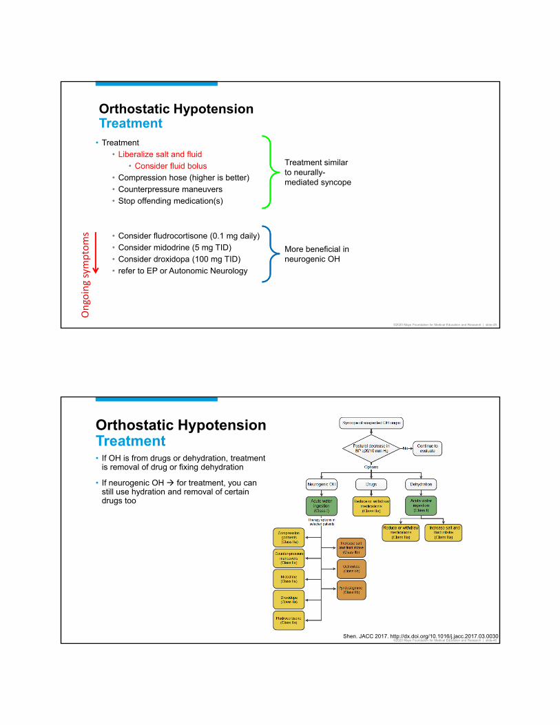
Orthostatic HypotensionTreatment
• Treatment• Liberalize salt and fluid
• Consider fluid bolus• Compression hose (higher is better)• Counterpressure maneuvers• Stop offending medication(s)
• Consider fludrocortisone (0.1 mg daily)• Consider midodrine (5 mg TID)• Consider droxidopa (100 mg TID)• refer to EP or Autonomic Neurology
Treatment similar to neurally-mediated syncope
More beneficial in neurogenic OH
Ongo
ing
sym
ptom
s
©2020 Mayo Foundation for Medical Education and Research | slide-46
Orthostatic HypotensionTreatment• If OH is from drugs or dehydration, treatment
is removal of drug or fixing dehydration
• If neurogenic OH for treatment, you can still use hydration and removal of certain drugs too
Shen. JACC 2017. http://dx.doi.org/10.1016/j.jacc.2017.03.0030
Page 24
©2020 Mayo Foundation for Medical Education and Research | slide-47
Case 3
• 70 year old man, who is touring Sedona, has abrupt syncope. He had maybe 1 second of warning symptoms (“curtains being lowered”) then was unconscious for 10 seconds before recovery. He had no other symptoms. There are no new medications. ECG shows new LBBB.
©2020 Mayo Foundation for Medical Education and Research | slide-48©2020 Mayo Foundation for Medical Education and Research | slide-48
Syncope BreakdownCardiac Arrhythmias
29%
17%54%
68%
10%
11%
5%
6%
Cardiac arrhythmias
Bradycardia
VT
SVT
Sorajja D. Circ 2009; 120:928.
Page 25
©2020 Mayo Foundation for Medical Education and Research | slide-49
Cardiac Arrhythmias
• SVT• Uncommon cause of syncope as its only symptom (usually HR > 115% of MPHR)
• Bradycardia (suspect if no warning symptoms)• If AV block, typically requires 2nd or 3rd degree AV block to cause syncope• Sinus bradycardia is unlikely especially if asymptomatic on monitoring
• Bundle branch block• If BBB, 50% have asystole and 50% have VT
©2020 Mayo Foundation for Medical Education and Research | slide-50
Case 3B
• 51 year old man with no complaints, and presents for his annual examination. He had syncope 4 months ago, but had a lot of alcohol that day.
Page 26
©2020 Mayo Foundation for Medical Education and Research | slide-51
Cardiac Arrhythmias
• NSVT rarely causes syncope, but obviously you are worried there was sustained VT previously
• Worrisome factors of NSVT• Polymorphic configuration (torsades de pointes)• RBBB VT• Prior MI• HCM• Low LVEF
• Not worrisome factors of NSVT• Non ischemic cardiomyopathy• Aortic stenosis (no additional risk for AS patients)
RBBB VT configuration Needs Further Evaluation
©2020 Mayo Foundation for Medical Education and Research | slide-52©2020 Mayo Foundation for Medical Education and Research | slide-52
ECG Features: What Bundle?
• RBBB predominantly (+) in V1 and V2:
• VT coming from LV
Similar to Right Bundle Branch Block
Page 27
©2020 Mayo Foundation for Medical Education and Research | slide-53
Cardiac ArrhythmiasEvaluation
• ECG
• Ambulatory monitoring (next slide)
• Echo: looking for depressed LVEF, ischemic cardiomyopathy, HCM• Do a stress ECHO if there is exertion-related syncope or suspect a relation to catecholamine
surge
• EP study
©2020 Mayo Foundation for Medical Education and Research | slide-54
Other Testing To ConsiderAmbulatory MonitoringLonger monitoring is more sensitive in detecting arrhythmias and conduction abnormalities, and then correlating findings to arrhythmias1
• Record now and analyze later• SmartWatch (< 1 minute)• Continuous ECG (Holter) monitor (1 - 2 days)• Event (loop) monitor (1 week - 30 days)• Patch monitor (3 - 30 days)• Implantable Loop Recorder (2 - 4 years)
• Record now and analyze now (real-time continuous monitoring transmitted to a station)• AliveCor (real-time only if patient still in arrhythmia)• Lead-based monitor (7-14 days)• Patch-based monitor (3-30 days)
Mobile Cardiac Outpatient Telemetry
1. Crawford. Circ. 1999; 100:8862. http://media.corporate-ir.net/media_files/IROL/25/251324/Images/Linq_AAA%20IMAGE-%20hi%20res.jpg
2
Page 28
©2020 Mayo Foundation for Medical Education and Research | slide-55
Cardiac Arrhythmias
• Treatment• VT
• Medical therapy / ICD• May need EP study / ablation
• SVT• Medical therapy• EP study / ablation
• Bradycardia (including tachy-brady syndrome) • Pacemaker
• Bundle branch block• EP study
• Medical therapy• Beta-blockers: Propranolol > metoprolol > sotalol• Calcium channel blockers (normal EF): diltiazem or verapamil• Amiodarone
©2020 Mayo Foundation for Medical Education and Research | slide-56
Loose Ends
6
©2020 Mayo Foundation for Medical Education and Research | slide-56
Page 29
©2020 Mayo Foundation for Medical Education and Research | slide-57
Syncope and ExerciseEvaluation• Evaluation is the same as for other etiologies
• Consider stress echocardiogram for exertional presyncope or syncope
• Consider cardiac MRI
• Consider tilt table for probable neurally-mediated physiology (syncope during recovery)• Establishes diagnosis and provides reassurance
• Ambulatory monitoring
©2020 Mayo Foundation for Medical Education and Research | slide-58
Syncope While Driving
• Prevalence of etiologies for syncope while driving is similar to syncope not while driving• Neurally-mediated syncope is still most common
• Recurrence is not predictable• 50% of recurrence occur after 6 months from initial event
• Driving restrictions vary from state to state• Know if it is your responsibility to report person to DMV• Arizona is driver self-report state
1. Sorajja D. Circ 2009; 120:928.
Page 30
©2020 Mayo Foundation for Medical Education and Research | slide-59
What Risk of Harm Is Acceptable?
• As a society, we accept a certain level of risk by letting higher risk drivers
• Young drivers: their risk is 0.007 or 0.7% per year• 2650 teenagers (aged 16-19) in the USA were killed by motor vehicle crashes1
• 292,000 teenagers injured in motor vehicle crashes1
• Elderly drivers: their risk is 0.005 or 0.5% per year• 5560 older adults (aged > 65 years) in the USA were killed by motor vehicle crashes• 214,000 older adults injured in motor vehicle crashes2
• The risk of young drivers causing an accident is about 142 times higher than what society accepts as the risk of the average driver losing consciousness and causing an accident
1. http://www.cdc.gov/motorvehiclesafety/teen_drivers/teendrivers_factsheet.html2. http://www.cdc.gov/motorvehiclesafety/older_adult_drivers/
©2020 Mayo Foundation for Medical Education and Research | slide-60
What Do You Need To Do For These Patients
• Check local laws and regulations
• Doctor-patient relationship may not be released in some countries without patient authorization
• Self-report by patients versus physician-report
• If you know withholding the information could endanger the patient or other parties, you cannot ethically withhold the information
• Reporting is done out of beneficence ---- not maleficence, therefore outweighing the principle of confidentiality
Page 31
©2020 Mayo Foundation for Medical Education and Research | slide-61
The EndThanks for the applause (if any)
©2020 Mayo Foundation for Medical Education and Research | slide-62
COPYRIGHT
Unauthorized duplication, distribution or exhibition of this program is an infringement of United States and International copyright laws.
Title 17, U.S. Copyright Code