ARTICLE Received 2 May 2013 | Accepted 21 Aug 2013 | Published 18 Sep 2013 MBNL1 and RBFOX2 cooperate to establish a splicing programme involved in pluripotent stem cell differentiation Julian P. Venables 1,2,3,4, *, Laure Lapasset 1,3,4,5,6, *, Gilles Gadea 3,4,7 , Philippe Fort 3,4,7 , Roscoe Klinck 8,9 , Manuel Irimia 10 , Emmanuel Vignal 3,4,7 , Philippe Thibault 8 , Panagiotis Prinos 8 , Benoit Chabot 8,9 , Sherif Abou Elela 8,9 , Pierre Roux 3,4,7 , Jean-Marc Lemaitre 3,4,5,6 & Jamal Tazi 1,3,4 Reprogramming somatic cells into induced pluripotent stem cells (iPSCs) has provided huge insight into the pathways, mechanisms and transcription factors that control differentiation. Here we use high-throughput RT–PCR technology to take a snapshot of splicing changes in the full spectrum of high- and low-expressed genes during induction of fibroblasts, from several donors, into iPSCs and their subsequent redifferentiation. We uncover a programme of concerted alternative splicing changes involved in late mesoderm differentiation and controlled by key splicing regulators MBNL1 and RBFOX2. These critical splicing adjustments arise early in vertebrate evolution and remain fixed in at least 10 genes (including PLOD2, CLSTN1, ATP2A1, PALM, ITGA6, KIF13A, FMNL3, PPIP5K1, MARK2 and FNIP1), implying that vertebrates require alternative splicing to fully implement the instructions of transcriptional control networks. DOI: 10.1038/ncomms3480 1 CNRS, Institut de Ge ´ne ´tique Mole ´culaire de Montpellier, Montpellier F-34293, France. 2 Institute of Genetic Medicine, Newcastle University, Newcastle- upon-Tyne NE1 3BZ, UK. 3 Universite ´ Montpellier 2, F-34293 Montpellier cedex 5, France. 4 Universite ´ Montpellier 1, F-34094 Montpellier cedex 5, France. 5 CNRS, Institut de Ge ´nomique Fonctionnelle de Montpellier, Montpellier F-34094, France. 6 INSERM, Montpellier F-34094, France. 7 CNRS, Centre de Recherche de Biochimie Macromole ´culaire de Montpellier, Montpellier F-34293, France. 8 Laboratoire de Ge ´nomique Fonctionnelle de l’Universite ´ de Sherbrooke, Sherbrooke, Quebec, Canada J1E 4K8. 9 De ´partement de microbiologie et d’infectiologie, Faculte ´ de me ´decine et des sciences de la sante ´, Universite ´ de Sherbrooke, Sherbrooke, Quebec, Canada J1E 4K8. 10 Banting and Best Department of Medical Research and Donnelly Centre, University of Toronto, Toronto, Ontario, Canada M5G 1L6. *These authors contributed equally to this work. Correspondence and requests for materials should be addressed to J.-M.L. (email: [email protected]) or to J.T. (email: [email protected]). NATURE COMMUNICATIONS | 4:2480 | DOI: 10.1038/ncomms3480 | www.nature.com/naturecommunications 1 & 2013 Macmillan Publishers Limited. All rights reserved.
Transcript
ARTICLE
Received 2 May 2013 | Accepted 21 Aug 2013 | Published 18 Sep 2013
MBNL1 and RBFOX2 cooperate to establisha splicing programme involved in pluripotentstem cell differentiationJulian P. Venables1,2,3,4,*, Laure Lapasset1,3,4,5,6,*, Gilles Gadea3,4,7, Philippe Fort3,4,7, Roscoe Klinck8,9,
Manuel Irimia10, Emmanuel Vignal3,4,7, Philippe Thibault8, Panagiotis Prinos8, Benoit Chabot8,9,
Sherif Abou Elela8,9, Pierre Roux3,4,7, Jean-Marc Lemaitre3,4,5,6 & Jamal Tazi1,3,4
Reprogramming somatic cells into induced pluripotent stem cells (iPSCs) has provided huge
insight into the pathways, mechanisms and transcription factors that control differentiation.
Here we use high-throughput RT–PCR technology to take a snapshot of splicing changes in
the full spectrum of high- and low-expressed genes during induction of fibroblasts, from
several donors, into iPSCs and their subsequent redifferentiation. We uncover a programme
of concerted alternative splicing changes involved in late mesoderm differentiation and
controlled by key splicing regulators MBNL1 and RBFOX2. These critical splicing adjustments
arise early in vertebrate evolution and remain fixed in at least 10 genes (including PLOD2,
CLSTN1, ATP2A1, PALM, ITGA6, KIF13A, FMNL3, PPIP5K1, MARK2 and FNIP1), implying that
vertebrates require alternative splicing to fully implement the instructions of transcriptional
control networks.
DOI: 10.1038/ncomms3480
1 CNRS, Institut de Genetique Moleculaire de Montpellier, Montpellier F-34293, France. 2 Institute of Genetic Medicine, Newcastle University, Newcastle-upon-Tyne NE1 3BZ, UK. 3 Universite Montpellier 2, F-34293 Montpellier cedex 5, France. 4 Universite Montpellier 1, F-34094 Montpellier cedex 5, France.5 CNRS, Institut de Genomique Fonctionnelle de Montpellier, Montpellier F-34094, France. 6 INSERM, Montpellier F-34094, France. 7 CNRS, Centre deRecherche de Biochimie Macromoleculaire de Montpellier, Montpellier F-34293, France. 8 Laboratoire de Genomique Fonctionnelle de l’Universite deSherbrooke, Sherbrooke, Quebec, Canada J1E 4K8. 9 Departement de microbiologie et d’infectiologie, Faculte de medecine et des sciences de la sante,Universite de Sherbrooke, Sherbrooke, Quebec, Canada J1E 4K8. 10 Banting and Best Department of Medical Research and Donnelly Centre, University ofToronto, Toronto, Ontario, Canada M5G 1L6. * These authors contributed equally to this work. Correspondence and requests for materials should beaddressed to J.-M.L. (email: [email protected]) or to J.T. (email: [email protected]).
Intense efforts are dedicated to the study of stem cells becauseof the potential medical benefits and the insights intodifferentiation that they offer1–4. Pluripotent stem cells can
be induced into ectoderm, endoderm and mesoderm, recapi-tulating the early events in embryogenesis5,6. Early differentiationinvolves the induction of cell line-specific transcription factors,whereas the late stages of differentiation are less wellcharacterized4,7–9. Alternative splicing is a means by which thegenome can control the expression of different protein isoformsthat can drive fundamental cellular changes and this is mostlyachieved through the differential expression of RNA-bindingproteins10–12. Ever since the discovery that alternative splicingcontrols Drosophila sex differentiation over 20 years ago, alter-native splicing has been suggested to be a potential mechanismwhereby different protein isoforms could be delivered toimplement mammalian differentiation13–16.
Several studies have also hinted at an involvement ofalternative splicing in pluripotency and pluripotent stem celldifferentiation along various pathways17,18. For example, differentisoforms of the forkhead transcription factor Foxp1 havedifferential effects on the induction of key pluripotency genessuch as Oct4 and Nanog19. Similarly, alternative splice forms ofDNMT3B are specific to stem cells, implying that layered andintegrated regulation of gene expression occurs at the levels oftranscription and splicing20. Current analyses of genome-widechanges in alternative splicing rely mostly on two techniques:microarrays and next-generation sequencing. However, thesetechniques are heavily biased towards the most highly expressedgenes and cannot quantify alternative splicing of medium- andlow-expressed genes that can be expressed at 10,000 times lowerlevels than housekeeping genes21. We recently developed aunique high-throughput RT–PCR platform to probe the entirerepertoire of over 3,000 alternative splice events annotated in theRefSeq database without bias against medium- and low-expressedgenes22, and further used this technique to probe the involve-ment of 81 different RNA-binding proteins in epithelial-to-mesenchymal transition by comparing the splicing profiles ofepithelial and mesenchymal tissues with those of the cells linestreated with siRNAs against the 81 proteins23.
Here we have used high-throughput RT–PCR to implicate theRNA-binding protein Muscleblind (MBNL1) in differentiation ofstem cells, and we show that essential induction of MBNL1 lateduring mesoderm differentiation controls a splicing programmethat induces and represses specific exons at the RNA level.
ResultsAlternative splicing programs in fibroblasts and stem cells. Toanalyse the programme of alternative splicing changes occurring inhuman pluripotent stem cells during stem cell induction, main-tenance and differentiation, we designed a large-scale screeningstrategy. Using high-throughput PCR, we previously screened theentire NCBI Reference Sequence (RefSeq) database for the pre-sence or absence of both of two predicted isoforms at thousands ofloci. From these, detected, alternatively spliced events we selected303 high-quality PCRs from across the alternative exons, usingonly purity and yield of the two PCR products in 10 different celllines as parameters We used these high-quality PCRs to studyalternative splicing in pluripotent stem cells in more detail becausethey occur in an essentially random and functionally diverse cross-section of genes22. Alternative splicing was studied in human skinfibroblasts and their induced pluripotent stem cells (iPSCs) andthen also in fibroblasts redifferentiated from those iPSCs24. Weobserved a reversible programme of alternative splicing (Fig. 1acorrelation R¼ � 0.88). Equally remarkably, the inclusion levels of15 of the 303 alternative exons shifted 450 percentage points
on average between pluripotent stem cells and their parentaland daughter fibroblasts (and 440% in both transitionsSupplementary Data 1). Such extreme shifts in alternative splicingare rarely described and are defined as ‘switch-like’25–27. Thus,using a randomly selected set of alternative splicing events we haveuncovered an unprecedented programme of reversible regulatedswitch-like alternative splicing in stem cells, whereby 5% of afunctionally random snapshot of all alternative splicing events canbe induced to shift reversibly from predominantly one isoform tonearly exclusively the other, between the fibroblastic andpluripotent states. Furthermore, there is a very good correlation(R¼ 0.824, Supplementary Fig. S2) between percent-spliced-invalues obtained with RNA-Seq data28 and our PCR-basedmethodology for those events in which RNA-seq data hadenough read depth to produce confident PSI estimates (104 outof 303 mapped events; Supplementary Data 1). To establish ifthese alternative splicing changes were a general feature of stemcells we performed manual end-point PCRs with the 15 switch-likesplicing events on three different pluripotent stem cells and theirresulting differentiated and parental fibroblasts. Samples includedthe well-characterized H9 embryonic stem cells and their redif-ferentiated fibroblasts. In all cases we observed a near completeand fully reversible shift from one isoform to the other betweenstem cells and fibroblasts (Fig. 1b).
Modulation of alternative splicing programs. To create a tool tounderstand the systems biology of splicing, we recently knockeddown 81 potential splicing factors in various cell lines andperformed RT–PCRs across 47 alternatively spliced regions indifferent genes23. Here we monitored these 47 splicing eventsusing RNA extracted from iPSCs and their parental fibroblasts24
with the aim of identifying the splicing factors involved inpluripotency and reprogramming (Supplementary Data 2).The splicing profile of these 47 PCRs showed an equivalentnear-perfect anti-correlation of R¼ � 0.91 in reversible stemcell induction and redifferentiation, demonstrating that thissmaller set of 47 alternative splice events can be used to investi-gate the splicing control mechanisms involved in stemnessand maintenance of pluripotency (Fig. 1c). The differences inpercent-spliced-in values, in these 47 alternative splice events,induced in pluripotency (iPSC values-original fibroblasts)were then compared with the differences induced when weknocked down 81 splicing potential factors (knockdown-control,Supplementary Data 3 and 4). Strikingly, the profile ofMBNL1 knockdown correlated most strongly (R¼ 0.6) withthe splicing profile of the induction of pluripotency (Fig. 2a,lines 1 and 2).
MBNL1 is a key gene involved in the myotonic dystrophies,DM1 and DM2, which were the first defined RNA diseases. Onenormal function of Muscleblind is to modulate splicing duringmuscle and heart development29–34. The second most correlatedknockdown was RBFOX2, which is a well-characterized splicingfactor that we and others have implicated in epithelial-to-mesenchymal transition and invasion22,23,34,35. To confirm theassociation of these two proteins with stem cell differentiation, weknocked down MBNL1 and RBFOX2 in fibroblasts, alone or incombination, and performed automated RT–PCR for the 303original alternative splicing events. We observed a significantcorrelation of splicing changes between individual MBNL1 andRBFOX2 knockdowns and the changes occurring upon inductionof the original fibroblasts into the pluripotent state (R¼ 0.4).Remarkably, the correlation was even higher (R¼ 0.6) betweensplicing changes associated with pluripotent stem cell derivationand those occurring in fibroblasts and a double-knockdownof MBNL1 and RBFOX2 (Fig. 2b). Manual PCR for the
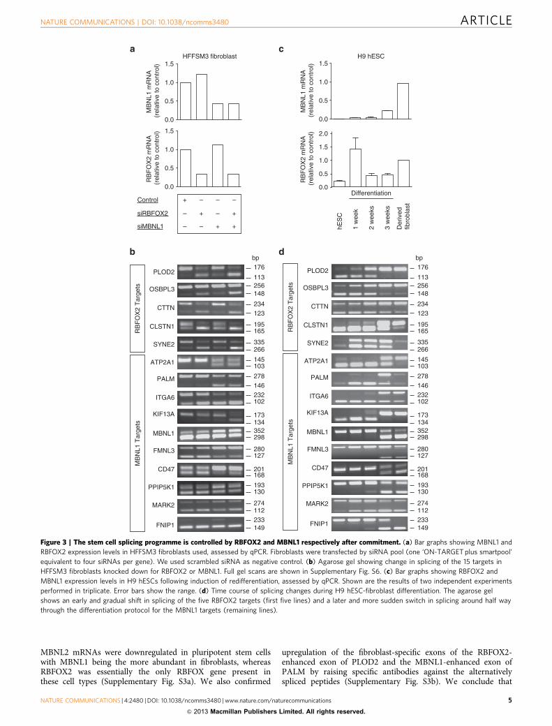
15 switch-like splice events further confirmed these results, withfive of the exons (PLOD2, OSBPL3, CTTN, CLSTN and SYNE2)strongly enhanced by RBFOX2 (that is, inclusion of these exonswas strongly inhibited by knockdown of RBFOX2), whereas theremaining 10 exons were either enhanced or inhibited by MBNL1(Fig. 3a,b).
Control of alternative splicing programs by MBNL1 andRBFOX2. One of the major questions in developmental biologyis to understand the events related to the specification of thedifferent cell lineages in the embryo. As pluripotent stem celldifferentiation is considered to occur in two phases (before andafter commitment)4, we sought to establish whether our splicingprogramme was associated with specification to committed stemcells or to the subsequent differentiation step. We studied alter-native splicing of the 15 switch-like targets in H9 human embryo-nic stem cells (hESCs) that differentiated into early ectoderm,endoderm and mesoderm precursors and surprisingly found onlyvery minor changes in alternative splicing of the 5 RBFOX2-controlled exons (first 5 lines of Supplementary Fig. S1a) and nochange at all for the 10 MBNL1-controlled exons. We confirmedthat the pluripotent stem cells had indeed committed to the three
germ layers using two marker genes for each type (SupplementaryFig. S1b, primers listed in Supplementary Table S1). To confirmthat the MBNL- and RBFOX-controlled splicing programme was alate event in differentiation we took samples of stem cells at 1, 2and 3 weeks into the 5-week differentiation protocol. qPCR ana-lysis demonstrated an early upregulation of RBFOX2 and a laterupregulation of MBNL1 (Fig. 3c). Consistent with this, manualend-point PCR demonstrated a gradual change in the fiveRBFOX2 targets, starting in the first week of differentiation,whereas the 10 MBNL1 targets changed later and more sharply,mostly between 2–3 weeks of differentiation (Fig. 3d). Changes insplicing were essentially complete after 3 weeks of differentiationconsistent with those changes occurring during the later stages ofmesoderm differentiation.
Next we checked whether MBNL1 and RBFOX2 were indeeddownregulated in pluripotent stem cells and we observed almostcomplete abrogation of MBNL1 expression and significantdownregulation of RBFOX2 at both the RNA and protein levelsin all stem cell types, irrespective of their provenance (Fig. 4;primers listed in Supplementary Table S1). Downregulation ofMBNL and RBFOX genes was also confirmed by retrospectiveanalysis of our previously published microarray data for allthree MBNL and RBFOX orthologues24. Both MBNL1 and
OSBPL3
CTTN
CLSTN1
PLOD2
FNIP1
MARK2
PPIP5K1
CD47
FMNL3
MBNL1
KIF13A
ITGA6
PALM
ATP2A1
SYNE2
hES
C
Der
ived
fibr
obla
st
Fib
robl
ast
iPS
C
HF
FS
M3
fibro
blas
t
Fib
robl
ast
iPS
C
Der
ived
fibr
obla
st
Der
ived
fibr
obla
st
–100 –50 50 100
–100
–50
50
100
Exon inclusion shift iPSC-parental fibroblast
Exo
n in
clus
ion
shift
der
ivat
ed fi
brob
last
-iPS
C
–100 –50 50 100
–100
–50
100
50E
xon
incl
usio
n sh
ift d
eriv
ated
fibr
obla
st-iP
SC
Exon inclusion shift iPSC-parental fibroblast
H9 Line 1 Line 2
113
256 148
176
123 195 165
234
bp
335 266145103
278146232102
173134
352298280127
201168
193130274112233149
Figure 1 | A reversible splicing programme in pluripotent stem cells. (a) Scatter plot showing reversible splicing changes in 303 exons in a diverse
array of genes. PCRs were performed on cDNA from parental fibroblasts, their induced pluripotent stem cells (iPSCs) and from re-differentiated fibroblasts
from these iPSCs. The transitions in splicing are shown on the x axis (iPSCs-parental fibroblasts) and y axis (re-differentiated fibroblasts-iPSCs)
respectively. (b) 15 switch-like splicing changes from (a) were assessed in the various fibroblast–to-stem cell transitions indicated. The upper and lower
bands on each gel represent the long (exon-included) and short (exon-omitted) isoforms respectively. Full gel scans are shown in Supplementary
Fig. 6. (c) The panel of 47 splice events from23 were also assayed for their splicing switches as in (a) to test their appropriateness for studying the splicing
Figure 2 | Implication of RBFOX2 and MBNL1 splicing factors in pluripotent stem cell induction. (a) Heat map showing percent-spliced-in shifts for the
47 alternate splicing events (x axis) between fibroblasts and iPSCs and when 81 different knockdowns were performed in two to five cell lines23. The
splicing factors are ordered according to the Pearson correlation (R) of their splicing profiles (k.d.—control, y axis) with that of pluripotent stem cell
induction (iPSC—parental fibroblast, x axis) on the first line. (b) Scatter plots comparing splicing changes for the 303 exons in pluripotent stem cell
induction with RBFOX2, MBNL1 and the double-knockdown (RBFOX2 and MBNL1) in HFFSM3 fibroblasts. Pearson correlations (R values) are shown.
MBNL2 mRNAs were downregulated in pluripotent stem cellswith MBNL1 being the more abundant in fibroblasts, whereasRBFOX2 was essentially the only RBFOX gene present inthese cell types (Supplementary Fig. S3a). We also confirmed
upregulation of the fibroblast-specific exons of the RBFOX2-enhanced exon of PLOD2 and the MBNL1-enhanced exon ofPALM by raising specific antibodies against the alternativelyspliced peptides (Supplementary Fig. S3b). We conclude that
MB
NL1
mR
NA
(re
lativ
e to
con
trol
)R
BF
OX
2 m
RN
A (
rela
tive
to c
ontr
ol)
HFFSM3 fibroblast
Control
siMBNL1
+
+ +
siRBFOX2 + +––
– ––
––
OSBPL3
CTTN
CLSTN1
PLOD2
FNIP1
MARK2
PPIP5K1
CD47
FMNL3
MBNL1
KIF13A
ITGA6
PALM
ATP2A1
SYNE2
RB
FO
X2
Tar
gets
MB
NL1
Tar
gets
H9 hESC
MB
NL1
mR
NA
(re
lativ
e to
con
trol
)R
BF
OX
2 m
RN
A (
rela
tive
to c
ontr
ol)
hES
C
1 w
eek
2 w
eeks
3 w
eeks
Differentiation
Der
ived
fibro
blas
t
OSBPL3
CTTN
CLSTN1
PLOD2
FNIP1
MARK2
PPIP5K1
CD47
FMNL3
MBNL1
KIF13A
ITGA6
PALM
ATP2A1
SYNE2
RB
FO
X2
Tar
gets
MB
NL1
Tar
gets
113256148
176
123
195165
234
bp
335266
145103
278146232102
173134352298
280127
201168
193130
274112233149
113256148
176
123
195165
234
bp
335266
145103
278146232102
173134352298
280127
201168
193130
274112233149
1.5
1.0
0.5
0.0
1.5
1.0
0.5
0.0
1.5
1.0
0.5
0.0
2.0
1.5
1.0
0.5
0.0
Figure 3 | The stem cell splicing programme is controlled by RBFOX2 and MBNL1 respectively after commitment. (a) Bar graphs showing MBNL1 and
RBFOX2 expression levels in HFFSM3 fibroblasts used, assessed by qPCR. Fibroblasts were transfected by siRNA pool (one ‘ON-TARGET plus smartpool’
equivalent to four siRNAs per gene). We used scrambled siRNA as negative control. (b) Agarose gel showing change in splicing of the 15 targets in
HFFSM3 fibroblasts knocked down for RBFOX2 or MBNL1. Full gel scans are shown in Supplementary Fig. S6. (c) Bar graphs showing RBFOX2 and
MBNL1 expression levels in H9 hESCs following induction of redifferentiation, assessed by qPCR. Shown are the results of two independent experiments
performed in triplicate. Error bars show the range. (d) Time course of splicing changes during H9 hESC-fibroblast differentiation. The agarose gel
shows an early and gradual shift in splicing of the five RBFOX2 targets (first five lines) and a later and more sudden switch in splicing around half way
through the differentiation protocol for the MBNL1 targets (remaining lines).
committed stem cells undergo a massive reversible reprogram-ming of splicing from inclusion or exclusion of exons to theopposite splice isoform during differentiation (45% of exonsassayed) and that the Muscleblind and Fox proteins (mostlyMBNL1 and RBFOX2) control the vast majority of thisprogramme.
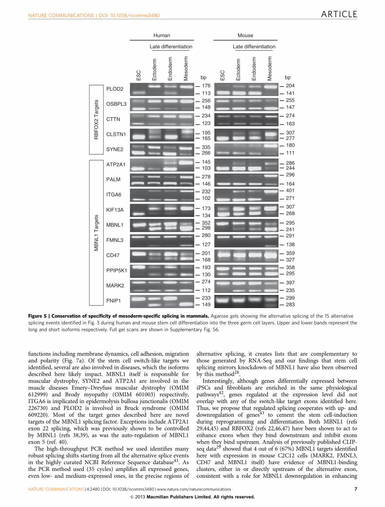
MBNL1 and RBFOX2 alternative splicing programs are con-served. To confirm the specificity of alternative splicing changesfor late mesoderm differentiation we compared splicing changesfor the 15 genes in mouse and human stem cells that we fullydifferentiated into late ectoderm, endoderm and mesoderm5. Inthis case we noted that 12 of the alternative splicing events (allexcluding OSBPL3, CTTN and CD47) shifted in human andmouse mesoderm and very few of these events changed inectoderm or endoderm (Fig. 5). Control markers for ectoderm,endoderm and mesoderm differentiation were verified by qPCR(Supplementary Fig. S4). We conclude that these 12 alternativesplice events undergo a conserved programme that qualitativelyregulates mesodermal gene expression. These results led us tosuspect that co-regulation of MBNL1 and RBFOX2 was necessaryfor the pluripotent stem cell splicing signature and for theirdifferentiation. To test this we inhibited their re-expression inpluripotent stem cells after 2 weeks of redifferentiation. Althoughwe did not observe any effect on the expression of specific mar-kers of pluripotency OCT4, NANOG and SOX2 (SupplementaryFig. S5a), we confirmed that splicing changes occurring lateduring differentiation can be reversed, either by individualknockdown of RBFOX2 and MBNL1 or by double-knockdown ofboth splicing factors (Fig. 6). The knockdowns in these sampleswere consistent with an early induction of RBFOX2 and a lateinduction of MBNL1.
Examination of the potential physiological roles of the 12switch-like exons in mammalian mesoderm revealed that nearlyall of them encode parts of proteins involved in membranedynamics, cell adhesion, migration and polarity (Fig. 7a). Tosupport the notion that the observed alternative isoforms haveimportant roles in controlling cellular morphology, we examinedtheir conservation further across evolution. All the genes probablyfulfil generic cell functions as they were already present in earlymetazoans or coelomates. Equally strikingly we found that 8 ofthe 12 alternatively spliced exons arose concurrently with geneduplications, mostly between jawless and jawed vertebrates(Fig. 7a). For the majority of genes, we found the presence ofalternative exons associated with alternative splicing afterduplications, whereas no exon homologue could be detectedbefore duplications, suggesting that exon gain and alternativesplicing were innovative features selected together to controldevelopment in jawed vertebrates. Similar findings on exonicsplicing elements and duplications were drawn from genome-wide studies36. Consistent with the function of these conservedsplicing events in membrane dynamics, cell adhesion, migrationand polarity, we observed a halving of cell size when eitherMBNL1 or RBFOX2 re-expression was inhibited during stemcell redifferentiation, countering the normal growth in cell sizeduring differentiation (Fig. 7b). Cells had less stress fibres withmore actin bundles (Supplementary Fig. S5b). Cells also hadreduced level of N-CADHERIN and interestingly increasedlevel of E-CADHERIN, an epithelial marker (Fig. 7c). Consistentwith E-CADHERIN re-expression, the RBFOX2 and MBNL1knockdowns trigger a reduction of the transcription factorTWIST, which represses the expression of E-CADHERIN37
(Supplementary Fig. S5c). We conclude therefore that coopera-tion between MBNL1 and RBFOX2 is essential for committedstem cell differentiation.
DiscussionGene expression programs are known to change during germ celldifferentiation. In this study we implicated a programme of geneticchanges, which occur at the level of alternative splicing of genesthat is essential for differentiation into one of the primary genelayers. The genes concerned are involved in important cellular
MB
NL1
mR
NA
(rel
ativ
e to
con
trol
)
0
0.5
1.0
1.5R
BF
OX
2 m
RN
A(r
elat
ive
to c
ontr
ol)
0
0.5
1.0
hES
C
Der
ived
fibr
obla
st
Fib
robl
ast
iPS
C
HF
FS
M3
fibro
blas
t
Fib
robl
ast
iPS
C
Der
ived
fibr
obla
st
Der
ived
fibr
obla
st
H9 Line 1 Line 2
MBNL1
RBFOX2
α TUBULIN
hES
C
Der
ived
fibr
obla
st
Fib
robl
ast
iPS
C
Fib
robl
ast
iPS
C
Der
ived
fibr
obla
st
Der
ived
fibr
obla
st
H9 Line 1 Line 2
kDa55
55
55
1.5
Figure 4 | Upregulation of RBFOX2 and MBNL1 expression after
commitment. (a) Bar graphs showing the expression level of MBNL1 and
RBFOX2 in parental fibroblasts, corresponding iPSCs, H9 hESCs and
fibroblast-differentiated cells (samples from Fig. 1b) assessed by qPCR.
(b) Western blots showing global expression of MBNL1 and RBFOX2.
Uncropped images of blots are shown in Supplementary Fig. S7.
functions including membrane dynamics, cell adhesion, migrationand polarity (Fig. 7a). Of the stem cell switch-like targets weidentified, several are also involved in diseases, which the isoformsdescribed here likely impact. MBNL1 itself is responsible formuscular dystrophy, SYNE2 and ATP2A1 are involved in themuscle diseases Emery–Dreyfuss muscular dystrophy (OMIM612999) and Brody myopathy (OMIM 601003) respectively,ITGA6 is implicated in epidermolysis bullosa junctionalis (OMIM226730) and PLOD2 is involved in Bruck syndrome (OMIM609220). Most of the target genes described here are noveltargets of the MBNL1 splicing factor. Exceptions include ATP2A1exon 22 splicing, which was previously shown to be controlledby MBNL1 (refs 38,39), as was the auto-regulation of MBNL1exon 5 (ref. 40).
The high-throughput PCR method we used identifies manyrobust splicing shifts starting from all the alternative splice eventsin the highly curated NCBI Reference Sequence database41. Asthe PCR method used (35 cycles) amplifies all expressed genes,even low- and medium-expressed ones, in the precise regions of
alternative splicing, it creates lists that are complementary tothose generated by RNA-Seq and our findings that stem cellsplicing mirrors knockdown of MBNL1 have also been observedby this method28.
Interestingly, although genes differentially expressed betweeniPSCs and fibroblasts are enriched in the same physiologicalpathways42, genes regulated at the expression level did notoverlap with any of the switch-like target exons identified here.Thus, we propose that regulated splicing cooperates with up- anddownregulation of genes43 to cement the stem cell-inductionduring reprogramming and differentiation. Both MBNL1 (refs29,44,45) and RBFOX2 (refs 22,46,47) have been shown to act toenhance exons when they bind downstream and inhibit exonswhen they bind upstream. Analysis of previously published CLIP-seq data29 showed that 4 out of 6 (67%) MBNL1 targets identifiedhere with expression in mouse C2C12 cells (MARK2, FMNL3,CD47 and MBNL1 itself) have evidence of MBNL1-bindingclusters, either in or directly upstream of the alternative exon,consistent with a role for MBNL1 downregulation in enhancing
Human Mouse
OSBPL3
CTTN
CLSTN1
FNIP1
MARK2
PPIP5K1
CD47
FMNL3
MBNL1
KIF13A
ITGA6
PALM
ATP2A1
SYNE2
PLOD2
Ect
oder
m
End
oder
m
Mes
oder
m
ES
C
Ect
oder
m
End
oder
m
Mes
oder
m
ES
C
Late differentiation Late differentiation
RB
FO
X2
Tar
gets
MB
NL1
Tar
gets
141 255 147
204
163
307277
274
bp
180
111
286244296
164401
271
307268
295241291
138
359327
358295
397
235
299283
113 256 148
176
123
195 165
234
bp
335 266
145103
278146
232102
173134352298280
127
201168
193130274
112
233149
Figure 5 | Conservation of specificity of mesoderm-specific splicing in mammals. Agarose gels showing the alternative splicing of the 15 alternative
splicing events identified in Fig. 3 during human and mouse stem cell differentiation into the three germ cell layers. Upper and lower bands represent the
long and short isoforms respectively. Full gel scans are shown in Supplementary Fig. S6.
these exons in stem cells (Supplementary Data 1). It is thereforelikely that binding sites for these proteins have been selected soonafter duplications, mostly between jawless and jawed vertebrates tocontrol exons important for cell differentiation in vertebrates. Insummary, our study reinforces the idea that splicing factors
determine the specific nature of a tissue and that the alternativesplicing process has been selected during evolution to control thelater stages of differentiation following cell cycle control andcommitment.
MethodsCell culture and in vitro differentiation assays. Human neonatal HFFSM3,line 1 and line 2 (ref. 24) fibroblasts (kind gift from J.M. Lemaitre, IGF Montpellier,France) were maintained in Dulbecco’s Modified Eagle Medium (DMEM, LifeTechnologies) containing 10% heat-inactivated fetal bovine serum (FBS, PAA),2 mM L-Glutamine, 1% penicillin and streptomycin (all from Life Technologies).H9 hESCs (WiCell Research) and iPSCs24 (kind gift from J.M. Lemaitre, IGFMontpellier, France) were maintained on matrigel (BD Biosciences) withchemically defined mTeSR (Stemcell Technologies) medium. E14 mouseEmbryonic Stem Cells (kind gift from T. Bouschet, IGF Montpellier, France) weremaintained on feeder-free culture on gelatin (Sigma-Aldrich) with chemicallydefined ESGRO complete PLUS clonal grade medium (Millipore).
In vitro differentiation. For the three germ layer differentiation inductions, H9hESCs were used and differentiated using the protocol described below and in aprevious study5.
Ectoderm model: H9 hESCs were seeded at 300,000 cells per well in six-wellplates pre-coated with matrigel (BD Biosciences) with chemically defined mTeSR(Stemcell Technologies) medium for 2 days. From days 3 to 9, H9 cells werecultured in the presence of N2 media (Life Technologies) and specific inhibitor ofSMAD pathway, SB431542 (TGFbeta receptor inhibitor) and Noggin (LifeTechnologies). The medium was changed daily from days 3 to 5 and every 2 daysfrom day 5 to day 9.
Endoderm model: H9 hESCs were seeded at 100,000 cells per well in six-wellplates pre-coated with matrigel (BD Biosciences) with chemically defined mTeSR(Stemcell Technologies) medium for 1 day. Differentiation was carried out inendoderm priming medium (DMEM 5.5 mM glucose, 10% KO-SR, 2 mMglutamine, 0.1 mM non essential amino-acid, 0,1 mM b-mercaptoethanol)supplemented with Activin A (100 ng ml� 1, Life Technologies), LY296002 (30 mM,Sigma-Aldrish) and FGF2 (10 ng ml� 1, Peprotech) from days 2 to 4, and Wnt3a(25 ng ml� 1, Life Technologies) from days 3 to 4. Then, cells were treated inhepatocyte-priming medium (DMEM 5.5 mM glucose, 2% KO-SR, 2 mMglutamine, 0.1 mM non essential amino-acid, 0,1 mM b-mercaptoethanol),supplemented with BMP4 (50 ng ml� 1, Life Technologies), FGF4 (25 ng ml� 1,Life Technologies) and 1% DMSO from days 5 to 9. Medium was changed daily inboth step of differentiation.
Mesoderm model: H9 hESCs were seeded at 100,000 cells cm� 2 in six-wellplates pre-coated with matrigel (BD Biosciences) with chemically definedmTeSR (Stemcell Technologies) medium for 1 day. Cells were cultured in DMEMor F12 medium supplemented with 10% FBS (Hyclone Laboratories), supple-mented with VEGF (20 ng ml� 1, Life Technologies). The medium was changedevery 2 days.
For early differentiation assay, differentiated cells were stopped at 10 days. Forlate differentiation assay, differentiated cells were re-seeded at 10 days and culturedin the same conditions for 10 more days.
For fibroblast differentiation, we used mesoderm model during 3 weeks duringwhich cells were re-seeded once a week. After 3 weeks, VEGF was removed andderivated fibroblasts were cultured as describe above.
The same protocols were used for mouse differentiation assays.
Knockdown of RBFOX2 and MBNL1. HFFSM3 fibroblasts and fibroblast-differ-entiated hESC H9 cells were transfected with siRNA ON-TARGET plus SMARTpools (Dharmacon, RBFOX2: L-020616-01-0005, MBNL1 L-014136-00-005), andscrambled siRNA (Dharmacon, D-001810-10-05). HFFSM3 fibroblasts were
MB
NL1
mR
NA
(re
lativ
e to
con
trol
)R
BF
OX
2 m
RN
A (
rela
tive
to c
ontr
ol)
0
1.5
0
20 days10 days
siMBNL1
+
+ +
siRBFOX2 + +
–
–
– –
–
–
–
+
+ +
+ +
–
–
– –
–
–
–
FNIP1
RB
FO
X2
Targ
ets
MB
NL1
Tar
gets
H9 hESC
Control
113256148
176
123195165
234
bp
335266
145103278146232102173134
352298280127201168193130274
112233149
MARK2
PPIP5K1
CD47
FMNL3
MBNL1
KIF13A
ITGA6
PALM
ATP2A1
SYNE2
CLSTN1
CTTN
OSBPL3
PLOD2
0.5
1.0
1.5
2.0
2.5
0.5
1.0
Figure 6 | Reversal of fibroblast splicing profile by inhibition of RBFOX2
and MBNL1 re-expression. (a) Bar graphs showing the knockdown
efficiency of RBFOX2 and MBNL1 in differentiating H9 hESCs at two
different time points, assessed by qPCR. Two treatments of siRNA pool
(one ‘ON-TARGET plus smartpool’ equivalent to 4 siRNAs per gene) were
applied at 2-day intervals before harvesting cells. The left hand and right
hand side represent siRNA pool applications commencing at either 7 or at
15 days after launching the differentiation. We used scrambled RNA as
negative control. Shown are means and s.d. of three independent
experiments performed in triplicate. (b) RT-PCRs for the 15 switch-like
exons were studied in the differentiating H9 hESCs knocked down for
RBFOX2 (siRBFOX2), MBNL1 (siMBNL1) or both. Full gel scans are shown in
transfected using INTERFERin (Polyplus Transfection). Fibroblast-differentiatedH9 cells were transfected twice every 2 days using DharmaFECT (ThermoScientific).
RNA preparation and analysis. RNA was isolated from cells using TRI reagent(Sigma-Aldrich). cDNA was generated by reverse transcription with MaximaReverse Transcriptase (Thermo Scientific). For qPCR assays, gene expressionanalysis was performed using 5 ng of cDNA and SYBR Green I Master (Roche).mRNA levels were normalized using the mean expression level of GAPDH, RPLO,RPL13A and PBGD. End-point RT–PCR was performed as in ref. 9. Briefly,reactions were carried out in the Eppendorf Mastercycler PCR Cycler. A first
cycle of 15 min at 95 �C was followed by 35 cycles of 30 s at 94 �C, 30 s at 55 �C, and1 min at 72 �C. The reaction was ended with the extension step of 10 min at72 �C. Visualization and analysis of amplified products were done using theLabChip HT DNA assay on an automated microfluidic station (Caliper,Hopkinton, MA, USA). For the 15 RBFOX2/MBNL1 selected targets, the amplifiedproducts were loaded on 1.5% agarose gel. All primers are detailed inSupplementary Table S1 and Supplementary Data 1.
Immunofluorescence analysis. Cells plated on cover slips were fixed for 10 min in3.7% formalin (in PBS) followed by a 2-min permeabilization with 0.1% TritonX-100 (in PBS) and incubation in PBS containing 0.1% bovine serum albumin.F-actin was revealed using rhodamin-conjugated phalloidin (Sigma-Aldrich) andnuclei were stained using Hoechst 33342 (Sigma-Aldrich). Cells were washed inPBS and water, mounted with DAKO mounting medium and observed under thefluorescence microscope.
Cell imaging was performed with a Leica DM6000 (Leica, Wetzlae, Germany)with PL APO grade oil � 20 objective. Images were captured with a Coolsnap HQ2camera (Roper Scientific Inc.) driven by Metamorph (Molecular Devices) andprocessed using FiJi software and cell surface area was determined using themeasure module of FiJi.
Immunoblot analysis. For whole-cell extracts, cells were lysed in RIPA buffer orurea buffer depending on the antibody used (RIPA extract for MBNL1, RBFOX2,a-TUBULIN; urea extract for PLOD2, PALM, actin). For RIPA extracts, cellswere lysed in RIPA buffer (50 mM Tris–HCl pH 8, 1% (w/v) NP-40, 1% (w/v) SDS,0.5% (w/v) Na-deoxycholate (DOC), 2 mM EDTA) supplemented with ‘proteaseinhibitor cocktail’ (Sigma), incubated for 30 min at 4 �C, centrifuged for 30 min at14,000 g and the protein amounts were quantifed using Bradford method. For ureaextracts, cells were lysed in urea buffer (63 mM Tris–HCl pH7.5, 2% (w/v) SDS, 5%(v/v) 2-mercaptoethanol, 8 M Urea), supplemented with ‘protease inhibitor cock-tail’. Cell lysates were next sonicated and centifuged. Supernatants were resolvedby SDS–PAGE and transferred to PVDF membranes (Amersham Biosciences).Membranes were blocked in PBS containing 5% (w/v) milk powder and 0.05%(v/v) Tween-20 (PBST) for 1 h and incubated with primary antibodies at 4 �Covernight. After washing with PBST, membranes were incubated with eitheranti-mouse or anti-rabbit secondary antibodies conjugated to horseradish perox-idase (HRP) for 1 h at room temperature. Signals were detected with ‘chemilu-minescent’ (Thermo Scientific), anti-RBFOX2 (1:100, ab51361, Abcam),anti-MBNL1 (gift from Wolfson Centre for Inherited Neuromuscular DiseaseMB1a-Mouse Anti-MBNL1 (ref. 48)), anti-b-actin (1:20,000, AC-74 clone, Sigma),anti-a tubulin (1:15,000, DM1A clone Sigma), anti-mouse IgG-HRP (1:10,000,0867428, MP Biomedicals) or anti-rabbit IgG-HRP (1:10,000, 0861202, MPBiomedicals). Polyclonal antibodies to human PLOD2 and PALM alternativepeptides were generated by CovalAb, Lyon-France by injecting rabbits withsynthetic peptides spanning amino acids 446-TLQREKDSPTPETFQMLSPPK-466 and 175-TVEKDKVTGETRVLSST-191, respectively. Antibodies wereaffinity-purified using a Sepharose bead column conjugated to the relevant peptide.
Gene ontology processSYNE2
Fibroblast migrationFMNL3
Cell migrationCLSTN1
Homophilic cell adhesionPLOD2
ECM organizationFNIP1
mTOR signallingMARK2
Cell polarityPALM Filopodium assemblyATP2A1 Transmembrane transportITGA6 Cell adhesionKIF13A Endosome-lysosome transport
MBNL1 RNA splicing
PPIP5K1 Inositol metabolism
AS splicing confirmed
Presence of the alternative exon
Incomplete data
Presence of orthologous gene and absence of AS exon
Duplications
siMBNL1pool
siRBFOX2pool
siDouble
Cel
l sur
face
(μm
2 )
0
5,000
10,000
15,000
20,000 ***
***
***
*** P < 0.0001
Control
BRACHYURYMIXL1N-CADHERINE-CADHERIN
siMBNL1pool
siRBFOX2pool
siDouble
Bony
verte
brat
es
Chord
ates
Mam
mal
sBat
racia
ns
Lam
prey
Ecdyz
ozoa
ns
Human
0.0
0.5
1.0
1.5
mR
NA
leve
l(r
elat
ive
to c
ontr
ol)
Control
Figure 7 | Alternative splicing of RBFOX2/MBNL1 target genes is
conserved in teleostomes and is required for fibroblast differentiation.
(a) Metazoan genomes deposited in Ensembl were searched for orthologs
and paralogs of each target gene and for the presence (yellow) or absence
(grey) of potential AS exons (Accession numbers in Supplementary
Table S2). Yellow indicates that alternatively spliced mRNAs were detected
in EST databases (orange indicates that EST number was too low to
conclude). Green indicates missing genomic and EST data. Duplications are
indicated by thick lines. (b) H9 hESCs were differentiated for 2 weeks and
then treated twice every 2 days with an siRNA pool against RBFOX2,
MBNL1 or both (siDouble). We used scrambled RNA as negative control
(Control). Cells were stained with phalloidin (anti-actin) and Hoechst stain
was used for nuclear staining to quantify the effect of RBFOX2 and/or
MBNL1 silencing on hESC-derived fibroblast morphology. Box-plots show
the average area of 33 cells for each condition. Whiskers correspond to the
5–95 percentiles, boxes, to the 25–75 percentiles and the band inside
the box, to the median. Statistical analysis was performed using an unpaired
t-test (***Po0.0001, n¼ 2). (c) Bar graph showing the expression levels
of differentiation markers BRACHYURY, MIXL1, N-CADHERIN and
E-CADHERIN in H9 hESCs after the knockdowns at 2 weeks into the
fibroblast differentiation, assessed by qPCR. Shown are the results of
two independent experiments performed in triplicate. Error bars show
References1. Park, I.-H. et al. Disease-specific induced pluripotent stem cells. Cell 134,
877–886 (2008).2. Robinton, D. A. & Daley, G. Q. The promise of induced pluripotent stem cells
in research and therapy. Nature 481, 295–305 (2012).3. Wu, S. M. & Hochedlinger, K. Harnessing the potential of induced pluripotent
stem cells for regenerative medicine. Nat. Cell Biol. 13, 497–505 (2011).4. Evseenko, D. et al. Mapping the first stages of mesoderm commitment during
differentiation of human embryonic stem cells. Proc. Natl Acad. Sci. USA 107,13742–13747 (2010).
5. Ramirez, J.-M. et al. Brief report: benchmarking human pluripotent stem cellmarkers during differentiation into the three germ layers unveils a strikingheterogeneity: all markers are not equal. Stem Cells 29, 1469–1474 (2011).
6. Kroon, E. et al. Pancreatic endoderm derived from human embryonicstem cells generates glucose-responsive insulin-secreting cells in vivo.Nat. Biotechnol. 26, 443–452 (2008).
7. Neph, S. et al. Circuitry and dynamics of human transcription factor regulatorynetworks. Cell 150, 1274–1286 (2012).
8. Loh, Y.-H. et al. The Oct4 and Nanog transcription network regulatespluripotency in mouse embryonic stem cells. Nat. Genet. 38, 431–440 (2006).
9. Boyer, L. A. et al. Core transcriptional regulatory circuitry in human embryonicstem cells. Cell 122, 947–956 (2005).
10. Maniatis, T. & Tasic, B. Alternative pre-mRNA splicing and proteomeexpansion in metazoans. Nature 418, 236–243 (2002).
11. Anko, M.-L. & Neugebauer, K. M. RNA-protein interactions in vivo: global getsspecific. Trends Biochem. Sci. 37, 255–262 (2012).
12. Barash, Y. et al. Deciphering the splicing code. Nature 465, 53–59 (2010).13. Venables, J. P., Tazi, J. & Juge, F. Regulated functional alternative splicing in
Drosophila. Nucleic Acids Res. 40, 1–10 (2012).14. Black, D. L. Mechanisms of alternative pre-messenger RNA splicing. Annu.
Rev. Biochem. 72, 291–336 (2003).15. Ule, J. et al. An RNA map predicting Nova-dependent splicing regulation.
Nature 444, 580–586 (2006).16. Xu, X. et al. ASF/SF2-regulated CaMKIIdelta alternative splicing temporally
reprograms excitation-contraction coupling in cardiac muscle. Cell 120, 59–72(2005).
17. Salomonis, N. et al. Alternative splicing regulates mouse embryonic stem cellpluripotency and differentiation. Proc. Natl Acad. Sci. USA 107, 10514–10519(2010).
18. Nelles, D. A. & Yeo, G. W. Alternative splicing in stem cell self-renewal anddiferentiation. Adv. Exp. Med. Biol. 695, 92–104 (2010).
19. Gabut, M. et al. An alternative splicing switch regulates embryonic stem cellpluripotency and reprogramming. Cell 147, 132–146 (2011).
20. Gopalakrishna-Pillai, S. & Iverson, L. E. A DNMT3B alternatively spliced exonand encoded peptide are novel biomarkers of human pluripotent stem cells.PLoS One 6, e20663 (2011).
21. Schwanhausser, B. et al. Global quantification of mammalian gene expressioncontrol. Nature 473, 337–342 (2011).
22. Venables, J. P. et al. Cancer-associated regulation of alternative splicing. Nat.Struct. Mol. Biol. 16, 670–676 (2009).
23. Venables, J. P. et al. RBFOX2 is an important regulator of mesenchymal tissue-specific splicing in both normal and cancer tissues. Mol. Cell Biol. 33, 396–405(2013).
24. Lapasset, L. et al. Rejuvenating senescent and centenarian human cells byreprogramming through the pluripotent state. Genes Dev. 25, 2248–2253 (2011).
25. Xing, Y. & Lee, C. Alternative splicing and RNA selection pressure—evolutionaryconsequences for eukaryotic genomes. Nat. Rev. Genet. 7, 499–509 (2006).
26. Xing, Y. & Lee, C. J. Protein modularity of alternatively spliced exons is associatedwith tissue-specific regulation of alternative splicing. PLoS Genet. 1, e34 (2005).
27. Wang, E. T. et al. Alternative isoform regulation in human tissuetranscriptomes. Nature 456, 470–476 (2008).
28. Han, H. et al. MBNL proteins repress ES-cell-specific alternative splicing andreprogramming. Nature 498, 241–245 (2013).
29. Wang, E. T. et al. Transcriptome-wide regulation of pre-mRNA splicing andmRNA localization by muscleblind proteins. Cell 150, 710–724 (2012).
30. Rinaldi, F. et al. Aberrant splicing and expression of the non muscle myosin heavy-chain gene MYH14 in DM1 muscle tissues. Neurobiol. Dis. 45, 264–271 (2012).
31. Lin, X. et al. Failure of MBNL1-dependent post-natal splicing transitions inmyotonic dystrophy. Hum. Mol. Genet. 15, 2087–2097 (2006).
32. Kalsotra, A. et al. A postnatal switch of CELF and MBNL proteins reprogramsalternative splicing in the developing heart. Proc. Natl Acad. Sci. USA 105,20333–20338 (2008).
33. Fugier, C. et al. Misregulated alternative splicing of BIN1 is associated withT tubule alterations and muscle weakness in myotonic dystrophy. Nat. Med. 17,720–725 (2011).
34. Venables, J. P., Vignal, E., Baghdiguian, S., Fort, P. & Tazi, J. Tissue-specificalternative splicing of Tak1 is conserved in deuterostomes. Mol. Biol. Evol. 29,261–269 (2012).
35. Shapiro, I. M. et al. An EMT-driven alternative splicing program occurs inhuman breast cancer and modulates cellular phenotype. PLoS Genet. 7,e1002218 (2011).
36. Zhang, Z. et al. Divergence of exonic splicing elements after gene duplicationand the impact on gene structures. Genome Biol. 10, R120 (2009).
37. Yang, J. et al. Twist, a master regulator of morphogenesis, plays an essential rolein tumor metastasis. Cell 117, 927–939 (2004).
38. Kimura, T. et al. Altered mRNA splicing of the skeletal muscle ryanodinereceptor and sarcoplasmic/endoplasmic reticulum Ca2þ -ATPase in myotonicdystrophy type 1. Hum. Mol. Genet. 14, 2189–2200 (2005).
39. Hino, S.-I. et al. Molecular mechanisms responsible for aberrant splicing ofSERCA1 in myotonic dystrophy type 1. Hum. Mol. Genet. 16, 2834–2843 (2007).
40. Gates, D. P., Coonrod, L. A. & Berglund, J. A. Autoregulated splicing ofmuscleblind-like 1 (MBNL1) Pre-mRNA. J. Biol. Chem. 286, 34224–34233 (2011).
41. NCBI Resource Coordinators. Database resources of the National Center forBiotechnology Information. Nucleic Acids Res. 41, D8–D20 (2013).
42. Boue, S., Paramonov, I., Barrero, M. J. & Izpisua Belmonte, J. C. Analysis ofhuman and mouse reprogramming of somatic cells to induced pluripotent stemcells. What is in the plate? PLoS One 5, e12664 (2010).
43. Pan, Q. et al. Revealing global regulatory features of mammalian alternativesplicing using a quantitative microarray platform. Mol. Cell 16, 929–941 (2004).
44. Goers, E. S., Purcell, J., Voelker, R. B., Gates, D. P. & Berglund, J. A. MBNL1binds GC motifs embedded in pyrimidines to regulate alternative splicing.Nucleic Acids Res. 38, 2467–2484 (2010).
45. Charizanis, K. et al. Muscleblind-like 2-mediated alternative splicing in thedeveloping brain and dysregulation in myotonic dystrophy. Neuron 75,437–450 (2012).
46. Yeo, G. W. et al. An RNA code for the FOX2 splicing regulator revealed bymapping RNA-protein interactions in stem cells. Nat. Struct. Mol. Biol. 16,130–137 (2009).
47. Zhang, C. et al. Defining the regulatory network of the tissue-specific splicingfactors Fox-1 and Fox-2. Genes Dev. 22, 2550–2563 (2008).
48. Holt, I. et al. Defective mRNA in myotonic dystrophy accumulates at theperiphery of nuclear splicing speckles. Genes Cells 12, 1035–1048 (2007).
AcknowledgementsThis work was supported by the collaborative laboratory Splicos therapeutics, andInstitut National du Cancer (Canceropole Ile-de-France). L.L. was supported by afellowship from Fondation pour la Recherche Medicale (FRM) grant (Equipe FRM2011 –no. DEQ20111223745, DCR20091217183), l’Association pour la Recherche contrele Cancer ARC (A09/4/5062 and SFI20111203957) and la Ligue regionale contre leCancer (LNCC PYRENEES-ORIENTALES 2010, 2012). This work was also funded bygrants from the Canadian Cancer Society and the Canadian Institute for HealthResearch. S. Abou Elela is the Canada Research Chair in RNA Biology and CancerGenomics. B. Chabot is the Canada Research Chair in Functional Genomics. Thisresearch using hESC has been authorized by the French Biomedical Agency (ABM) forJM Lemaitre Laboratory, (NOR: SJSB0721833S). We thank Mathieu Durand and ElvyLapointe for technical assistance. We thank Wolfson Centre for inherited NeuromuscularDisease for providing anti-MBNL1 antibody. We thank T. Bouschet for the gift of E14mouse ES line. We are grateful to S. Alaoui from Covalab for design of immunogenicpeptides and production of specific antibodies against PLOD2 and PALM splicingisoforms. We thank B.J. Blencowe for comments on the manuscript. We acknowledgeMontpellier RIO Imaging (MRI) for the imaging acquisition and analysis.
Author contributionsL.L. perfomed pluripotent stem cell experiments, differentiation assays and siRNAexperiments on pluripotent stem cells. J.P.V. performed RT-PCR and G.G. performedsiRNA experiments on fibroblasts. L.L. and G.G. performed immunofluorescenceexperiments, qPCR and analysis. E.V. contributed to the analysis of cell area and sta-tistical analysis. J.P.V., P.T., R.K., P.P. and B.C. designed high-throughput RT–PCRexperiments. J.P.V., R.K., P.T., P.P., B.C. and S.A.E. contributed to the analysis of thedata. M.I. analysed RNA-seq and CLIP-seq data. P.F. analysed splicing evolution andgene ontology. J.P.V., L.L., G.G., P.R., J.-M.L. and J.T. were involved in study design andmanuscript preparation, performed experiments and analysed data. All authors discussedthe results and commented on the manuscript.
Additional informationSupplementary information accompanies this paper at http://www.nature.com/naturecommunications
Competing financial interests: The authors declare no competing financial interests.
Reprints and permission information is available online at http://npg.nature.com/reprintsandpermissions/
How to cite this article: Venables, J. P. et al. MBNL1 and RBFOX2 cooperate to establisha splicing programme involved in pluripotent stem cell differentiation. Nat. Commun.4:2480 doi: 10.1038/ncomms3480 (2013).