+ All Categories
Home > Documents > MECHANICS OF RESPIRATORY PUMPS - Brown University€¦ · 1980, 1988; Graham, 1997). Gas exchange...

MECHANICS OF RESPIRATORY PUMPS - Brown University€¦ · 1980, 1988; Graham, 1997). Gas exchange...

Date post: 26-Jul-2018
Category:
Upload: doanhuong
View: 216 times
Download: 0 times
Share this document with a friend
31
Comp. by:bala Date:10/8/05 Time:18:35:55 Stage:First Proof File Path://spsind002s/ serials/PRODENV/000000~1/00E256~2/S00000~1/00D23E~1/000000~2/000009578.3D Proof by:Subha QC by:Alakesh ProjectAcronym:bs:FP Volume:23001 UNCORRECTED PROOF 1 MECHANICS OF RESPIRATORY PUMPS ELIZABETH L. BRAINERD LARA A. FERRYGRAHAM I. Introduction II. Aquatic Respiratory Pumps A. TwoPhase Pump in Actinopterygian Fishes B. TwoPhase Pump in Elasmobranch Fishes C. Ram Ventilation D. Gill Ventilation in Lamprey and Hagfish III. Aerial Respiratory Pumps A. Evolutionary History and Biomechanical Challenges B. Air Ventilation Mechanics IV. Future Directions I. INTRODUCTION To facilitate oxygen uptake and carbon dioxide excretion, fishes ventilate their gas exchange surfaces with water or air. Because water and air diVer substantially in their density, viscosity, and oxygen content, the biomechani- cal problems associated with aquatic and aerial ventilation also diVer. Nonetheless, aerial and aquatic respiratory pumps do share one biomechan- ical challenge stemming from the fact that muscles only generate force in the direction of shortening (Brainerd, 1994b). It is a simple matter for muscle contraction to generate positive pressure and force fluid out of a cavity, but respiratory pumps also require an expansive phase to refill the cavity with new fluid. Some biomechanical trickery is necessary for muscle shortening to cause the expansion of a cavity and the generation of subambient pressure. This trickery generally takes the form of a lever system or occasionally elastic recoil, as is described for aquatic and aerial respiratory pumps in Sections II and III below. 1 Tuna : Volume 23 Copyright # 2005 Elsevier Inc. All rights reserved FISH PHYSIOLOGY DOI: 10.1016/S1546-5098(05)23001-7
Transcript

Comp. by:bala Date:10/8/05 Time:18:35:55 Stage:First Proof File Path://spsind002s/serials/PRODENV/000000~1/00E256~2/S00000~1/00D23E~1/000000~2/000009578.3DProof by:Subha QC by:Alakesh ProjectAcronym:bs:FP Volume:23001

UNCORRECTEDPROOF

1

MECHANICS OF RESPIRATORY PUMPS

ELIZABETH L. BRAINERD

LARA A. FERRY‐GRAHAM

I. Introduction

II. Aquatic Respiratory Pumps

A. Two‐Phase Pump in Actinopterygian Fishes

B. Two‐Phase Pump in Elasmobranch Fishes

C. Ram Ventilation

D. Gill Ventilation in Lamprey and Hagfish

III. Aerial Respiratory Pumps

A. Evolutionary History and Biomechanical Challenges

B. Air Ventilation Mechanics

IV. Future Directions

I. INTRODUCTION

To facilitate oxygen uptake and carbon dioxide excretion, fishes ventilate

their gas exchange surfaces with water or air. Because water and air diVersubstantially in their density, viscosity, and oxygen content, the biomechani-

cal problems associated with aquatic and aerial ventilation also diVer.Nonetheless, aerial and aquatic respiratory pumps do share one biomechan-

ical challenge stemming from the fact that muscles only generate force in the

direction of shortening (Brainerd, 1994b). It is a simple matter for muscle

contraction to generate positive pressure and force fluid out of a cavity, but

respiratory pumps also require an expansive phase to refill the cavity with

new fluid. Some biomechanical trickery is necessary for muscle shortening to

cause the expansion of a cavity and the generation of subambient pressure.

This trickery generally takes the form of a lever system or occasionally

elastic recoil, as is described for aquatic and aerial respiratory pumps in

Sections II and III below.

1

Tuna : Volume 23 Copyright # 2005 Elsevier Inc. All rights reservedFISH PHYSIOLOGY DOI: 10.1016/S1546-5098(05)23001-7

Comp. by:bala Date:10/8/05 Time:18:35:56 Stage:First Proof File Path://spsind002s/serials/PRODENV/000000~1/00E256~2/S00000~1/00D23E~1/000000~2/000009578.3DProof by:Subha QC by:Alakesh ProjectAcronym:bs:FP Volume:23001

UNCORRECTEDPROOF

The primary biomechanical problems in the design of aquatic respiratory

pumps stem from the physical and chemical properties of water: high density

(1000 kg m�3 for fresh water), high viscosity (1.0 � 10�3 Pa s for fresh water

at 20 �C), and low oxygen content (from 0.4% by volume in seawater at 30 �Cto 1% by volume in fresh water at 0 �C when in equilibrium with air). To

minimize the work of ventilation, the high density of water dictates that the

respiratory medium should undergo as little acceleration and deceleration as

possible, the high viscosity dictates that fluid velocities should be low, and

the low oxygen content dictates that oxygen extraction eYciency should be

high. The unidirectional flow, countercurrent gas exchange system of ray‐finned and cartilaginous fishes is well designed to meet these requirements

(Hughes and Shelton, 1962). Buccal and opercular pumps, as described in

Section II, generally work together to produce unidirectional flow of water

over the gills, but some interesting cases of momentary flow reversal have

recently been discovered (Summers and Ferry‐Graham, 2001).

In contrast to water, air has low density (1.2 kg m�3 at 20 �C), lowviscosity (0.02 � 10�3 Pa s at 20 �C), and high oxygen content (21% by

volume). Aerial gas exchange is a primitive characteristic for ray‐finnedfishes that was lost in basal euteleosts and that has re‐evolved at least 38

times and possibly as many as 67 times within acanthomorph fishes (Liem,

1980, 1988; Graham, 1997). Gas exchange organs include lungs, respiratory

gas bladders, skin, gills, and various air‐breathing organs (ABOs) such as the

labyrinth organs of anabantoids (Liem, 1980; Graham, 1997). The bio-

mechanical challenges for aerial respiratory pumps stem from predation risk

(because fishes are vulnerable when they go to the surface to breathe and

thus must limit their time there), hydrostatic pressure, buoyancy, surface

tension, and mechanical conflicts between breathing and feeding. As

described in Section III, the solutions to these problems are diverse.

II. AQUATIC RESPIRATORY PUMPS

In fish gills, the exchange of dissolved gases between water and blood

occurs on the surface of tiny, leaf‐like projections—the secondary lamellae.

Water is pumped over the secondary lamellae in a direction opposite to the

direction of the blood moving through the vessels of the secondary lamellae

(Hughes and Shelton, 1962). This countercurrent flow of water and blood

produces much greater oxygen extraction from the water than would be

produced by concurrent flow. When the flow is concurrent, water and blood

quickly reach diVusion equilibrium and no more oxygen can be extracted. In

countercurrent flow, even though diVusion is occurring, the partial pressure

of oxygen in the water is always slightly higher than the partial pressure of

2 ELIZABETH L. BRAINED AND LARA A. FERRY-GRAHAM

Comp. by:bala Date:10/8/05 Time:18:35:56 Stage:First Proof File Path://spsind002s/serials/PRODENV/000000~1/00E256~2/S00000~1/00D23E~1/000000~2/000009578.3DProof by:Subha QC by:Alakesh ProjectAcronym:bs:FP Volume:23001

UNCORRECTEDPROOF

the oxygen in the blood, allowing extraction of a high percentage of the

oxygen from the water. Countercurrent gas exchange results in oxygen

partial pressures that are higher in the blood leaving the lamellae and

entering the body than in the water exiting the gill slits. Fishes are the only

vertebrates that can achieve such high percentages of oxygen extraction from

their respiratory medium (Piiper and Scheid, 1992).

The eYciency of countercurrent exchange depends on the ability of the

aquatic respiratory pumps to produce unidirectional flow of water over the

gills. In both actinopterygian and elasmobranch fishes, unidirectional flow is

achieved with a two‐phase pump system.

A. Two‐Phase Pump in Actinopterygian Fishes

The two‐phase pump models of aquatic ventilation come from the pio-

neering work of G. M. Hughes (Hughes and Shelton, 1958; Hughes, 1960a,b,

1966, 1970, 1978a,b; Hughes and Ballintijn, 1965; Hughes and Umezawa,

1968; Hughes and Morgan, 1973). In Hughes’s models, the buccal and

opercular cavities are depicted as pistons (Figure 1.1). The movement of a

piston to increase or decrease the volume inside a chamber mimics the

expansion and compression of the buccal and opercular cavities during

normal ventilation. In the two‐phase model, the suction pump phase begins

with the opercular cavity compressed and just beginning to expand, causing

the pressure inside to be lower than ambient and somewhat lower than the

pressure in the buccal cavity (Figure 1.1, stage 1). This expansion of the

opercular cavity results in water being drawn into the mouth, over the gills,

and into the opercular cavity. At the start of the pressure pump phase, the

buccal cavity begins to compress while the opercular cavity continues

to expand (Figure 1.1, stage 2). Subsequently, the buccal cavity reaches

Fig. 1.1. The two‐phase pump model of aquatic ventilation as developed by Hughes (1960a,b):

stage 1, start of suction pump phase; stage 2, transition from suction to pressure pump; stage 3,

pressure pump phase; stage 4, transition from pressure to suction pump phase. During the stage

4 transition, pressure may be momentarily higher in the opercular than in the buccal cavity.

Flow reversal may result from the pressure reversal, or adduction of the gill bars may pose

enough resistance to block backflow. (Adapted from Ferry‐Graham, 1999, Figure 6, p. 1507 and

Summers and Ferry‐Graham, 2002, Figure 4 p. 96].)

1. RESPIRATION 3

Comp. by:bala Date:10/8/05 Time:18:35:56 Stage:First Proof File Path://spsind002s/serials/PRODENV/000000~1/00E256~2/S00000~1/00D23E~1/000000~2/000009578.3DProof by:Subha QC by:Alakesh ProjectAcronym:bs:FP Volume:23001

UNCORRECTEDPROOF

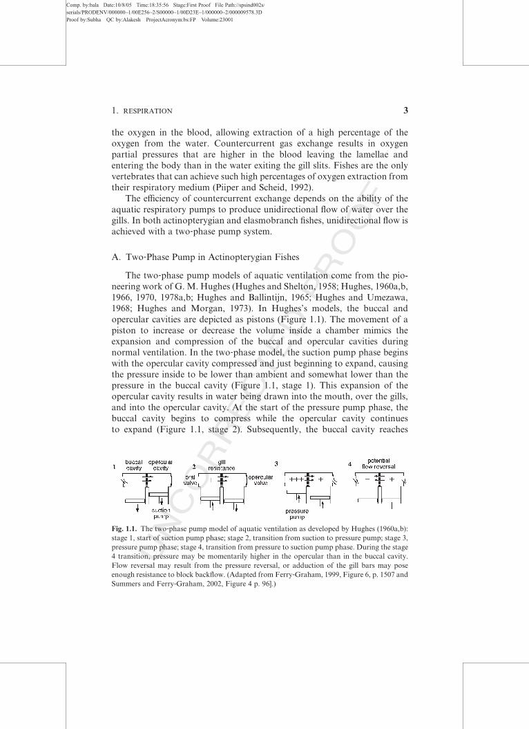
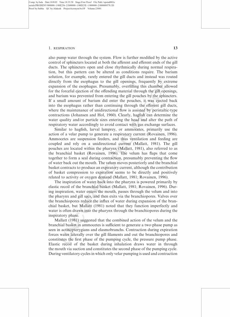
maximal compression before the opercular cavity, thereby maintaining

higher pressure in the buccal cavity and maintaining unidirectional flow as

water exits the opercular valves (Figure 1.1, stage 3). Just as the pressure

pump ends and the suction pump starts again, there is a brief moment of

pressure reversal in which opercular pressure is higher than buccal pressure

(Figure 1.1, stage 4). This pressure reversal may, in some circumstances,

produce brief reversals of flow (see later discussion), but overall the eVect ofthe two‐phase pump is to produce flow over the gills that is unidirectional

and continuous, albeit highly pulsatile (Hughes, 1960b; Piiper and Schuman,

1967; Scheid and Piiper, 1971, 1976; Malte, 1992; Malte and Lomholt, 1998;

Piiper, 1998).

The suction and pressure pumps are powered by abduction and adduc-

tion of the opercula, suspensoria, and hyoid apparatus. To generate buccal

and opercular expansion and create the subambient pressures of the suction

pump, each of these functional units acts as a lever system to convert muscle

shortening into abduction of skeletal elements. The motor pattern of the

two‐phase aquatic respiratory pump is summarized in Figure 1.2 (Liem,

1985). Starting with the with the pressure phase (P in Figure 1.2) the

adductor mandibulae muscle fires (becomes active) to reduce the gape of

the mouth, which in many fishes is sealed with a flap‐like oral valve that

closes in response to superambient pressure in the buccal cavity. Then, the

geniohyoideus fires to protract and elevate the hyoid apparatus, and the

adductor arcus palatini fires to adduct the suspensorium, thereby compres-

sing the buccal cavity. Increased pressure in the buccal cavity drives water

across the gills and into the opercular cavity, and at the end of the pressure

pump phase, the adductor operculi contracts and water is forced out the

opercular valve. At the beginning of the suction pump phase (S in Figure

1.2), the levator operculi fires to open the mouth by a small amount and the

levator arcus palatini fires to abduct the suspensorium. After a slight delay,

the dilator operculi fires to abduct the operculum, and the pressure in the

opercular chamber falls below buccal pressure and water is drawn over the

gills. The branchiostegal rays fan out during opercular expansion to main-

tain the opercular valve seal. Then the adductor mandibulae fires and the

pressure phase starts again.

The slight delay between the start of buccal expansion and the firing of

the dilator operculi leads to the potential for a momentary pressure reversal

(Figure 1.1, stage 4). The available data to date for teleosts suggest that while

pressure reversals do occur, concomitant flow reversals likely do not occur

(Hughes and Shelton, 1958; Saunders, 1961). Lauder (1984) demonstrated

that the gill bars adduct during the pressure reversal, momentarily increasing

the resistance between the buccal and opercular cavities. By placing plastic

spacers on the gill bars to prevent them from closing fully during normal

4 ELIZABETH L. BRAINED AND LARA A. FERRY-GRAHAM

Comp. by:bala Date:10/8/05 Time:18:35:56 Stage:First Proof File Path://spsind002s/serials/PRODENV/000000~1/00E256~2/S00000~1/00D23E~1/000000~2/000009578.3DProof by:Subha QC by:Alakesh ProjectAcronym:bs:FP Volume:23001

UNCORRECTEDPROOF

respiration, Lauder was able to observe flow reversals. When the spacers

were absent, flow reversals were not observed (Lauder, 1984).

The two‐phase aquatic respiratory pump model has been found to apply

to most teleost species studied to date, including the following freshwater

fishes: trout Salmo trutta, tench Tinca tinca, and roach Leuciscus rutilus

(Hughes and Shelton, 1958); white sucker catfish Catostomus commersoni

and brown bullhead catfish Ictalurus nebulosus (Saunders, 1961); and

carp Cyprinus carpio (Saunders, 1961; Ballintijn, 1969a,b). Pelagic and semi‐pelagic marine species studied include the horse mackerel Trachurus

Fig. 1.2. Functional morphology of gill ventilation in an anabantoid,Heleostoma temmincki. P,

the pressure pump phase (stage 3 of Figure 1.1). Note that buccal pressure always exceeds

opercular pressure. S, suction pump phase (stage 1 of Figure 1.1). Note that opercular pressure

is lower than buccal pressure. (From Liem, 1985, Figure 11–2, p. 187.)

1. RESPIRATION 5

Comp. by:bala Date:10/8/05 Time:18:35:56 Stage:First Proof File Path://spsind002s/serials/PRODENV/000000~1/00E256~2/S00000~1/00D23E~1/000000~2/000009578.3DProof by:Subha QC by:Alakesh ProjectAcronym:bs:FP Volume:23001

UNCORRECTEDPROOF

trachurus, herring Clupea harengus, whiting Gadus merlangus, conger eel Con-

ger conger, rockling Onos mustela, great pipefish Syngnathus acus, and wrasse

Crenilabrus melops (Hughes, 1960a). Benthic marine species also appear to fit

this model: stickleback Gasterosteus aculeatus (Anker, 1978; Elshoud, 1978),

bullhead sculpin Cottus bubalis, butterfly blenny Blennius ocellaris, grey

gurnard Trigla gurnardus, and dragonet Callionymus lyra (Hughes, 1960a).

Morphological evidence combinedwith opportunistic observation of live speci-

mens suggests that the two‐phase pump is also used by the bowfin Amia calva

(Liem, 1985) and coelacanth Latimeria chalumnae (Hughes, 1995).

Even the morphologically bizarre flatfishes appear to fit this model

(Hughes, 1960a; Liem et al., 1985). With one eye having migrated to the

opposite side of the head, they rest on the substrate on the ‘‘blind side,’’

which can be either the left or the right side of the body. When flatfishes are

at rest and buried in mud or sand, the two‐phase pump is modified such that

water generally exits from only the eyed side (Yazdani and Alexander, 1967;

Kerstens et al., 1979). During activity or when exposed to hypoxia, water

exits from both sides (SteVensen et al., 1981a; Liem et al., 1985), and during

extreme hypoxia, flatfishes will even raise their heads up above the substrate,

presumably to reduce the resistance encountered by the exhaled water

(SteVensen et al., 1981a).

For reasons that are unclear, some teleosts have gill slits that are restrict-

ed to a small hole; the rest of the opercular valve and the branchiostegal rays

are covered with skin. Some of the fishes with tiny gill openings are all

tetraodontiforms (puVerfishes, triggerfishes and their allies), some pleuro-

nectiforms (flatfishes), synbranchiform and elopomorph eels, some anten-

nariids (anglerfishes), and some gasterosteiforms (pipefish and seahorses).

The puVers, anglerfishes, flatfishes, and seahorses jet water out of their gill

openings at the start of locomotion or when handled (Brainerd et al., 1997;

E.L.B., personal observation). It is possible that the function of reduced gill

slits is to increase the velocity of these water jets, but a more thorough survey

of opercular valve morphology and function is needed to draw any firm

conclusions.

B. Two‐Phase Pump in Elasmobranch Fishes

It was once thought that a countercurrent gas exchange system does

not exist in cartilaginous fishes because they often exhibit lower oxygen

extraction eYciencies relative to bony fishes (Millen et al., 1966; Piiper and

Schuman, 1967). Elasmobranchs diVer morphologically from actinoptery-

gian fishes in several ways with regard to respiratory features. Most notably,

they have five or more gill slits on each side of the head compared with the

single opercular opening in ray‐finned fishes. The parabranchial chambers in

6 ELIZABETH L. BRAINED AND LARA A. FERRY-GRAHAM

Comp. by:bala Date:10/8/05 Time:18:35:56 Stage:First Proof File Path://spsind002s/serials/PRODENV/000000~1/00E256~2/S00000~1/00D23E~1/000000~2/000009578.3DProof by:Subha QC by:Alakesh ProjectAcronym:bs:FP Volume:23001

UNCORRECTEDPROOF

elasmobranchs, which are homologous with the opercular chamber of acti-

nopterygians, are similarly separated by septa along their length internally.

Early work by several authors proposed that the septa, to which the lamellae

are attached, might interfere with the flow of water and force concurrent

exchange during at least part of the respiratory cycle (Hughes and Shelton,

1962; Piiper and Schuman, 1967). Piiper and Schuman (1967) proposed a

‘‘multi‐capillary’’ model, much like gas exchange in birds, to explain the

observed partial pressures of oxygen in the blood and the ventilatory water.

Further work, however, rejected this view on the grounds that the partial

pressure of oxygen in the arterial blood was higher than that of the expired

water in the Scyliorhinus stellaris, as can only be achieved with a counter-

current gas exchange system (Piiper and Baumgarten‐Schumann, 1968).

Further investigations support the notion of a countercurrent gas exchanger

in elasmobranchs (Grigg, 1970; Scheid and Piiper, 1976; De Vries and De

Jager, 1984), and the countercurrent exchange model presently serves to

describe gas exchange in all aquatic‐breathing fishes, even hagfish (Malte

and Lomholt, 1998) and lamprey (Mallatt, 1981, 1996).

The respiratory pump in elasmobranchs is a two‐phase pump that is very

similar to the actinopterygian two‐phase pump (Figure 1.3A) (Hughes,

1960b; Hughes and Ballintijn, 1965). Recent work on several elasmobranch

species has demonstrated, however, that flow reversals are only partially

prevented by the action of the gill bars, and that flow reversals may be

widespread among species and body types (Figure 1.3B) (Ferry‐Graham,

1999; Summers and Ferry‐Graham, 2001, 2002). It is only with the applica-

tion of technologies recently made available to biologists that we have been

able to observe directly the path and pattern of water flow during ventila-

tion. The pioneers in this field had to rely on pressure recordings taken inside

the respiratory chambers to infer patterns of water flow. Further, move-

ments of any pertinent anatomical features, because they are internal,

could only be inferred from electromyographic recordings indicating when

the muscles were electrically active, but not necessarily performing actual

movements. The addition of sonomicrometry to this field has allowed the

determination of the physical position of important morphological elements.

Sonomicrometry, combined with the use of endoscopy to visualize anatomi-

cal elements in action and the movement of the ventilatory water, has

confirmed that although the core elements of Hughes’s elasmobranch mod-

els are correct, small diVerences exist, at least among the species originally

studied and those studied more recently (Ferry‐Graham, 1999; Summers and

Ferry‐Graham, 2001, 2002). The most important of these is the observation

that the gill bars do close, but not for the entire duration of the pressure

reversal period (Figure 1.4). Thus, water does flow back over the gills and

into the oral chamber.

1. RESPIRATION 7

Comp. by:bala Date:10/8/05 Time:18:35:56 Stage:First Proof File Path://spsind002s/serials/PRODENV/000000~1/00E256~2/S00000~1/00D23E~1/000000~2/000009578.3DProof by:Subha QC by:Alakesh ProjectAcronym:bs:FP Volume:23001

UNCORRECTEDPROOF

Fig. 1.3. Representative traces showing pressure reversals in (A) Cephaloscyllium ventriosum

and (B) Leucoraja erinacea. The data from L. erinacea show much longer pressure reversals

(indicated by negative pressure diVerential). Individuals of C. ventriosum also sometimes showed

reversals of this magnitude and duration, although they were not as common. Squalus acanthias

(data not shown) also showed both types of reversal profiles. L. erinacea did not exhibit profiles

as in (A). Profiles from C. ventriosum and L. erinacea sometimes lacked a pressure reversal;

S. acanthias profiles always had a reversal of some nature.

Fig. 1.4. Modifications to the two‐phase pump model in elasmobranchs verified by sonometric

data and direct observation of anatomical elements and water flow inside the oral and para-

branchial chambers using endoscopy (Summers and Ferry‐Graham, 2002). Specific modifica-

tions are indicated with text on the figures at each time interval. The mouth may be slightly open

in stage 4, depending on the species. (Adapted from Ferry‐Graham, 1999, Figure 7, p. 1508 and

Summers and Ferry‐Graham, 2002, Figure 4, p. 96.)

8 ELIZABETH L. BRAINED AND LARA A. FERRY-GRAHAM

Comp. by:bala Date:10/8/05 Time:18:35:57 Stage:First Proof File Path://spsind002s/serials/PRODENV/000000~1/00E256~2/S00000~1/00D23E~1/000000~2/000009578.3DProof by:Subha QC by:Alakesh ProjectAcronym:bs:FP Volume:23001

UNCORRECTEDPROOF

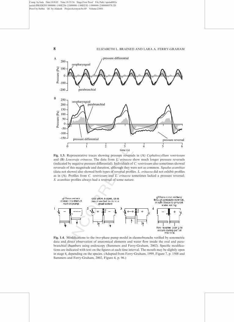
Flow reversals have been diYcult to detect since they are typically not

apparent externally. Valves normally prevent water from exiting the mouth

or entering through the gill slits in most species. Water was never observed

exiting the mouth in the swellshark Cephaloscyllium ventriosum (Ferry‐Graham, 1999; Summers and Ferry‐Graham, 2002), and it only rarely exited

the mouth in the skates Leucoraja erinacea and Raja clavata (Hughes, 1960b;

Summers and Ferry‐Graham, 2001, 2002). Water exited the mouth more

frequently in the dogfish Squalus acanthias, but not for the entire portion of

the pressure reversal period and not during every pressure reversal (Summers

and Ferry‐Graham, 2002). Water never entered through the gills slits in any

species studied. This is likely due to the fact that the reversals are fairly small

in nature and short in duration. For example, water did not exit the mouth

of most L. erinacea, even when the mouth was open and flow reversals were

directly observed at the gills (Summers and Ferry‐Graham, 2002).

Bidirectional flow has been observed, and tends to be much more obvi-

ous, at the spiracles of some elasmobranchs. Spiracles are openings on the

dorsal surface of the head that lead directly to the oral chamber and channel

water toward the gills. Recent comparative analyses suggest that the spiracle

is a derived feature within elasmobranchs (Summers and Ferry‐Graham,

2002), but this analysis depends strongly on the placement of the batoids

within any given elasmobranch phylogeny, and the position of Batoidea is

still in flux (Shirai, 1996; Douady et al., 2003). The presence of the spiracle is

not tightly correlated with a benthic habitat, as C. ventriosum, a derived

carchariniform shark, is largely benthic but lacks spiracles, and S. acanthias,

a basal squaliform shark, spends much of its time in open water and has

fairly large spiracles. However, the use of the spiracle as the exclusive

ventilatory aperture has been observed only in benthic species.

Water was seen to enter and exit the spiracle in L. erinacea when the skate

was resting on the bottom (Summers and Ferry‐Graham, 2001), and was also

seen on occasion in R. clavata in earlier studies (Hughes, 1960b). In contrast,

no consistent pattern of exclusive spiracular use was observed in the non‐benthic dogfish, S. acanthias. Skates tend to rest or even bury themselves in

the substrate, and thus the mouth is not or cannot be used to draw in a current

of water for respiration during these periods of time. Outflow through the gills

may be similarly reduced to prevent stirring up sediment upon discharge.

Although distantly related, the sturgeon, Acipenser transmontanus, provides

some evidence for this notion via the evolution of convergent structures. The

sturgeon inhabits and forages in largely silty benthic habitats. Despite its

reduced spiracles, enlarged openings on the dorsal regions of the gill slits

serve to both draw in and expel water for respiration (Burggren, 1978). Other

benthic fishes, such asC. ventriosum, in which the spiracles are so reduced that

they are presumed to be nonfunctional, have been observed propped up on

1. RESPIRATION 9

Comp. by:bala Date:10/8/05 Time:18:35:57 Stage:First Proof File Path://spsind002s/serials/PRODENV/000000~1/00E256~2/S00000~1/00D23E~1/000000~2/000009578.3DProof by:Subha QC by:Alakesh ProjectAcronym:bs:FP Volume:23001

UNCORRECTEDPROOF

their pectoral fins or with their neurocrania rotated dorsally during periods of

very active buccal pumping, thereby increasing the exposure of the mouth to

the surrounding water (L.A.F.G., personal observation).

The physiological consequences of flow reversals, whether the reversals

be inadvertent, as during the switch from pressure to suction pump, or

apparently deliberate, as in spiracular breathing, may not be as grave as

some researchers have suggested. Most species can tolerate large, experi-

mentally induced ineYciencies in gas exchange (Malte, 1992), and it is likely

that natural flow reversals decrease as oxygen demand increases and the

respiratory pumps work harder.

The kinematics of ventilation in elasmobranchs are highly variable

(Hughes, 1960b, 1978a; Hughes and Ballintijn, 1965). Much of this variation

may be driven by physiological requirements, such as oxygen demand. For

example, increases in ventilatory stroke volume are likely achieved by in-

creases in the compression and subsequent expansion of the oral and para-

branchial chambers. When a fish is at rest and the oral and parabranchial

chambers are compressed to a lesser degree, the two‐pump system can break

down. Several scenarios have been documented, ranging from double

pressure reversals to a complete failure of the suction pump to operate.

Figure 1.5 depicts a scenario in which the pressure reversal is extreme.

Sonometric and endoscopic data show that the gill bars are closed during

stages 1 and 2 of such sequences, preventing prolonged reversals in water

flow. However, water is also not flowing from anterior to posterior, as the

suction pump is insuYcient to generate flow. Variations of this pattern exist

such that pressure reversals are seen at stages 4, 1, and 2 (Summers and

Ferry‐Graham, 2002), and just 4 and 2 (Ferry‐Graham, 1999), whereby the

suction pump presumably manages to create some anterior‐to‐posterior flowbetween pressure reversals.

Fig. 1.5. A general scenario depicting a complete failure of the suction pump to generate

anterior‐to‐posterior water flow verified by sonometric data and direct observation of anatomi-

cal elements and water flow inside the oral and parabranchial chambers using endoscopy

(Summers and Ferry‐Graham, 2002). The mouth may be slightly open in stage 4, depending

on the species. (Adapted from Ferry‐Graham, 1999, Figure 7, p. 1508 and Summers and Ferry‐Graham, 2002, Figure 4, p. 96.)

10 ELIZABETH L. BRAINED AND LARA A. FERRY-GRAHAM

Comp. by:bala Date:10/8/05 Time:18:35:57 Stage:First Proof File Path://spsind002s/serials/PRODENV/000000~1/00E256~2/S00000~1/00D23E~1/000000~2/000009578.3DProof by:Subha QC by:Alakesh ProjectAcronym:bs:FP Volume:23001

UNCORRECTEDPROOF

C. Ram Ventilation

During ram ventilation, a respiratory current is generated by the loco-

motor eVorts of the fish. In fast‐swimming fishes, water enters the oral cavity

and passes over the gills as long as the fish holds its mouth and opercular

valves open.

Many fishes are able to buccal pump when needed but switch to ram

ventilation at appropriate swimming speeds. Facultative ram ventilation has

been documented in paddlefish Polyodon spathula (Burggren and Bemis,

1992; Sanderson et al., 1994), sandtiger sharks Odontaspis (¼ Eugomphodus

or Carcharias) taurus (von Wahlert, 1964), leopard sharks Triakis semifas-

ciata (Hughes 1960b), a variety of salmonids (Roberts, 1978; SteVensen,1985), several pelagic species such as mackerel Scomber scombrus, blue

runner Caranx crysos, bluefish Pomatomus saltatrix, scup Stenotomus cry-

sops, and the halfmoon Medialuna californica (Roberts, 1975), and shark-

suckers Echeneis naucrates and remoras Remora remora when attached

to a fast‐swimming shark or aquatic mammal (Muir and Buckley, 1967;

SteVensen and Lomholt, 1983; SteVensen, 1985). Interestingly, a number of

species, including some that routinely move into open water habitats, never

switch to ram ventilation. An apparent inability to perform ram ventilation

has been documented in the striped mullet Mugil cephalus and in basses and

rockfishes of the genera Paralabrax and Sebastes (Roberts, 1975). In facul-

tative ram ventilation, the switch from buccal pumping to ram ventilation is

triggered by a mechanoreceptor that is stimulated by flow velocity (Roberts

and Rowell, 1988); benthic fishes may lack this reflex altogether (Roberts,

1978). Switching from active pumping to passive ram ventilating is estimated

to save about 10% of the total energy expenditure during high‐speed loco-

motion, although these calculations are only rough estimates (Brown and

Muir, 1970; Roberts, 1978; SteVensen, 1985).In contrast, pelagic fishes such as the scombrids (tuna and mackerel,

primarily tuna), istiophorids (sailfish), and xiphiids (swordfish) are obligate

ram ventilators. Their branchial anatomy is so severely reduced that they

cannot generate a suYcient respiratory current using the buccal pump. There

is a great deal of fusion of both the gill filaments and the lamellae in all of

these families of fishes as well as in the dolphinfish Coryphaena hippurus

(Muir and Kendall, 1968). Lamellae on adjacent filaments may be fused to

one another along their facing edges, and in some adjacent filaments may

even be fused along part of their length. Water passes through small slits or

openings where fusion is incomplete. The reason for the fusion is not entirely

clear, but it occurs widely among fast‐swimming oceanic fish, and there

appears to be greater fusion in more‐derived species. Possible advantages

of fusion include (1) restricting access by parasites to the gill tissues,

1. RESPIRATION 11

Comp. by:bala Date:10/8/05 Time:18:35:58 Stage:First Proof File Path://spsind002s/serials/PRODENV/000000~1/00E256~2/S00000~1/00D23E~1/000000~2/000009578.3DProof by:Subha QC by:Alakesh ProjectAcronym:bs:FP Volume:23001

UNCORRECTEDPROOF

(2) increasing the rigidity of the structure so that it does not collapse and can

therefore extract the greatest amount of oxygen possible, and (3) reducing

the velocity of water flow over the lamellae to increase oxygen extraction

(Muir and Kendall, 1968). Interestingly, similar fusion is found in A. calva,

which lives in stagnant marshes, further suggesting that enhanced oxygen

extraction may be a primary function of the fusion (Bevelander, 1934).

D. Gill Ventilation in Lamprey and Hagfish

In the two groups of extant jawless fishes, the anatomy of the respiratory

pumps is markedly diVerent from that of gnathostome fishes. Nonetheless,

water flow through the oropharynx in lampreys and hagfishes is largely

unidirectional and countercurrent gas exchange occurs (Mallatt, 1981,

1996; Malte and Lomholt, 1998).

The respiratory structures of hagfishes consist of pairs of sacs or

pouches, anywhere from 6 to 14 depending on the species, that house the

gill lamellae. The lamellae are the primary gas exchange surfaces (Malte and

Lomholt, 1998). The skin of the hagfish is also quite permeable, but, except

when scavenging on carcasses and other large food falls, hagfish are largely

buried in the sediment with only their nostrils and tentacles exposed

(SteVensen et al., 1984). Water reaches the pouches through aVerent ductsoriginating in the posterior portion of the pharynx and exits through eVerentducts that lead to external gill openings on either side of the animal. In some

species, the eVerent ducts fuse to form one common opening to the sur-

rounding medium. Water enters the pharynx through the mouth or the

nostril and is pumped into the aVerent ducts by the action of the velum

(Malte and Lomholt, 1998). The velum is a muscular structure situated at

the dorsal midline of the rostral portion of the pharynx that serves to

contract the chamber and pump water posteriorly. As a result, the flow

entering the nostril is pulsatile and the frequency is highly variable, ranging

from 0.01 to 1.3 Hz (SteVensen et al., 1984), with the higher frequencies

recorded from hagfish under warmer experimental conditions.

Based on anatomical studies, it was long thought that the velum alone

was responsible for generating the respiratory current, and hagfish had little

ability to alter the path of water once in the head. One of the first studies to

examine hagfish anatomy in action was a cineradiographic study (Johansen

and Hol, 1960). In this study, the researchers used barium and hypaque dyes

that fluoresce under radiographic light to follow the path of the respiratory

currents in live animals after introducing the contrast agents at either the

mouth or the nostril. This foundational, and unequalled, study revealed that

hagfish do use pumping of the velum to generate respiratory water flow

through the head. However, the gill pouches themselves are muscular and

12 ELIZABETH L. BRAINED AND LARA A. FERRY-GRAHAM

Comp. by:bala Date:10/8/05 Time:18:35:58 Stage:First Proof File Path://spsind002s/serials/PRODENV/000000~1/00E256~2/S00000~1/00D23E~1/000000~2/000009578.3DProof by:Subha QC by:Alakesh ProjectAcronym:bs:FP Volume:23001

UNCORRECTEDPROOF

also pump water through the system. Flow is further modified by the active

control of sphincters located at both the aVerent and eVerent ends of the gillducts. The sphincters open and close rhythmically during normal respira-

tion, but this pattern can be altered as conditions require. The barium

solution, for example, rarely entered the gill ducts and instead was routed

directly from the esophagus to the gill openings, frequently by extreme

expansion of the esophagus. Presumably, overfilling this chamber allowed

for the forceful ejection of the oVending material through the gill openings,

and barium was prevented from entering the gill pouches by the sphincters.

If a small amount of barium did enter the pouches, it was ejected back

into the esophagus rather than continuing through the eVerent gill ducts,where the maintenance of unidirectional flow is assisted by peristaltic‐typecontractions (Johansen and Hol, 1960). Clearly, hagfish can determine the

water quality and/or particle sizes entering the head and alter the path of

respiratory water accordingly to avoid contact with gas exchange surfaces.

Similar to hagfish, larval lamprey, or ammocetes, primarily use the

action of a velar pump to generate a respiratory current (Rovainen, 1996).

Ammocetes are suspension feeders, and thus ventilation and feeding are

coupled and rely on a unidirectional current (Mallatt, 1981). The gill

pouches are located within the pharynx (Mallatt, 1981), also referred to as

the branchial basket (Rovainen, 1996). The velum has flaps that come

together to form a seal during contraction, presumably preventing the flow

of water back out the mouth. The velum moves posteriorly and the branchial

basket contracts to produce an expiratory current, although the contribution

of basket compression to expiration seems to be directly and positively

related to activity or oxygen demand (Mallatt, 1981; Rovainen, 1996).

The inspiration of water back into the pharynx is powered primarily by

elastic recoil of the branchial basket (Mallatt, 1981; Rovainen, 1996). Dur-

ing inspiration, water enters the mouth, passes through the velum and into

the pharynx and gill sacs, and then exits via the branchiopores. Valves over

the branchiopores reduce the influx of water during expansion of the bran-

chial basket, but Mallatt (1981) noted that they function imperfectly and

water is often drawn into the pharynx through the branchiopores during the

inspiratory phase.

Mallatt (1981) suggested that the combined action of the velum and the

branchial basket in ammocetes is suYcient to generate a two‐phase pump as

seen in actinopterygians and elasmobranchs. Contraction during expiration

forces water laterally over the gill filaments and out the branchiopores and

constitutes the first phase of the pumping cycle, the pressure pump phase.

Elastic recoil of the basket during inhalation draws water in through

the mouth via suction and constitutes the second phase of the pumping cycle.

During ventilatory cycles inwhich only velar pumping is used and contraction

1. RESPIRATION 13

Comp. by:bala Date:10/8/05 Time:18:35:58 Stage:First Proof File Path://spsind002s/serials/PRODENV/000000~1/00E256~2/S00000~1/00D23E~1/000000~2/000009578.3DProof by:Subha QC by:Alakesh ProjectAcronym:bs:FP Volume:23001

UNCORRECTEDPROOF

of the basket does not contribute to water flow, the suction pump is not

suYcient to generate substantial lateral flow across the gills. As noted previ-

ously, there is detectable backflow during the suction pump phase where

water is drawn in through the branchiopores. This backflow period can be

lengthy, persisting for up to half of the complete ventilatory cycle.

During metamorphosis from ammocete larva to adult lamprey, the

velum is extensively remodeled. Many adult lamprey are parasitic, feeding

by attaching their rasping mouth parts onto the sides of fishes with a sucker‐like structure. Therefore, the mouth and anterior portions of the head are

largely unavailable for respiration, and water both enters and exits the gill

sacs via the external branchiopores. In adults, the velum presumably func-

tions to prevent the rostral flow of water and maintain ventilation separate

from feeding, while contraction and elastic recoil of the branchial basket

exclusively generate the respiratory current (Mallatt, 1981; Rovainen, 1996).

III. AERIAL RESPIRATORY PUMPS

A. Evolutionary History and Biomechanical Challenges

Lungs are present in basal members of Actinopterygii and Sarcopterygii

but not in Chondrichthyes; therefore, it is most parsimonious to conclude

that lungs arose in stem osteichthians and have been retained as a primitive

character in actinopterygians and sarcopterygians. Within Actinopterygii,

paired lungs are present only in Polypteriformes, and an unpaired lung,

homologous with paired lungs and termed a gas bladder, is present in other

basal actinopterygians (Liem, 1988; Graham, 1997). The pneumatic duct

connecting the gas bladder to the pharynx was lost in euteleosts, probably in

stem acanthomorphs, and buoyancy control became the primary function of

the gas bladder. Thus, the physoclistous swim bladder of euteleosts is

homologous with the physostomous gas bladders of basal actinopterygians

and with the lungs of tetrapods.

The physostomous gas bladder lost and regained its respiratory function

several times in the evolutionary history of basal actinopterygians and

teleosts (Liem, 1989b). However, once the pneumatic duct was lost, the swim

bladder did not regain its respiratory function in any euteleosts. Instead,

various other kinds of ABOs evolved, such as the suprabranchial chambers

of Channa and Monopterus, the branchial diverticulae of Clarias and ana-

bantoids, and the stomach and intestinal modifications of some siluriforms

(Graham, 1997).

All air‐breathing fishes are bimodal or trimodal breathers (Graham,

1997). They retain gills as important sites of CO2 excretion and ion

14 ELIZABETH L. BRAINED AND LARA A. FERRY-GRAHAM

Comp. by:bala Date:10/8/05 Time:18:35:58 Stage:First Proof File Path://spsind002s/serials/PRODENV/000000~1/00E256~2/S00000~1/00D23E~1/000000~2/000009578.3DProof by:Subha QC by:Alakesh ProjectAcronym:bs:FP Volume:23001

UNCORRECTEDPROOF

exchange, and the gills also absorb oxygen when the water is not hypoxic. In

addition, the skin is often an important site of gas exchange, both in water

(SteVensen et al., 1981b) and when fishes emerge during ‘‘terrestrial trespas-

sing’’ (Liem, 1987). In severely hypoxic water, some air‐breathing fishes may

actually lose oxygen to the water through their gills and skin if the oxygen

derived from air breathing causes the blood to have a higher oxygen tension

than the surrounding water. This apparent ineYciency results from the fact

that blood from most ABOs flows back to the heart and gills before being

redistributed to the rest of the body. This seemingly maladaptive system is

one of several lines of evidence that led to the myocardial oxygenation

theory (Farmer, 1997), in which selection for increased oxygen delivery to

the heart muscle is proposed as a primary selection force in the evolution of

air breathing.

Aerial respiratory pumps face biomechanical challenges that result from

the interaction of air and water. Within lungs and gas bladders, pressure

generated by aerial pumps must overcome the surface tension of the air–

liquid interface. However, surface tension is probably quite low, as surfac-

tants are produced by the epithelia of gas bladders and lungs (Liem, 1988).

Hydrostatic pressure also aVects aerial respiratory pumps. If a fish takes an

air breath with its body at an angle with the surface of the water, as is usually

the case, then the aerial pump pressure must exceed the hydrostatic pressure

at the deepest part of the gas‐filled space (Figure 1.6). On the other hand,

hydrostatic pressure may also assist breathing by contributing to exhalation.

Fig. 1.6. The eVect of hydrostatic pressure on air breathing. When a fish approaches the surface

at an angle, hydrostatic pressure at the caudal end of the lungs or gas bladder may assist

expiration but will also oppose inspiration.

1. RESPIRATION 15

Comp. by:bala Date:10/8/05 Time:18:35:58 Stage:First Proof File Path://spsind002s/serials/PRODENV/000000~1/00E256~2/S00000~1/00D23E~1/000000~2/000009578.3DProof by:Subha QC by:Alakesh ProjectAcronym:bs:FP Volume:23001

UNCORRECTEDPROOF

Air breathing strongly aVects the buoyancy of fishes, and this coupling

between respiration and buoyancy places a constraint on the volume of air

that can be held in a gas exchange organ. Fishes are vulnerable to both aerial

and aquatic predators when they come to the surface to breathe (Kramer

and Graham, 1976). Presumably there is selection to breathe as infrequently

as possible, which should favor high‐volume gas exchange organs. However,

too much air would result in positive buoyancy—a condition that traps

fishes on the surface and increases their vulnerability to predators. There-

fore, the upper limit on the size of aerial gas exchange organs is constrained

by the need to avoid positive buoyancy.1 In addition, air‐breathing fishes

have fine control over their gas volume and manage their buoyancy at

slightly negative, neutral, or slightly positive, depending on their behavioral

needs at any given moment in time (E.L.B., personal observation). In most

cases, total gas volume is probably regulated on the basis of buoyancy,

whereas tidal volume and breath frequency vary with metabolic needs.

B. Air Ventilation Mechanics

Unlike ourselves and other amniotes, fishes lack the intercostal and/or

diaphragmatic muscles necessary for aspiration breathing. Instead, almost

all air‐breathing fishes use buccal pump breathing, in which expansions and

compressions of the buccopharyngeal cavity ventilate the gas exchange

organs (Liem, 1985). As described previously for aquatic ventilation, the

hyoid apparatus and suspensorium act as lever systems to convert muscle

shortening into buccal cavity expansion, thereby generating subambient

pressure and drawing air in through the mouth. As the mouth closes, the

hyoid protracts and the suspensorium adducts, generating superambient

pressure and forcing air into the gas exchange organ. Aquatic ventilation,

suction feeding, and aquatic coughing all involve buccopharyngeal

expansion and compression, and the evolution of aerial buccal pumps ap-

pears to have occurred by modifying and combining these basic behaviors

(McMahon, 1969; Liem, 1980, 1985; Brainerd, 1994a).

In most basal actinopterygian and basal teleost fishes, the respiratory gas

bladder is ventilated with a four‐stroke buccal pump, named by analogy

with the piston movements in four‐stroke internal combustion engines

(Brainerd et al., 1993; Brainerd, 1994a). A four‐stroke air breath begins as

the fish approaches the surface and transfers gas from the gas bladder into

the buccal cavity. Hydrostatic pressure, elastic recoil of the gas bladder or

1One could imagine a scenario in which fishes might experience selection for added bone mass

to oVset a larger lung, if selection for infrequent air breathing were suYciently strong. One

possible group in which to look for this eVect would be the armored catfishes.

AU1

16 ELIZABETH L. BRAINED AND LARA A. FERRY-GRAHAM

Comp. by:bala Date:10/8/05 Time:18:35:59 Stage:First Proof File Path://spsind002s/serials/PRODENV/000000~1/00E256~2/S00000~1/00D23E~1/000000~2/000009578.3DProof by:Subha QC by:Alakesh ProjectAcronym:bs:FP Volume:23001

UNCORRECTEDPROOF

body wall, and active expansion of the buccal cavity, thereby sucking gas out

of the gas bladder, may all contribute to the transfer phase of expiration

(Liem, 1988; Brainerd, 1994a). After gas transfer, the buccal cavity com-

presses and expired gas is expelled either out the mouth (Amia) or out the

opercular valves (all others). With the fish still at the surface, the mouth then

opens and the buccal cavity expands to inspire fresh air, whereupon the

mouth closes and the buccal cavity compresses to pump the fresh air into the

gas bladder. Thus, the four strokes of this buccal pump are (1) gas transfer,

(2) expulsion, (3) inspiration, and (4) compression (Figure 1.7). Four‐strokebreathing has been observed in basal actinopterygians, Amia and Lepisos-

teus, and in basal teleosts, Arapaima, Gymnarchus, Notopterus, Pangasius

(Rahn et al., 1971; Liem, 1988, 1989b; Brainerd, 1994a), andMegalops (E.L.

B., personal observation).

In contrast to the four‐stroke buccal pump of actinopterygians, lepido-

sirenid lungfishes ventilate their lungs with a two‐stroke buccal pump2

(Bishop and Foxon, 1968; McMahon, 1969; Brainerd et al., 1993; Brainerd,

2No data are available on air ventilation in the only extant, non‐lepidosirenid lungfish,Neoceratodus, but observations of an Australian lungfish taking air breaths in a public aquariumsuggest that they may use a four‐stroke pump (E.L.B., personal observation).

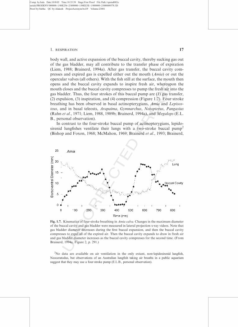
Fig. 1.7. Kinematics of four‐stroke breathing in Amia calva. Changes in the maximum diameter

of the buccal cavity and gas bladder were measured in lateral projection x‐ray videos. Note that

gas bladder diameter decreases during the first buccal expansion, and then the buccal cavity

compresses to expel all of the expired air. Then the buccal cavity expands to draw in fresh air

and gas bladder diameter increases as the buccal cavity compresses for the second time. (From

Brainerd, 1994a, Figure 2, p. 291.)

1. RESPIRATION 17

Comp. by:bala Date:10/8/05 Time:18:35:59 Stage:First Proof File Path://spsind002s/serials/PRODENV/000000~1/00E256~2/S00000~1/00D23E~1/000000~2/000009578.3DProof by:Subha QC by:Alakesh ProjectAcronym:bs:FP Volume:23001

UNCORRECTEDPROOF

1994a). With the snout of the lungfish protruding slightly from the surface of

the water, the mouth opens and the buccal cavity expands to draw in fresh

air. While the buccal cavity is expanding, exhalation of air from the lungs

begins, driven by hydrostatic pressure, elastic recoil of the lungs and body

wall, and possibly the contraction of smooth muscle in the lung walls.

Neither buccal suction nor contraction of body musculature contributes to

expiration (Figure 1.8). Buccal expansion generally continues beyond the

end of expiration, and then buccal compression forces gas into the lungs

(Figure 1.9). Because the buccal cavity does not compress after exhalation in

two‐stroke breathing, expired gas mixes with fresh air in the buccal cavity,

and then this mixed gas is pumped into the lungs. In contrast, all of the

expired gas is expelled from the buccal cavity in four‐stroke breathing beforefresh air is inspired and pumped into the gas bladder (Figure 1.7).

The two‐stroke buccal pump is present in amphibians as well as in lepi-

dosirenid lungfishes (Brainerd et al., 1993), whereas the four‐stroke buccal

pump is typical of actinopterygian fishes. This phylogenetic pattern indicates

that two‐stroke breathing is the ancestral condition for Sarcopterygii, where-

as four‐stroke breathing is the ancestral condition for Actinopterygii. The

ancestral condition for Osteichthyes cannot be determined, because no extant

outgroups to Osteichthyes breathe air (Brainerd, 1994a).

The kinematics of the two‐ and four‐stroke buccal pumps resemble

kinematics associated with gill ventilation, suction feeding, and aquatic

coughing (Brainerd, 1994a). Four‐stroke breathing, suction feeding, and

Fig. 1.8. Buccal and pleuroperitoneal (abdominal) pressure during an air breath in Protopterus

aethiopicus. Note that pleuroperitoneal pressure decreases during exhalation, indicating a slight

contribution of body wall elastic recoil to exhalation, but buccal pressure does not decrease,

indicating that buccal expansion does not contribute to exhalation. (From Brainerd et al., 1993,

Figure 8, p. 176.)

18 ELIZABETH L. BRAINED AND LARA A. FERRY-GRAHAM

Comp. by:bala Date:10/8/05 Time:18:35:59 Stage:First Proof File Path://spsind002s/serials/PRODENV/000000~1/00E256~2/S00000~1/00D23E~1/000000~2/000009578.3DProof by:Subha QC by:Alakesh ProjectAcronym:bs:FP Volume:23001

UNCORRECTEDPROOF

aquatic coughing are all fast movements. The two complete buccal expan-

sion–compression cycles of four‐stroke breathing occur in under 1 s, with

some fishes completing each cycle in less than 100 ms. The gas transfer and

expiration phases may have arisen by modification of the aquatic cough, in

which the buccal cavity is expanded with the mouth closed. The inspiration

and compression phases may have arisen by modification of the movements

associated with suction feeding.

The two‐stroke buccal pump of lungfishes more closely resembles the

aquatic ventilatory pump in its movements and timing (McMahon, 1969;

Brainerd, 1994a). In four‐stroke breathing, gill ventilation stops well before

each air breath, but in lungfishes, gill ventilation continues as the fish

approaches the surface of the water, and the buccal expansion associated

with the air breath follows smoothly from the previous gill ventilation cycle

(Brainerd, 1994a). The buccal cavity expands more during an air breath than

during an aquatic breath, but otherwise the movements are very similar

(McMahon, 1969). Aquatic breathing resumes immediately after the buccal

compression phase of the air breath, without missing a beat in the aquatic

ventilatory rhythm (Brainerd, 1994a).

Although the four‐stroke buccal pump is typical for actinopterygians,

two alternative ventilatory mechanisms have been described. In polypterid

fishes, the patterns of buccal expansion and air transfer are similar to

four‐stroke, but elastic recoil of the ganoid scale jacket produces subambient

Fig. 1.9. Kinematics of two‐stroke breathing in Lepidosiren. Changes in the maximum diameter

of the buccal cavity and gas bladder were measured in lateral projection x‐ray videos. Note that,

in comparison to four‐stroke breathing (Figure 7), the buccal cavity expands and compresses

only once, and therefore some of the expired air is pumped back into the lungs. (From Brainerd,

1994a, Figure 3, p. 293.)

1. RESPIRATION 19

Comp. by:bala Date:10/8/05 Time:18:35:59 Stage:First Proof File Path://spsind002s/serials/PRODENV/000000~1/00E256~2/S00000~1/00D23E~1/000000~2/000009578.3DProof by:Subha QC by:Alakesh ProjectAcronym:bs:FP Volume:23001

UNCORRECTEDPROOF

pressure in the body cavity whereby air is aspirated into the lungs

(Figure 1.10) (Brainerd et al., 1989). Two euteleosts, Gymnotus and Hopler-

ythrinus, ventilate their gas bladders in a manner that is completely diVerentfrom any other actinopterygians (Farrell and Randall, 1977; Liem, 1989b).

An air breath starts with a large buccal expansion at the surface of the water

(Figure 1.11). Then the fish sinks below the surface and compresses the

buccal cavity to pump the air into its esophagus, which expands greatly,

and the esophagus gradually empties into the gas bladder through the

Fig. 1.10. Recoil aspiration in Polypterus. Frames from an x‐ray video of lung ventilation in

Polypterus senegalis, lateral projection. The left frame is at the end of expiration, and the middle

and right frames show inspiration. Note that the mouth is wide open as the lungs refill with air,

indicating that the fish is inhaling by aspiration breathing, rather than buccal pumping (a mouth

seal is necessary for buccal pumping).

Fig. 1.11. Esophageal pump in Gymnotus carapo. Frames from an x‐ray video of lung ventila-

tion in lateral projection. Frames 1–4 show inspiration and frames 5–8 show expiration. See text

for explanation. Abbreviations: b, buccal cavity; e, esophagus; g, gas bladder; g’, anterior

chamber of the gas bladder. (Adapted from Liem, 1989b, Figure 8, p. 346.)

AU2

20 ELIZABETH L. BRAINED AND LARA A. FERRY-GRAHAM

Comp. by:bala Date:10/8/05 Time:18:36:00 Stage:First Proof File Path://spsind002s/serials/PRODENV/000000~1/00E256~2/S00000~1/00D23E~1/000000~2/000009578.3DProof by:Subha QC by:Alakesh ProjectAcronym:bs:FP Volume:23001

UNCORRECTEDPROOF

pneumatic duct. The fish remains submerged and expiration ensues in re-

verse of inspiration; gas moves first into the esophagus and then into the

buccal cavity and finally is released as bubbles. This mechanism results in

relatively small tidal volumes (Figure 1.11), whereas two‐ and four‐strokebreathing and recoil aspiration exchange between 50 and 100% of the gas

bladder volume with each breath.

The loss of the pneumatic duct in stem acanthomorphs, presumably

through lack of selection for air breathing, apparently produced an evolu-

tionary constraint that prevented the subsequent recruitment of the swim

bladder for gas exchange. Nonetheless, air breathing has evolved many times

in higher teleosts, most commonly through the use of relatively unmodified

buccal, opercular, pharyngeal, and/or branchial surfaces for gas exchange.

In these cases, slight modifications of the expansive phase of the aquatic

respiratory pump or suction feeding pump are used to draw in a bubble of

air at the surface, and then the buccal and/or opercular cavities remain

expanded to retain the bubble after submergence (Graham, 1997).

In some teleosts, more elaborate ABOs have evolved. A common theme

is the evolution of a suprabranchial chamber (SBC) that may be a relatively

simple space dorsal and caudal to the opercular cavity, as in Monopterus, or

that may contain elaborate structures that increase the surface area for gas

exchange, such as the labyrinth organ of anabantoids, the respiratory tree of

Channa, and the respiratory fans and trees of Clarias (Graham, 1997). The

dorsal location of the SBC makes biomechanical sense since inspired air will

tend to rise up into the chamber and displace the gas or water that is present.

Ventilation of the suprabranchial chamber is accomplished by one of two

mechanisms, named monophasic and diphasic by Peters (1978), and re-

named triphasic and quadruphasic by Liem to reflect the number of phases

recognizable with EMG and cineradiography (Liem, 1980, 1985, 1989a).

Triphasic ventilation is eVective when the SBC has both anterior and poste-

rior openings, as in anabantoids. The three phases are as follows: (1) a

preparatory phase in which the buccal cavity compresses to expel water,

(2) an expansive phase in which the buccal cavity expands to draw in fresh

air through the mouth, and (3) a compressive phase in which the buccal

cavity compresses to force fresh air into the SBC. The SBC is a rigid

structure encased in bone, so the addition of fresh air forces the old gas

out of the chamber, thus creating a unidirectional draft of air through the

SBC (Liem, 1980).

Muscle activity during the triphasic pump is nearly identical to activity

during suction feeding. The levator operculi (LO), levator arcus palatini

(LAP), and sternohyoideus (SH) are active during expansion, and the ad-

ductor arcus palatini (AAP), adductor mandibulae (AM), and geniohyoi-

deus (GH) are active during compression. One interesting diVerence is that

AU3

1. RESPIRATION 21

Comp. by:bala Date:10/8/05 Time:18:36:00 Stage:First Proof File Path://spsind002s/serials/PRODENV/000000~1/00E256~2/S00000~1/00D23E~1/000000~2/000009578.3DProof by:Subha QC by:Alakesh ProjectAcronym:bs:FP Volume:23001

UNCORRECTEDPROOF

the dilator operculi (DO) is active during the expansive phase of suction

feeding but only becomes active at the end of the compressive phase of

triphasic ventilation when bubbles are released through the opercular valve

(Liem, 1985).

Quadruphasic ventilation is more complex and is bidirectional (Liem,

1980). The four phases are as follows (Figure 1.12): (1) a preparatory phase

in which the buccal cavity compresses to expel water, (2) a reversal phase in

which activity in the DO abducts the operculum rapidly, activity in the SH

Fig. 1.12. Quadruphasic ventilation of the SBC in an anabantoid, Heleostoma temmincki.

Drawings traced from an x‐ray video of lung ventilation in lateral projection. See text for

explanation. Abbreviations: sac, suprabranchial air chamber; sb, swim bladder. (From Liem,

1980, Figure 5, p. 66.)

22 ELIZABETH L. BRAINED AND LARA A. FERRY-GRAHAM

Comp. by:bala Date:10/8/05 Time:18:36:00 Stage:First Proof File Path://spsind002s/serials/PRODENV/000000~1/00E256~2/S00000~1/00D23E~1/000000~2/000009578.3DProof by:Subha QC by:Alakesh ProjectAcronym:bs:FP Volume:23001

UNCORRECTEDPROOF

retracts and depresses the hyoid apparatus, and a current of water is drawn

into the posterior opening of the SBC, forcing gas forward through the

anterior opening of the SBC and into the buccal cavity whence it is expelled,

(3) an expansive phase in which the buccal cavity expands to draw in fresh

air through the mouth, and (4) a compressive phase in which the buccal

cavity compresses to force fresh air into the SBC. Muscle activity in phases 3

and 4 is identical to suction feeding, including the activity of the DO during

expansion. The muscle activity of the reversal phase is identical to muscle

activity during the aquatic cough, which is normally used to create a rostrad

current of water to clear debris from the gills (Liem, 1980).

Most species of air‐breathing fishes with an SBC use either triphasic or

quadruphasic ventilation, but anabantoids are able to use both (Liem,

1989a). The quadruphasic pump relies on a current of water for expiration,

so this pump works only when fishes are submerged; the triphasic pump

works when fishes are in or out of water. Most air breathers that use

buccopharyngeal surfaces or an SBC for gas exchange either expel the air

bubble before feeding or lose the air bubble in the process of feeding. In

anabantoids and clariids, however, air is not lost from the SBC during

feeding. Valves separate the SBC from the buccal and opercular cavities,

eVectively decoupling feeding and air breathing. Liem (1989a) proposed this

decoupling as an explanation for the relatively diverse types of food items

eaten by anabantoids and clariids, compared to the limited diets of channids

and synbranchiforms.

Some air‐breathing teleosts, particularly the catfishes and loaches,

specialize parts of the digestive tract for gas exchange. In loricariid and

trichomycterid catfishes, part of the stomach is thin walled and highly

vascularized, and air breathing has been described for loricariids (Gradwell,

1971). Loricariids release air from their stomachs while resting on the

bottom; the air escapes either out the mouth or out from under the opercu-

lum. Soon thereafter, the fish darts to the surface and grabs a bubble of air

in the buccal cavity and forces it into the stomach. Loaches, family

Cobitidae, and armored catfishes in the family Callichthyidae use the intes-

tine for gas exchange. In both groups, the region of the intestine just

proximal to the anus is thin walled and vascularized. Armored catfishes

have been demonstrated to ventilate the intestine unidirectionally (Gee and

Graham, 1978). A fish darts to the surface and grabs a bubble of air, and as

it forces the air into the esophagus, a bubble emerges simultaneously from

the anus. The armor of the catfish may play a role in this simultaneous

expulsion of air. It is highly unlikely that air just pumped into the esophagus

travels to the distal end of the GI tract that quickly, but the armor may limit

the total volume of the body to the extent that air forced in the front end

increases the pressure in the whole peritoneal cavity, thus forcing air out the

AU4

AU5

AU6

1. RESPIRATION 23

Comp. by:bala Date:10/8/05 Time:18:36:00 Stage:First Proof File Path://spsind002s/serials/PRODENV/000000~1/00E256~2/S00000~1/00D23E~1/000000~2/000009578.3DProof by:Subha QC by:Alakesh ProjectAcronym:bs:FP Volume:23001

UNCORRECTEDPROOF

anus. Air presumably is then transported by peristalsis to the distal intestine

for gas exchange.

IV. FUTURE DIRECTIONS

Work to date has yielded a fairly complete understanding of the func-

tional morphology and basic mechanics of aquatic and aerial respiratory

pumps in fishes, but many rich and interesting areas for future research

remain. Most of the work reviewed here was done before the experimental

techniques of sonomicrometry and endoscopy became available. Applica-

tion of these techniques to the study of water flow in the pharynx has yielded

some unexpected results, such as the discovery of substantial flow reversals

during gill ventilation in elasmobranchs (Ferry‐Graham and Summers,

1999) and the discovery of crossflow filtration in suspension feeding fishes

(Sanderson et al., 2001). Further application of sonomicrometry to quantify

shape changes of the pharynx and endoscopy to measure fluid flow could

yield the data necessary for the production of more sophisticated and

quantitative models of gill ventilation and gas exchange.

Sonomicrometry could also be applied to study the length changes of

respiratory muscles during gill ventilation. Most work on whole muscle

function has focused on high‐performance locomotor activities (reviewed

in Biewener, 2002). The study of cranial muscles during gill ventilation could

yield information on the behavior of muscles when the strongest selection is

likely to act on energetic eYciency rather than on maximizing force or

power. This work may also relate to the function of muscles that perform

multiple tasks with markedly diVerent performance requirements. The mus-

cles of the gill ventilation pump are also used for suction feeding, a function

that presumably requires high power output from the muscles (because the

muscles do work to accelerate water into the mouth). Are breathing and

suction feeding achieved by diVerent muscle fiber types? How are these fiber

types activated? Does the presence of a large volume of inactive fast fibers in

a dual‐use muscle reduce the energetic eYciency of gill ventilation (due to the

inertia and viscosity of the extra muscle mass)? Might this be a source of

balancing selection on the size of muscles used for suction feeding?

Finally, as in almost all areas of fish biomechanics, studies of ventilation

have focused primarily on adult fishes, with little attention paid to develop-

ment and ontogeny. At small body sizes, water flow across the gills will be

dominated by viscous forces (due to low Reynolds number), which will

increase the work of breathing and also decrease the convective transport

of oxygenated water to the surfaces of the secondary lamellae. However, this

eVect is balanced by the eYcacy of diVusion over small distances. Small fish

AU7

24 ELIZABETH L. BRAINED AND LARA A. FERRY-GRAHAM

Comp. by:bala Date:10/8/05 Time:18:36:00 Stage:First Proof File Path://spsind002s/serials/PRODENV/000000~1/00E256~2/S00000~1/00D23E~1/000000~2/000009578.3DProof by:Subha QC by:Alakesh ProjectAcronym:bs:FP Volume:23001

UNCORRECTEDPROOF

larvae absorb oxygen across their body and yolk sac surfaces; only at larger

sizes do fish need gills at all. Mathematical modeling, combined with mor-

phological and kinematic data, may provide the most insight into changes in

the biomechanics of ventilation over the lifetimes of fishes.

ACKNOWLEDGMENTS

We are grateful to Karel Liem for reading and commenting on an earlier version of this

chapter. Thanks to Harvard University Press, Blackwell Publishing, Springer‐Verlag GmbH,

Thomson Publishing Services, and the Society for Integrative and Comparative Biology for

permission to reprint figures. This material is based in part on work supported by the National

Science Foundation under Grant Nos. 9875245 and 0316174 to E.L.B. and 0320972 to L.A.F.G.

REFERENCES

Anker, G. C. (1978). Analyses of respiration and feeding movements of the three‐spinedstickleback, Gasterosteus aculeatus L. Neth. J. Zool. 28, 485–523.

Ballintijn, C. M. (1969a). Movement pattern and eYciency of the respiratory pump of the carp

(Cyprinus carpio L.). J. Exp. Biol. 50, 593–613.

Ballintijn, C. M. (1969b). Muscle co‐ordination of the respiratory pump of the carp (Cyprinus

carpio L.). J. Exp. Biol. 50, 569–591.

Bevelander, G. (1934). The gills of Amia calva specialized for respiration in an oxygen deficient

habitat. Copeia 1934, 123–127.

Biewener, A. A. (2002). Future directions for the analysis of musculoskeletal design and

locomotor performance. J. Morphol. 252, 38–51.

Bishop, I. R., and Foxon, G. E. H. (1968). The mechanism of breathing in the South American

lungfish, Lepidosiren paradoxa; a radiological study. J. Zool. (Lond.) 154, 263–271.

Brainerd, E. L. (1994a). The evolution of lung‐gill bimodal breathing and the homology of

vertebrate respiratory pumps. Amer. Zool. 34, 289–299.

Brainerd, E. L. (1994b). Mechanical design of polypterid fish integument for energy storage

during recoil aspiration. J. Zool.(Lond.) 232, 7–19.

Brainerd, E. L., Liem, K. F., and Samper, C. T. (1989). Air ventilation by recoil aspiration in

polypterid fishes. Science 246, 1593–1595.

Brainerd, E. L., Ditelberg, J. S., and Bramble, D. M. (1993). Lung ventilation in salamanders

and the evolution of vertebrate air‐breathing mechanisms. Biol. J. Linn. Soc. 49, 163–183.

Brainerd, E. L., Page, B. N., and Fish, F. E. (1997). Opercular jetting during fast starts by

flatfishes. J. Exp. Biol. 200, 1179–1188.

Brown, C. E., and Muir, B. S. (1970). Analysis of ram ventilation of fish gills with application to

skipjack tuna (Katsuwonus pelamis). J. Fish. Res. Bd. Can. 27, 1637–1652.

Burggren, W. W. (1978). Gill ventilation in the sturgeon, Acipenser transmontanus: Unusual

adaptations for bottom dwelling. Resp. Physiol. 34, 153–170.

Burggren, W. W., and Bemis, W. E. (1992). Metabolism and ram gill ventilation in juvenile

paddlefish Polyodon spathula (Chondrostei: Polyodontidae). Phys. Zool. 65, 515–539.

De Vries, R., and De Jager, S. (1984). The gill in the spiny dogfish, Squalus acanthias: Respira-

tory and nonrespiratory function. J. Anat. 169, 1–29.

1. RESPIRATION 25

Comp. by:bala Date:10/8/05 Time:18:36:00 Stage:First Proof File Path://spsind002s/serials/PRODENV/000000~1/00E256~2/S00000~1/00D23E~1/000000~2/000009578.3DProof by:Subha QC by:Alakesh ProjectAcronym:bs:FP Volume:23001

UNCORRECTEDPROOF

Douady, C. J., Dosay, M., Shivji, M. S., and Stanhope, M. J. (2003). Molecular phylogenetic

evidence refuting the hypothesis of Batoidea (rays and skates) as derived sharks. Mol.

Phylogenet. Evol. 26, 215–221.

Elshoud, G. C. A. (1978). Respiration in the three‐spined stickleback, Gasterosteus aculeatus L.:

An electromyographic approach. Neth. J. Zool. 28, 524–544.

Farmer, C. (1997). Did lungs and the intracardiac shunt evolve to oxygenate the heart in

vertebrates? Paleobiology 23, 358–372.

Ferry‐Graham, L., and Summers, A. P. (1999). Kinematics of ventilation in the little skate,

Leucoraja erinacea, as indicated by sonomicrometry. Bull. Mt. Desert Isl. Biol. Lab. 38,

97–100.

Ferry‐Graham, L. A. (1999). Mechanics of ventilation in swellsharks, Cephaloscyllium ventrio-

sum (Scyliorhinidae). J. Exp. Biol. 202, 1501–1510.

Gee, J., and Graham, J. (1978). Respiratory and hydrostatic functions of the intestine of the

catfishes, Hoplosternum thoracatum and Brochis splendens. J. Exp. Biol. 74, 1–16.

Gradwell, N. (1971). A photographic analysis of the air breathing behavior of the catfish,

Plecostomus punctatus. Can. J. Zool. 49, 1089–1094.

Graham, J. B. (1997). ‘‘Air‐Breathing Fishes: Evolution, Diversity, and Adaptation.’’ Academic

Press, New York.

Grigg, G. C. (1970). Water flow through the gills of Port Jackson sharks. J. Exp. Biol. 52,

565–568.

Hughes, G. M. (1960a). A comparative study of gill ventilation in marine teleosts. J. Exp. Biol.

37, 28–45.

Hughes, G. M. (1960b). The mechanism of gill ventilation in the dogfish and skate. J. Exp. Biol.

37, 11–27.

Hughes, G. M. (1966). The dimensions of fish gills in relation to their function. J. Exp. Biol. 45,

177–195.

Hughes, G. M. (1970). A comparative approach to fish respiration. Experientia 26, 113–122.

Hughes, G. M. (1978a). On the respiration of Torpedo marmorata. J. Exp. Biol. 73, 85–105.

Hughes, G. M. (1978b). Some features of gas transfer in fish. Bull. Inst. Math. & Appns. 14,

39–43.

Hughes, G. M. (1995). The gills of the coelacanth, Latimeria chalumnae: A study in relation to

body size. Phil. Trans. Roy. Soc. London B 347, 427–438.

Hughes, G. M., and Ballintijn, C. M. (1965). The muscular basis of the respiratory pumps in the

dogfish (Scyliorhinus canicula). J. Exp. Biol. 43, 363–383.

Hughes, G. M., andMorgan, M. (1973). The structure of fish gills in relation to their respiratory

function. Biol. Rev. 48, 419–475.

Hughes, G. M., and Shelton, G. (1958). The mechanism of gill ventilation in three freshwater

teleosts. J. Exp. Biol. 35, 807–823.

Hughes, G. M., and Shelton, G. (1962). Respiratory mechanisms and their nervous control in

fish. Adv. Comp. Physiol. Biochem. 1, 275–364.

Hughes, G. M., and Umezawa, S. (1968). Oxygen consumption and gill water flow in the dogfish

Scyliorhinus canicula L. J. Exp. Biol. 49, 557–564.

Johansen, K., and Hol, R. (1960). A cineradiographic study of respiration in Myxine glutinosa

L. J. Exp. Biol. 37, 474–480.

Kerstens, A., Lomholt, J. P., and Johansen, K. (1979). The ventilation, extraction and oxygen

uptake in undisturbed flounders, Platichthys flesus: Responses to hypoxia acclimation. J.

Exp. Biol. 83, 169–179.

Kramer, D. L., and Graham, J. B. (1976). Synchronous air breathing, a social component of

respiration in fishes. Copeia 1976, 689–697.

26 ELIZABETH L. BRAINED AND LARA A. FERRY-GRAHAM

Comp. by:bala Date:10/8/05 Time:18:36:01 Stage:First Proof File Path://spsind002s/serials/PRODENV/000000~1/00E256~2/S00000~1/00D23E~1/000000~2/000009578.3DProof by:Subha QC by:Alakesh ProjectAcronym:bs:FP Volume:23001

UNCORRECTEDPROOF

Lauder, G. V. (1984). Pressure and water flow patterns in the respiratory tract of the bass

(Micropterus salmoides). J. Exp. Biol. 113, 151–164.

Liem, K. F. (1980). Air ventilation in advanced teleosts: Biomechanical and evolutionary

aspects. In ‘‘Environmental Physiology of Fishes’’ (Ali, M. A., Ed.), pp. 57–91. Plenum

Press, New York.

Liem, K. F. (1985). Ventilation. In ‘‘Functional Vertebrate Morphology’’ (Hildebrand, M.,

Bramble, D. M., Liem, K. F., and Wake, D. B., Eds.), pp. 186–209. Harvard University

Press, Cambridge, MA.

Liem, K. F. (1987). Functional design of the air ventilation apparatus and overland excursions

by teleosts. Fieldiana, Zool. 37, 29.

Liem, K. F. (1988). Form and function of lungs: The evolution of air breathing mechanisms.

Amer. Zool. 28, 739–759.

Liem, K. F. (1989a). Functional design and diversity in the feeding morphology and ecology of

air‐breathing teleosts. Forschr. Zool. 35, 487–500.

Liem, K. F. (1989b). Respiratory gas bladders in teleosts: Functional conservatism and mor-

phological diversity. Am. Zool. 29, 333–352.

Liem, K. F., Wallace, J. W., and Whalen, G. (1985). Flatfishes breathe symmetrically: An

experimental reappraisal. Exp. Biol. 44, 159–172.

Mallatt, J. (1981). The suspension feeding mechanism of the larval lamprey. J. Zool. (Lond.)

194, 103–142.

Mallatt, J. (1996). Ventilation and the origin of jawed vertebrates: A new mouth. Zool. J. Linn.

Soc. 117, 329–404.

Malte, H. (1992). EVect of pulsatile flow on gas exchange in the fish gill: Theory and experimen-

tal data. Resp. Physiol. 88, 51–62.

Malte, H., and Lomholt, J. P. (1998). Ventilation and gas exchange. In ‘‘The Biology of Hag-

fishes’’ (Jorgensen, J. M., Lomholt, J. P., Weber, R. E., and Malte, H., Eds.), pp. 223–234.

Chapman and Hall Ltd., London.

McMahon, B. R. (1969). A functional analysis of aquatic and aerial respiratory movements of

an African lungfish, Protopterus aethiopicus, with reference to the evolution of the lung‐ventilation mechanism in vertebrates. J. Exp. Biol. 51, 407–430.

Millen, J. E., Murdaugh, H. V., Jr., Hearn, D. C., and Robin, E. D. (1966). Measurement of gill

water flow in Squalus acanthias using the dye‐dilution technique. Am. J. Physiol. 211, 11–14.

Muir, B. S., and Buckley, R. M. (1967). Gill ventilation in Remora remora. Copeia 581–586.

Muir, B. S., and Kendall, J. I. (1968). Structural modifications in the gills of tuna and some

other oceanic fishes. Copeia 389–398.

Peters, H. M. (1978). On the mechanism of air ventilation in anabantoids (Pisces: Teleostei).

Zoomorphologie 89, 93–123.

Piiper, J. (1998). Branchial gas transfer models. Comp. Biochem. Physiol. A 119, 125–130.

Piiper, J., and Baumgarten‐Schumann, D. (1968). EVectiveness of O2 and CO2 exchange in the

gills of the dogfish (Scyliorhinus stellaris). Resp. Physiol. 5, 338–349.

Piiper, J., and Schuman, D. (1967). EYciency of O2 exchange in the gills of the dogfish,

Scyliorhinus stellaris. Resp. Physiol. 2, 135–148.

Piiper, J., and Scheid, P. (1992). Gas exchange in vertebrates through lungs, gills, and skin.News

Physiol. Sci. 7, 199–203.

Rahn, H., Rahn, K. B., Howell, B. J., Gans, C., and Tenney, S. M. (1971). Air breathing of the

garfish (Lepisosteus osseus). Resp. Physiol. 11, 285–307.

Roberts, J. L. (1975). Active branchial and ram gill ventilation in fishes. Biol. Bull. 148, 85–105.

Roberts, J. L. (1978). Ram gill ventilation in fish. In ‘‘The Physiological Ecology of Tunas’’

(Sharp, G. D., and Dizon, A. E., Eds.), pp. 83–88. Academic Press, New York.

1. RESPIRATION 27

Comp. by:bala Date:10/8/05 Time:18:36:01 Stage:First Proof File Path://spsind002s/serials/PRODENV/000000~1/00E256~2/S00000~1/00D23E~1/000000~2/000009578.3DProof by:Subha QC by:Alakesh ProjectAcronym:bs:FP Volume:23001

UNCORRECTEDPROOF

Roberts, J. L., and Rowell, D. M. (1988). Periodic respiration of gill‐breathing fishes. Can.

J. Zool. 66, 182–190.

Rovainen, C. M. (1996). Feeding and breathing in lampreys. Brain Behav. Evol. 48, 297–305.

Sanderson, S. L., Cech, J. J., and Cheer, A. Y. (1994). Paddlefish buccal flow velocity during

ram suspension feeding and ram ventilation. J. Exp. Biol. 186, 145–156.

Sanderson, S. L., Cheer, A. Y., Goodrich, J. S., Graziano, J. D., and Callan, W. T. (2001).

Crossflow filtration in suspension‐feeding fishes. Nature (Lond.) 412, 439–441.

Saunders, R. L. (1961). The irrigation of the gills in fishes: I. Studies of the mechanism of

branchial irrigation. Can. J. Zool. 39, 637–653.

Scheid, P., and Piiper, J. (1971). Theoretical analysis of respiratory gas equilibration in water

passing through fish gills. Resp. Physiol. 13, 305–318.

Scheid, P., and Piiper, J. (1976). Quantitative functional analysis of branchial gas transfer:

Theory and application to Scyliorhinus stellaris (Elasmobranchii). In ‘‘Respiration of

Amphibious Vertebrates’’ (Hughes, G. M., Ed.), pp. 17–38. Academic Press, New York.

Shirai, S. (1996). Phylogenetic interrelationships of neoselachians (Chondrichthyes: Euselachii).

In ‘‘Interrelationships of Fishes’’ (Stiassney, M. L. J., Parenti, L. R., and Johnson, G. P.,

Eds.), pp. 9–34. Academic Press, San Diego, CA.

SteVensen, J. F. (1985). The transition between branchial pumping and ram ventilation in fishes:

Energetic consequences and dependence on oxygen tension. J. Exp. Biol. 114, 141–150.

SteVensen, J. F., Johansen, K., Sindberg, C. D., Sorenson, J. H., and Moller, J. L. (1984).

Ventilation and oxygen consumption in the hagfish, Myxine glutinosa L. J. Exp. Mar. Biol.

Ecol. 84, 173–178.

SteVensen, J. F., and Lomholt, J. P. (1983). Energetic cost of active branchial ventilation in the

sharksucker, Echeneis naucrates. J. Exp. Biol. 103, 185–192.

SteVensen, J. F., Lomholt, J. P., and Johansen, K. (1981a). Gill ventilation and O2 extraction

during graded hypoxia in two ecologically distinct species of flatfish, the flounder,

Platichthys flesus, and the plaice Pleuronectes platessa. Env. Biol. Fish. 7, 157–163.

SteVensen, J. F., Lomholt, J. P., and Johansen, K. (1981b). The relative importance of skin

oxygen uptake in the naturally buried plaice, Pleuronectes platessa, exposed to graded

hypoxia. Resp. Physiol. 44, 269–275.

Summers, A. P., and Ferry‐Graham, L. A. (2001). Ventilatory modes and mechanics of the

hedgehog skate (Leucoraja erinacea): Testing the continuous flow model. J. Exp. Biol. 204,

1577–1587.

Summers, A. P., and Ferry‐Graham, L. A. (2002). Respiration in elasmobranchs: New models

of aquatic ventilation. In ‘‘Vertebrate Biomechanics and Evolution’’ (Bels, V. L., Gasc,

J. P., and Casinos, A., Eds.), pp. 87–100. Bios Scientific Publishers Ltd., Oxford.

von Wahlert, G. (1964). Passive respiration in sharks. Naturwissenschaften 51, 297.

Yazdani, G. M., and Alexander, R. M. (1967). Respiratory currents of flatfish. Nature 213,

96–97.

FURTHER READING

Farrell, A. P., and Randall, D. J. (1978). Air‐breathing mechanics in two Amazonian teleosts,

Arapaima gigas and Hoplerythrinus unitaeniatus. Can. J. Zool. 56, 939–945.

28 ELIZABETH L. BRAINED AND LARA A. FERRY-GRAHAM

Comp. by:bala Date:10/8/05 Time:18:36:01 Stage:First Proof File Path://spsind002s/serials/PRODENV/000000~1/00E256~2/S00000~1/00D23E~1/000000~2/000009578.3DProof by:Subha QC by:Alakesh ProjectAcronym:bs:FP Volume:23001

UNCORRECTEDPROOF

Author Query Form

Journal: Fish Physiology

Volume No.23 Chapter No.: 1

_____________________________________________________Dear Author,

During the preparation of your manuscript for typesetting some questions have arisen.These are listed below. Please check your typeset proof carefully and mark anycorrections in the margin of the proof or compile them as a separate list. This form shouldthen be returned with your marked proof/list of corrections to Elsevier Science.

Disk use

In some instances we may be unable to process the electronic file of your article and/orartwork. In that case we have, for efficiency reasons, proceeded by using the hard copy ofyour manuscript. If this is the case the reasons are indicated below:

Disk damaged Incompatible file format LaTeX file for non-La-TeX journal

Virus infected Discrepancies between electronic file and (peer-reviewed,therefore definitive) hard copy.

Other: ...................................................

We have proceeded as follows:

Manuscript scanned Manuscript keyed in Artwork scanned

Files only partly used (parts processed differently:.......................................................)

Bibliography

If discrepancies were noted between the literature list and the text references, thefollowing may apply:

The references listed below were noted in the text but appear to be missing from yourliterature list. Please complete the list or remove the references from the text.

Uncited references: This section comprises references which occur in the reference listbut not in the body of the text. Please position each reference in the text or, alternatively,delete it. Any reference not dealt with will be retained in this section.

Query Refs. Details Required Author’s response

AU1 Does the change from "include"to "involve" preserve your mean-ing?

AU2 Please check date of Farrell andRandall; 1978 in Refs.

AU3 Spell out EMG, or okay as is?

AU4 I’ve changed this from "a SBC"to "an SBC", assuming the ab-breviation is pronounced "essbee cee". Okay?

Comp. by:bala Date:10/8/05 Time:18:36:01 Stage:First Proof File Path://spsind002s/serials/PRODENV/000000~1/00E256~2/S00000~1/00D23E~1/000000~2/000009578.3DProof by:Subha QC by:Alakesh ProjectAcronym:bs:FP Volume:23001

UNCORRECTEDPROOF

AU5 Would perhaps "dedicate" be amore accurate verb than "spe-cialize"? If so, please alsochange "for" to "to".

AU6 Spell out GI?

AU7 Edit okay?

Comp. by:bala Date:10/8/05 Time:18:36:01 Stage:First Proof File Path://spsind002s/serials/PRODENV/000000~1/00E256~2/S00000~1/00D23E~1/000000~2/000009578.3DProof by:Subha QC by:Alakesh ProjectAcronym:bs:FP Volume:23001

UNCORRECTEDPROOF

Typesetter Query

Attention authors: Please address every typesetter query below. Failure to do so mayresult in references being deleted from your proofs. Thanks for your cooperation.

Query Refs. Details Required Author’s response

TS1 Please provide volume number for thefollowing references.Muir and Buckley (1967).Muir and Kendall (1968).


Recommended