64
MEDICAL TREATMENT UTILIZATION SCHEDULE (MTUS) OCCUPATIONAL INTERSTITIAL LUNG DISEASE GUIDELINE OCTOBER 2015
MEDICAL TREATMENT UTILIZATION SCHEDULE (MTUS)
OCCUPATIONAL INTERSTITIAL LUNG DISEASE GUIDELINE
OCTOBER 2015
2
Proposed Occupational Interstitial Lung Disease Guideline MTUS – 8 C.C.R. § 9792.23.11 (Public Forum – October 2015)
CONTRIBUTORS TO THE OCCUPATIONAL INTERSTITIAL LUNG DISEASE GUIDELINE Editor-in-Chief: Kurt T. Hegmann, MD, MPH, FACOEM, FACP Assistant Editors: Jeremy J. Biggs, MD, MSPH Matthew A. Hughes, MD, MPH, FACOEM Evidence-based Practice Interstitial Lung Disease Panel Chairs: Francesca K. Litow, MD, MPH, FACOEM Edward Lee Petsonk, MD, CM, FACP Evidence-based Practice Interstitial Lung Disease Panel Members: Bruce K. Bohnker, MD, MPH, FACOEM Carl A. Brodkin, MD, MPH, FACOEM Clayton T. Cowl, MD, MS, FACOEM Tee L. Guidotti, MD, MPH, FACOEM Philip Harber, MD, MPH, FACOEM Panel Consultant: Mary C. Townsend, DrPH Managing Editors: Production: Marianne Dreger, MA Research: Julie A. Ording, MPH This Chapter of the Medical Treatment Utilization Schedule is based on American College of Occupational and Environmental Medicine (ACOEM) Occupational Practice Guidelines published and copyrighted by the Reed Group Ltd. Copyright © 2008-2015 by Reed Group, Ltd. Reprinted from ACOEM’s Occupational Practice Guidelines, with permission from Reed Group, Ltd., www.mdguidelines.com. All rights reserved. Commercial use prohibited. Licenses may be purchased from Reed Group, Ltd. at www.mdguidelines.com.
3
Proposed Occupational Interstitial Lung Disease Guideline MTUS – 8 C.C.R. § 9792.23.11 (Public Forum – October 2015)
Research Conducted By: Jeremy J. Biggs, MD, MSPH Matthew A. Hughes, MD, MPH, FACOEM Matthew S. Thiese, PhD, MSPH Ulrike Ott, PhD, MSPH Atim C. Effiong, MPH Leslie M. Cepeda-Echeverria Tessa Langley Deborah G. Passey, MS William Caughey, MS Kylee Fon Tokita, BS Riann Robbins, BS Alzina Koric, MPP Jeremiah L. Dortch, BS Specialty Society and Society Representative Listing ACOEM acknowledges the following organizations and their representatives who served as reviewers of the Occupational Interstitial Lung Disease Guideline. Their contributions are greatly appreciated. By listing the following individuals or organizations, it does not infer that these individuals or organizations support or endorse the Occupational Interstitial Lung Disease Guideline developed by ACOEM. American College of Chest Physicians Stephen A. Mette, MD, FCCP, FACP Other External Reviewers: Stephen Frangos, MD, MPH, FACOEM Charles Yarborough, MD, MPH, FACOEM These panel members represent expertise in occupational medicine, internal medicine, preventive medicine, pulmonary medicine, allergy and immunology, toxicology, aerospace medicine, and epidemiology. As required for quality guidelines (Institute of Medicine’s (IOM) Standards for Developing Trustworthy Clinical Practice Guidelines and Appraisal of Guidelines for Research and Evaluation (AGREE)), a detailed application process captured conflicts of interest.
4
Proposed Occupational Interstitial Lung Disease Guideline MTUS – 8 C.C.R. § 9792.23.11 (Public Forum – October 2015)
TABLE OF CONTENTS Overview……………………………………………………………………..…………..…………...…...5 Complications and Comorbid Conditions……………………………….…………………..…...........7 Impact………………………………………………………………………………………………...…...7 Etiologic Agents…………………………………………………………………………………………………8 Initial Assessment………………………………………………...……………………………………………10 Medical History…………………………………………………………………………………..…………......11 Physical Examination………………………………………………………………...…………..........…13 Diagnostic Approach…………………………..………………………………...……………..…….……........14 Summary Tables
Table 2. Recommendations for Diagnostic Testing of Occupational ILD.…………...………….15 Table 3. Recommendations for Management of Occupational ILD…………………………….....15
Diagnostic Testing Spirometry ………………………………………………………………….....……...………...15 Chest Radiographs…………………………………………………………………..…...…....24 High Resolution Computed Tomography Scans………………………………………..………31 Carbon Monoxide Diffusing Capacity (DLCO)…………………………………..……….......36 Biological Sampling Sputum Samples and Bronchoalveolar Lavage……………………….....……...…….40 Management………………………………………………………………..…………………...……..44 Pharmacological Treatment…………………………………….…………………………….46 Exposure Assessment……………………………………………….…………………..........46 6-Minute Walk Test and Distance-Saturation Product………………………………….....47 Decision-Making Process-Disposition-Fitness for Duty/Return to Work………………....49 Flow Chart for Work Disposition Determinations for Workers with Occupational ILD………….51 Algorithm 1. Diagnostic Testing of Occupational ILD…………………………...………...……….52 Appendix 1. Chest Radiographs……………………………………….….……………..….……….53 Appendix 2. Low Quality/Supplementary Studies……………………………….……....…………54 References………………………………………………………………………………...….….…….58
5
Proposed Occupational Interstitial Lung Disease Guideline MTUS – 8 C.C.R. § 9792.23.11 (Public Forum – October 2015)
OVERVIEW These guidelines and recommendations are intended to guide the clinician in an evidence-based approach to occupational lung diseases. The guidelines focus on the “traditional” inorganic dust-related diseases (e.g., silicosis, asbestosis, and coal workers’ pneumoconiosis (CWP)). They do not cover the immunologically mediated diseases such as chronic beryllium disease (CBD) or hypersensitivity pneumonitis (HP). Written recommendations for each topic have been researched and developed. Although clinical medicine remains both a science and an art, occupational exposure history, presentation, and diagnostic screening test results form the foundation for diagnosis and treatment plans. Interstitial lung diseases (ILDs) are a heterogeneous group of more than 100 diseases that inflame and/or scar the lung parenchyma and which are classified together because of similar clinical, roentgenographic, physiologic, and/or pathologic features.(1-3) Although the etiology of many ILDs is currently unknown, those that are occupationally-induced are preventable.(4, 5) The term “Occupational ILD” describes diverse pathophysiologies that are analogous to those that occur with non-occupational ILD. Occupational ILD can be similar to non-occupational ILD from a functional viewpoint. Both have progressive fibrotic changes and may share common physiologic sequelae. Although both ILD and occupational ILD may have common structural abnormalities, and be similar physiologically, there are critical differences in the processes that lead to the fibrosis (i.e., exposures) which may affect the clinical findings.(6) According to the National Occupational Exposure Survey, there are millions of workers potentially exposed to substances known to cause occupational ILD. OCCUPATIONALLY-RELATED INTERSTITIAL LUNG DISEASE Occupational lung disease is often classified into several different categories, of which occupational ILD is one of the main categories and obstructive airways diseases such as, work-related asthma and occupational chronic obstructive pulmonary disease (COPD) is another. However, because most occupational dusts are not homogeneous in size, they may deposit and trigger inflammatory effects in airways, as well as, alveoli. Inflammatory responses may result in airflow limitation in both large and small airways with changes in lung volumes as the lung parenchymal tissue becomes stiffened and scarred.(7, 8) There is often some degree of overlap in which exposures that cause ILD may also affect airways. For example, exposures triggering hypersensitivity pneumoconiosis may also affect airways, e.g., many dust exposures result in airways inflammation.(5) ILD describes disorders affecting the lung interstitium, or fabric of connective tissue that supports the many pulmonary structures, surrounds the air spaces, provides the microscopic separation of blood from air with minimal impedance to diffusion, serves as a conduit and fluid channel for lymphatic drainage and the migration of immune cells, and collects and sequesters a fraction of insoluble particles that deposit in the lung.(9) Acute injury to the interstitium is manifested mostly by edema and inflammation, while chronic injury is characterized by fibrosis, the end stage of chronic inflammation. ILD sometimes referred to as “pulmonary fibrosis” or “interstitial fibrosis” is a group of chronic, generally irreversible conditions manifested by a
6
Proposed Occupational Interstitial Lung Disease Guideline MTUS – 8 C.C.R. § 9792.23.11 (Public Forum – October 2015)
vigorous immune and/or inflammatory response and exuberant fibroblast activity that results in excessive collagen deposition.(10, 11) Occupationally-related ILDs fall into four often clinically overlapping categories: • Pneumoconiosis is defined as the non-neoplastic reaction of the lungs to inhaled mineral or
organic dusts and the resultant alteration of pulmonary tissue structure.(4, 11) Hundreds of types of pneumoconioses have been identified, but only three are common and, therefore, reasonably feasible for guidelines: silicosis, asbestosis, and CWP.(4, 12) In these conditions, the radiological characteristics result from the accumulation of inflammatory and fibrotic responses triggered by dust deposition.
• Hypersensitivity Pneumonitis (HP), also called extrinsic allergic alveolitis, is a large family of
disorders of immune response to inhaled antigens or low-molecular weight chemicals, often associated with granulomatous pathological changes.(4) Agents include animal proteins, plant proteins, bacteria, fungi, and diisocyanates. HPs tend to be highly specific to occupation or environmental settings. In agricultural workers, the most common HP is an immune response to spores of a thermophilic actinomycete bacteria and is often called “farmer’s lung.” Farmer’s lung is one of the most frequent forms of HP but there are many others including Bird fancier's lung, hot tub lung, humidifier lung, and mushroom picker's disease.(13)
• Other Granulomatous Diseases are chronic immune and foreign-body responses to antigens
in the lung (which may be dusts and, therefore, also considered pneumoconioses). Prominent examples include beryllium (beryllium disease) or, rarely, to cobalt in cemented tungsten carbide (hard metal disease).(14-17) The tissue response is mediated by immune mechanisms and may not localize to an area of dust accumulation. This may manifest in systemic, body-wise disease manifestations. These disorders are uncommon, problems develop at different exposure levels in different people, and the clinical presentations are variable.
• Diffuse Interstitial Fibrosis is a response to severe lung injury including irritant inhalation
injury (e.g., diffuse alveolar injury related to nitrogen oxides). Diffuse interstitial fibrosis should be distinguished from more common idiopathic interstitial fibrosis either of the “usual interstitial pneumonia” or the “nonspecific interstitial pneumonia” types. Advanced forms of all of the occupational ILDs may have a similar clinical presentation to diffuse interstitial fibrosis.
Occupational ILDs have varied latency periods, usually years in the case of pneumoconioses, and present predominantly or exclusively with pulmonary manifestations. There are few exceptions where extra-pulmonary symptoms and signs may develop (e.g., rare cases of beryllium disease, silica-associated autoimmune disease or renal disease).(4, 18)
7
Proposed Occupational Interstitial Lung Disease Guideline MTUS – 8 C.C.R. § 9792.23.11 (Public Forum – October 2015)
COMPLICATIONS AND COMORBID CONDITIONS Chronic bronchitis, defined by chronic sputum production, is common among workers exposed to silica. It has been reported that exposure to silica at levels below those associated with simple silicosis has been associated with chronic airflow limitation and/or mucus hypersecretion and/or pathologic emphysema.(19) Several studies have suggested that patients with silicosis have increased risk for lung cancer. However, it is not clear whether silica exposure in the absence of silicosis carries increased risk for lung cancer and if so, at what dose. The International Agency for Research on Cancer (IARC) reclassified silica as a Group I substance (“carcinogenic to humans”) in October 1996.(19) Silicosis may also progress to massive, accreted fibrotic zones in the lung (“conglomerative silicosis”) that result in respiratory failure, pulmonary hypertension, and cor pulmonale with right heart failure. Silica exposure is associated with a variety of systemic and pulmonary conditions.(18) Comorbid conditions are common with asbestos-related disease. Individuals with asbestosis experience variable rates of disease progression, ranging from mild to severe respiratory impairment. Persistent and progressive dyspnea and wheezing are associated with accelerated loss of ventilatory capacity.(20) Pleural thickening, in the form of discreet pleural plaques (calcified or uncalcified) or diffuse pleural thickening, is most common and characteristic of prior asbestos exposures. These findings help to identify past asbestos exposures, including when overt parenchymal disease is not evident. Non-malignant asbestos-related pleural effusion may also be an early manifestation in some cases. Asbestos exposure is associated with an increased risk for lung cancer (with far greater risk, or interaction, with cigarette smoking), mesothelioma (involving pleural or peritoneal serosal membranes), laryngeal and colon cancer.(21) Pneumothoraces have also been reported to spontaneously occur.(22) Coal workers’ pneumoconiosis (CWP) is often associated with bronchitis and some degree of airways obstruction. CWP may progress to large intrathoracic fibrotic masses, usually visible on chest x-rays in the upper and mid lung fields (“progressive massive fibrosis”), which are associated with severe respiratory impairment. CWP is associated with an elevated risk of autoimmune disorders, principally rheumatoid arthritis (aka, “Caplan’s syndrome”). Thus, workers with CWP may have associated autoimmune disorders and develop systemic clinical manifestations.(23) HP often begins with wheezing and airways obstruction. Untreated and unmanaged, it may progress to respiratory insufficiency and profound impairment. Pigeon breeders’ lung famously is associated with clubbing, unlike most hypersensitivity pneumonitides.(24) Hard metal disease is an immune-mediated pneumoconiosis associated with airway hyper-reactivity. It is often accompanied by cobalt-induced reversible airways disease. Clinical presentations typically include recurring, severe episodes of bronchospasm, with this entity sometimes called “hard metal asthma.”(25)
8
Proposed Occupational Interstitial Lung Disease Guideline MTUS – 8 C.C.R. § 9792.23.11 (Public Forum – October 2015)
Giant cell interstitial pneumonia is a rare disorder associated with cobalt in cemented tungsten carbide (hard metal disease)(26) Giant cell interstitial pneumonia is a pathological diagnosis in which interstitial fibrosis is accompanied by activated macrophages that fill alveoli and is part of a dysfunctional foreign body reaction.(27) IMPACT Although the prevalences of pneumoconioses in the United States have declined, especially after institution of modern dust regulations and changes in industry practices, they and other occupational ILDs remain a substantial risk in the U.S. workforce. Silicosis is still the most common occupational disease worldwide with estimates of “3,600-7,300 cases per year in the United States from 1987 to 1996.”(28) Silicosis currently causes approximately 150 annual deaths in the United States. Asbestosis continues to be seen as a legacy disease in older workers. Occasional new cases of asbestosis are seen in younger workers, for example, those engaged in insulation removal without proper preventive measures including respiratory protection, engineering controls (e.g., exhaust ventilation) and work practices (e.g., wet processes).(29) CWP, which was disappearing for decades, has been rising in prevalence in recent years.(30, 31) Other ILDs (e.g., flock workers’ lung and indium lung) tend to be localized due to specific, regional occupations and are not generally monitored closely. Certain surveillance information is available through National Institute for Occupational Safety and Health (NIOSH) reports and trends in work-related lung diseases from the Work-Related Lung Disease (WoRLD) Surveillance System (available at: www2.cdc.gov/drds/WorldReportData/). ETIOLOGIC AGENTS Occupational ILDs are most commonly associated with mineral and metal dusts, fibers, organic dusts and persistent antigens, reactive low molecular-weight compounds that act as antigens when inhaled into the lungs, and toxic gases that cause deep lung injury. While most of these ILDs are rare outside of occupational settings, some may occur with sufficient non-occupational exposures in uncontrolled settings (e.g., hobbies). Pharmaceuticals are especially known for triggering ILD in non-occupational settings. Table 1 contains potential examples of exposures that may increase risk of occupational ILDs if there is sufficient frequency, intensity and duration of exposures, especially if not well controlled. Table 1. Etiologic Agents for Occupational ILDs* Exposure Category Agents Industries Example Processes Inorganic mineral dusts
Non-fibrous Crystalline silica Silicates (including talc, kaolin, diatomaceous earth, mica, mixed dusts)
Mining, oil and gas, construction, foundry, pottery, manufacturing
Drilling, mining, excavating, abrasive blasting, grinding, cutting
Fibrous Asbestos, mineral fibers Power plant, foundry, demolition
Removal of old asbestos-containing construction materials (e.g., insulation)
Carbonaceous Coal, graphite Mining, electricity generation and storage, metals
Coal mining/ handling, battery manufacture, pencil making
Metals Beryllium, tin, cobalt, indium, barium
Nuclear, aircraft, tools, electronics
Machining, grinding, smelting, metal product manufacturing
9
Proposed Occupational Interstitial Lung Disease Guideline MTUS – 8 C.C.R. § 9792.23.11 (Public Forum – October 2015)
Toxic and inflammatory
PVC fumes, paraquat, diisocyanates
Plastics, chemicals Construction, freezer/refrigerator insulation, weed killing
Organic dusts Fungi, bacteria, plant and animal proteins
Wood and food products, animal rearing, farming
Cleaning, water sprays, shredding
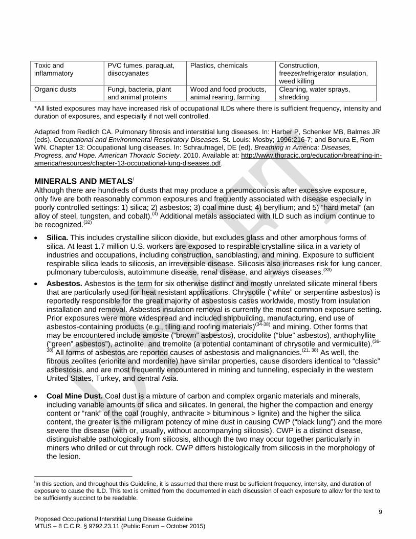
*All listed exposures may have increased risk of occupational ILDs where there is sufficient frequency, intensity and duration of exposures, and especially if not well controlled. Adapted from Redlich CA. Pulmonary fibrosis and interstitial lung diseases. In: Harber P, Schenker MB, Balmes JR (eds). Occupational and Environmental Respiratory Diseases. St. Louis: Mosby; 1996:216-7; and Bonura E, Rom WN. Chapter 13: Occupational lung diseases. In: Schraufnagel, DE (ed). Breathing in America: Diseases, Progress, and Hope. American Thoracic Society. 2010. Available at: http://www.thoracic.org/education/breathing-in-america/resources/chapter-13-occupational-lung-diseases.pdf.
MINERALS AND METALS i Although there are hundreds of dusts that may produce a pneumoconiosis after excessive exposure, only five are both reasonably common exposures and frequently associated with disease especially in poorly controlled settings: 1) silica; 2) asbestos; 3) coal mine dust; 4) beryllium; and 5) “hard metal” (an alloy of steel, tungsten, and cobalt).(4) Additional metals associated with ILD such as indium continue to be recognized.(32)
• Silica. This includes crystalline silicon dioxide, but excludes glass and other amorphous forms of silica. At least 1.7 million U.S. workers are exposed to respirable crystalline silica in a variety of industries and occupations, including construction, sandblasting, and mining. Exposure to sufficient respirable silica leads to silicosis, an irreversible disease. Silicosis also increases risk for lung cancer, pulmonary tuberculosis, autoimmune disease, renal disease, and airways diseases.(33)
• Asbestos. Asbestos is the term for six otherwise distinct and mostly unrelated silicate mineral fibers that are particularly used for heat resistant applications. Chrysotile (“white” or serpentine asbestos) is reportedly responsible for the great majority of asbestosis cases worldwide, mostly from insulation installation and removal. Asbestos insulation removal is currently the most common exposure setting. Prior exposures were more widespread and included shipbuilding, manufacturing, end use of asbestos-containing products (e.g., tiling and roofing materials)(34-38) and mining. Other forms that may be encountered include amosite (“brown” asbestos), crocidolite (“blue” asbestos), anthophyllite (“green” asbestos”), actinolite, and tremolite (a potential contaminant of chrysotile and vermiculite).(36-
38) All forms of asbestos are reported causes of asbestosis and malignancies.(21, 38) As well, the fibrous zeolites (erionite and mordenite) have similar properties, cause disorders identical to “classic” asbestosis, and are most frequently encountered in mining and tunneling, especially in the western United States, Turkey, and central Asia.
• Coal Mine Dust. Coal dust is a mixture of carbon and complex organic materials and minerals,
including variable amounts of silica and silicates. In general, the higher the compaction and energy content or “rank” of the coal (roughly, anthracite > bituminous > lignite) and the higher the silica content, the greater is the milligram potency of mine dust in causing CWP (“black lung”) and the more severe the disease (with or, usually, without accompanying silicosis). CWP is a distinct disease, distinguishable pathologically from silicosis, although the two may occur together particularly in miners who drilled or cut through rock. CWP differs histologically from silicosis in the morphology of the lesion.
iIn this section, and throughout this Guideline, it is assumed that there must be sufficient frequency, intensity, and duration of exposure to cause the ILD. This text is omitted from the documented in each discussion of each exposure to allow for the text to be sufficiently succinct to be readable.
10
Proposed Occupational Interstitial Lung Disease Guideline MTUS – 8 C.C.R. § 9792.23.11 (Public Forum – October 2015)
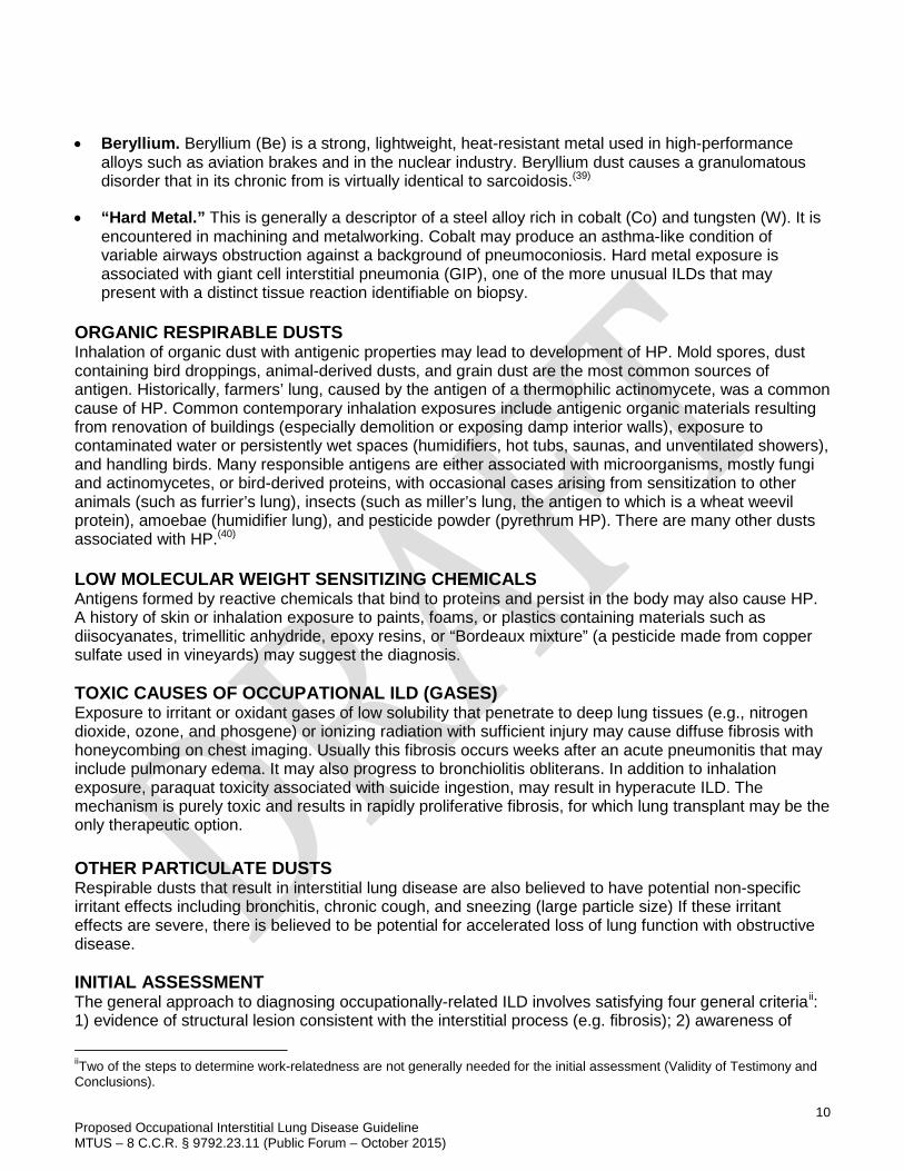
• Beryllium. Beryllium (Be) is a strong, lightweight, heat-resistant metal used in high-performance alloys such as aviation brakes and in the nuclear industry. Beryllium dust causes a granulomatous disorder that in its chronic from is virtually identical to sarcoidosis.(39)
• “Hard Metal.” This is generally a descriptor of a steel alloy rich in cobalt (Co) and tungsten (W). It is
encountered in machining and metalworking. Cobalt may produce an asthma-like condition of variable airways obstruction against a background of pneumoconiosis. Hard metal exposure is associated with giant cell interstitial pneumonia (GIP), one of the more unusual ILDs that may present with a distinct tissue reaction identifiable on biopsy.
ORGANIC RESPIRABLE DUSTS Inhalation of organic dust with antigenic properties may lead to development of HP. Mold spores, dust containing bird droppings, animal-derived dusts, and grain dust are the most common sources of antigen. Historically, farmers’ lung, caused by the antigen of a thermophilic actinomycete, was a common cause of HP. Common contemporary inhalation exposures include antigenic organic materials resulting from renovation of buildings (especially demolition or exposing damp interior walls), exposure to contaminated water or persistently wet spaces (humidifiers, hot tubs, saunas, and unventilated showers), and handling birds. Many responsible antigens are either associated with microorganisms, mostly fungi and actinomycetes, or bird-derived proteins, with occasional cases arising from sensitization to other animals (such as furrier’s lung), insects (such as miller’s lung, the antigen to which is a wheat weevil protein), amoebae (humidifier lung), and pesticide powder (pyrethrum HP). There are many other dusts associated with HP.(40) LOW MOLECULAR WEIGHT SENSITIZING CHEMICALS Antigens formed by reactive chemicals that bind to proteins and persist in the body may also cause HP. A history of skin or inhalation exposure to paints, foams, or plastics containing materials such as diisocyanates, trimellitic anhydride, epoxy resins, or “Bordeaux mixture” (a pesticide made from copper sulfate used in vineyards) may suggest the diagnosis. TOXIC CAUSES OF OCCUPATIONAL ILD (GASES) Exposure to irritant or oxidant gases of low solubility that penetrate to deep lung tissues (e.g., nitrogen dioxide, ozone, and phosgene) or ionizing radiation with sufficient injury may cause diffuse fibrosis with honeycombing on chest imaging. Usually this fibrosis occurs weeks after an acute pneumonitis that may include pulmonary edema. It may also progress to bronchiolitis obliterans. In addition to inhalation exposure, paraquat toxicity associated with suicide ingestion, may result in hyperacute ILD. The mechanism is purely toxic and results in rapidly proliferative fibrosis, for which lung transplant may be the only therapeutic option. OTHER PARTICULATE DUSTS Respirable dusts that result in interstitial lung disease are also believed to have potential non-specific irritant effects including bronchitis, chronic cough, and sneezing (large particle size) If these irritant effects are severe, there is believed to be potential for accelerated loss of lung function with obstructive disease. INITIAL ASSESSMENT The general approach to diagnosing occupationally-related ILD involves satisfying four general criteriaii: 1) evidence of structural lesion consistent with the interstitial process (e.g. fibrosis); 2) awareness of
iiTwo of the steps to determine work-relatedness are not generally needed for the initial assessment (Validity of Testimony and Conclusions).
11
Proposed Occupational Interstitial Lung Disease Guideline MTUS – 8 C.C.R. § 9792.23.11 (Public Forum – October 2015)
epidemiological or workplace studies with evidence of an agent-disease relationships; 3) evidence of exposure to an agent known to cause occupational ILD (e.g., asbestos), including sufficient dose to cause the disease; and 4) exclusion of alternative diagnoses as less likely. In practice, evidence of a structural lesion is usually demonstrated by chest x-ray and/or high resolution CT (HRCT) scan of the chest and lungs. Consideration of alternative diagnoses may require additional clinical tests and even biopsy. Biopsies are rarely necessary for the positive diagnosis of occupational ILD. Testing may be needed for beryllium disease. Clinical determination of causation by a particular agent may be satisfied by the occupational history and these initial steps. Conclusive evidence of causation may in some cases require considerably greater investigation. MEDICAL HISTORY The occupational history is usually specific for occupational ILD. Identification of a past, significant exposure usually suggests the diagnosis. Yet, in addition to describing the most recent work, it is essential to describe prior work due to the long latencies associated with some exposures. Patients with ILD of all types usually present with shortness of breath and cough. Unfortunately, those clinical symptoms are nonspecific and may be of limited value for recognition, diagnosis, and confirmation of either non-occupational or occupational ILD without additional objective testing. The presence of a comorbid condition that is associated with interstitial disease such as rheumatologic, autoimmune, inflammatory bowel, connective tissue disease (aka, collagen-vascular disorders), or drug reactions may render occupational causes less likely. However, in the case of some pneumoconioses, there may be confounding autoimmune pathology that may be related to work exposures. CWP and silicosis, in particular, are associated with an increased incidence of rheumatoid arthritis and, in the case of silicosis, systemic sclerosis, autoimmune vasculitis, and nephropathy. Occupational ILD affects both genders and workers of all ethnic backgrounds, although most are men due to the occupational distributions and pneumoconioses are much more prevalent in some racial/ethnic populations presumably due to greater exposures.(41, 42) While genetic factors have been identified and associated with immune mediated pneumoconioses, heredity has not been demonstrated to play a major role in ILD.(26) The time since first exposure (latency) to development of clinically apparent ILD varies by exposure, but some generalizations can be made. Pneumoconioses typically become clinically apparent over a period of years, exceptions are rare and include accelerated silicosis and CWP associated with high exposure levels. In HP, sensitization may occur in the first few weeks after beginning exposure, yet in others, it may be delayed for months or years. The acute, predominant airways symptoms of HP or acute beryllium disease develop in a sensitized individual over days to weeks and progress over weeks to interstitial inflammation and ultimately to fibrosis, but may rarely also be hyperacute or sudden in onset, similar to some eosinophilic pneumonias or some drug-induced pneumonitis. Differential diagnosis of an acute influenza-like or febrile disorder should include HP in a patient with a history of exposure to inhaled antigens. However, it may also suggest rheumatological or autoimmune lung disease and infection (mycoplasma, Legionella spp., or, rarely, diffuse mycosis) as a cause of interstitial disease, the latter especially in a host with a compromised immune system. A history of exposure to birds should also raise the possibility of other diseases including psittacosis. While there are no well-established risk factors for development of HP, personal susceptibility may play a role. Personal risk factors may play an important role in idiopathic interstitial fibrosis (usual interstitial pneumonia), which has a strong genetic component; a small subset of sarcoidosis are thought to be familial. Tuberous sclerosis, neurofibromatosis, and metabolic diseases affecting the lung, such as Gaucher’s disease, are hereditary but are individually rare. Other genetic impacts and interactions are not well defined.
12
Proposed Occupational Interstitial Lung Disease Guideline MTUS – 8 C.C.R. § 9792.23.11 (Public Forum – October 2015)
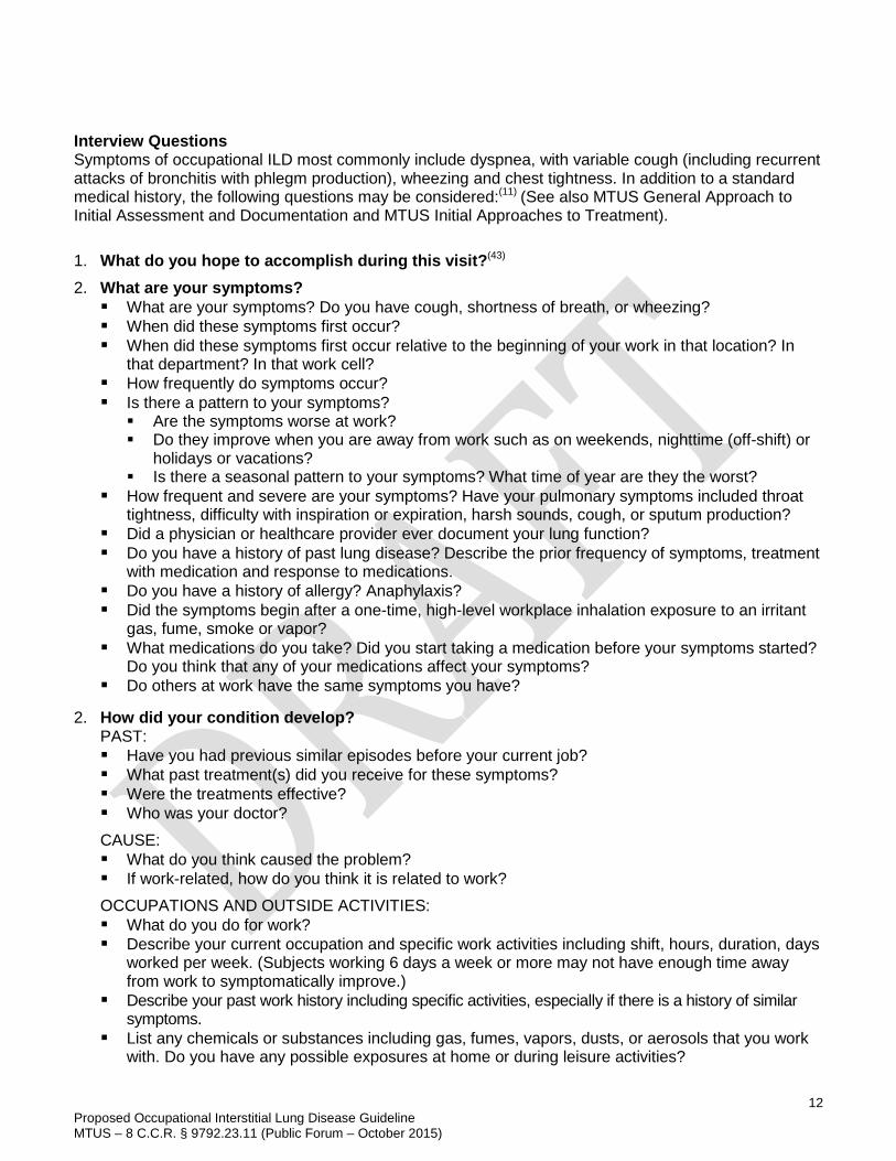
Interview Questions Symptoms of occupational ILD most commonly include dyspnea, with variable cough (including recurrent attacks of bronchitis with phlegm production), wheezing and chest tightness. In addition to a standard medical history, the following questions may be considered:(11) (See also MTUS General Approach to Initial Assessment and Documentation and MTUS Initial Approaches to Treatment).
1. What do you hope to accomplish during this visit?(43)
2. What are your symptoms? What are your symptoms? Do you have cough, shortness of breath, or wheezing? When did these symptoms first occur? When did these symptoms first occur relative to the beginning of your work in that location? In
that department? In that work cell? How frequently do symptoms occur? Is there a pattern to your symptoms? Are the symptoms worse at work? Do they improve when you are away from work such as on weekends, nighttime (off-shift) or
holidays or vacations? Is there a seasonal pattern to your symptoms? What time of year are they the worst?
How frequent and severe are your symptoms? Have your pulmonary symptoms included throat tightness, difficulty with inspiration or expiration, harsh sounds, cough, or sputum production?
Did a physician or healthcare provider ever document your lung function? Do you have a history of past lung disease? Describe the prior frequency of symptoms, treatment
with medication and response to medications. Do you have a history of allergy? Anaphylaxis? Did the symptoms begin after a one-time, high-level workplace inhalation exposure to an irritant
gas, fume, smoke or vapor? What medications do you take? Did you start taking a medication before your symptoms started?
Do you think that any of your medications affect your symptoms? Do others at work have the same symptoms you have?
2. How did your condition develop?
PAST: Have you had previous similar episodes before your current job? What past treatment(s) did you receive for these symptoms? Were the treatments effective? Who was your doctor?
CAUSE: What do you think caused the problem? If work-related, how do you think it is related to work?
OCCUPATIONS AND OUTSIDE ACTIVITIES: What do you do for work? Describe your current occupation and specific work activities including shift, hours, duration, days
worked per week. (Subjects working 6 days a week or more may not have enough time away from work to symptomatically improve.)
Describe your past work history including specific activities, especially if there is a history of similar symptoms.
List any chemicals or substances including gas, fumes, vapors, dusts, or aerosols that you work with. Do you have any possible exposures at home or during leisure activities?

13
Proposed Occupational Interstitial Lung Disease Guideline MTUS – 8 C.C.R. § 9792.23.11 (Public Forum – October 2015)
List any “secondary jobs” or concurrent occupations that may involve exposure to chemicals or substances including gas, fumes, vapors, dusts, or aerosols.
What is the work area’s room size, specific ventilation, other co-worker reports, exhaust hoods, remodeling, and recent change in processes? Are there Material Safety Data Sheets and industrial hygiene reports available?
Were there changes in work processes in the period preceding the onset of symptoms?iii Does your employer provide protective equipment at work, such as masks or respirators? How
often do you use them? Are they required? When were you last fit tested? Are your symptoms constant or do they come and go? Does anything seem to make the problem worse or better? Do symptoms develop within minutes
of specific activities or exposures at work? Describe when your symptoms first started? Was there an event at the time the symptoms
started? Have your symptoms changed over time since then? How? Do your symptoms limit your work performance and if so, how? Describe your living environment including any hobbies, crafts, pets, family members who work
with chemicals, family members who smoke, living near an industrial plant, or living near congested traffic area.(4, 44)
NON-OCCUPATIONAL ACTIVITIES: What is your lifetime exposure to tobacco? Second-hand exposure? What has your lifetime exposure been to other inhaled substances, marijuana, hookah, spice,
etc? What are your leisure activities (e.g., woodworking, gardening, welding etc.)? Do you have a second job (moonlighting)?
3. How do these symptoms limit you? Are there any activities that you can no longer perform? Do you feel very short of breath during exercise? Do you feel more short of breath when doing normal daily activities? How long have your activities been limited?
4. Do you have other medical problems? Do you have headaches, fatigue, malaise, weight loss, changes in appetite, fever, physical
abilities and exercise intolerance? Do you have any autoimmune, infectious, or metabolic diseases? Do you have any allergies? Do you have any other respiratory diseases or conditions? Do you smoke? Does someone else in your environment smoke? Do you use other drugs, including marijuana? Do you have diabetes, kidney disease, or HIV/AIDS? Have you ever had cancer?
PHYSICAL EXAMINATION Other references provide detailed guidance on pulmonary examination.(45, 46) In general, an occupational pulmonary physical examination should include the following elements: Vital signs, including measured respiratory rate.
iiiSymptoms of cough or dyspnea that develop or worsen after a worker starts a new job or after new materials are introduced on a job are suggestive (a substantial period – from months to years – can elapse between initial exposure and development of symptoms).
14
Proposed Occupational Interstitial Lung Disease Guideline MTUS – 8 C.C.R. § 9792.23.11 (Public Forum – October 2015)
Overall functional abilities, including ease of movement, walking and changing positions while assessing breathlessness.
Assessment of respiratory status with quiet respirations (e.g., rate, depth, use of accessory muscles, nasal flaring).
Inspection for stigmata of pulmonary disease as well as potential etiologies including mucous membrane abnormalities, nasal polyps/swelling, clubbing (asbestosis, idiopathic pulmonary fibrosis, some hypersensitivity pneumonitides), nasal crease line, and anterior-posterior diameter. While of limited sensitivity, clubbing, if present, may be useful in the diagnosis of asbestosis and idiopathic pulmonary fibrosis (IPF).
Palpation primarily for chest wall abnormalities, tracheal deviation or tactile fremitus. Percussion for resonance to identify aeration, diaphragm level, suggestion for fluid interface or
consolidation. Auscultation for inspiration to expiration ratio, adventitious breath sounds including crackles,
wheeze (often a secondary manifestation of HP and a primary manifestation of eosinophilic pneumonia) and pleural rubs, as well as timing, location and persistence of lung findings.
Cardiac examination with attention to findings of cor pulmonale and heart failure. Dermal examination for signs of disease, i.e., erythema nodosum (sarcoidosis).(11)
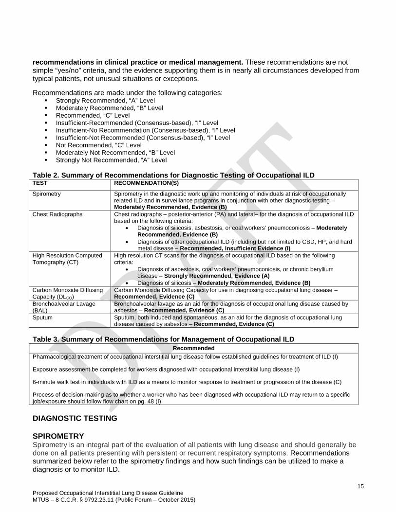
DIAGNOSTIC APPROACH The diagnoses of silicosis, asbestosis and CWP are typically made clinically, based on occupational history of sufficient exposure with appropriate latency, objective radiographic evidence (chest radiograph and/or HRCT), assessment of pulmonary function (including consistent changes in ventilatory capacity, static lung volumes or gas-exchange), and consideration of alternative differential diagnoses. While some reviews have recommended a surgical biopsy for diagnosis of non-occupational ILD, in the setting of an appropriate clinical presentation, several studies have established the diagnosis of ILD by HRCT at 70%.(11) The diagnosis of most occupational ILDs may be suggested when the patient belongs to a group at high risk. The diagnosis is usually made from the combination of occupational exposure history and imaging studies, often a chest x-ray alone. The most common challenges in differential diagnosis include: 1) distinguishing between occupational interstitial disease and idiopathic pulmonary fibrosis, 2) identifying the responsible agent in a case of mixed-dust pneumoconiosis or HP, 3) identifying the agent when the history is unclear, and 4) differentiating between sarcoidosis and beryllium disease, generally using immunologic testing. In a worker with a typical clinical picture (including exposure history, latency, and radiographic presentation), lung biopsy is rarely needed to provide a diagnosis of occupational ILD. Pathologic examination of lung tissue may at times be required in atypical settings, particularly to exclude treatable non-occupational disorders or malignancy. As in non-occupational settings, by using an interdisciplinary approach, including HRCT, to reach a diagnosis results in a lung biopsy being rarely helpful unless clinical or radiographic features are inconclusive or atypical.(11) SUMMARY TABLES: RECOMMENDATIONS AND EVIDENCE Table 2 summarizes the recommendations from the Evidence-based Practice ILD Panel for diagnostic testing for occupational ILD. Table 3 summarizes the recommendations for management of occupational ILD. The recommendations are based on critically appraised higher quality research evidence and on expert consensus observing First Principles when higher quality evidence was unavailable or inconsistent. The reader is cautioned to utilize the more detailed indications, specific appropriate diagnoses, temporal sequencing, prior testing or treatment, and contraindications that are elaborated in more detail for each test or treatment in the body of this Guideline in using these
15
Proposed Occupational Interstitial Lung Disease Guideline MTUS – 8 C.C.R. § 9792.23.11 (Public Forum – October 2015)
recommendations in clinical practice or medical management. These recommendations are not simple “yes/no” criteria, and the evidence supporting them is in nearly all circumstances developed from typical patients, not unusual situations or exceptions. Recommendations are made under the following categories:
Strongly Recommended, “A” Level Moderately Recommended, “B” Level Recommended, “C” Level Insufficient-Recommended (Consensus-based), “I” Level Insufficient-No Recommendation (Consensus-based), “I” Level Insufficient-Not Recommended (Consensus-based), “I” Level Not Recommended, “C” Level Moderately Not Recommended, “B” Level Strongly Not Recommended, “A” Level
Table 2. Summary of Recommendations for Diagnostic Testing of Occupational ILD TEST RECOMMENDATION(S)
Spirometry Spirometry in the diagnostic work up and monitoring of individuals at risk of occupationally related ILD and in surveillance programs in conjunction with other diagnostic testing – Moderately Recommended, Evidence (B)
Chest Radiographs Chest radiographs – posterior-anterior (PA) and lateral– for the diagnosis of occupational ILD based on the following criteria:
• Diagnosis of silicosis, asbestosis, or coal workers’ pneumoconiosis – Moderately Recommended, Evidence (B)
• Diagnosis of other occupational ILD (including but not limited to CBD, HP, and hard metal disease – Recommended, Insufficient Evidence (I)
High Resolution Computed Tomography (CT)
High resolution CT scans for the diagnosis of occupational ILD based on the following criteria:
• Diagnosis of asbestosis, coal workers’ pneumoconiosis, or chronic beryllium disease – Strongly Recommended, Evidence (A)
• Diagnosis of silicosis – Moderately Recommended, Evidence (B) Carbon Monoxide Diffusing Capacity (DLCO)
Carbon Monoxide Diffusing Capacity for use in diagnosing occupational lung disease – Recommended, Evidence (C)
Bronchoalveolar Lavage (BAL)
Bronchoalveolar lavage as an aid for the diagnosis of occupational lung disease caused by asbestos – Recommended, Evidence (C)
Sputum Sputum, both induced and spontaneous, as an aid for the diagnosis of occupational lung disease caused by asbestos – Recommended, Evidence (C)
Table 3. Summary of Recommendations for Management of Occupational ILD
Recommended Pharmacological treatment of occupational interstitial lung disease follow established guidelines for treatment of ILD (I) Exposure assessment be completed for workers diagnosed with occupational interstitial lung disease (I) 6-minute walk test in individuals with ILD as a means to monitor response to treatment or progression of the disease (C) Process of decision-making as to whether a worker who has been diagnosed with occupational ILD may return to a specific job/exposure should follow flow chart on pg. 48 (I) DIAGNOSTIC TESTING SPIROMETRY Spirometry is an integral part of the evaluation of all patients with lung disease and should generally be done on all patients presenting with persistent or recurrent respiratory symptoms. Recommendations summarized below refer to the spirometry findings and how such findings can be utilized to make a diagnosis or to monitor ILD.
16
Proposed Occupational Interstitial Lung Disease Guideline MTUS – 8 C.C.R. § 9792.23.11 (Public Forum – October 2015)
Spirometry is the most commonly performed of the pulmonary function tests (PFTs). Since spirometry is often the only PFT performed in the occupational setting, it is frequently simply called a “PFT.” Spirometry measures the volumes and rates of flow during forced exhalation after a maximal inhalation. In the occupational setting, a calibrated volume or flow measuring device is used to monitor ventilatory function and to identify existing or incipient lung disorders involving the airways, lungs, and chest wall.(47, 48) The forced vital capacity (FVC) reflects the capacity of the lung to hold air after a maximal inspiration and is the primary indicator of the presence of possible restrictive impairment. The FVC is reduced, or “restricted,” when compliance of the lung is decreased, or when chest wall expansion or neuromuscular function are limited. Though the FVC may also be reduced in airway diseases that result in airway closure and trapping air in the lungs, the FVC reduction usually will not be accompanied by an equal reduction in the FEV1, so the ratio of FEV1/FVC is reduced in purely obstructive disorders. In contrast, in a purely restrictive disorder, both FVC and FEV1 are reduced by a similar degree, yielding a normal or high FEV1/FVC ratio.(49-51) In interpreting the results of spirometry, it is important to consider all aspects of the worker’s health, including exposures, smoking status, and other conditions including adiposity that may affect the results. Spirometry patterns are generally not specific for any one type or cause of occupational ILD. However, spirometry provides important information regarding the functional status of the lungs, and is useful in initial assessment, evaluating prognosis, and monitoring the effectiveness of exposure controls and other therapeutic interventions. Spirometry is used for several distinct purposes: 1) routine surveillance testing to identify workers requiring more detailed evaluation; 2) as a key component in the diagnosis of occupational and other ILDs; 3) as a factor in considering work ability and appropriate assignments; 4) for monitoring course over time; and 5) as part of the assessment of compensable impairment. The appropriate criteria should be selected for each case. Recommendation: Spirometry for Occupational Interstitial Lung Disease Diagnosis and Surveillance Spirometry is moderately recommended in the diagnostic work-up and monitoring of individuals at risk of occupationally related interstitial lung diseases and in surveillance programs in conjunction with other diagnostic testing.
Strength of Evidence – Moderately Recommended, Evidence (B) Level of Confidence – High
Indications – Diagnostic: Patients with history and/or chest radiography consistent with ILD and workplace exposure consistent with plausible etiologies (e.g., worker complaining of chronic or intermittent cough, shortness of breath, or decreased physical abilities).(52) Reliable results may not be achieved in the presence of symptomatic upper or lower respiratory infections or painful disorders of the chest or mouth. (49) Thus, spirometry should generally be postponed if there has been recent surgery, respiratory infections, or recent cardiac problems. Indications – Surveillance: For workers in occupations with exposures that are either known or thought to be associated with development of occupational ILD, the American College of Occupational and Environmental Medicine (ACOEM), NIOSH and the American Thoracic Society (ATS) currently recommend that a decrement in FEV1 over time that is at least 15% more than that expected due to aging should trigger further medical evaluation of the worker.(47, 50) Such longitudinal evaluation should only be undertaken when spirometry tests are of adequate technical quality. It is recommended to perform periodic serial spirometry testing to assist in earlier determination of pulmonary decline.(47-49, 53) Harms – Minimal. Benefits – Provide physiologic evidence for occupational ILD, and differentiate between obstructive and restrictive patterns of lung function.
17
Proposed Occupational Interstitial Lung Disease Guideline MTUS – 8 C.C.R. § 9792.23.11 (Public Forum – October 2015)
Technique – Diagnostic spirometry testing should be performed using recommended equipment and procedures by an appropriately trained technician in accordance with recommendations or requirements of Occupational Safety and Health Administration (OSHA), NIOSH, and Mine Safety and Health Administration (MSHA). When diagnostic spirometry is abnormal, testing should first be repeated on another occasion, if possible, to ensure that a worker was maximally inhaling, blasting out hard, and exhaling fully during the test. If results remain abnormal, short term reversibility of the spirometry results should be assessed, most often by repeating the spirometry testing after the individual has undergone a standardized short-acting bronchodilator inhalation protocol. ACOEM recommends that when performing occupational spirometry, technicians strive to meet ATS/ERS criteria for a valid test, that is, recording three or more acceptable curves, with the largest FVC and largest FEV1 repeated to within 0.15 L (150 mL).(50) Once a satisfactory test has been recorded for the worker, diagnostic interpretation may compare his/her largest results with normal ranges derived from appropriate similar populations.(49, 54, 55) Interpretation – There are several steps in the interpretation of spirometry testing performed as part of the evaluation of workers at risk of occupational ILD. First, the interpreter must review and comment on test quality and determine whether within and between manoeuvre acceptability criteria were met. If the test is considered adequate for interpretation, then assess reference values (often called normal or predicted values) against which to compare the worker’s results must be selected based on studies of asymptomatic and otherwise healthy persons of similar age, height, gender, and race/ethnicity. For workers in the U.S., ACOEM,(50) American Thoracic Society/European Respiratory Society (ATS/ERS),(56)
OSHA,(51) and AMA Guides to the Evaluation of Permanent Impairment(57) recommend the use of reference values from the National Health and Nutrition Examination Survey (NHANES) III study, which included large numbers of subjects of varying race/ethnicities.(50) Measured worker results are compared to the NHANES III predicted/normal values that are specific for the tested individual’s age, gender, self-reported race/ethnicity, and measured height. For Asian Americans, for whom there are no NHANES III reference values at this time, the worker’s FVC and FEV1 results should be compared to race-adjusted reference values. These adjusted values are obtained by determining the reference values (i.e., the predicted value and the Lower Limit of the Normal (LLN)) for a Caucasian of the same age, height, and gender and then multiplying those FVC and FEV1 predicted and LLN values by a scaling factor of 0.88.(50, 51, 58) If this correction is omitted for Asian Americans, workers may be erroneously labeled with restrictive impairments. No other groups at this time are recognized as needing race-adjustment of reference values. Since 1991, the ATS (1991, 2005), and more recently ACOEM (2000, 2011) and OSHA (2013) have recommended interpreting test results using two steps after verifying adequate test quality. The first measurement to be assessed is the FEV1/FVC. If the worker's measured ratio is below the predicted LLN ratio, the worker has airways obstruction. The severity of obstruction is assessed by comparing the worker's measured FEV1 to the appropriate predicted or reference value. Percent of predicted is calculated, with decreasing values indicating worsening severity of obstruction. The second step in interpretation of results is to assess the worker's vital capacity relative to the normal range for individuals with the worker's characteristics. Percent predicted values for FVC are also used clinically to assess restrictive ventilatory impairment (e.g., in various workers’ compensation systems). Since the FVC is the measure of vital capacity obtained from the spirometric forced expiratory maneuver, the measured FVC is compared to the lower limit of normal for the worker's FVC. If the results fall below the lower limit, it is interpreted as having possible restrictive impairment and may need further tests of pulmonary function and/or imaging studies to confirm a true restrictive impairment. Severity of a possible restrictive impairment also may be assessed using percent of predicted FEV1 as recommended by the ATS/ERS – “Mild: FEV1 >70% of predicted, Moderate: FEV1 60-69% of predicted, Moderately Severe: FEV1 50-59% of predicted, Severe: FEV1 35-49% of predicted, Very Severe: FEV1 <35% of predicted.”(56)
18
Proposed Occupational Interstitial Lung Disease Guideline MTUS – 8 C.C.R. § 9792.23.11 (Public Forum – October 2015)
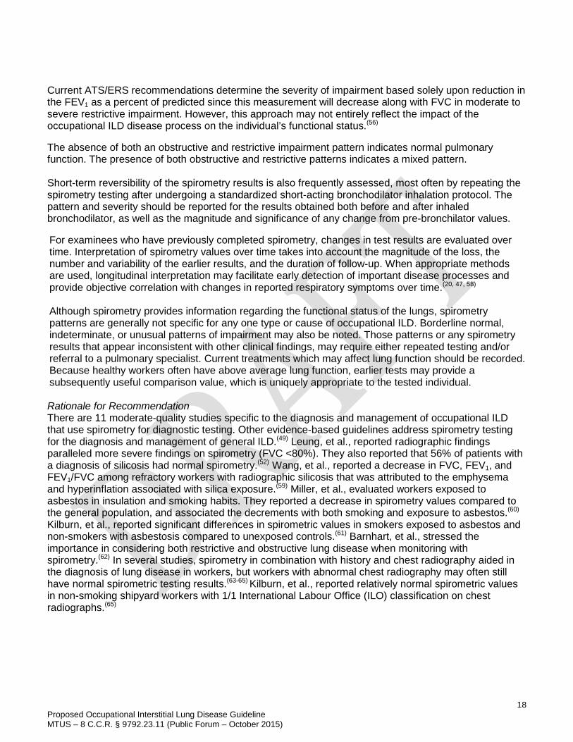
Current ATS/ERS recommendations determine the severity of impairment based solely upon reduction in the FEV1 as a percent of predicted since this measurement will decrease along with FVC in moderate to severe restrictive impairment. However, this approach may not entirely reflect the impact of the occupational ILD disease process on the individual’s functional status.(56) The absence of both an obstructive and restrictive impairment pattern indicates normal pulmonary function. The presence of both obstructive and restrictive patterns indicates a mixed pattern. Short-term reversibility of the spirometry results is also frequently assessed, most often by repeating the spirometry testing after undergoing a standardized short-acting bronchodilator inhalation protocol. The pattern and severity should be reported for the results obtained both before and after inhaled bronchodilator, as well as the magnitude and significance of any change from pre-bronchilator values.
For examinees who have previously completed spirometry, changes in test results are evaluated over time. Interpretation of spirometry values over time takes into account the magnitude of the loss, the number and variability of the earlier results, and the duration of follow-up. When appropriate methods are used, longitudinal interpretation may facilitate early detection of important disease processes and provide objective correlation with changes in reported respiratory symptoms over time.(20, 47, 58) Although spirometry provides information regarding the functional status of the lungs, spirometry patterns are generally not specific for any one type or cause of occupational ILD. Borderline normal, indeterminate, or unusual patterns of impairment may also be noted. Those patterns or any spirometry results that appear inconsistent with other clinical findings, may require either repeated testing and/or referral to a pulmonary specialist. Current treatments which may affect lung function should be recorded. Because healthy workers often have above average lung function, earlier tests may provide a subsequently useful comparison value, which is uniquely appropriate to the tested individual. Rationale for Recommendation There are 11 moderate-quality studies specific to the diagnosis and management of occupational ILD that use spirometry for diagnostic testing. Other evidence-based guidelines address spirometry testing for the diagnosis and management of general ILD.(49) Leung, et al., reported radiographic findings paralleled more severe findings on spirometry (FVC <80%). They also reported that 56% of patients with a diagnosis of silicosis had normal spirometry.(52) Wang, et al., reported a decrease in FVC, FEV1, and FEV1/FVC among refractory workers with radiographic silicosis that was attributed to the emphysema and hyperinflation associated with silica exposure.(59) Miller, et al., evaluated workers exposed to asbestos in insulation and smoking habits. They reported a decrease in spirometry values compared to the general population, and associated the decrements with both smoking and exposure to asbestos.(60) Kilburn, et al., reported significant differences in spirometric values in smokers exposed to asbestos and non-smokers with asbestosis compared to unexposed controls.(61) Barnhart, et al., stressed the importance in considering both restrictive and obstructive lung disease when monitoring with spirometry.(62) In several studies, spirometry in combination with history and chest radiography aided in the diagnosis of lung disease in workers, but workers with abnormal chest radiography may often still have normal spirometric testing results.(63-65) Kilburn, et al., reported relatively normal spirometric values in non-smoking shipyard workers with 1/1 International Labour Office (ILO) classification on chest radiographs.(65)

19
Proposed Occupational Interstitial Lung Disease Guideline MTUS – 8 C.C.R. § 9792.23.11 (Public Forum – October 2015)
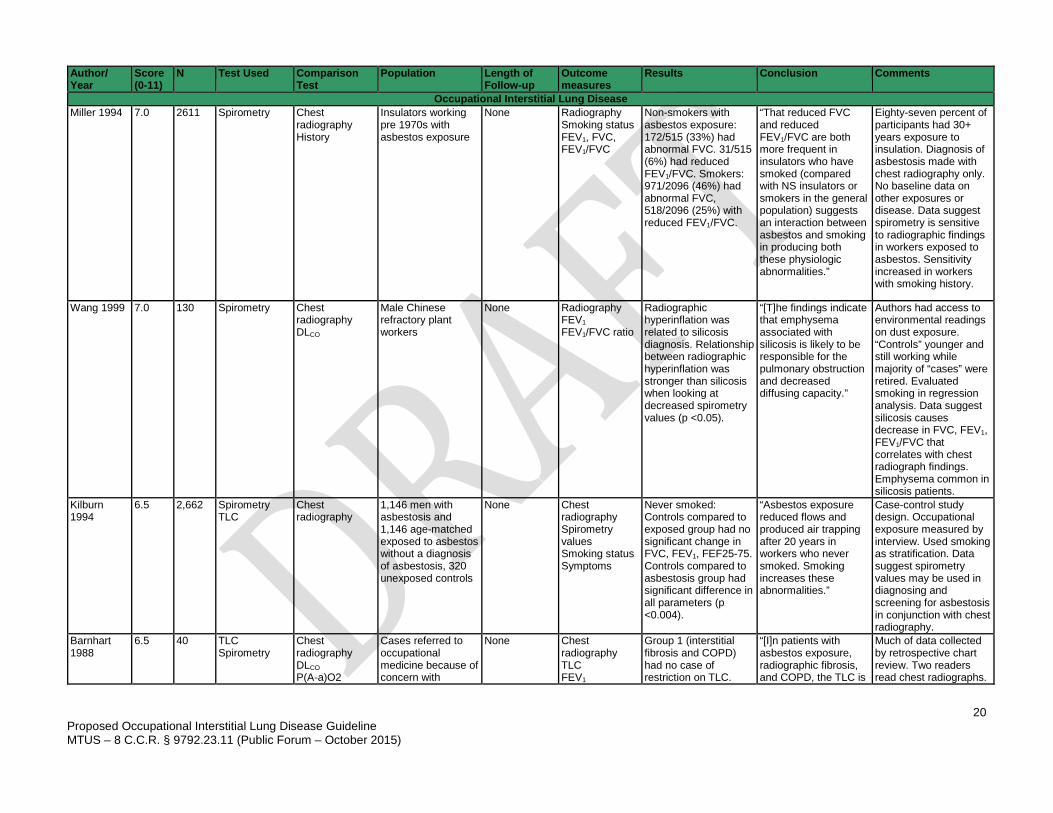
Spirometry is not invasive, has few adverse effects, and is low to moderate cost. Thus, it is highly recommended, although the evidence base is moderate, as part of a diagnostic work up, and monitoring of occupational ILDs. Evidence for the Use of Spirometry There are 11 moderate-quality diagnostic studies incorporated into this analysis.(7, 52, 59-67) There are 7 other studies in Appendix 2.(47, 48, 54, 68-71)
20
Proposed Occupational Interstitial Lung Disease Guideline MTUS – 8 C.C.R. § 9792.23.11 (Public Forum – October 2015)
Author/ Year
Score (0-11)
N Test Used Comparison Test
Population Length of Follow-up
Outcome measures
Results Conclusion Comments
Occupational Interstitial Lung Disease Miller 1994 7.0 2611 Spirometry Chest
radiography History
Insulators working pre 1970s with asbestos exposure
None Radiography Smoking status FEV1, FVC, FEV1/FVC
Non-smokers with asbestos exposure: 172/515 (33%) had abnormal FVC. 31/515 (6%) had reduced FEV1/FVC. Smokers: 971/2096 (46%) had abnormal FVC, 518/2096 (25%) with reduced FEV1/FVC.
“That reduced FVC and reduced FEV1/FVC are both more frequent in insulators who have smoked (compared with NS insulators or smokers in the general population) suggests an interaction between asbestos and smoking in producing both these physiologic abnormalities.”
Eighty-seven percent of participants had 30+ years exposure to insulation. Diagnosis of asbestosis made with chest radiography only. No baseline data on other exposures or disease. Data suggest spirometry is sensitive to radiographic findings in workers exposed to asbestos. Sensitivity increased in workers with smoking history.
Wang 1999 7.0 130 Spirometry Chest radiography DLCO
Male Chinese refractory plant workers
None Radiography FEV1 FEV1/FVC ratio
Radiographic hyperinflation was related to silicosis diagnosis. Relationship between radiographic hyperinflation was stronger than silicosis when looking at decreased spirometry values (p <0.05).
“[T]he findings indicate that emphysema associated with silicosis is likely to be responsible for the pulmonary obstruction and decreased diffusing capacity.”
Authors had access to environmental readings on dust exposure. “Controls” younger and still working while majority of “cases” were retired. Evaluated smoking in regression analysis. Data suggest silicosis causes decrease in FVC, FEV1, FEV1/FVC that correlates with chest radiograph findings. Emphysema common in silicosis patients.
Kilburn 1994
6.5 2,662 Spirometry TLC
Chest radiography
1,146 men with asbestosis and 1,146 age-matched exposed to asbestos without a diagnosis of asbestosis, 320 unexposed controls
None Chest radiography Spirometry values Smoking status Symptoms
Never smoked: Controls compared to exposed group had no significant change in FVC, FEV1, FEF25-75. Controls compared to asbestosis group had significant difference in all parameters (p <0.004).
“Asbestos exposure reduced flows and produced air trapping after 20 years in workers who never smoked. Smoking increases these abnormalities.”
Case-control study design. Occupational exposure measured by interview. Used smoking as stratification. Data suggest spirometry values may be used in diagnosing and screening for asbestosis in conjunction with chest radiography.
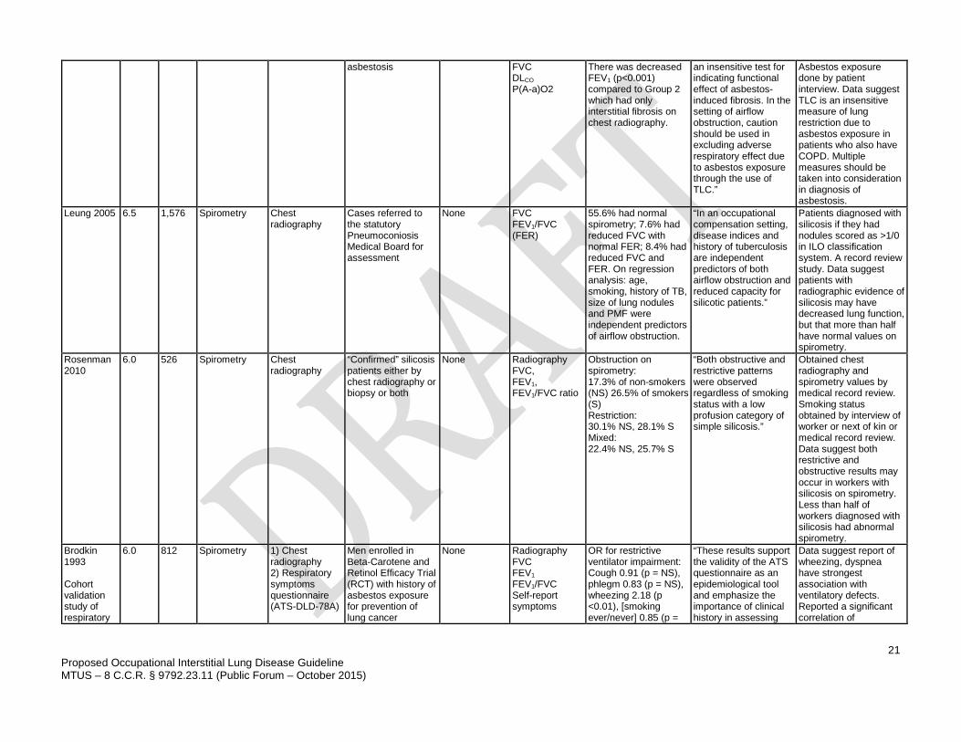
Barnhart 1988
6.5 40 TLC Spirometry
Chest radiography DLCO P(A-a)O2
Cases referred to occupational medicine because of concern with
None Chest radiography TLC FEV1
Group 1 (interstitial fibrosis and COPD) had no case of restriction on TLC.
“[I]n patients with asbestos exposure, radiographic fibrosis, and COPD, the TLC is
Much of data collected by retrospective chart review. Two readers read chest radiographs.
21
Proposed Occupational Interstitial Lung Disease Guideline MTUS – 8 C.C.R. § 9792.23.11 (Public Forum – October 2015)
asbestosis FVC DLCO P(A-a)O2
There was decreased FEV1 (p<0.001) compared to Group 2 which had only interstitial fibrosis on chest radiography.
an insensitive test for indicating functional effect of asbestos-induced fibrosis. In the setting of airflow obstruction, caution should be used in excluding adverse respiratory effect due to asbestos exposure through the use of TLC.”
Asbestos exposure done by patient interview. Data suggest TLC is an insensitive measure of lung restriction due to asbestos exposure in patients who also have COPD. Multiple measures should be taken into consideration in diagnosis of asbestosis.
Leung 2005 6.5 1,576 Spirometry Chest radiography
Cases referred to the statutory Pneumoconiosis Medical Board for assessment
None FVC FEV1/FVC (FER)
55.6% had normal spirometry; 7.6% had reduced FVC with normal FER; 8.4% had reduced FVC and FER. On regression analysis: age, smoking, history of TB, size of lung nodules and PMF were independent predictors of airflow obstruction.
“In an occupational compensation setting, disease indices and history of tuberculosis are independent predictors of both airflow obstruction and reduced capacity for silicotic patients.”
Patients diagnosed with silicosis if they had nodules scored as >1/0 in ILO classification system. A record review study. Data suggest patients with radiographic evidence of silicosis may have decreased lung function, but that more than half have normal values on spirometry.
Rosenman 2010
6.0 526 Spirometry Chest radiography
“Confirmed” silicosis patients either by chest radiography or biopsy or both
None Radiography FVC, FEV1, FEV1/FVC ratio
Obstruction on spirometry: 17.3% of non-smokers (NS) 26.5% of smokers (S) Restriction: 30.1% NS, 28.1% S Mixed: 22.4% NS, 25.7% S
“Both obstructive and restrictive patterns were observed regardless of smoking status with a low profusion category of simple silicosis.”
Obtained chest radiography and spirometry values by medical record review. Smoking status obtained by interview of worker or next of kin or medical record review. Data suggest both restrictive and obstructive results may occur in workers with silicosis on spirometry. Less than half of workers diagnosed with silicosis had abnormal spirometry.
Brodkin 1993 Cohort validation study of respiratory
6.0 812 Spirometry 1) Chest radiography 2) Respiratory symptoms questionnaire (ATS-DLD-78A)
Men enrolled in Beta-Carotene and Retinol Efficacy Trial (RCT) with history of asbestos exposure for prevention of lung cancer
None Radiography FVC FEV1 FEV1/FVC Self-report symptoms
OR for restrictive ventilator impairment: Cough 0.91 (p = NS), phlegm 0.83 (p = NS), wheezing 2.18 (p <0.01), [smoking ever/never] 0.85 (p =
“These results support the validity of the ATS questionnaire as an epidemiological tool and emphasize the importance of clinical history in assessing
Data suggest report of wheezing, dyspnea have strongest association with ventilatory defects. Reported a significant correlation of

22
Proposed Occupational Interstitial Lung Disease Guideline MTUS – 8 C.C.R. § 9792.23.11 (Public Forum – October 2015)
questionnaire
NS), parenchymal small opacities 1.41 (p <0.001), pleural thickening 1.06 (p = NS).
respiratory status.” radiographic findings with ventilatory defects.
Kilburn 1985
6.0 257 Spirometry Chest radiography DLCO Symptoms
Male shipyard workers
None Radiography FVC, FEV1, FeF25-27, FEF75-
85 DLCO Symptoms
14/43 (33%) nonsmokers had 1/1 radiographs with normal spirometry values. Current and ex-smokers had a downward trend in same values.
“These shipyard workers had minimal to moderate asbestosis with much pleural disease and little functional impairment when compared to a smoking-specific reference population.”
Used PA and lateral chest radiographs with 3 different B readers to diagnosis asbestosis in shipyard workers. Included smoking as a variable. Data suggest earlier asbestosis does not cause a significant drop in FEV1, FVC in older shipyard workers.
Non-Occupational Interstitial Lung Disease Aaron 1999 5.5 1,831 Spirometry Helium dilution
Plethysmograph Uncertain None TLC
VC FEV1 FVC FEV1/FVC
Sensitivity: 193/225 (86%); Specificity: 1,329/1606 (83%); PPV: 193/470 (41%); NPV: 1,329/1,361 (97.6%)
“[T]he accuracy with which spirometric measurement of FVC and expiratory flow rates can diagnose the presence of a restrictive impairment. Patients whose FVC fall above the 95% CI of the predicted value are very unlikely to have a restrictive impairment, and in these patients… measurement of lung volumes can be avoided.”
Uncertain what type of patients included in study. Does not appear to have any occupationally-related cases. Data suggest spirometry is useful in ruling out a restrictive lung disease diagnosis.
Boros 2004 4.0 1,173 Spirometry Whole body plethysmo-graphy
Mean age 44.3 – with HP (74), sarcoidosis (568), pulmonary fibrosis (194), connective tissue disease (51), and pneumoconiosis (23)
None TLC VC
882/1,173 (75.2%) both indices were above (LLN), 267/1,173 (22.8%) TLC was markedly reduced, 209/1,173 (17.8%) VC reduced (p <0.01), 185/1,173 (15.8%) had both indices reduced.
“Our results indicate that the spirometric measurement of VC is not enough for the detection of restriction, and may result in missing the diagnosis of diminished lung volume in almost 10% of patients. Thus in order to assess lung function reliably in ILD patients, the measurement of TLC seems to be essential.”
How each patient was originally diagnosed not described. Small number of pneumoconiosis patients. No separation of results based on diagnosis. Making this study difficult to assess in terms of occupational lung disease. Data suggest that in generalized ILD patients both VC and TLC is useful.

23
Proposed Occupational Interstitial Lung Disease Guideline MTUS – 8 C.C.R. § 9792.23.11 (Public Forum – October 2015)
Other Sircar 2007 4.5 1,730 Spirometry None Coal miners 12 years FEV1
Death Odds ratios: Compared to below 30ml/year loss. 1.39 (0.99-1.97) 60ml/year to 90ml/year 1.90 (1.32-2.76) more than 90ml/year loss of FEV1.
“Risk of death increases in individuals with rates of decline above about 60ml/year and is statistically significant with declines of 90ml or more. These results should be useful to healthcare providers in assessing lung function declines observed in individuals.”
A retrospective review of cross-sectional studies. Cause of death determined by death certificates. Data suggest serial FEV1 in coal miners with lung disease may aid in the management of the disease. If there is a loss of over 90ml/year then the risk of death increases.
24
Proposed Occupational Interstitial Lung Disease Guideline MTUS – 8 C.C.R. § 9792.23.11 (Public Forum – October 2015)
CHEST RADIOGRAPHS Chest radiographs are part of the usual evaluation of patients with respiratory symptoms. They historically have been used to investigate the relationship between exposure to respirable particles (dusts) and disease,(72) and are widely used for diagnosing and monitoring ILD. Chest radiographs show opacities which represent the accumulation of dust and the body’s reaction to the exposure.(73-77) Of the ILDs, some have more easily identifiable lesions supporting a diagnosis with radiographic testing than others. Many diseases require consideration of clinical findings, occupational history, and radiographic findings for the diagnosis.(78, 79) Silicosis and CWP, while distinct diseases, have similar radiographic appearances that generally necessitate a well-focused occupational history to help differentiate between the two disorders. Radiographs should be interpreted by a physician with appropriate training, experience, and skills in interpretation of radiographs for diagnosis of ILD. To document the patterns and severity of radiographic appearances of pneumoconiosis, radiographs are often interpreted according to the International Labour Organization (ILO) classification.(80) The size, shape and number of the opacities recorded using the ILO classification system have been shown to be related to the amount and composition of dust retained in the lung.(73, 74, 81-85) Comparison of radiographic appearances with associated pathology and lung dust content in a group of coal workers have been reported.(73) ILO classification of pneumoconiosis is recommended for worker screening and epidemiological purposes.(80, 86) Recommendation: Posterior-Anterior (PA) and Lateral Chest Radiographs Chest radiographs – posterior-anterior (PA) and lateral – are recommended for the diagnosis of occupational interstitial lung disease based on the following criteria. 1. Diagnosis of silicosis, asbestosis, or coal workers’ pneumoconiosis (CWP).
Strength of Evidence – Moderately Recommended, Evidence (B) Level of Confidence – High
2. Other occupational ILD – including but not limited to chronic beryllium disease (CBD), HP,
and hard metal disease.
Strength of Evidence – Recommended, Insufficient Evidence (I) Level of Confidence – Moderate
Performed – Chest radiographs should be performed by trained technicians and according to the ACR-SPR Practice Guidelines for the performance of chest radiography.(87) Physicians who interpret chest radiographs for diagnosis or medical surveillance of occupational lung disease should have appropriate training, experience, and skills. Indications – To assist in the diagnosis of ILD in workers.(88, 89) Harms – Small amount of radiation exposure 0.1mSV.(87) Benefits – Provides structural anatomic information about the lung parenchyma and pleura that informs the differential diagnosis of occupational ILD and also provides information about the extent of involvement and progression of disease. Advantages and Limitations – Chest radiographs are widely available and relatively inexpensive. Radiographs may assist in the diagnosis of occupational lung diseases, but cases will often need additional testing and history.(85, 88, 89) Rationale for Recommendations
25
Proposed Occupational Interstitial Lung Disease Guideline MTUS – 8 C.C.R. § 9792.23.11 (Public Forum – October 2015)
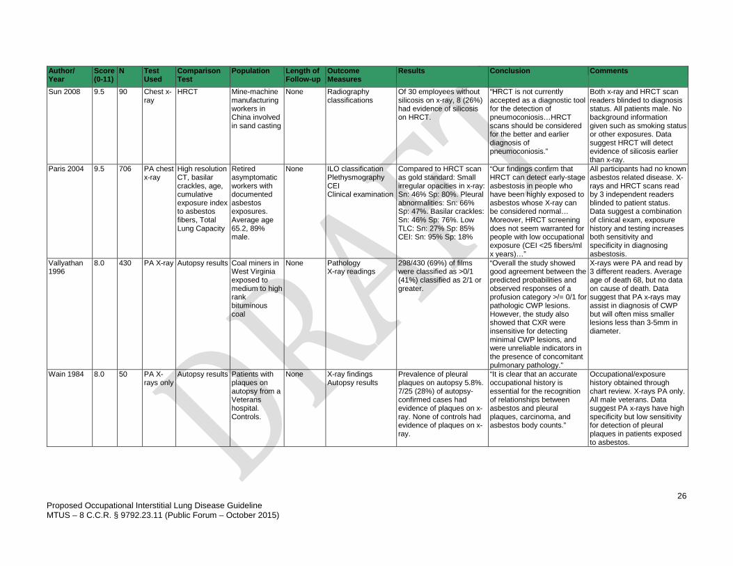
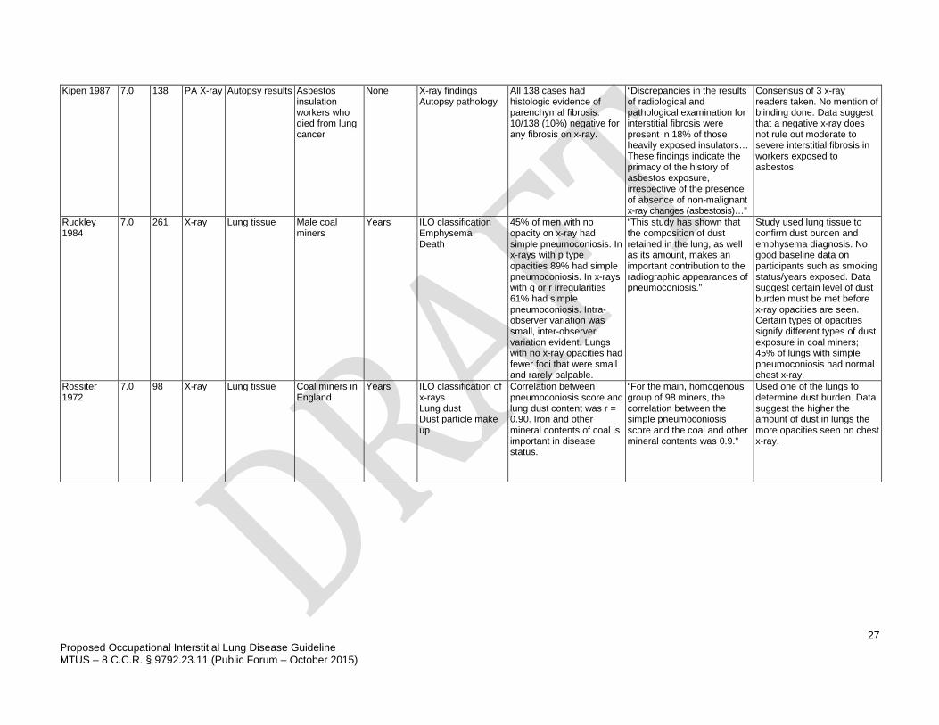
There are studies evaluating the use of chest radiographs in diagnosis of occupational ILDs. The majority of the high and moderate quality studies are done in populations exposed to coal, silica, and asbestos. Paris, et al., reported the use of total lung capacity (TLC) in combination with high exposure, basilar crackles on exam and positive x-ray findings for diagnosing asbestosis to a sensitivity of 76% and specificity of 57%.(90) A study comparing PA x-rays to autopsy results in veterans exposed to asbestos recommended x-ray in the diagnosis of pleural plaques.(91) Ruckley, et al., compared chest x-rays within four years of death to the autopsy lung tissue in coal miners reported important correlations in the type of lesions seen on x-ray and the degree of exposure. They also reported that certain types of opacities (p in the ILO classification) are more common in miners with emphysema. However, they also reported that up to 45% of patients with evidence of simple pneumoconiosis had no findings on x-ray.(73) In 1987, a follow-up study also reported fibrotic lesions in lungs in x-rays classified as normal.(75) Another study in coal workers reported benefit in using x-rays in the diagnosis of CWP, but also reported that x-rays often missed lesions if they were less than 3-5mm in diameter.(92) Other studies of coal miners also reported a strong correlation between ILO readings and dust burden in lung tissue.(77) Other studies also reported findings on x-ray and comparisons to other diagnostic tests and recommended x-rays in the diagnosis of ILDs.(64, 81-83, 88, 93-95) Sun, et al., published data on silicosis that supports the use of both x-ray and high resolution CT scans (HRCT).(96) Evidence for the Use of Chest Radiographs There are 4 high-(90-92, 96) and 13 moderate-quality(64, 73-75, 77, 81, 86, 88, 93-95, 97, 98) studies incorporated into this analysis. There is 1 low-quality study in Appendix 2.(83)
26
Proposed Occupational Interstitial Lung Disease Guideline MTUS – 8 C.C.R. § 9792.23.11 (Public Forum – October 2015)
Author/ Year
Score (0-11)
N Test Used
Comparison Test
Population Length of Follow-up
Outcome Measures
Results Conclusion Comments
Sun 2008 9.5 90 Chest x-ray
HRCT Mine-machine manufacturing workers in China involved in sand casting
None Radiography classifications
Of 30 employees without silicosis on x-ray, 8 (26%) had evidence of silicosis on HRCT.
“HRCT is not currently accepted as a diagnostic tool for the detection of pneumoconiosis…HRCT scans should be considered for the better and earlier diagnosis of pneumoconiosis.”
Both x-ray and HRCT scan readers blinded to diagnosis status. All patients male. No background information given such as smoking status or other exposures. Data suggest HRCT will detect evidence of silicosis earlier than x-ray.
Paris 2004 9.5 706 PA chest x-ray
High resolution CT, basilar crackles, age, cumulative exposure index to asbestos fibers, Total Lung Capacity
Retired asymptomatic workers with documented asbestos exposures. Average age 65.2, 89% male.
None ILO classification Plethysmography CEI Clinical examination
Compared to HRCT scan as gold standard: Small irregular opacities in x-ray: Sn: 46% Sp: 80%. Pleural abnormalities: Sn: 66% Sp: 47%. Basilar crackles: Sn: 46% Sp: 76%. Low TLC: Sn: 27% Sp: 85% CEI: Sn: 95% Sp: 18%
“Our findings confirm that HRCT can detect early-stage asbestosis in people who have been highly exposed to asbestos whose X-ray can be considered normal… Moreover, HRCT screening does not seem warranted for people with low occupational exposure (CEI <25 fibers/ml x years)…”
All participants had no known asbestos related disease. X-rays and HRCT scans read by 3 independent readers blinded to patient status. Data suggest a combination of clinical exam, exposure history and testing increases both sensitivity and specificity in diagnosing asbestosis.
Vallyathan 1996
8.0 430 PA X-ray Autopsy results Coal miners in West Virginia exposed to medium to high rank bituminous coal
None Pathology X-ray readings
298/430 (69%) of films were classified as >0/1 (41%) classified as 2/1 or greater.
“Overall the study showed good agreement between the predicted probabilities and observed responses of a profusion category >/= 0/1 for pathologic CWP lesions. However, the study also showed that CXR were insensitive for detecting minimal CWP lesions, and were unreliable indicators in the presence of concomitant pulmonary pathology.”
X-rays were PA and read by 3 different readers. Average age of death 68, but no data on cause of death. Data suggest that PA x-rays may assist in diagnosis of CWP but will often miss smaller lesions less than 3-5mm in diameter.
Wain 1984 8.0 50 PA X-rays only
Autopsy results Patients with plaques on autopsy from a Veterans hospital. Controls.
None X-ray findings Autopsy results
Prevalence of pleural plaques on autopsy 5.8%. 7/25 (28%) of autopsy-confirmed cases had evidence of plaques on x-ray. None of controls had evidence of plaques on x-ray.
“It is clear that an accurate occupational history is essential for the recognition of relationships between asbestos and pleural plaques, carcinoma, and asbestos body counts.”
Occupational/exposure history obtained through chart review. X-rays PA only. All male veterans. Data suggest PA x-rays have high specificity but low sensitivity for detection of pleural plaques in patients exposed to asbestos.
27
Proposed Occupational Interstitial Lung Disease Guideline MTUS – 8 C.C.R. § 9792.23.11 (Public Forum – October 2015)
Kipen 1987 7.0 138 PA X-ray Autopsy results Asbestos insulation workers who died from lung cancer
None X-ray findings Autopsy pathology
All 138 cases had histologic evidence of parenchymal fibrosis. 10/138 (10%) negative for any fibrosis on x-ray.
“Discrepancies in the results of radiological and pathological examination for interstitial fibrosis were present in 18% of those heavily exposed insulators… These findings indicate the primacy of the history of asbestos exposure, irrespective of the presence of absence of non-malignant x-ray changes (asbestosis)…”
Consensus of 3 x-ray readers taken. No mention of blinding done. Data suggest that a negative x-ray does not rule out moderate to severe interstitial fibrosis in workers exposed to asbestos.
Ruckley 1984
7.0 261 X-ray Lung tissue Male coal miners
Years ILO classification Emphysema Death
45% of men with no opacity on x-ray had simple pneumoconiosis. In x-rays with p type opacities 89% had simple pneumoconiosis. In x-rays with q or r irregularities 61% had simple pneumoconiosis. Intra-observer variation was small, inter-observer variation evident. Lungs with no x-ray opacities had fewer foci that were small and rarely palpable.
“This study has shown that the composition of dust retained in the lung, as well as its amount, makes an important contribution to the radiographic appearances of pneumoconiosis.”
Study used lung tissue to confirm dust burden and emphysema diagnosis. No good baseline data on participants such as smoking status/years exposed. Data suggest certain level of dust burden must be met before x-ray opacities are seen. Certain types of opacities signify different types of dust exposure in coal miners; 45% of lungs with simple pneumoconiosis had normal chest x-ray.
Rossiter 1972
7.0 98 X-ray Lung tissue Coal miners in England
Years ILO classification of x-rays Lung dust Dust particle make up
Correlation between pneumoconiosis score and lung dust content was r = 0.90. Iron and other mineral contents of coal is important in disease status.
“For the main, homogenous group of 98 miners, the correlation between the simple pneumoconiosis score and the coal and other mineral contents was 0.9.”
Used one of the lungs to determine dust burden. Data suggest the higher the amount of dust in lungs the more opacities seen on chest x-ray.
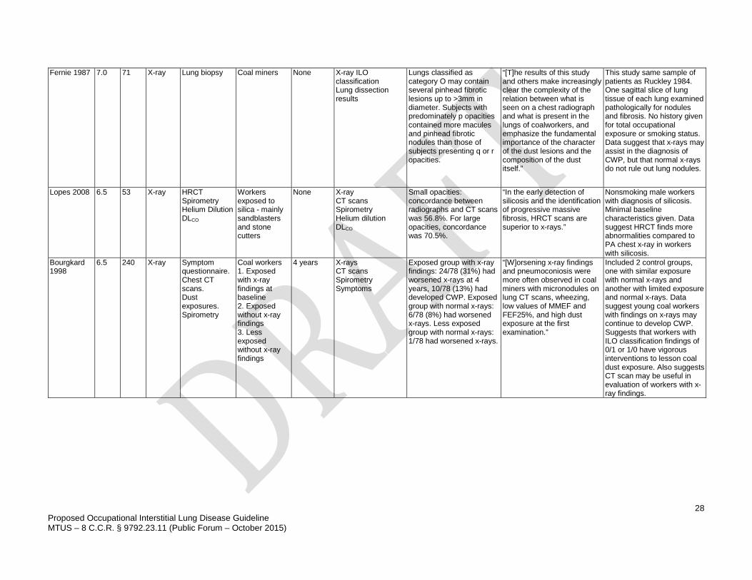
28
Proposed Occupational Interstitial Lung Disease Guideline MTUS – 8 C.C.R. § 9792.23.11 (Public Forum – October 2015)
Fernie 1987 7.0 71 X-ray Lung biopsy Coal miners None X-ray ILO classification Lung dissection results
Lungs classified as category O may contain several pinhead fibrotic lesions up to >3mm in diameter. Subjects with predominately p opacities contained more macules and pinhead fibrotic nodules than those of subjects presenting q or r opacities.
“[T]he results of this study and others make increasingly clear the complexity of the relation between what is seen on a chest radiograph and what is present in the lungs of coalworkers, and emphasize the fundamental importance of the character of the dust lesions and the composition of the dust itself.”
This study same sample of patients as Ruckley 1984. One sagittal slice of lung tissue of each lung examined pathologically for nodules and fibrosis. No history given for total occupational exposure or smoking status. Data suggest that x-rays may assist in the diagnosis of CWP, but that normal x-rays do not rule out lung nodules.
Lopes 2008 6.5 53 X-ray HRCT Spirometry Helium Dilution DLCO
Workers exposed to silica - mainly sandblasters and stone cutters
None X-ray CT scans Spirometry Helium dilution DLCO
Small opacities: concordance between radiographs and CT scans was 56.8%. For large opacities, concordance was 70.5%.
“In the early detection of silicosis and the identification of progressive massive fibrosis, HRCT scans are superior to x-rays.”
Nonsmoking male workers with diagnosis of silicosis. Minimal baseline characteristics given. Data suggest HRCT finds more abnormalities compared to PA chest x-ray in workers with silicosis.
Bourgkard 1998
6.5 240 X-ray Symptom questionnaire. Chest CT scans. Dust exposures. Spirometry
Coal workers 1. Exposed with x-ray findings at baseline 2. Exposed without x-ray findings 3. Less exposed without x-ray findings
4 years X-rays CT scans Spirometry Symptoms
Exposed group with x-ray findings: 24/78 (31%) had worsened x-rays at 4 years, 10/78 (13%) had developed CWP. Exposed group with normal x-rays: 6/78 (8%) had worsened x-rays. Less exposed group with normal x-rays: 1/78 had worsened x-rays.
“[W]orsening x-ray findings and pneumoconiosis were more often observed in coal miners with micronodules on lung CT scans, wheezing, low values of MMEF and FEF25%, and high dust exposure at the first examination.”
Included 2 control groups, one with similar exposure with normal x-rays and another with limited exposure and normal x-rays. Data suggest young coal workers with findings on x-rays may continue to develop CWP. Suggests that workers with ILO classification findings of 0/1 or 1/0 have vigorous interventions to lesson coal dust exposure. Also suggests CT scan may be useful in evaluation of workers with x-ray findings.
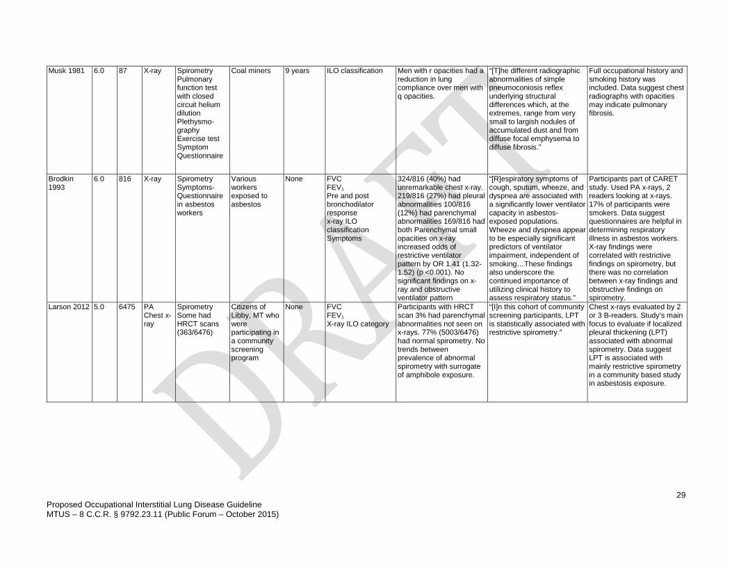
29
Proposed Occupational Interstitial Lung Disease Guideline MTUS – 8 C.C.R. § 9792.23.11 (Public Forum – October 2015)
Musk 1981 6.0 87 X-ray Spirometry Pulmonary function test with closed circuit helium dilution Plethysmo-graphy Exercise test Symptom Questionnaire
Coal miners 9 years ILO classification Men with r opacities had a reduction in lung compliance over men with q opacities.
“[T]he different radiographic abnormalities of simple pneumoconiosis reflex underlying structural differences which, at the extremes, range from very small to largish nodules of accumulated dust and from diffuse focal emphysema to diffuse fibrosis.”
Full occupational history and smoking history was included. Data suggest chest radiographs with opacities may indicate pulmonary fibrosis.
Brodkin 1993
6.0 816 X-ray Spirometry Symptoms-Questionnaire in asbestos workers
Various workers exposed to asbestos
None FVC FEV1 Pre and post bronchodilator response x-ray ILO classification Symptoms
324/816 (40%) had unremarkable chest x-ray. 219/816 (27%) had pleural abnormalities 100/816 (12%) had parenchymal abnormalities 169/816 had both Parenchymal small opacities on x-ray increased odds of restrictive ventilator pattern by OR 1.41 (1.32-1.52) (p <0.001). No significant findings on x-ray and obstructive ventilator pattern
“[R]espiratory symptoms of cough, sputum, wheeze, and dyspnea are associated with a significantly lower ventilator capacity in asbestos-exposed populations. Wheeze and dyspnea appear to be especially significant predictors of ventilator impairment, independent of smoking…These findings also underscore the continued importance of utilizing clinical history to assess respiratory status.”
Participants part of CARET study. Used PA x-rays, 2 readers looking at x-rays. 17% of participants were smokers. Data suggest questionnaires are helpful in determining respiratory illness in asbestos workers. X-ray findings were correlated with restrictive findings on spirometry, but there was no correlation between x-ray findings and obstructive findings on spirometry.
Larson 2012 5.0 6475 PA Chest x-ray
Spirometry Some had HRCT scans (363/6476)
Citizens of Libby, MT who were participating in a community screening program
None FVC FEV1 X-ray ILO category
Participants with HRCT scan 3% had parenchymal abnormalities not seen on x-rays. 77% (5003/6476) had normal spirometry. No trends between prevalence of abnormal spirometry with surrogate of amphibole exposure.
“[I]n this cohort of community screening participants, LPT is statistically associated with restrictive spirometry.”
Chest x-rays evaluated by 2 or 3 B-readers. Study’s main focus to evaluate if localized pleural thickening (LPT) associated with abnormal spirometry. Data suggest LPT is associated with mainly restrictive spirometry in a community based study in asbestosis exposure.
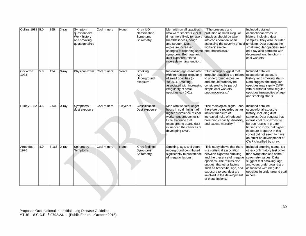
30
Proposed Occupational Interstitial Lung Disease Guideline MTUS – 8 C.C.R. § 9792.23.11 (Public Forum – October 2015)
Collins 1988 5.0 895 X-ray Symptom questionnaire, Work history and smoking questionnaires
Coal miners None X-ray ILO classification Symptoms Spirometry
Men with small opacities who were smokers 2 or 3 times more likely to report breathlessness, cough and sputum. Dust exposure increased changes of reporting same symptoms. Both age and dust exposure related inversely to lung function.
“[T]he presence and profusion of small irregular opacities should be taken into consideration when assessing the severity of coal workers’ simple pneumoconiosis.”
Included detailed occupational exposure history, including dust samples. They also included smoking. Data suggest the small irregular opacities seen on x-ray also correlate with decreased lung function in coal workers.
Cockcroft 1983
5.0 124 X-ray Physical exam Coal miners Years Smoking Age Underground exposure
Increasing age associated with increasing irregularity of small opacities (p <0.001). Smoking associated with increasing irregularity of small opacities (p <0.01).
“Our findings suggest that irregular opacities are related to underground exposure and should probably be considered to be part of simple coal workers’ pneumoconiosis.”
Included detailed occupational exposure history, and smoking status. Data suggest the irregular opacities may signify CWP with or without small regular opacities irrespective of age and smoking status.
Hurley 1982 4.5 2,600 X-ray Symptoms, dust exposure
Coal miners 10 years Classification Dust exposure
Men who worked longer hours in coalmining had higher prevalence of coal worker pneumoconiosis. Little evidence that exposures to quartz dust influenced the chances of developing CWP.
“The radiological signs…can therefore be regarded as an indirect measure of increased risks of reduced breathing capacity, disability, and excess mortality.”
Included detailed occupational exposure history, including dust samples. Data suggest that overall coal dust exposure burden results in greater findings on x-ray, but higher exposure to quartz in this cohort did not seem to have an effect on development of CWP classified by x-ray.
Amandus 1976
4.0 6,166 X-ray Spirometry Symptoms
Coal miners None X-ray findings Symptoms Spirometry
Smoking, age, and years underground contributed significantly to prevalence of irregular lesions.
“This study shows that there is a statistical association between cigarette smoking and the presence of irregular opacities. The results also suggest that other factors such as bronchitis, age, and exposure to coal dust are involved in the development of these lesions.”
Included smoking status. No other confirmatory test other than symptoms and some spirometry values. Data suggest that smoking, age, and years underground are associated with irregular opacities in underground coal miners.
31
Proposed Occupational Interstitial Lung Disease Guideline MTUS – 8 C.C.R. § 9792.23.11 (Public Forum – October 2015)
HIGH RESOLUTION COMPUTED TOMOGRAPHY (HRCT) SCANS Since the late 1980s, CT scans have been used in diagnosis of ILD. Contemporary practice is to use high resolution CT scanning (HRCT) for pulmonary evaluation. Several studies have reported both greater sensitivity and specificity compared to chest x-ray in detecting both parenchymal and airway changes.(99-110) However, with the newer technologies, it is becoming more difficult to separate between subnormal radiological findings that may occur in normal working populations, especially as the working population ages and these findings must be evaluated in context of exposures and other comorbidities. Although grading systems for HRCT have been proposed, there is currently no widely adopted counterpart to the ILO Classification system for chest x-rays. Although useful in diagnosis of occupational ILD, HRCT is not considered an essential part of the evaluation if there are existing radiographs documenting occupational ILD consistent with the worker’s exposure. On the other hand, if there are atypical features, subtle abnormalities on routine radiography, and/or competing causes for the findings, then an HRCT may be quite helpful in confirming or excluding a diagnosis of occupational ILD. Recommendation: High Resolution CT Scan High resolution CT scans are recommended for the diagnosis of occupational interstitial lung disease based on the following criteria: 1. Diagnosis of coal workers’ pneumoconiosis, asbestosis, or chronic beryllium disease.
Strength of Evidence – Strongly Recommended, Evidence (A) Level of Confidence – High
2. Diagnosis of silicosis.
Strength of Evidence – Moderately Recommended, Evidence (B) Level of Confidence – High
Performed – CT scans should be performed by trained technicians and according to the American College of Radiology (ACR) guidelines. Readers of CT scans for occupational lung disease should have appropriate training and experience. They are generally performed in the supine position, but prone imaging may be of use in certain circumstances, for example, detection of subtle peripheral and/or basilar findings.(102) There is also evidence to support scanning the entire thorax in patients with asbestosis to look for apical disease.(111) Indications – To assist in the diagnosis of occupationally-related ILD in workers suspected of having pathology.(106) HRCT scanning is recommended when the findings make occupational ILD reasonably likely and when the chest radiograph alone is insufficient. Although useful in diagnosis of occupational ILD, HRCT is not an essential part of the evaluation if chest radiographs document an occupational ILD that is consistent with the worker’s exposure. On the other hand, if there are atypical features, subtle abnormalities on routine radiography, and/or competing causes for the findings, then an HRCT may be quite helpful in confirming or excluding a diagnosis of occupational ILD. Harms – Radiation exposure 7.0 mSV, potential diagnosis of other (neoplastic) lung findings which may prompt invasive studies that carry inherent risks (e.g., thoracotomy, biopsy).(87) Benefits – Provides detailed information regarding structural parenchymal and pleural changes to support differential diagnosis of occupational ILD. Advantages and Limitations – CT scans are moderately costly and have increased radiation exposure compared to chest radiography.(112) Many of the findings on CT scan may also be related to other health conditions such as idiopathic pulmonary fibrosis, therefore, the findings must be considered in context
32
Proposed Occupational Interstitial Lung Disease Guideline MTUS – 8 C.C.R. § 9792.23.11 (Public Forum – October 2015)
with clinical history and work-related exposures. HRCT may demonstrate patterns of structural abnormality that may permit specific categorization of occupational ILD particularly as silicosis, with a high degree of diagnostic certainty. Rationale for Recommendations There are 5 high- and 8 moderate-quality studies evaluating the use of HRCT scans in the diagnosis of occupational ILDs. Many of the studies did not include baseline smoking status, which may make drawing conclusions more difficult. Gamsu, et al., conducted HRCT scans both in the prone and supine positions at maximal inspiration. They compared HRCT scan results to biopsy results and chest radiography. They reported greater specificity of asbestosis diagnosis with at least two findings on HRCT scan.(102) Several other moderate-quality studies reported greater sensitivity by HRCT scan compared to chest radiography in the detection of abnormalities associated with a diagnosis of asbestosis.(100, 103, 107, 109) Collins, et al. reported that HRCT scans may detect CWP at earlier stages than chest radiography, but that the workers with HRCT findings and normal chest radiographs did not have any physiological abnormalities.(106) Gevenois, et al. also reported greater detection of abnormalities on HRCT compared to chest radiography in low grade CWP.(99) Other studies also reported HRCT detecting more findings compared to chest radiography in worker’s exposed to coal dust.(105) Evidence for the Use of HRCT There are 5 high-(99, 102, 104, 106, 113) and 9 moderate-quality(100, 101, 103, 105, 107, 111, 112, 114, 115) studies incorporated into this analysis.
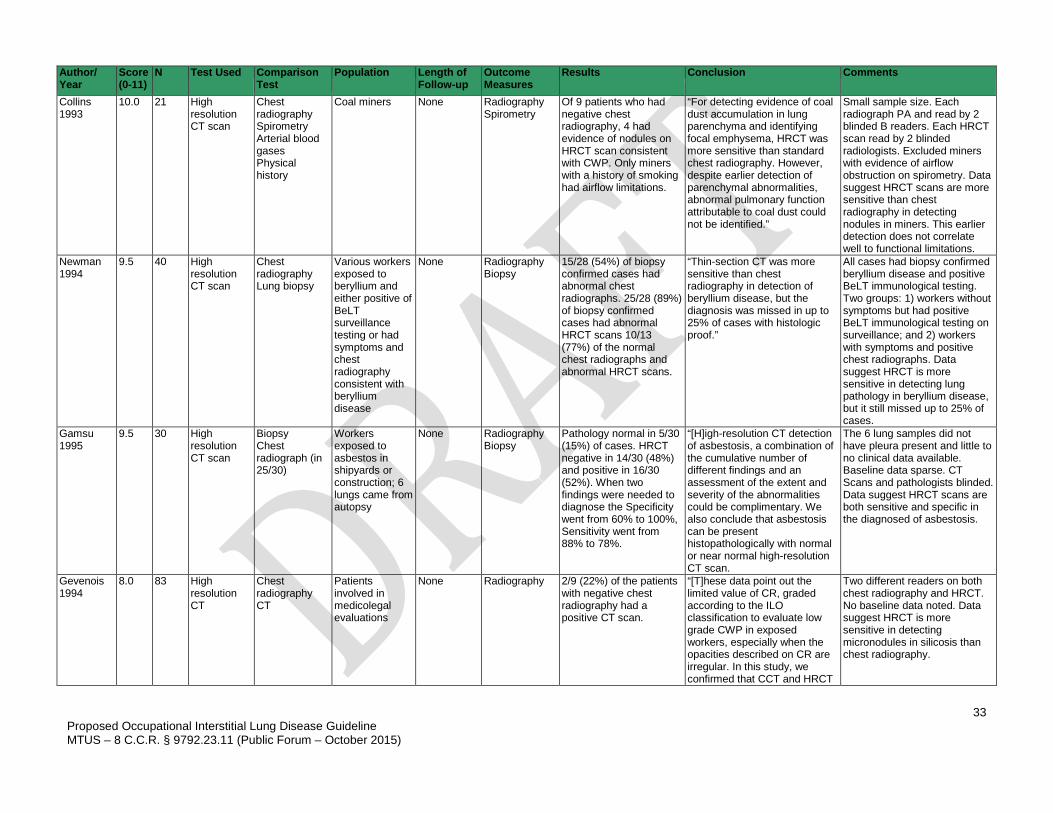
33
Proposed Occupational Interstitial Lung Disease Guideline MTUS – 8 C.C.R. § 9792.23.11 (Public Forum – October 2015)
Author/ Year
Score (0-11)
N Test Used Comparison Test
Population Length of Follow-up
Outcome Measures
Results Conclusion Comments
Collins 1993
10.0 21 High resolution CT scan
Chest radiography Spirometry Arterial blood gases Physical history
Coal miners None Radiography Spirometry
Of 9 patients who had negative chest radiography, 4 had evidence of nodules on HRCT scan consistent with CWP. Only miners with a history of smoking had airflow limitations.
“For detecting evidence of coal dust accumulation in lung parenchyma and identifying focal emphysema, HRCT was more sensitive than standard chest radiography. However, despite earlier detection of parenchymal abnormalities, abnormal pulmonary function attributable to coal dust could not be identified.”
Small sample size. Each radiograph PA and read by 2 blinded B readers. Each HRCT scan read by 2 blinded radiologists. Excluded miners with evidence of airflow obstruction on spirometry. Data suggest HRCT scans are more sensitive than chest radiography in detecting nodules in miners. This earlier detection does not correlate well to functional limitations.
Newman 1994
9.5 40 High resolution CT scan
Chest radiography Lung biopsy
Various workers exposed to beryllium and either positive of BeLT surveillance testing or had symptoms and chest radiography consistent with beryllium disease
None Radiography Biopsy
15/28 (54%) of biopsy confirmed cases had abnormal chest radiographs. 25/28 (89%) of biopsy confirmed cases had abnormal HRCT scans 10/13 (77%) of the normal chest radiographs and abnormal HRCT scans.
“Thin-section CT was more sensitive than chest radiography in detection of beryllium disease, but the diagnosis was missed in up to 25% of cases with histologic proof.”
All cases had biopsy confirmed beryllium disease and positive BeLT immunological testing. Two groups: 1) workers without symptoms but had positive BeLT immunological testing on surveillance; and 2) workers with symptoms and positive chest radiographs. Data suggest HRCT is more sensitive in detecting lung pathology in beryllium disease, but it still missed up to 25% of cases.
Gamsu 1995
9.5 30 High resolution CT scan
Biopsy Chest radiograph (in 25/30)
Workers exposed to asbestos in shipyards or construction; 6 lungs came from autopsy
None Radiography Biopsy
Pathology normal in 5/30 (15%) of cases. HRCT negative in 14/30 (48%) and positive in 16/30 (52%). When two findings were needed to diagnose the Specificity went from 60% to 100%, Sensitivity went from 88% to 78%.
“[H]igh-resolution CT detection of asbestosis, a combination of the cumulative number of different findings and an assessment of the extent and severity of the abnormalities could be complimentary. We also conclude that asbestosis can be present histopathologically with normal or near normal high-resolution CT scan.
The 6 lung samples did not have pleura present and little to no clinical data available. Baseline data sparse. CT Scans and pathologists blinded. Data suggest HRCT scans are both sensitive and specific in the diagnosed of asbestosis.
Gevenois 1994
8.0 83 High resolution CT
Chest radiography CT
Patients involved in medicolegal evaluations
None Radiography 2/9 (22%) of the patients with negative chest radiography had a positive CT scan.
“[T]hese data point out the limited value of CR, graded according to the ILO classification to evaluate low grade CWP in exposed workers, especially when the opacities described on CR are irregular. In this study, we confirmed that CCT and HRCT
Two different readers on both chest radiography and HRCT. No baseline data noted. Data suggest HRCT is more sensitive in detecting micronodules in silicosis than chest radiography.
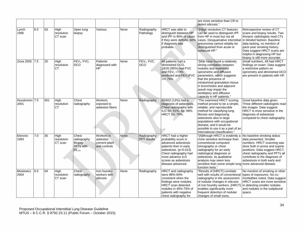
34
Proposed Occupational Interstitial Lung Disease Guideline MTUS – 8 C.C.R. § 9792.23.11 (Public Forum – October 2015)
are more sensitive than CR to detect silicosis.”
Lynch 1995
8.0 63 High resolution CT scan
Open lung biopsy
Various None Radiography Pathology
HRCT was able to distinguish between HP and IPF in 90% of cases if they were definite, 60% if diagnosis was probable.
“[H]igh resolution CT features can be used to distinguish IPF from HP in most but not all cases. Desquamative interstitial pneumonia cannot reliably be distinguished from acute or subacute HP.”
Retrospective review of CT scans and biopsy results. Two thoracic radiologists read CTs in blinded fashion. Baseline data lacking, no mention of pack-year smoking history. Data suggest HRCT scans are helpful in diagnosing HP but biopsy is still more accurate.
Ziora 2005 7.5 20 High resolution CT
FEV1, FVC, DCO
Patients diagnosed with HP
None FEV1, FVC, DCO
All patients had a diminished DCO. 19/20 (95%) had FVC and FEV1 <70% predicted and FEV1/FVC >/= 75%.
“[W]e have found a relatively strong correlation between nodules and examined spirometric and diffusion parameters, which suggests that the presence of intraluminal granulation tissue in bronchioles and adjacent aveoli may impair the ventilatory and diffusion capacity in HP patients.”
Small numbers. All had HRCT findings on exam. Data suggest a restrictive pattern on spirometry and diminished DCO are present in patients with HP.
Huuskonen 2001
7.5 651 High resolution CT
Chest radiography
Workers exposed to asbestos fibers
None Radiography 85/602 (14%) had a diagnosis of asbestosis. Chest radiography with ILO Sn: 51%, Sp: 89%. HRCT Sn: 70%.
“The examined HRCT scoring method proved to be a simple, reliable, and reproducible method for classifying lung fibrosis and diagnosing asbestosis also in large populations with occupational disease, and it would be possible to use it as a part of an international classification.”
Good baseline data given. Three different radiologists read the images. Data suggest HRCT is more sensitive in the diagnosis of asbestosis compared to chest radiography.
Eterovic 1993
7.0 35 High resolution CT scan
Chest radiography Biopsy PFTs with DLCO
Workers in asbestos cement plant and controls
None Radiography PFT results
HRCT had a higher probability score in advanced asbestosis patients then in early asbestosis. (p=0.013) Chest radiography had more advance ILO scores as asbestosis disease advanced.
“[A]lthough HRCT is evidently a more sensitive technique than conventional computed tomography or chest radiography for an early radiological diagnosis or asbestosis, its qualitative analysis may seem less sensitive than some simple lung function tests.”
No baseline smoking status data presented. Smaller numbers. HRCT scanning was done both in prone and supine positions. Data suggest HRCT, chest radiography and PFTs all contribute to the diagnosis of asbestosis in both early and more advanced disease.
Mosiewicz 2004
6.5 64 High resolution CT scan
Chest radiography
Iron foundry workers with silicosis
None Radiography HRCT and radiography were 88%-94% consistent when the findings were nodules. HRCT scan detected nodules in 45%-75% of patients with negative chest radiography for
“Results of [HRCT] correlate well with results of conventional radiography in the assessment of nodular changes in silicosis of iron foundry workers. [HRCT] enables significantly more frequent detection of nodular changes of small sizes,
No mention of smoking or other types of exposures. No co-morbidities noted. Data suggest HRCT scans are more sensitive in detecting smaller nodules and nodules in the subpleural space.
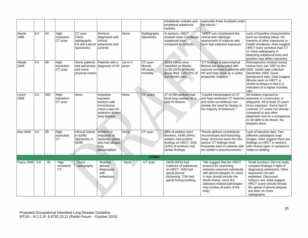
35
Proposed Occupational Interstitial Lung Disease Guideline MTUS – 8 C.C.R. § 9792.23.11 (Public Forum – October 2015)
intralobular nodules and peripheral subpleural nodules.
especially those localized under the pleura.”
Aberle 1988
6.0 63 High resolution CT scan
CT scan Chest radiography PA and Lateral Spirometry
Workers diagnosed with clinical asbestosis and controls
None Radiography Spirometry
In workers, HRCT showed more Curvilinear subpleural lines compared to controls.
“HRCT can complement the clinical and radiologic assessment of subjects who have had asbestos exposure.”
Lack of baseline characteristics such as smoking status. No mention of other exposures or health conditions. Data suggest HRCT more sensitive than CT or chest radiography in detecting subpleural lines and position may affect outcomes.
Hanak 2008
4.5 69 High resolution CT scan
Some patients had spirometry and some physical exams
Patients with a diagnosis of HP
Up to 9 years
CT scan- fibrosis All-cause mortality
26/69 (38%) were classified as fibrotic. 11/26 (42%) of fibrotic group died. 1/43 (2%) of non-fibrotic died.
“CT findings of parenchymal fibrosis are associated with reduced survival in patients with HP and may serve as a useful prognostic indicator.”
Retrospective medical record review from Jan 1997 to Dec 2002. Death data collected December 2006. Good background data. Data suggest fibrosis seen on HRCT is similar to biopsy in that it is indicative of a higher mortality rate.
Lynch 1988
4.0 260 High resolution CT scan
None Asbestos exposed workers with inconclusive chest x-rays for asbestos related lung disease
None CT scans 27 of 260 workers had focal lung masses for a total 43 lesions.
“Careful interpretation of CT and high-resolution CT features and close surveillance can obviate the need for biopsy in the majority of instances.”
All workers exposed to asbestos in construction or shipyards. All at least 10 years since exposure. Some had IV contrast. CT scans not directly compared to any other diagnostic tool so a comparison is not able to be drawn. No biopsies done.
Han 2000 4.0 85 High resolution CT
Clinical history in 53/85, Spirometry in 53/85
Welders in shipyards or assembly plants who had alleged lung abnormalities
None CT scan 79% of welders were smokers. 54/85 (64%) welders had positive findings on HRCT. 6/43 (14%) of smokers had similar findings.
“Poorly-defined centrilobular micronodules and branching linear structures were the thin-section CT findings most frequently seen in patients with arc-welder’s pneumoconiosis.”
Lack of baseline data. Two different radiologists read images. Data suggest there are findings on HRCT in workers with clinical signs or symptoms relate to welding.
OTHER Topcu 2000 5.0 26 High
resolution CT
Chest radiography
Workers already diagnosed with asbestosis
None CT scan 24/26 (92%) had evidence of asbestosis on HRCT. 9/26 had apical pleural thickening. 7/26 had apical honeycombing.
“We suggest that the HRCT protocol for examining asbestos-exposed individuals with pleural plaques on chest X-rays should include the whole thorax, since the asbestos-related pathologies may involve all parts of the lung.”
Small numbers. Did not really compare findings in light of diagnosing asbestosis. Other exposures not well explained. Discussed tobacco use. Data suggest HRCT scans should include the apices if pleural plaques are seen on chest radiographs.
36
Proposed Occupational Interstitial Lung Disease Guideline MTUS – 8 C.C.R. § 9792.23.11 (Public Forum – October 2015)
MAGNETIC RESONANCE IMAGING (MRI) OF THE CHEST There is no recommendation regarding the role of MRI of the lung in the diagnosis of occupational lung disease. MRI is not currently used as a primary diagnostic tool for occupational ILD. PET/CT SCANS OF THE CHEST PET/CT scans are beyond the scope of this guideline. These are generally used in cases with questions of mass lesions or invasion of chest wall and not used either for surveillance or first-line diagnosis of occupational ILD. CARBON MONOXIDE DIFFUSING CAPACITY (DLCO) DLCO is a measurement of carbon monoxide transfer from inspired gas to pulmonary capillary blood. DLCO is a product of two measurements during breath holding at full inhalation, carbon monoxide uptake from the alveolar gas space, and the accessible alveolar volume. The single-breath diffusion capacity testing is a common method for measuring diffusing capacity of the lung.(103, 116, 117) The lung volume during breath holding is measured simultaneously by dilution of a non-absorbable gas such as helium or methane.(118) DLCO measures CO transfer from the inspired air to the pulmonary capillary blood and this includes all the following steps:
1. Bulk flow delivery of CO to the airways and alveolar spaces; 2. Mixing and diffusion of CO in the alveolar ducts, air sacs and alveoli; 3. Transfer of CO across the gaseous to liquid interface of the alveolar membrane; 4. Mixing and diffusion of CO in the lung parenchyma and alveolar capillary plasma; 5. Diffusion across the red cell membrane and within the interior of the red blood cell; and 6. Chemical reaction with constituents of blood hemoglobin.(119)
DLCO has long been used in the diagnosis of lung disease in both the non-occupational and occupational setting. It has been reported to be a sensitive indicator of gas exchange, being abnormal in patients with ILD, pulmonary vascular lung disease, and emphysema.(120) However, although DLCO may be a useful test for assessing the presence of ILD in general, it is not diagnostic for any specific type of ILD. The measurement of carboxyhemoglobin (COHb) levels and hemoglobin (Hb) concentrations for adjustment of DLCO results is important for correct interpretation of both individual and group studies of DLCO and should be performed whenever possible.(121, 122) Recommendation: Carbon Monoxide Diffusing Capacity (DLCO) Carbon monoxide diffusing capacity is recommended for use in diagnosing occupational lung disease.
Strength of Evidence – Recommended, Evidence (C) Level of Confidence – High
Performed – DLCO should be performed according to the ATS/ERS statement published in 2005. It is recommended that at least two DLCO tests should be performed and the average reported. It is recommended that the two measurements for the DLCO agree within 10%.(119) It is important to obtain smoking status as cigarette smoking may cause measureable baseline levels of CO causing an increased back-pressure and carboxyhemoglobin.(119) Indications – DLCO may be used to help in diagnosing gas exchange abnormalities in patients with lung disease.(123) Harms – None. Benefits – Accurate assessment of gas exchange abnormalities in patients with lung disease.
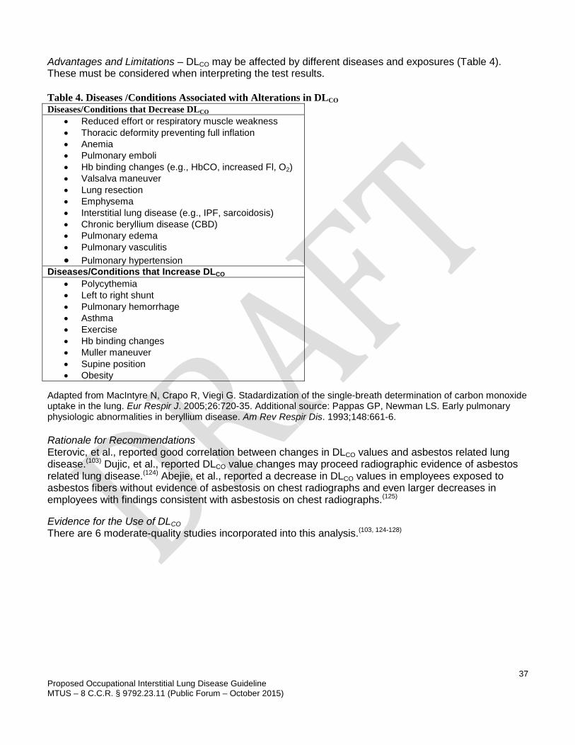
37
Proposed Occupational Interstitial Lung Disease Guideline MTUS – 8 C.C.R. § 9792.23.11 (Public Forum – October 2015)
Advantages and Limitations – DLCO may be affected by different diseases and exposures (Table 4). These must be considered when interpreting the test results. Table 4. Diseases /Conditions Associated with Alterations in DLCO Diseases/Conditions that Decrease DLCO
• Reduced effort or respiratory muscle weakness • Thoracic deformity preventing full inflation • Anemia • Pulmonary emboli • Hb binding changes (e.g., HbCO, increased Fl, O2) • Valsalva maneuver • Lung resection • Emphysema • Interstitial lung disease (e.g., IPF, sarcoidosis) • Chronic beryllium disease (CBD) • Pulmonary edema • Pulmonary vasculitis • Pulmonary hypertension
Diseases/Conditions that Increase DLCO • Polycythemia • Left to right shunt • Pulmonary hemorrhage • Asthma • Exercise • Hb binding changes • Muller maneuver • Supine position • Obesity
Adapted from MacIntyre N, Crapo R, Viegi G. Stadardization of the single-breath determination of carbon monoxide uptake in the lung. Eur Respir J. 2005;26:720-35. Additional source: Pappas GP, Newman LS. Early pulmonary physiologic abnormalities in beryllium disease. Am Rev Respir Dis. 1993;148:661-6. Rationale for Recommendations Eterovic, et al., reported good correlation between changes in DLCO values and asbestos related lung disease.(103) Dujic, et al., reported DLCO value changes may proceed radiographic evidence of asbestos related lung disease.(124) Abejie, et al., reported a decrease in DLCO values in employees exposed to asbestos fibers without evidence of asbestosis on chest radiographs and even larger decreases in employees with findings consistent with asbestosis on chest radiographs.(125) Evidence for the Use of DLCO There are 6 moderate-quality studies incorporated into this analysis.(103, 124-128)
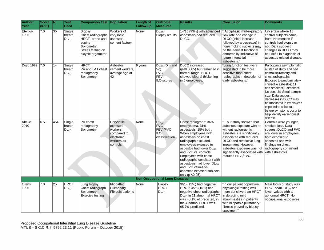
38
Proposed Occupational Interstitial Lung Disease Guideline MTUS – 8 C.C.R. § 9792.23.11 (Public Forum – October 2015)
Author/ Year
Score (0-11)
N Test Used
Comparison Test Population Length of Follow-up
Outcome Measures
Results Conclusion Comments
Eterovic 1993
7.0 35 Single breath DLCO
Biopsy Chest radiographs HRCT- prone and supine Spirometry Stress testing on bicycle ergometer
Workers of chrysotile asbestos cement factory
None DLCO Biopsy results
14/15 (93%) with advanced asbestosis had reduced DLCO.
“[A] biphasic mid-expiratory flow rate and change in DLCO (initial increase followed by a decrease) in non-smoking subjects may be the earliest functional abnormality indicative of future interstitial asbestosis.”
Uncertain where 13 control subjects came from. No mention if controls had biopsy or not. Data suggest changes in DLCO may be useful in diagnosis of asbestos related disease.
Dujic 1992 7.0 14 Single breath DLCO
HRCT PA and LAT chest radiography Spirometry
Asbestos cement workers, average age of 42
9 years DLCO (Dm and Vc) FVC FEV1 ILO scores
DLCO increased (p<0.0005) but remained in normal range. HRCT showed pleural thickening in 6 employees.
“Lung function test were suggested to be more sensitive than chest radiographs in detection of early asbestosis.”
Participants asymptomatic at start of study and had normal spirometry and chest radiographs. Exposed to predominately chrysotile asbestos; 11 non-smokers, 3 smokers. No controls. Small sample size. Data suggest decreases in DLCO may be monitored in employees exposed to asbestos before symptoms occur to help identify earlier onset disease.
Abejie 2010
6.5 454 Single breath DLCO
PA chest radiography Spirometry
Chrysotile exposed workers compared to electronic workers as controls
None DLCO FVC FEV1/FVC ILO classification
Chest radiograph: 36% emphysema, 31% asbestosis, 15% both. When employees with asbestosis on chest radiograph excluded, employees exposed to asbestos had lower DLCO and FVC vs. controls. Employees with chest radiographs consistent with asbestosis had lower DLCO and FVC values vs. asbestos exposed subjects only (p <0.05).
“…our study showed that asbestos exposure with or without radiographic asbestosis is significantly associated with reduced DLCO and restrictive lung impairment. However, asbestos exposure was not significantly associated with reduced FEV1/FVC.
Controls were younger, smoked less. Data suggest DLCO and FVC are lower in employees both exposed to asbestos and with findings on chest radiography consistent with asbestosis.
Non-Occupational Lung Diseases Orens 1995
7.0 25 HRCT DLCO
Lung biopsy Chest radiograph Spirometry Exercise testing
Idiopathic Pulmonary Fibrosis patients
None Biopsy HRCT DLCO
3/25 (12%) had negative HRCT; 4/25 (16%) had negative chest radiographs. DLCO in 21 abnormal HRCT was 46.1% of predicted, in the 4 normal HRCT was 65.7% predicted.
“In our patient population, physiologic testing was more sensitive than HRCT in detecting mild abnormalities in patients with idiopathic pulmonary fibrosis proved by biopsy specimen.”
Main focus of study was HRCT scan. DLCO had lower values with an abnormal HRCT. No occupational exposures.

39
Proposed Occupational Interstitial Lung Disease Guideline MTUS – 8 C.C.R. § 9792.23.11 (Public Forum – October 2015)
Sette 2004 7.0 82 Single breath DLCO
HRCT Chest radiography Exercise testing Spirometry
Asbestos cement workers Chrysotile miners
None Images Gas exchange values
16/82 (20%) had normal pulmonary gas exchange values.
“[D]ual semiquantitative and qualitative thin-section CT classification of lung parenchymal abnormalities can be used successfully to estimate the individual likelihood of pulmonary gas exchange impairment.”
Gas exchange impairment was defined as DLCO <70% predicted.
Boros 2010 6.0 830 Single breath DLCO
Chest x-ray Spirometry Whole body plethysmography Static lung compliance
Patients with sarcoidosis
None DLCO values 772/830 had normal lung volumes. 75% had parenchymal involvement on chest x-ray. 123/830 (14.8%) had low DLCO values.
“Static lung compliance and DLCO concern different aspects of respiratory pathophysiology.”
ERS reference values used for DLCO. No occupational lung disease.
40
Proposed Occupational Interstitial Lung Disease Guideline MTUS – 8 C.C.R. § 9792.23.11 (Public Forum – October 2015)
BIOLOGICAL SAMPLING SPUTUM SAMPLES AND BRONCHOALVEOLAR LAVAGE (BAL) If insufficient clinical objective evidence is obtained from physical examination, chest radiographs and spirometry, additional testing including biological sampling may be indicated to confirm the diagnosis of occupational ILD. The following discussion includes specific indications for biological sampling for each major category of occupational ILD. Bronchoalveolar lavage (BAL) has been suggested as a potentially important diagnostic tool in the evaluation of exposure to asbestos and other occupational lung diseases.(129-131) This method of testing has been used in the diagnosis of lower respiratory tract disease prior to the use of HRCT of the chest.(132) Collection of sputum is simpler, less invasive and less expensive than BAL.(133) Sputum collection is done by having the patient cough to attempt to produce the sputum from deep within the lungs. It is recommended that each sample be at least 15mL to help increase the sensitivity of the sample. Inhaled asbestos fibers that are coated with iron-containing mucoprotein and imbedded in lung tissue are referred to as asbestos bodies (AB). Ferruginous bodies (FB) result from the deposition of an iron-rich protein layer at the cell-particle interface of any type of fiber and when asbestos is verified they are called asbestos bodies.(134) Ferruginous/asbestos bodies are detectible by light microscopy, whereas asbestos fibers are detected with electron microscopy. Recommendation: Bronchoalveolar Lavage Bronchoalveolar lavage is recommended as an aid for the diagnosis of occupational lung disease caused by asbestos. 1. Diagnosis of asbestos-related occupational interstitial lung disease.
Strength of Evidence – Recommended, Evidence (C) Level of Confidence – Low
Performed – BAL should be performed according to the ATS guidelines on performance of BAL for ILD.(132) Indications – To assist in the diagnosis of occupationally-related asbestos interstitial lung disease.(129, 132,
135, 136) Harms – Low incidence of paroxysmal coughing, vomiting, syncope. Benefits – Support for diagnosis (though not required given modern testing i.e., HRCT). Advantages and Limitations – Smoking is an important confounder in the assessment of BAL fluid (BALF) as it may interfere with cellular profiles of the lavage. BAL has been reported to be more beneficial in diagnosing occupational lung disease in non-smoking populations.(132) Presence of specific fibers or dusts in asbestos exposure, coal or silica does not discriminate well between exposure and disease.(132) The type of asbestos fiber may also influence the results with reports of less ABs found with chrysotile exposure.(133, 137) Differences in sampling, preparation and counting techniques, definitions of reference populations and expression of results have previously caused major difficulties in comparing results from different laboratories.(138) Rationale for Recommendation
41
Proposed Occupational Interstitial Lung Disease Guideline MTUS – 8 C.C.R. § 9792.23.11 (Public Forum – October 2015)
Teschler, et al., reported a greater sensitivity with BAL compared to sputum among a selected sample group.(133) Vathesatogkit, et al., reported more FBs detected in the BALF of exposed versus unexposed subjects, and also reported a decrease in spirometry and DLCO in subjects with FBs in their BALF.(130) BAL is a high cost procedure with moderate risk of adverse events, but has fewer adverse events also costing less when compared to open lung biopsy. Therefore, it is recommended in select cases. Recommendation: Sputum Sampling Sputum, both induced and spontaneous, is recommended as an aid for the diagnosis of occupational lung disease caused by asbestos. 1. Diagnosis of asbestos-related occupational interstitial lung disease.
Strength of Evidence – Recommended, Evidence (C) Level of Confidence – Low
Harms – Paroxysmal coughing, vomiting, syncope. Benefits – Support for diagnosis (though not current given modern imaging techniques such as HRCT). Rationale for Recommendation Sputum has been less reliable than BAL samples largely because of inability to obtain quality specimens.(134) However, sputum has the advantages of being a noninvasive and less expensive method when compared to BAL, thoracoscopic or open lung biopsy. Overall, the sensitivity of identifying asbestos bodies in sputum is poor but specificity is reportedly high.(135, 139, 140) Collection of sputum is simpler when compared to BAL and biopsy. It is also less expensive and has fewer adverse effects. ABs in sputum is considered a highly specific marker of asbestos exposure, but it is considered insensitive.(133, 141) In a study of 11,000 sputum samples from the general population, no false-positive samples were reported.(142) Sulotto, et al., reported ABs found in workers exposed to both chrysotile and amphibole fibers, while there was no direct correlation between ABs in sputum samples and asbestos related disease.(141) Evidence for the Use of Bronchial Alveolar Lavage (BAL) and Sputum There are 4 moderate-quality studies on BAL(130, 133, 137, 143) and 4 moderate-quality studies on sputum incorporated into this analysis.(135, 139-141) There is 1 low-quality study and 2 other studies in Appendix 2.(129, 131, 134)

42
Proposed Occupational Interstitial Lung Disease Guideline MTUS – 8 C.C.R. § 9792.23.11 (Public Forum – October 2015)
Author/ Year
Score (0-11)
N Test Used
Comparison Test
Population Length of Follow-up
Outcome Measures
Results Conclusion Comments
BAL Teschler 1996 7.5 135 BAL Sputum
tissue samples
Workers exposed to asbestos dust: Group 1 classified as high exposure, Group 2 as medium, Group 3 as occasional exposure
None Asbestos bodies (light microscopy, at 400x) in BAL and sputum in all subjects. Lung tissue in 21 subjects.
33% of subjection in group 1, 68% in group 2; 45% in group 3 had ABs in BAL but not Sputum. Open lung biopsy had ABs in all samples. Samples with less than 1,000 ABs/cm3 had no Abs in sputum samples.
“…many subjects with positive BAL fluid analysis had negative sputum results. These findings suggest that BAL is the superior of the two methods for assessing lung AB content.”
Tissue samples done only on 21 subjects. Data suggest BAL is more sensitive than Sputum in detecting FBs in subjects. No correlation is made between FBs and disease burden.
Vathesatogkit 2004
7.0 60 BAL Chest radiography HRCT scan Spirometry DLCO
Utility workers and controls
None Asbestos bodies (light microscopy, at 40x) Respiratory symptoms Chest radiographs HRCT scans Spirometry DLCO
AB found in 10/30 subjects (33%) and 0/30 controls. AB positive subjects had reduced FEV1 and diffusion capacity (p <0.05). HRCT scans showed higher prevalence of parenchymal disease (p <0.05).
“In asbestos-exposed subjects, the presence of AB in BAL cytospin slides should be viewed as a clinically important finding, and their HRCT scans should be reviewed carefully for evidence of interstitial lung disease.”
Two blinded pathologist read slides for AB. Data suggest detection of Asbestos bodies in utility workers represents an indicator of exposure, but not necessarily related to asbestos diseases.
Corhay 1990 4.5 121 BAL Chest radiography Spirometry DLCO
Steel workers and controls (white collar workers)
5 year repeat BAL in 7 subjects. Others, none.
Asbestos bodies (light microscopy, at 200x)
Chest radiographs normal in 65 steel workers. ABs found in 38/65 (58.5%) of steel workers and 6% of controls. Smoking habits and presence of COPD did not influence AB counts.
“This study shows that steel workers may be subject to a nontrivial exposure to asbestos in an industrial plan environment.”
Not compared to tissue samples. No sputum samples taken. Data suggest steel workers may be exposed to asbestos as part of their job.
Karjalainen 1994
4.0 156 BAL Exposure data
Exposed workers
None Asbestos bodies (light microscopy, at 200x)
Concentration of >\= AB/ml found in 85% exposed to asbestos, and 7% of those not likely exposed. Patients with asbestosis (n = 9) showed higher average concentrations of AB (median 13) than patients with pleural disease only (median 2.4).
“…the correlation between AB concentration and exposure history was greater than in earlier studies on workers exposed to chrysotile.”
No other biological testing done other than BAL. Broke analyses down by type of job. Data suggest higher concentrations of ABs seem to correlate with higher exposure and more significant disease but the correlation is not linear.
Sputum Alexopoulos 2011
7.0 39 Induced Sputum
Broncho-alveolar lavage Chest
Romanian brake factory workers without
None Total number and vitality of cells Number of dust
In the six workers who reported using PPE none had asbestos bodies in IS or BALF.
IS “usefulness for screening of workers should be further evaluated because
At least 15 years of exposure to asbestos at >5 fibers per mL. Chest radiographs </=

43
Proposed Occupational Interstitial Lung Disease Guideline MTUS – 8 C.C.R. § 9792.23.11 (Public Forum – October 2015)
radiography Spirometry ECG
pneumo-coniosis
cells Iron laden macrophages Asbestos bodies (AB)
14/39 (36%) had AB in BALF. Of those 7/14 (50%) has AB in IS.
the inflammatory response in our study lacks specificity since it might have been induced [by] asbestos, dust and smoking.”
1/0 ILO classification by two physicians. BAL performed in right middle lobe. Sputum induction done by inhaling saline then asked to cough. Study suggests IS may be helpful in proving insight for both inhalation of dusts and inflammatory processes in lung.
McLarty 1980 6.0 674 Sputum Chest radiography Spirometry Smoking status
Exposed workers in insulation
10 years Ferruginous bodies Chest radiography Spirometry
Workers with ferruginous bodies and irregular small opacities was correlated (p<0.001). Workers with ferruginous bodies and restriction on spirometry was correlated (p<0.02).
“Clinically, the presence of ferruginous bodies in the sputum was found to be significantly related to radiographic findings of interstitial pulmonary and pleural fibrosis and to spirometric findings of restrictive lung disease.”
Both spontaneous and aerosol-induced sputum specimens used. Data suggest sputum samples not only show asbestos exposure, but may be correlated with radiological changes and spirometry findings.
Paris 2002 4.5 223 Sputum Exposure data
Exposed workers, brake and textile
None Asbestos body (light microscopy, at 160x) Exposure data
118/223 (53%) sputum samples.
“It is clear that a negative mineralogical sputum examination cannot therefore, exclude the reality of even high occupational exposure.”
No other diagnostic tests used. Data suggest a negative result on sputum cannot exclude asbestos exposure.
Sulotto 1997 4.0 142 Sputum Spirometry Chest radiography
Exposed workers in textile
Up to 5 years
Ferruginous bodies (light microscopy, at 400x) Spirometry
Asbestos-related diseases were present in 58% of subjects. ABs were found in 94 smears (21%) and in at least 1 specimen in 44.4% of subjects.
“…our study confirms the utility of obtaining several specimens from each subject in order to increase the probability of asbestos body identification.”
Collection of sputum samples for 3 weeks or less. Minimum amount of specimens was 2. Data suggest multiple sputum samples beneficial up to 4 in identifying FBs in sputum in exposed workers.
44 Proposed Occupational Interstitial Lung Disease Guideline MTUS – 8 C.C.R. § 9792.23.11 (Public Forum– October 2015)
MANAGEMENT OF OCCUPATIONAL INTERSTITIAL LUNG DISEASE Management of workers diagnosed with occupational ILD consists of the coordinated use of five strategies:
1. General management of restrictive lung disease due to interstitial fibrosis. 2. Specific management of the underlying disease. 3. Specific management of comorbidities. 4. Prevention of further loss of lung function and major complications. 5. Evaluation of work capacity and fitness for duty.
The general management of restrictive lung disease due to interstitial fibrosis consists of supporting oxygenation. This includes use of supplemental oxygen if desaturation is documented during exertion or sleep. In advanced or rapidly progressive cases, evaluation for lung transplantation should be performed. The paucity of therapeutic options reflects the irreversibility of fibrosis, once it is established. Extensive fibrosis, which may occur following recovery from diffuse alveolar damage by toxic inhalation, is refractory to direct management. Fibrosis associated with pneumoconioses and autoimmune processes tends to progress through stages, ultimately reaching a similar “end stage” condition characterized by restrictive disease, pulmonary hypertension, cor pulmonale, congestive heart failure, and lung infections due to loss of host defense mechanisms. ILD, as it advances, is often associated with a chronic dry cough, which may require suppression particularly when it interferes with sleep. Specific management of the underlying disease is more critical for a good outcome than general management of fibrotic lung disease. Systemic glucocorticosteroids (aka “steroids”) may be effective when used judiciously in HP and beryllium disease. Steroids are rarely used for other pneumoconiosis, although some modest improvements have been documented (e.g., in silicosis, asbestosis, and CWP). Yet adverse effects of steroids are considerable.(144, 145) Treatment options that may be proposed for rheumatologic ILD (e.g., systemic sclerosis), such as cytotoxic drugs or immunotherapy, are not known to have any benefit in occupational ILD or idiopathic ILD. Bronchodilators and inhaled corticosteroids may have a role in the presence of an accompanying airways effect, as in HP, cobalt-induced asthma, or dust-related airway diseases. Among fibrotic lung diseases, asbestosis and IPF are associated with a high rate of lung cancer. Screening by imaging through helical high-resolution CT scanning has been recommended for cigarette smokers, who are of course another high-risk group. Although it has not been validated in asbestosis specifically, this screening modality (and possibly others in the future) may also reasonably be considered in cases of asbestosis.(146) Screening for colon cancer has been recommended for patients with asbestosis.(22) Silicosis also confers a risk for lung cancer, but not as great as asbestosis and without known risk for other malignancies. Specific management of comorbidities is important in occupational ILD, particularly for silicosis. Silicosis is sometimes complicated by opportunistic infections, particularly tuberculosis and atypical mycobacteria. The resulting “silicotuberculosis” may be refractory to management and may require highly individualized and prolonged multi-drug treatment. Coexisting airways disease is managed with standard treatment approaches. Preventing further loss of lung function by preventing respiratory comorbidity is essential, as the natural history of occupational ILD is an accelerated decline in lung function, often with sporadic incremental drops due to decompensation and exacerbation following which the patient usually does not return to baseline. (See also MTUS Cornerstones of Disability Prevention and Management). Patients with ILD require immunization against pneumococcal pneumonia and influenza. Respiratory infections are recommended to be treated aggressively, with a low threshold for hospitalization if the ILD is advanced.
45 Proposed Occupational Interstitial Lung Disease Guideline MTUS – 8 C.C.R. § 9792.23.11 (Public Forum– October 2015)
Pulmonary rehabilitation may be effective even in the complex settings of occupational respiratory diseases (e.g., asthma), providing sustained improvement of functional capacity, and reducing health care utilization.(147, 148) No studies have made direct comparisons between different systems of rehabilitation.(149) Preventing further loss of lung function by avoiding provocative exposures is essential and has implications for fitness for duty in work that involves airborne exposures. Smoking, of any variety, including exposure to sidestream smoke, should be strictly avoided, as the resulting respiratory irritation further compromises lung function. Avoidance of airway irritants, including fragrances, alcohols and aldehydes, solvents, and dusts may help some patients to preserve lung function, prevent episodes of shortness of breath, and to reduce the propensity to cough. On the other hand, low-level exposure, when easily tolerated by the patient, is not necessarily a contraindication to continued work, although as discussed below, monitoring is recommended to assure early recognition of disease acceleration or cardiopulmonary complications. Evaluation of work capacity and fitness for duty is an important function when the patient is capable of working. A fitness-for-duty evaluation should be performed with detailed knowledge of workplace exposures. The worker should be identified as fit for duty, fit for duty with accommodation, or unfit for duty. Workers who are thought to be fit for duty with accommodation should have the recommended work limitations identified in as much detail as necessary to support an appropriate job placement (i.e., “light duty” is not sufficient). Patients who are unfit for duty should generally be further evaluated using their state’s system and/or the relevant edition of the American Medical Association’s AMA Guides to the Evaluation of Permanent Impairment, which provides detailed guidance on respiratory impairment,iv or the relevant guidelines for state or federal programs (e.g., reference the extensive procedures specified in the Code of Federal Regulations (CFR).(150) Medical removal is a strategy used to permit an individual to avoid further exposures that might lead to progression or resulting in earlier impairment. “Medical removal” is the decision to move a worker to an alternative work assignment in order to protect them from a potential occupational hazard. It applies when the worker is believed to be unusually susceptible to exposure levels below existing occupational exposure limits (usually the OSHA or MSHA permissible exposure limits (PELs) or NIOSH-recommended exposure limits (RELs)) and ongoing potential exposures are judged to represent an excessive health risk to the individual. Workers who have developed evidence of pneumoconiosis, particularly with fewer than 20 years of exposure, may be particularly susceptible and should be considered for recommending medical removal. Whenever medical removal is contemplated due to the recognition of an occupational disease, it is essential to concurrently analyze ongoing exposures in the applicable working environments, and to identify potential explanations for the failure of primary protection. If a worker has minor impairment(s) and when current exposures have been consistently shown to be well-controlled during all tasks, there may be no compelling rationale for medical removal. In such cases, it is reasonable for the affected worker to continue working in the assignment if both the worker and the employer will carefully avoid sporadic conditions that have potential for exposure at greater than minimal acceptable levels. In such instances, it is important to monitor dust levels and control measures as well as periodically reevaluate the worker’s health. For affected workers, participation in professionally administered personal respiratory protection programs may be especially useful under mildly dusty conditions, near the PEL, or in moderately dusty conditions near the PEL where there is no spillover of dust and dust levels are low where workers wear their respirators. Respirators may not be completely protective in cases of exposure to high airborne particulate levels. Additionally, periodic medical monitoring is important for individuals with symptoms or findings of occupational lung effects who continue to experience workplace exposures. Progressing ILD may make the worker intolerant of respirators, especially when moderately severe or worse, due to the increased work of breathing and increased dead space.(151) Therefore, queries about compliance to be
ivStates have adopted a wide range of editions of the AMA Guides to the Evaluation of Permanent Impairment.
46 Proposed Occupational Interstitial Lung Disease Guideline MTUS – 8 C.C.R. § 9792.23.11 (Public Forum– October 2015)
sure that the worker is not removing the respirator during work for reasons of communication, discomfort, or health (e.g., expectoration), thus defeating its purpose. Maintenance of work capacity and fitness for duty through exercise is important to prevent deconditioning. This is also important for patients who are unfit for work so that they may retain capacity for activities of daily living. Pulmonary rehabilitation programs, as for COPD and asthma, have not been shown to have a benefit for restrictive lung disease. However, in cases of mixed disease or when depression or lack of adherence is an issue, participation in rehabilitation programs may provide motivation, peer support, and better monitoring and control of comorbid conditions, such as airways disease. PHARMACOLOGICAL TREATMENT Recommendation: Management of Occupational Interstitial Lung Disease (Pharmacological Treatment) It is recommended that the pharmacological treatment of occupational interstitial lung disease follow established guidelines for treatment of interstitial lung disease.
Strength of Evidence – Recommended, Insufficient Evidence (I) Level of Confidence – Moderate
Benefits – Accurate identification of etiologic agents for occupational ILD and provision of data to support evidence-based decision making regarding personal protective equipment and return to work. Pharmacologic Treatment of Occupational ILD The goal of pharmacologic treatment of occupational ILD primarily addresses symptoms and limitations. It cannot reduce fibrosis. The pharmacologic treatment of occupational ILD does not differ from the treatment of ILD that is not work related. Workers with clinical findings consistent with a given type of occupational ILD should be referred to a physician with training and experience in medical management of that condition. EXPOSURE ASSESSMENT Recommendation: Management of Occupational ILD (Exposure Assessment) It is recommended that an exposure assessment be completed for workers diagnosed with occupational interstitial lung disease.
Strength of Evidence – Recommended, Insufficient Evidence (I) Level of Confidence – Moderate
Benefits – Accurate identification of etiologic agents for occupational ILD and provision of data to support evidence-based decision making regarding personal protective equipment and return to work. Exposure Assessment for Workers with Occupational ILD Exposure data from industrial hygiene surveys and Safety Data Sheets (formerly known as Material Safety Data Sheets) and other sources such as area or personal monitoring data should be reviewed and considered for each worker diagnosed with occupational ILD. It is recommended that those evaluating workers with occupational ILD should request this information from the worker’s employer(s) rather than relying solely on the worker’s self-reported exposures. Additional data such as medical surveillance records from periodic examinations performed in compliance with OSHA standards may also be available for review to support past evaluation of pulmonary status. Rationale for Recommendations Exposure assessment data are necessary to determine past and present exposures to specific agents, to ascertain the degree of respiratory hazards that exist, and to identify appropriate personal protective equipment to reduce exposure. In addition, as continued occupational exposure to certain agents such as beryllium would not be advisable for workers who have developed occupational ILD, identification of this exposure is essential for fitness for duty/return to work decision-making. The ability of a worker to use appropriate personal protective equipment to protect from further exposure is dependent upon pulmonary function and the physical demands of the job. Generally speaking, workers with severe to
47 Proposed Occupational Interstitial Lung Disease Guideline MTUS – 8 C.C.R. § 9792.23.11 (Public Forum– October 2015)
very severe respiratory impairment may not have sufficient inspiratory capacity to work while wearing respirators that increase the work of breathing (such as half-or full-face filtering respirators), and likewise may not be able to perform the functions of an occupation requiring moderate physical activity. 6-MINUTE WALK TEST AND DISTANCE-SATURATION PRODUCT The 6-minute walk test (6MWT) is described as a prognostic tool for patients with various pulmonary diseases, although this is not a diagnostic test.(152-162) The test measures the distance a patient can walk on a flat, hard surface in a period of 6 minutes. Results provide objective measurement of the pulmonary system, as well as the cardiovascular, musculoskeletal, and nervous systems. The distance-saturation product (DSP) is the product of the distance walked during the 6MWT and the lowest oxygen saturation during the test.(155) This has been reported to be a more reliable indicator of prognosis with lung disease than either parameter alone.(153) The 6MWT is relatively inexpensive to perform, and is accessible in most clinical settings. Current studies support that the 6MWT is useful in research settings to evaluate grouped data, and in individuals with non-occupational ILD. The 6MWT may be useful for monitoring individuals with ILD, to assess individual performance over time. The presence of peripheral vascular disease, muscle weakness, deconditioning, and nutritional status are other important determinants of functional performance that may impact the results of the 6MWT. Although the 6MWT result correlates with performance, it may not provide sufficient information to assess maximum exercise performance. The 6MWT is not a substitute for maximal exercise testing, and thus may not provide sufficient information for decision-making regarding an individual worker’s functional ability to perform the duties of a specific occupation or position, or for determination of impairment.(159, 161-181) Therefore, referral to a physician with skills and expertise in evaluating workers with ILD is generally indicated for assessment for fitness for duty for moderately strenuous jobs, particularly if the ILD is more than mild. Recommendation: 6-Minute Walk Test The 6-minute walk test is recommended in individuals with interstitial lung disease as a means to monitor response to treatment or progression of the disease.
Strength of Evidence – Recommended, Evidence (C) Level of Confidence – Moderate
Technique – The walking course should be 30 meters or more. The corridor should be marked off every 3 meters. Treadmills are not recommended as the patient cannot pace themselves and studies have reported significant differences between treadmill 6MWT and hallway 6MWT.(152, 182) Pulse oximetry is optional for the 6MWT but required for DSP testing. It is recommended to use both the walking distance and the body weight as it has been shown to correlate closer with lung function, anaerobic threshold, and maximal oxygen uptake.(183, 184) It is recommended that the patient walk alone, including pushing their own oxygen tank as this more accurately represents their independent function.(185) Absolute contraindications for the 6MWT include:
1. History of unstable angina. 2. Heart attack within the previous month.
Relative contraindications for the 6MWT include:
1. Resting tachycardia (>120 beats/minute) 2. Uncontrolled hypertension.(152, 185)
Reasons for immediately stopping the test are chest pain, intolerable dyspnea, leg cramps, staggering, excessive diaphoresis, and pale or ashen appearance.(152, 185) An example of a reference equation for the 6-minute walk distance in healthy adults is “6MWD pred = 218+(5.14 x height (cm)-5.32 x age (years)) – 1.8 x height (cm)) + (51.31 x sex) where sex = 1 for males, 0 for females.”(186) Other gender-specific reference equations are also available.(187)
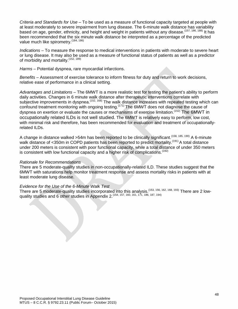
48 Proposed Occupational Interstitial Lung Disease Guideline MTUS – 8 C.C.R. § 9792.23.11 (Public Forum– October 2015)
Criteria and Standards for Use – To be used as a measure of functional capacity targeted at people with at least moderately to severe impairment from lung disease. The 6-minute walk distance has variability based on age, gender, ethnicity, and height and weight in patients without any disease.(157, 186, 188) It has been recommended that the six minute walk distance be interpreted as a percentage of the predicted value much like spirometry.(184, 186) Indications – To measure the response to medical interventions in patients with moderate to severe heart or lung disease. It may also be used as a measure of functional status of patients as well as a predictor of morbidity and mortality.(152, 189) Harms – Potential dyspnea, rare myocardial infarctions. Benefits – Assessment of exercise tolerance to inform fitness for duty and return to work decisions, relative ease of performance in a clinical setting. Advantages and Limitations – The 6MWT is a more realistic test for testing the patient’s ability to perform daily activities. Changes in 6 minute walk distance after therapeutic interventions correlate with subjective improvements in dyspnea.(152, 168) The walk distance increases with repeated testing which can confound treatment monitoring with ongoing testing.(171) The 6MWT does not diagnose the cause of dyspnea on exertion or evaluate the causes or mechanisms of exercise limitation.(152) The 6MWT in occupationally related ILDs is not well studied. The 6MWT is relatively easy to perform, low cost, with minimal risk and therefore, has been recommended for evaluation and treatment of occupationally-related ILDs. A change in distance walked >54m has been reported to be clinically significant.(158, 185, 190) A 6-minute walk distance of <350m in COPD patients has been reported to predict mortality.(191) A total distance under 200 meters is consistent with poor functional capacity, while a total distance of under 350 meters is consistent with low functional capacity and a higher risk of complications.(192) Rationale for Recommendations There are 5 moderate-quality studies in non-occupationally-related ILD. These studies suggest that the 6MWT with saturations help monitor treatment response and assess mortality risks in patients with at least moderate lung disease. Evidence for the Use of the 6-Minute Walk Test There are 5 moderate-quality studies incorporated into this analysis.(153, 156, 162, 168, 193) There are 2 low-quality studies and 6 other studies in Appendix 2.(154, 157, 160, 161, 171, 186, 187, 194)
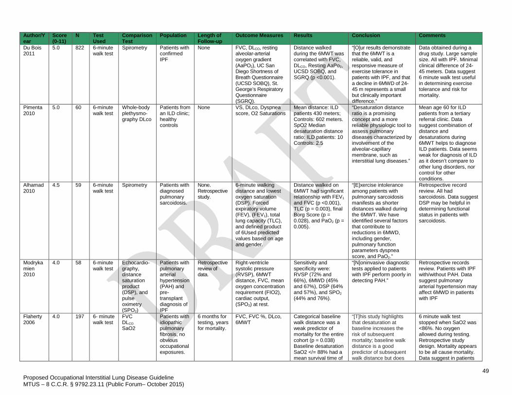
49 Proposed Occupational Interstitial Lung Disease Guideline MTUS – 8 C.C.R. § 9792.23.11 (Public Forum– October 2015)
Author/Year
Score (0-11)
N Test Used
Comparison Test
Population Length of Follow-up
Outcome Measures Results Conclusion Comments
Du Bois 2011
5.0 822 6-minute walk test
Spirometry Patients with confirmed IPF
None FVC, DLCO, resting alveolar-arterial oxygen gradient (AaPO2), UC San Diego Shortness of Breath Questionnaire (UCSD SOBQ), St. George’s Respiratory Questionnaire (SGRQ).
Distance walked during the 6MWT was correlated with FVC, DLCO, Resting AaPo2, UCSD SOBQ, and SGRQ (p <0.001).
“[O]ur results demonstrate that the 6MWT is a reliable, valid, and responsive measure of exercise tolerance in patients with IPF, and that a decline in 6MWD of 24-45 m represents a small but clinically important difference.”
Data obtained during a drug study. Large sample size. All with IPF. Minimal clinical difference of 24-45 meters. Data suggest 6 minute walk test useful in determining exercise tolerance and risk for mortality.
Pimenta 2010
5.0 60 6-minute walk test
Whole-body plethysmo-graphy DLco
Patients from an ILD clinic; healthy controls
None VS, DLco, Dyspnea score, O2 Saturations
Mean distance: ILD patients 430 meters; Controls: 602 meters. SpO2 Median desaturation distance ratio: ILD patients: 10 Controls: 2.5
“Desaturation distance ratio is a promising concept and a more reliable physiologic tool to assess pulmonary diseases characterized by involvement of the alveolar-capillary membrane, such as interstitial lung diseases.”
Mean age 60 for ILD patients from a tertiary referral clinic. Data suggest combination of distance and desaturations during 6MWT helps to diagnose ILD patients. Data seems weak for diagnosis of ILD as it doesn’t compare to other lung disorders, nor control for other conditions.
Alhamad 2010
4.5 59 6-minute walk test
Spirometry Patients with diagnosed pulmonary sarcoidosis.
None. Retrospective study.
6-minute walking distance and lowest oxygen saturation (DSP). Forced expiratory volume (FEV), (FEV1), total lung capacity (TLC), and defined product of 6Used predicted values based on age and gender.
Distance walked on 6MWT had significant relationship with FEV1 and FVC (p <0.001), TLC (p = 0.003), final Borg Score (p = 0.028), and PaO2 (p = 0.005).
“[E]xercise intolerance among patients with pulmonary sarcoidosis manifests as shorter distances walked during the 6MWT. We have identified several factors that contribute to reductions in 6MWD, including gender, pulmonary function parameters dyspnea score, and PaO2.”
Retrospective record review. All had sarcoidosis. Data suggest DSP may be helpful in determining functional status in patients with sarcoidosis.
Modrykamien 2010
4.0 58 6-minute walk test
Echocardio-graphy, distance saturation product (DSP), and pulse oximetry (SPO2)
Patients with pulmonary arterial hypertension (PAH) and pre-transplant diagnosis of IPF
Retrospective review of data.
Right-ventricle systolic pressure (RVSP), 6MWT distance, FVC, mean oxygen concentration requirement (FIO2), cardiac output, (SPO2) at rest.
Sensitivity and specificity were: RVSP (72% and 66%), 6MWD (45% and 67%), DSP (64% and 57%), and SPO2 (44% and 76%).
“[N]oninvasive diagnostic tests applied to patients with IPF perform poorly in detecting PAH.”
Retrospective records review. Patients with IPF with/without PAH. Data suggest pulmonary arterial hypertension may affect 6MWD in patients with IPF
Flaherty 2006
4.0 197 6- minute walk test
FVC DLCO SaO2
Patients with idiopathic pulmonary fibrosis, no obvious occupational exposures.
6 months for testing, years for mortality.
FVC, FVC %, DLco, 6MWT
Categorical baseline walk distance was a weak predictor of mortality for the entire cohort (p = 0.038) Baseline desaturation SaO2 </= 88% had a mean survival time of
“[T]his study highlights that desaturation at baseline increases the risk of subsequent mortality; baseline walk distance is a good predictor of subsequent walk distance but does
6 minute walk test stopped when SaO2 was <86%. No oxygen allowed during testing. Retrospective study design. Mortality appears to be all cause mortality. Data suggest in patients

50 Proposed Occupational Interstitial Lung Disease Guideline MTUS – 8 C.C.R. § 9792.23.11 (Public Forum– October 2015)
3.21 years vs. 6.83 years (p = 0.006)
not reliably predict risk of subsequent mortality…”
with IPF, desaturations at baseline, not 6MWD is a better predictor of subsequent mortality.
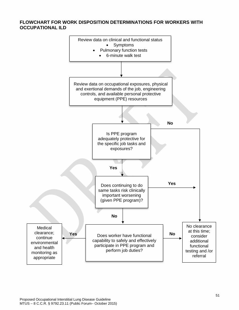
51 Proposed Occupational Interstitial Lung Disease Guideline MTUS – 8 C.C.R. § 9792.23.11 (Public Forum– October 2015)
FLOWCHART FOR WORK DISPOSITION DETERMINATIONS FOR WORKERS WITH OCCUPATIONAL ILD
Review data on clinical and functional status • Symptoms
• Pulmonary function tests • 6-minute walk test
Review data on occupational exposures, physical and exertional demands of the job, engineering
controls, and available personal protective equipment (PPE) resources
Is PPE program
adequately protective for the specific job tasks and
exposures?
Does worker have functional capability to safely and effectively participate in PPE program and
perform job duties?
Does continuing to do
same tasks risk clinically important worsening
(given PPE program)?
No clearance at this time;
consider additional functional
testing and /or referral
Yes
Yes
Yes Medical
clearance; continue
environmental and health
monitoring as appropriate
No
No
No
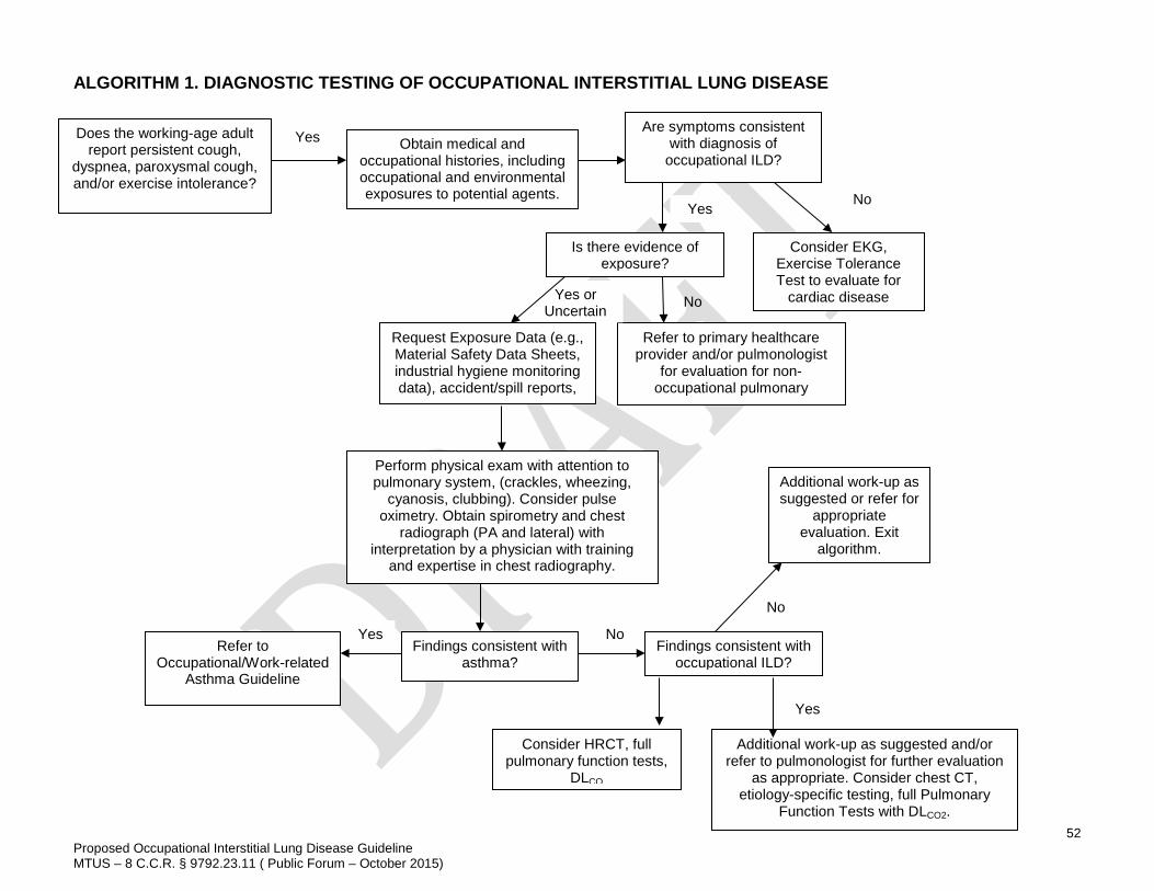
52
Proposed Occupational Interstitial Lung Disease Guideline MTUS – 8 C.C.R. § 9792.23.11 ( Public Forum – October 2015)
ALGORITHM 1. DIAGNOSTIC TESTING OF OCCUPATIONAL INTERSTITIAL LUNG DISEASE
Does the working-age adult report persistent cough,
dyspnea, paroxysmal cough, and/or exercise intolerance?
Obtain medical and occupational histories, including occupational and environmental exposures to potential agents.
Yes Are symptoms consistent
with diagnosis of occupational ILD?
Consider EKG, Exercise Tolerance Test to evaluate for
cardiac disease
Is there evidence of exposure?
Yes No
No
Refer to primary healthcare provider and/or pulmonologist
for evaluation for non-occupational pulmonary
di
Yes or Uncertain
Request Exposure Data (e.g., Material Safety Data Sheets, industrial hygiene monitoring data), accident/spill reports,
Perform physical exam with attention to pulmonary system, (crackles, wheezing,
cyanosis, clubbing). Consider pulse oximetry. Obtain spirometry and chest
radiograph (PA and lateral) with interpretation by a physician with training
and expertise in chest radiography.
Findings consistent with asthma?
Findings consistent with occupational ILD?
Refer to Occupational/Work-related
Asthma Guideline
Yes No
Additional work-up as suggested or refer for
appropriate evaluation. Exit
algorithm.
No
Additional work-up as suggested and/or refer to pulmonologist for further evaluation
as appropriate. Consider chest CT, etiology-specific testing, full Pulmonary
Function Tests with DLCO2.
Yes
Consider HRCT, full pulmonary function tests,
DLCO
53
Proposed Occupational Interstitial Lung Disease Guideline MTUS – 8 C.C.R. § 9792.23.11 ( Public Forum – October 2015)
APPENDIX 1. CHEST RADIOGRAPHS The International Labour Organization (ILO) pneumoconioses classification system provides specific ratings for opacity size, shape and number seen on routine chest radiographics, and is most commonly used globally and in the United States.(81) It is a descriptive method that standardizes the interpretation and reporting of both the type and degree of changes on chest x-ray. However, it does not provide diagnostic criteria for the pneumoconioses. The Coal Workers’ X-Ray Surveillance Program was established under the Federal Coal Mine Health and Safety Act of 1969 (P.L. 91-173), which was amended by the Federal Mine Safety and Health Act of 1977 [30 USC 843]. In 2014, the MSHA extended coverage to surface, as well as, underground miners. Currently, mandatory x-rays include the following:
• An initial chest x-ray within 6 months of beginning employment, • Another chest x-ray 3 years after the initial examination, • A third chest x-ray 2 years following the second one if a miner is still engaged in underground
coal mining and if the second chest x-ray shows evidence of category 1 or higher pneumoconiosis according to the ILO classification.(195)
In addition to these mandatory chest x-rays, mine operators are required to offer an opportunity for periodic, voluntary chest x-rays approximately every 5 years. The chest x-rays obtained under the Coal Workers’ X-Ray Surveillance Program are submitted to and become the property of NIOSH.
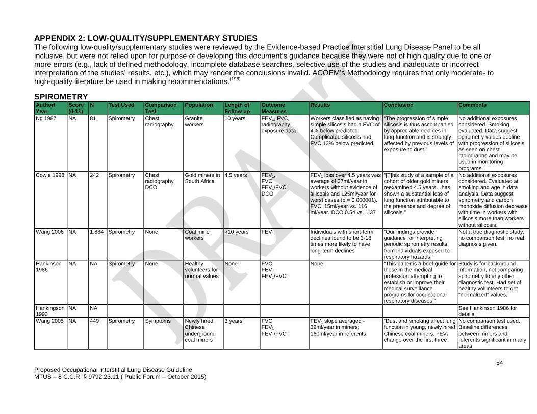
54
Proposed Occupational Interstitial Lung Disease Guideline MTUS – 8 C.C.R. § 9792.23.11 ( Public Forum – October 2015)
APPENDIX 2: LOW-QUALITY/SUPPLEMENTARY STUDIES The following low-quality/supplementary studies were reviewed by the Evidence-based Practice Interstitial Lung Disease Panel to be all inclusive, but were not relied upon for purpose of developing this document’s guidance because they were not of high quality due to one or more errors (e.g., lack of defined methodology, incomplete database searches, selective use of the studies and inadequate or incorrect interpretation of the studies’ results, etc.), which may render the conclusions invalid. ACOEM’s Methodology requires that only moderate- to high-quality literature be used in making recommendations.(196) SPIROMETRY Author/ Year
Score (0-11)
N Test Used Comparison Test
Population Length of Follow up
Outcome Measures
Results Conclusion Comments
Ng 1987 NA 81 Spirometry Chest radiography
Granite workers
10 years FEV1, FVC, radiography, exposure data
Workers classified as having simple silicosis had a FVC of 4% below predicted. Complicated silicosis had FVC 13% below predicted.
“The progression of simple silicosis is thus accompanied by appreciable declines in lung function and is strongly affected by previous levels of exposure to dust.”
No additional exposures considered. Smoking evaluated. Data suggest spirometry values decline with progression of silicosis as seen on chest radiographs and may be used in monitoring programs.
Cowie 1998 NA 242 Spirometry Chest radiography DCO
Gold miners in South Africa
4.5 years FEV1, FVC FEV1/FVC DCO
FEV1 loss over 4.5 years was average of 37ml/year in workers without evidence of silicosis and 125ml/year for worst cases (p = 0.000001). FVC: 15ml/year vs. 116 ml/year. DCO 0.54 vs. 1.37
“[T]his study of a sample of a cohort of older gold miners reexamined 4.5 years…has shown a substantial loss of lung function attributable to the presence and degree of silicosis.”
No additional exposures considered. Evaluated at smoking and age in data analysis. Data suggest spirometry and carbon monoxide diffusion decrease with time in workers with silicosis more than workers without silicosis.
Wang 2006 NA 1,884 Spirometry None Coal mine workers
>10 years FEV1 Individuals with short-term declines found to be 3-18 times more likely to have long-term declines
“Our findings provide guidance for interpreting periodic spirometry results from individuals exposed to respiratory hazards.”
Not a true diagnostic study, no comparison test, no real diagnosis given.
Hankinson 1986
NA NA Spirometry None Healthy volunteers for normal values
None FVC FEV1 FEV1/FVC
None “This paper is a brief guide for those in the medical profession attempting to establish or improve their medical surveillance programs for occupational respiratory diseases.”
Study is for background information, not comparing spirometry to any other diagnostic test. Had set of healthy volunteers to get “normalized” values.
Hankingson 1993
NA NA See Hankinson 1986 for details
Wang 2005 NA 449 Spirometry Symptoms Newly hired Chinese underground coal miners
3 years FVC FEV1 FEV1/FVC
FEV1 slope averaged -39ml/year in miners; 160ml/year in referents
“Dust and smoking affect lung function in young, newly hired Chinese coal miners. FEV1 change over the first three
No comparison test used. Baseline differences between miners and referents significant in many areas.
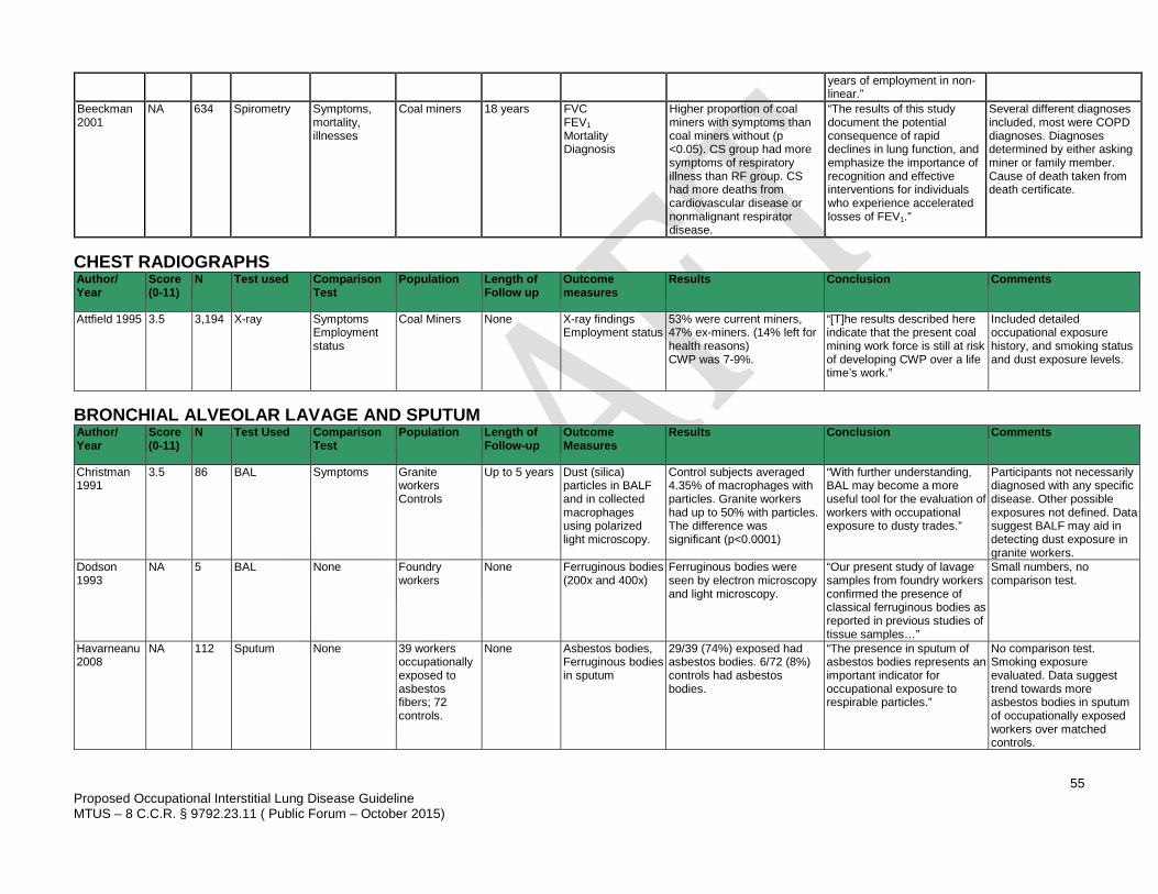
55
Proposed Occupational Interstitial Lung Disease Guideline MTUS – 8 C.C.R. § 9792.23.11 ( Public Forum – October 2015)
years of employment in non-linear.”
Beeckman 2001
NA 634 Spirometry Symptoms, mortality, illnesses
Coal miners 18 years FVC FEV1 Mortality Diagnosis
Higher proportion of coal miners with symptoms than coal miners without (p <0.05). CS group had more symptoms of respiratory illness than RF group. CS had more deaths from cardiovascular disease or nonmalignant respirator disease.
“The results of this study document the potential consequence of rapid declines in lung function, and emphasize the importance of recognition and effective interventions for individuals who experience accelerated losses of FEV1.”
Several different diagnoses included, most were COPD diagnoses. Diagnoses determined by either asking miner or family member. Cause of death taken from death certificate.
CHEST RADIOGRAPHS
BRONCHIAL ALVEOLAR LAVAGE AND SPUTUM
Author/ Year
Score (0-11)
N Test used Comparison Test
Population Length of Follow up
Outcome measures
Results Conclusion Comments
Attfield 1995 3.5 3,194 X-ray Symptoms Employment status
Coal Miners None X-ray findings Employment status
53% were current miners, 47% ex-miners. (14% left for health reasons) CWP was 7-9%.
“[T]he results described here indicate that the present coal mining work force is still at risk of developing CWP over a life time’s work.”
Included detailed occupational exposure history, and smoking status and dust exposure levels.
Author/ Year
Score (0-11)
N Test Used Comparison Test
Population Length of Follow-up
Outcome Measures
Results Conclusion Comments
Christman 1991
3.5 86 BAL Symptoms Granite workers Controls
Up to 5 years Dust (silica) particles in BALF and in collected macrophages using polarized light microscopy.
Control subjects averaged 4.35% of macrophages with particles. Granite workers had up to 50% with particles. The difference was significant (p<0.0001)
“With further understanding, BAL may become a more useful tool for the evaluation of workers with occupational exposure to dusty trades.”
Participants not necessarily diagnosed with any specific disease. Other possible exposures not defined. Data suggest BALF may aid in detecting dust exposure in granite workers.
Dodson 1993
NA 5 BAL None Foundry workers
None Ferruginous bodies (200x and 400x)
Ferruginous bodies were seen by electron microscopy and light microscopy.
“Our present study of lavage samples from foundry workers confirmed the presence of classical ferruginous bodies as reported in previous studies of tissue samples…”
Small numbers, no comparison test.
Havarneanu 2008
NA 112 Sputum None 39 workers occupationally exposed to asbestos fibers; 72 controls.
None Asbestos bodies, Ferruginous bodies in sputum
29/39 (74%) exposed had asbestos bodies. 6/72 (8%) controls had asbestos bodies.
“The presence in sputum of asbestos bodies represents an important indicator for occupational exposure to respirable particles.”
No comparison test. Smoking exposure evaluated. Data suggest trend towards more asbestos bodies in sputum of occupationally exposed workers over matched controls.
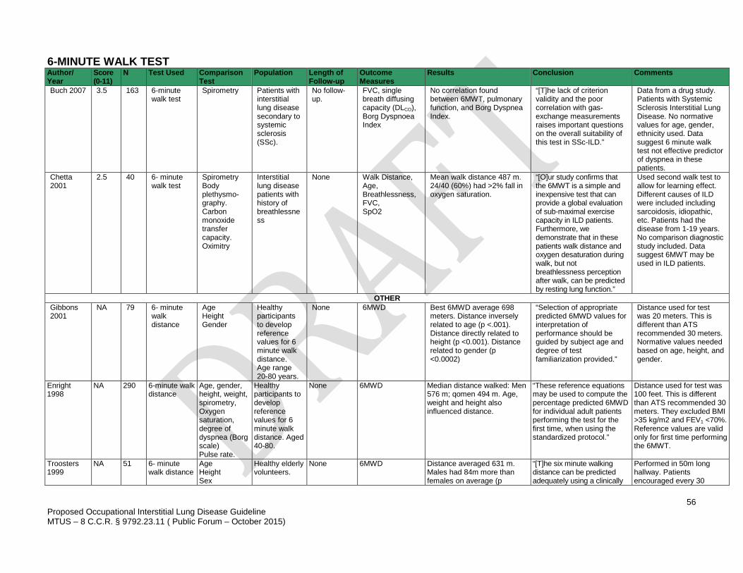
56
Proposed Occupational Interstitial Lung Disease Guideline MTUS – 8 C.C.R. § 9792.23.11 ( Public Forum – October 2015)
6-MINUTE WALK TEST Author/ Year
Score (0-11)
N Test Used Comparison Test
Population Length of Follow-up
Outcome Measures
Results Conclusion Comments
Buch 2007 3.5 163 6-minute walk test
Spirometry Patients with interstitial lung disease secondary to systemic sclerosis (SSc).
No follow-up.
FVC, single breath diffusing capacity (DLCO), Borg Dyspnoea Index
No correlation found between 6MWT, pulmonary function, and Borg Dyspnea Index.
“[T]he lack of criterion validity and the poor correlation with gas-exchange measurements raises important questions on the overall suitability of this test in SSc-ILD.”
Data from a drug study. Patients with Systemic Sclerosis Interstitial Lung Disease. No normative values for age, gender, ethnicity used. Data suggest 6 minute walk test not effective predictor of dyspnea in these patients.
Chetta 2001
2.5 40 6- minute walk test
Spirometry Body plethysmo-graphy. Carbon monoxide transfer capacity. Oximitry
Interstitial lung disease patients with history of breathlessness
None Walk Distance, Age, Breathlessness, FVC, SpO2
Mean walk distance 487 m. 24/40 (60%) had >2% fall in oxygen saturation.
“[O]ur study confirms that the 6MWT is a simple and inexpensive test that can provide a global evaluation of sub-maximal exercise capacity in ILD patients. Furthermore, we demonstrate that in these patients walk distance and oxygen desaturation during walk, but not breathlessness perception after walk, can be predicted by resting lung function.”
Used second walk test to allow for learning effect. Different causes of ILD were included including sarcoidosis, idiopathic, etc. Patients had the disease from 1-19 years. No comparison diagnostic study included. Data suggest 6MWT may be used in ILD patients.
OTHER Gibbons 2001
NA 79 6- minute walk distance
Age Height Gender
Healthy participants to develop reference values for 6 minute walk distance. Age range 20-80 years.
None 6MWD Best 6MWD average 698 meters. Distance inversely related to age (p <.001). Distance directly related to height (p <0.001). Distance related to gender (p <0.0002)
“Selection of appropriate predicted 6MWD values for interpretation of performance should be guided by subject age and degree of test familiarization provided.”
Distance used for test was 20 meters. This is different than ATS recommended 30 meters. Normative values needed based on age, height, and gender.
Enright 1998
NA 290 6-minute walk distance
Age, gender, height, weight, spirometry, Oxygen saturation, degree of dyspnea (Borg scale) Pulse rate.
Healthy participants to develop reference values for 6 minute walk distance. Aged 40-80.
None 6MWD Median distance walked: Men 576 m; qomen 494 m. Age, weight and height also influenced distance.
“These reference equations may be used to compute the percentage predicted 6MWD for individual adult patients performing the test for the first time, when using the standardized protocol.”
Distance used for test was 100 feet. This is different than ATS recommended 30 meters. They excluded BMI >35 kg/m2 and FEV1 <70%. Reference values are valid only for first time performing the 6MWT.
Troosters 1999
NA 51 6- minute walk distance
Age Height Sex
Healthy elderly volunteers.
None 6MWD Distance averaged 631 m. Males had 84m more than females on average (p
“[T]he six minute walking distance can be predicted adequately using a clinically
Performed in 50m long hallway. Patients encouraged every 30
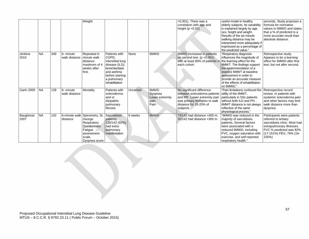
57
Proposed Occupational Interstitial Lung Disease Guideline MTUS – 8 C.C.R. § 9792.23.11 ( Public Forum – October 2015)
Weight <0.001). There was a correlation with age and height (p <0.01)
useful model in healthy elderly subjects. Its variability is explained largely by age, sex, height and weight. Results of the six minute walking distance may be interpreted more adequately if expressed as a percentage of the predicted value.”
seconds. Study proposes a formula for normative values in 6MWD and states that a % of predicted is a more accurate result than absolute distance.
Jenkins 2010
NA 349 6- minute walk distance
Repeated 6- minute walk distance maximum of 4 weeks after first.
Patients with COPD, interstitial lung disease (ILD), bronchiectasis and asthma before starting a pulmonary rehabilitation
None 6MWD 6MWD increased in patients on second test. (p <0.001) with at least 80% of patients in each cohort.
“Respiratory diagnosis influences the magnitude of the learning effect for the 6MWT. The findings support the recommendation of a practice 6MWT at baseline assessment in order to provide an accurate measure of the effects of rehabilitation on 6MWD.”
Retrospective study. Appears to be a learning effect for 6MWD after first test, but not after second.
Garin 2009 NA 128 6- minute walk distance
Mortality Patients with scleroderma and or idiopathic pulmonary fibrosis
Uncertain 6MWD Dyspnea Lower extremity pain Pain
No significant difference between scleroderma patients and IPF. Lower extremity pain was primary limitation to walk distance for 15-20% of subjects.
“Pain limitations confound the utility of the 6MWT, particularly in SSc patients without both ILD and PH… 6MWT distance is not always reflective of the same physiological process.”
Retrospective record review. In patients with systemic scleroderma pain and other factors may limit walk distance more than dyspnea.
Baughman 2007
NA 142 6-minute walk distance
Spirometry, St. George Respiratory Questionnaire Fatigue assessment scale, Dyspnea score
Sarcoidosis patients, 130/142 (92%) had extra-pulmonary manifestation
6 weeks 6MWD 73/142 had distance <400 m; 32/142 had distance <300 m.
“6MWD was reduced in the majority of sarcoidosis patients. Several factors were associated with a reduced 6MWD, including FVC, oxygen saturation with exercise, and self-reported respiratory health.”
Participants were patients referred to tertiary sarcoidosis clinic. Most had extrapulmonary illnesses. FVC % predicted was 82% (17-151%) FEV1 76% (16-155%)
58
Proposed Occupational Interstitial Lung Disease Guideline MTUS – 8 C.C.R. § 9792.23.11 ( Public Forum – October 2015)
REFERENCES
1. Raghu G, Collard H, Egan J, et al. An official ATS/ERS/JRS/ALAT statement: idiopathic pulmonary fibrosis: evidence-based guidelines for diagnosis and management. Am J Respir Crit Care Med. 2011;183:788-824. 2. Ryu JH, Daniels CE, Hartman TE, Yi ES. Diagnosis of interstitial lung diseases. Mayo Clin Proc. 2007;82(8):976-86. 3. King TE, Jr. Clinical advances in the diagnosis and therapy of the interstitial lung diseases. Am J Respir Crit Care Med. 2005;172(3):268-79. 4. Bates DV, Gotsch AR, Brooks S, Landrigan PJ, Hankinson JL, Merchant JA. Prevention of occupational lung disease. Task Force on Research and Education for the Prevention and Control of Respiratory Diseases. Chest. 1992;102(3 Suppl):257S-76S. 5. American Thoracic Society, European Respiratory Society. American Thoracic Society/European Respiratory Society International Multidisciplinary Consensus Classification of the Idiopathic Interstitial Pneumonias. Am J Respir Crit Care Med. 2002;165(2):277-304. 6. Petsonk EL, Rose C, Cohen R. Coal mine dust lung disease. New lessons from old exposure. Am J Respir Crit Care Med. 2013;187(11):1178-85. 7. Boros PW, Franczuk M, Wesolowski S. Value of spirometry in detecting volume restriction in interstitial lung disease patients. Spirometry in interstitial lung diseases. Respiration. 2004;71(4):374-9. 8. Stansbury RC, Beeckman-Wagner LA, Wang ML, Hogg JP, Petsonk EL. Rapid decline in lung function in coal miners: evidence of disease in small airways. Am J Ind Med. 2013;56(9):1107-12. 9. Allen B, Garland B. Patient's page. Interstitial lung disease. South Med J. 2007;100(6):619. 10. Olson A, Schwarz, M, Roman J. Interstitial lung disease. In: Schraufnagel D, ed. Breathing in America: Diseases, Progress, and Hope: American Thoracic Society; 2010:99-108. 11. Wells A, Hirani N, and on behalf of the British Thoracic Society Interstitial Lung Disease Guideline Group, a subgroup of the British Thoracic Society Standards of Care Committee, in collaboration with the Thoracic Society of Australia, and New Zealand and the Irish Thoracic Society. Interstitial lung disease guideline: the British Thoracic Society in collaboration with the Thoracic Society of Australia and New Zealand and the Irish Thoracic Society. Thorax. 2008;63(Suppl V):v1-58. 12. Glenn D. Current issues surrounding silica. Professional Safety. 2008;53(2):37-46. 13. Schenker MB, ed. Respiratory health hazards in agriculture. American Thoracic Society Consensus Report. Am J Respir Crit Care Med. 1998;158(suppl 4):S1-76. 14. Cummings KJ, Deubner DC, Day GA, et al. Enhanced preventive programme at a beryllium oxide ceramics facility reduces beryllium sensitisation among new workers. Occup Environ Med. 2007;64(2):134-40. 15. Infante PF, Newman LS. Beryllium exposure and chronic beryllium disease. Lancet. 2004;363(9407):415-6. 16. Silver K, Kukulka G, Gorniewicz J, Rayman K, Sharp R. Genetic susceptibility testing for beryllium: worker knowledge, beliefs, and attitudes. Am J Ind Med. 2011;54(7):521-32. 17. Thomas CA, Bailey RL, Kent MS, Deubner DC, Kreiss K, Schuler CR. Efficacy of a program to prevent beryllium sensitization among new employees at a copper-beryllium alloy processing facility. Public Health Rep. 2009;124 (Suppl 1):112-24. 18. Steenland K, Brown D. Mortality study of gold miners exposed to silica and nonasbestiform amphibole minerals: an update with 14 more years of follow-up. Am J Ind Med. 1995;27(2):217-29. 19. American Thoracic Society. Adverse effects of crystalline silica exposure. Am J Respir Crit Care Med. 1997;155:761-5. 20. Brodkin CA, Barnhart S, Checkoway H, Balmes J, Omenn GS, Rosenstock L. Longitudinal pattern of reported respiratory symptoms and accelerated ventilatory loss in asbestos-exposed workers. Chest. 1996;109(1):120-6. 21. Straif K, Benbrahim-Tallaa L, Baan R, et al. A review of human carcinogens-Part C: metals, arsenic, dusts, and fibres. Lancet Oncology. 2009;10(5):453-4. 22. American Thoracic Society. Diagnosis and initial management of nonmalignant diseases related to asbestos. Am J Respir Crit Care Med. 2004;170:691-715. 23. Caplan A. Correlation of radiological category with lung pathology in coal-workers' pneumoconiosis. Br J Ind Med. 1962;19171-9. 24. Patel AM, Ryu JH, Reed CE. Hypersensitivity pneumonitis: current concepts and future questions. J Allergy Clin Immunol. 2001;108(5):661-70. 25. Lison D, Lauwerys R, Demedts M, Nemery B. Experimental research into the pathogenesis of cobalt/hard metal lung disease. Eur Respir J. 1996;9(5):1024-8. 26. Naqvi AH, Hunt A, Burnett BR, Abraham JL. Pathologic spectrum and lung dust burden in giant cell interstitial pneumonia (hard metal disease/cobalt pneumonitis): review of 100 cases. Arch Environ Occup Health. 2008;63(2):51-70. 27. Choi JW, Lee KS, Chung MP, Han J, Chung MJ, Park JS. Giant cell interstitial pneumonia: high-resolution CT and pathologic findings in four adult patients. AJR Am J Roentgenol. 2005;184(1):268-72. 28. Rosenman KD, Reilly MJ, Henneberger PK. Estimating the total number of newly-recognized silicosis cases in the United States. Am J Ind Med. 2003;44(2):141-7. 29. Mazurek J, Wood J. Asbestosis-Related Years of Potential Life Lost Before Age 65 Years -- United States, 1968-2005. MMWR. 2008;57(49):1321-5. 30. Mazurek J, Laney A, Wood J. Coal Workers' Pneumoconiosis-Related Years of Potential Life Lost Before Age 65 Years -- United States, 1968-2006. MMWR. 2009;58(50):1412-6. 31. Loomis D. Basic protections are still lacking. Occup Environ Med. 2010;67(6):361.
59
Proposed Occupational Interstitial Lung Disease Guideline MTUS – 8 C.C.R. § 9792.23.11 ( Public Forum – October 2015)
32. Cummings KJ, Nakano M, Omae K, et al. Indium lung disease. Chest. 2012;141(6):1512-21. 33. Department of Health and Human Services, Centers for Disease Control and Prevention, National Institute for Occupational Safety and Health. Publication Number 2002-129 -- NIOSH Hazard Review: Health Effects of Occupational Exposure to Respirable Crystalline Silica. Available at: http://www.cdc.gov/niosh/docs/2002-129/. 2002 (April). 34. Hueper W. Section IV. Human exposure to asbestos: community studies. Occupational and nonoccupational exposures to asbestos. Annals New York Academy of Sciences. 1965;184-95. 35. Wolff H, Vehmas T, Oksa P, Rantanen J, Vainio H. Asbestos, asbestosis, and cancer: Helsinki Criteria update 2014: Recommendations. Consensus Report. Scand J Work Environ Health. 2014;Online-first article. 36. Agency for Toxic Substances and Disease Registry. What Is Asbestos? . 2008. Retrieved December 22, 2014 from: http://www.atsdr.cdc.gov/asbestos/more_about_asbestos/what_is_asbestos. 37. Agency for Toxic Substances and Disease Registry. Toxicological Profile for Asbestos. 2001. Retrieved December 22, 2014 from: http://www.atsdr.cdc.gov/toxprofiles/tp61.pdf. 38. World Health Organization. International Programme on Chemical Safety. Environmental Health Criteria 53. Asbestos and Other Natural Mineral Fibers. Geneva; 1986. 39. Mayer AS, Hamzeh N, Maier LA. Sarcoidosis and chronic beryllium disease: similarities and differences. Semin Respir Crit Care Med. 2014;35(3):316-29. 40. Hypersensitivity Pneumonitis Update (Review). Diffuse Lung Disease & Interstitial Lung Disease, Society Journal 1. Available at: http://pulmccm.org/2012/review-articles/hypersensitivity-pneumonitis-2012-update-review-chest/. 2012. 41. U.S. Department of Health and Human Services, National Institute for Occupational Safety and Health, Division of Respiratory Disease Studies. Work-related Lung Disease Surveillance Report 1999. DHHS (NIOSH) Number 2000-105. Available at: http://www.cdc.gov/niosh/docs/2000-105/pdfs/2000-105.pdf. 1999. 42. Mapel DW, Coultas DB, James DS, Hunt WC, Stidley CA, Gilliland FD. Ethnic differences in the prevalence of nonmalignant respiratory disease among uranium miners. Am J Public Health. 1997;87(5):833-8. 43. Cowl CT. Occupational asthma: review of assessment, treatment, and compensation. Chest. 2011;139(3):674-81. 44. Blanc P, Balmes J. History and physical examination. In: Harber P, Schenker M, J B, eds. Occupational and Environmental Respiratory Diseases. St. Louis, MO: Mosby; 1995:28-38. 45. Bickley L, Szilagyi P, Bates B. Bates' Guide to Physical Examination and History Taking: Lippincott Williams & Wilkins; 2009. 46. LeBlond R, Brown D, DeGowin R. DeGowin's Diagnostic Examination, Ninth Edition: McGraw-Hill Professional; 2008. 47. Wang ML, Avashia BH, Petsonk EL. Interpreting periodic lung function tests in individuals: the relationship between 1- to 5-year and long-term FEV1 changes. Chest. 2006;130(2):493-9. 48. Beeckman LA, Wang ML, Petsonk EL, Wagner GR. Rapid declines in FEV1 and subsequent respiratory symptoms, illnesses, and mortality in coal miners in the United States. Am J Respir Crit Care Med. 2001;163(3 Pt 1):633-9. 49. Miller MR, Crapo R, Hankinson J, et al. General considerations for lung function testing. Eur Respir J. 2005;26(1):153-61. 50. Townsend M, Occupational and Environmental Lung Disorders Committee. Spirometry in the occupational health setting - 2011 update. JOEM. 2011;53(5):569-84. 51. U.S. Department of Labor, Occupational Safety and Health Administration. OSHA Publication No. 3637-2013. Spirometry Testing in Occupational Health Programs. Best Practices for Healthcare Professionals. Available at: https://www.osha.gov/Publications/OSHA3637.pdf. 2013. 52. Leung CC, Chang KC, Law WS, et al. Determinants of spirometric abnormalities among silicotic patients in Hong Kong. Occup Med (Lond). 2005;55(6):490-3. 53. Townsend MC. Evaluating pulmonary function change over time in the occupational setting. J Occup Environ Med. 2005;47(12):1307-16. 54. Hankinson JL. Pulmonary function testing in the screening of workers: guidelines for instrumentation, performance, and interpretation. J Occup Med. 1986;28(10):1081-92. 55. Hankinson JL, Bang KM. Acceptability and reproducibility criteria of the American Thoracic Society as observed in a sample of the general population. Am Rev Respir Dis. 1991;143(3):516-21. 56. Pellegrino R, Viegi G, Brusasco V, et al. Interpretative strategies for lung function tests. Eur Respir J. 2005;26(5):948-68. 57. Rondinelli R. AMA Guides to the Evaluation of Permanent Impairment, Sixth Edition. Chicago, IL: American Medical Association; 2007. 58. Redlich CA, Tarlo SM, Hankinson JL, et al. Official American Thoracic Society technical standards: spirometry in the occupational setting. Am J Respir Crit Care Med. 2014;189(8):983-93. 59. Wang X, Yano E. Pulmonary dysfunction in silica-exposed workers: a relationship to radiographic signs of silicosis and emphysema. Am J Ind Med. 1999;36(2):299-306. 60. Miller A, Lilis R, Godbold J, Chan E, Wu X, Selikoff IJ. Spirometric impairments in long-term insulators. Relationships to duration of exposure, smoking, and radiographic abnormalities. Chest. 1994;105(1):175-82. 61. Kilburn KH, Warshaw RH. Airways obstruction from asbestos exposure. Effects of asbestosis and smoking. Chest. 1994;106(4):1061-70. 62. Barnhart S, Hudson LD, Mason SE, Pierson DJ, Rosenstock L. Total lung capacity. An insensitive measure of impairment in patients with asbestosis and chronic obstructive pulmonary disease? Chest. 1988;93(2):299-302.
60
Proposed Occupational Interstitial Lung Disease Guideline MTUS – 8 C.C.R. § 9792.23.11 ( Public Forum – October 2015)
63. Rosenman KD, Reilly MJ, Gardiner J. Results of spirometry among individuals in a silicosis registry. J Occup Environ Med. 2010;52(12):1173-8. 64. Brodkin CA, Barnhart S, Anderson G, Checkoway H, Omenn GS, Rosenstock L. Correlation between respiratory symptoms and pulmonary function in asbestos-exposed workers. Am Rev Respir Dis. 1993;148(1):32-7. 65. Kilburn KH, Warshaw R, Thornton JC. Asbestosis, pulmonary symptoms and functional impairment in shipyard workers. Chest. 1985;88(2):254-9. 66. Aaron SD, Dales RE, Cardinal P. How accurate is spirometry at predicting restrictive pulmonary impairment? Chest. 1999;115(3):869-73. 67. Sircar K, Hnizdo E, Petsonk E, Attfield M. Decline in lung function and mortality: implications for medical monitoring. Occup Environ Med. 2007;64(7):461-6. 68. Ng TP, Chan SL, Lam KP. Radiological progression and lung function in silicosis: a ten year follow up study. Br Med J (Clin Res Ed). 1987;295(6591):164-8. 69. Cowie RL. The influence of silicosis on deteriorating lung function in gold miners. Chest. 1998;113(2):340-3. 70. Hankinson JL, Wagner GR. Medical screening using periodic spirometry for detection of chronic lung disease. Occup Med. 1993;8(2):353-61. 71. Wang ML, Wu ZE, Du QG, et al. A prospective cohort study among new Chinese coal miners: the early pattern of lung function change. Occup Environ Med. 2005;62(11):800-5. 72. Hourihane DO, Lessof L, Richardson PC. Hyaline and calcified pleural plaques as an index of exposure to asbestos. Br Med J. 1966;1(5495):1069-74. 73. Ruckley VA, Fernie JM, Chapman JS, et al. Comparison of radiographic appearances with associated pathology and lung dust content in a group of coalworkers. Br J Ind Med. 1984;41(4):459-67. 74. Musk AW, Cotes JE, Bevan C, Campbell MJ. Relationship between type of simple coalworkers' pneumoconiosis and lung function. A nine-year follow-up study of subjects with small rounded opacities. Br J Ind Med. 1981;38(4):313-20. 75. Fernie JM, Ruckley VA. Coalworkers' pneumoconiosis: correlation between opacity profusion and number and type of dust lesions with special reference to opacity type. Br J Ind Med. 1987;44(4):273-7. 76. Uragoda CG. Graphite pneumoconiosis and its declining prevalence in Sri Lanka. J Trop Med Hyg. 1989;92(6):422-4. 77. Rossiter CE. Relation between content and composition of coalworkers' lungs and radiological appearances. Br J Ind Med. 1972;29(1):31-44. 78. Kim KI, Kim CW, Lee MK, et al. Imaging of occupational lung disease. Radiographics. 2001;21(6):1371-91. 79. Epler GR. Clinical overview of occupational lung disease. Radiol Clin North Am. 1992;30(6):1121-33. 80. International Labour Office. Guidelines for the use of the ILO International Classification of Radiographs of Pneumoconioses, Revised edition 2011. Geneva: International Labour Office; 2011. 81. Cockcroft A, Lyons JP, Andersson N, Saunders MJ. Prevalence and relation to underground exposure of radiological irregular opacities in South Wales coal workers with pneumoconiosis. Br J Ind Med. 1983;40(2):169-72. 82. Attfield M, Morring K. An investigation into the relationship between coal workers’ pneumoconiosis and dust exposure in U.S. coal miners. Am Ind Hyg Assoc. 1992;53:486-92. 83. Attfield MD, Seixas NS. Prevalence of pneumoconiosis and its relationship to dust exposure in a cohort of U.S. bituminous coal miners and ex-miners. Am J Ind Med. 1995;27(1):137-51. 84. Stark P, Jacobson F, Shaffer K. Standard imaging in silicosis and coal worker's pneumoconiosis. Radiol Clin North Am. 1992;30(6):1147-54. 85. Gefter WB, Conant EF. Issues and controversies in the plain-film diagnosis of asbestos-related disorders in the chest. J Thorac Imaging. 1988;3(4):11-28. 86. Lopes A, Mogami R, Capone D, Tessarollo B, Lopes De Melo P, Jansen J. High-resolution computed tomography in silicosis: correlation with chest radiography and pulmonary function tests. J Bras Pneumol. 2008;34(5):264-72. 87. American College of Radiology (ACR), Society for Pediatric Radiology (SPR). ACR–SPR Practice Guideline for the Performance of Chest Radiography. Available at: http://www.acr.org/~/media/ACR/Documents/PGTS/guidelines/Chest_Radiography.pdf. 2011. 88. Kipen HM, Lilis R, Suzuki Y, Valciukas JA, Selikoff IJ. Pulmonary fibrosis in asbestos insulation workers with lung cancer: a radiological and histopathological evaluation. Br J Ind Med. 1987;44(2):96-100. 89. Laney AS, Attfield MD. Coal workers' pneumoconiosis and progressive massive fibrosis are increasingly more prevalent among workers in small underground coal mines in the United States. Occup Environ Med. 2010;67(6):428-31. 90. Paris C, Benichou J, Raffaelli C, et al. Factors associated with early-stage pulmonary fibrosis as determined by high-resolution computed tomography among persons occupationally exposed to asbestos. Scand J Work Environ Health. 2004;30(3):206-14. 91. Wain SL, Roggli VL, Foster WL, Jr. Parietal pleural plaques, asbestos bodies, and neoplasia. A clinical, pathologic, and roentgenographic correlation of 25 consecutive cases. Chest. 1984;86(5):707-13. 92. Vallyathan V, Brower PS, Green FH, Attfield MD. Radiographic and pathologic correlation of coal workers' pneumoconiosis. Am J Respir Crit Care Med. 1996;154(3 Pt 1):741-8. 93. Hurley JF, Burns J, Copland L, Dodgson J, Jacobsen M. Coalworkers' simple pneumoconiosis and exposure to dust at 10 British coalmines. Br J Ind Med. 1982;39(2):120-7. 94. Bourgkard E, Bernadac P, Chau N, Bertrand JP, Teculescu D, Pham QT. Can the evolution to pneumoconiosis be suspected in coal miners? A longitudinal study. Am J Respir Crit Care Med. 1998;158(2):504-9.
61
Proposed Occupational Interstitial Lung Disease Guideline MTUS – 8 C.C.R. § 9792.23.11 ( Public Forum – October 2015)
95. Amandus HE, Lapp NL, Jacobson G, Reger RB. Significance of irregular small opacities in radiographs of coalminers in the USA. Br J Ind Med. 1976;33(1):13-7. 96. Sun J, Weng D, Jin C, et al. The value of high resolution computed tomography in the diagnostics of small opacities and complications of silicosis in mine machinery manufacturing workers, compared to radiography. J Occup Health. 2008;50(5):400-5. 97. Larson TC, Lewin M, Gottschall EB, Antao VC, Kapil V, Rose CS. Associations between radiographic findings and spirometry in a community exposed to Libby amphibole. Occup Environ Med. 2012;69(5):361-6. 98. Collins HP, Dick JA, Bennett JG, et al. Irregularly shaped small shadows on chest radiographs, dust exposure, and lung function in coalworkers' pneumoconiosis. Br J Ind Med. 1988;45(1):43-55. 99. Gevenois PA, Pichot E, Dargent F, Dedeire S, Vande Weyer R, De Vuyst P. Low grade coal worker's pneumoconiosis. Comparison of CT and chest radiography. Acta Radiol. 1994;35(4):351-6. 100. Huuskonen O, Kivisaari L, Zitting A, Taskinen K, Tossavainen A, Vehmas T. High-resolution computed tomography classification of lung fibrosis for patients with asbestos-related disease. Scand J Work Environ Health. 2001;27(2):106-12. 101. Hanak V, Golbin JM, Hartman TE, Ryu JH. High-resolution CT findings of parenchymal fibrosis correlate with prognosis in hypersensitivity pneumonitis. Chest. 2008;134(1):133-8. 102. Gamsu G, Salmon CJ, Warnock ML, Blanc PD. CT quantification of interstitial fibrosis in patients with asbestosis: a comparison of two methods. AJR Am J Roentgenol. 1995;164(1):63-8. 103. Eterovic D, Dujic Z, Tocilj J, Capkun V. High resolution pulmonary computed tomography scans quantified by analysis of density distribution: application to asbestosis. Br J Ind Med. 1993;50(6):514-9. 104. Newman LS, Buschman DL, Newell JD, Jr., Lynch DA. Beryllium disease: assessment with CT. Radiology. 1994;190(3):835-40. 105. Mosiewicz J, Myslinski W, Zlomaniec G, Czabak-Garbacz R, Krupski W, Dzida G. Diagnostic value of high resolution computed tomography in the assessment of nodular changes in pneumoconiosis in foundry workers in Lublin. Ann Agric Environ Med. 2004;11(2):279-84. 106. Collins LC, Willing S, Bretz R, Harty M, Lane E, Anderson WH. High-resolution CT in simple coal workers' pneumoconiosis. Lack of correlation with pulmonary function tests and arterial blood gas values. Chest. 1993;104(4):1156-62. 107. Aberle DR, Gamsu G, Ray CS, Feuerstein IM. Asbestos-related pleural and parenchymal fibrosis: detection with high-resolution CT. Radiology. 1988;166(3):729-34. 108. Bergin CJ, Castellino RA, Blank N, Moses L. Specificity of high-resolution CT findings in pulmonary asbestosis: do patients scanned for other indications have similar findings? AJR Am J Roentgenol. 1994;163(3):551-5. 109. Akira M, Yokoyama K, Yamamoto S, et al. Early asbestosis: evaluation with high-resolution CT. Radiology. 1991;178(2):409-16. 110. Afeltra A, Zennaro D, Garzia P, et al. Prevalence of interstitial lung involvement in patients with connective tissue diseases assessed with high-resolution computed tomography. Scand J Rheumatol. 2006;35(5):388-94. 111. Topcu F, Bayram H, Simsek M, et al. High-resolution computed tomography in cases with environmental exposure to asbestos in Turkey. Respiration. 2000;67:139-45. 112. Han D, Goo JM, Im JG, Lee KS, Paek DM, Park SH. Thin-section CT findings of arc-welders' pneumoconiosis. Korean J Radiol. 2000;1(2):79-83. 113. Lynch DA, Newell JD, Logan PM, King TE, Jr., Muller NL. Can CT distinguish hypersensitivity pneumonitis from idiopathic pulmonary fibrosis? AJR Am J Roentgenol. 1995;165(4):807-11. 114. Ziora D, Jastrzebski D, Lubina M, Wojdala A, Kozielski J. High-resolution computed tomography in hypersensitivity pneumonitis - correlation with pulmonary function. Ann Agric Environ Med. 2005;12(1):31-4. 115. Lynch DA, Gamsu G, Ray CS, Aberle DR. Asbestos-related focal lung masses: manifestations on conventional and high-resolution CT scans. Radiology. 1988;169(3):603-7. 116. Punjabi NM, Shade D, Patel AM, Wise RA. Measurement variability in single-breath diffusing capacity of the lung. Chest. 2003;123(4):1082-9. 117. Gaensler EA, Smith AA. Attachment for automated single breath diffusing capacity measurement. Chest. 1973;63(2):136-45. 118. Hughes JM, Pride NB. Examination of the carbon monoxide diffusing capacity (DL(CO)) in relation to its KCO and VA components. Am J Respir Crit Care Med. 2012;186(2):132-9. 119. Macintyre N, Crapo RO, Viegi G, et al. Standardisation of the single-breath determination of carbon monoxide uptake in the lung. Eur Respir J. 2005;26(4):720-35. 120. Johnson DC. Importance of adjusting carbon monoxide diffusing capacity (DLCO) and carbon monoxide transfer coefficient (KCO) for alveolar volume. Respir Med. 2000;94(1):28-37. 121. Frey TM, Crapo RO, Jensen RL, Elliott CG. Diurnal variation of the diffusing capacity of the lung: is it real? Am Rev Respir Dis. 1987;136(6):1381-4. 122. American Thoracic Society. Single-breath carbon monoxide diffusing capacity (transfer factor). Recommendations for a standard technique - 1995 update. Am J Respir Crit Care Med. 1995;152:2185-98. 123. Kaminsky DA, Whitman T, Callas PW. DLCO versus DLCO/VA as predictors of pulmonary gas exchange. Respir Med. 2007;101(5):989-94. 124. Dujic Z, Tocilj J, Boschi S, Saric M, Eterovic D. Biphasic lung diffusing capacity: detection of early asbestos induced changes in lung function. Br J Ind Med. 1992;49(4):260-7.
62
Proposed Occupational Interstitial Lung Disease Guideline MTUS – 8 C.C.R. § 9792.23.11 ( Public Forum – October 2015)
125. Abejie BA, Wang X, Kales SN, Christiani DC. Patterns of pulmonary dysfunction in asbestos workers: a cross-sectional study. J Occup Med Toxicol. 2010;512. 126. Orens JB, Kazerooni EA, Martinez FJ, et al. The sensitivity of high-resolution CT in detecting idiopathic pulmonary fibrosis proved by open lung biopsy. A prospective study. Chest. 1995;108(1):109-15. 127. Sette A, Neder JA, Nery LE, et al. Thin-section CT abnormalities and pulmonary gas exchange impairment in workers exposed to asbestos. Radiology. 2004;232(1):66-74. 128. Boros PW, Enright PL, Quanjer PH, Borsboom GJ, Wesolowski SP, Hyatt RE. Impaired lung compliance and DL,CO but no restrictive ventilatory defect in sarcoidosis. Eur Respir J. 2010;36(6):1315-22. 129. Dodson RF, O'Sullivan M, Corn CJ, Garcia JG, Stocks JM, Griffith DE. Analysis of ferruginous bodies in bronchoalveolar lavage from foundry workers. Br J Ind Med. 1993;50(11):1032-8. 130. Vathesatogkit P, Harkin TJ, Addrizzo-Harris DJ, Bodkin M, Crane M, Rom WN. Clinical correlation of asbestos bodies in BAL fluid. Chest. 2004;126(3):966-71. 131. Christman JW, Emerson RJ, Hemenway DR, Graham WG, Davis GS. Effects of work exposure, retirement, and smoking on bronchoalveolar lavage measurements of lung dust in Vermont granite workers. Am Rev Respir Dis. 1991;144(6):1307-13. 132. Meyer KC, Raghu G, Baughman RP, et al. An official American Thoracic Society clinical practice guideline: the clinical utility of bronchoalveolar lavage cellular analysis in interstitial lung disease. Am J Respir Crit Care Med. 2012;185(9):1004-14. 133. Teschler H, Thompson AB, Dollenkamp R, Konietzko N, Costabel U. Relevance of asbestos bodies in sputum. Eur Respir J. 1996;9(4):680-6. 134. Havarneanu D, Alexandrescu I, Popa D. The risk assessment in occupational exposure to asbestos dusts through sputum cytologic examination. J Prev Med. 2008;16(3-4):46-53. 135. Alexopoulos EC, Bouros D, Dimadi M, Serbescu A, Bakoyannis G, Kokkinis FP. Comparative analysis of induced sputum and bronchoalveolar lavage fluid (BALF) profile in asbestos exposed workers. J Occup Med Toxicol. 2011;623. 136. Pairon JC, Martinon L, Iwatsubo Y, et al. Retention of asbestos bodies in the lungs of welders. Am J Ind Med. 1994;25(6):793-804. 137. Corhay JL, Delavignette JP, Bury T, Saint-Remy P, Radermecker MF. Occult exposure to asbestos in steel workers revealed by bronchoalveolar lavage. Arch Environ Health. 1990;45(5):278-82. 138. De Vuyst P, Karjalainen A, Dumortier P, et al. Guidelines for mineral fibre analyses in biological samples: report of the ERS Working Group. European Respiratory Society. Eur Respir J. 1998;11(6):1416-26. 139. McLarty JW, Greenberg SD, Hurst GA, et al. The clinical significance of ferruginous bodies in sputa. J Occup Med. 1980;22(2):92-6. 140. Paris C, Galateau-Salle F, Creveuil C, et al. Asbestos bodies in the sputum of asbestos workers: correlation with occupational exposure. Eur Respir J. 2002;20(5):1167-73. 141. Sulotto F, Capellaro E, Chiesa A, Villari S, Bontempi S, Scansetti G. Relationship between asbestos bodies in sputum and the number of specimens. Scand J Work Environ Health. 1997;23(1):48-53. 142. Modin BE, Greenberg SD, Buffler PA, Lockhart JA, Seitzman LH, Awe RJ. Asbestos bodies in a general hospital/clinic population. Acta Cytol. 1982;26(5):667-77. 143. Karjalainen A, Anttila S, Mantyla T, Taskinen E, Kyyronen P, Tukiainen P. Asbestos bodies in bronchoalveolar lavage fluid in relation to occupational history. Am J Ind Med. 1994;26(5):645-54. 144. Sharma S, Pane J, Verma K. Effect of prednisolone treatment in chronic silicosis. Am Rev Respir Dis. 1991;143 (4 Pt 1):814-21. 145. Goodman G, Kaplan P, Stachura I, et al. Acute silicosis responding to corticosteroid therapy. Chest. 1992;101(2):366-70. 146. Wood DE, Eapen GA, Ettinger DS, et al. Lung cancer screening. J Natl Compr Canc Netw. 2012;10(2):240-65. 147. Ochmann U, Jorres RA, Nowak D. Long-term efficacy of pulmonary rehabilitation: a state-of-the-art review. J Cardiopulm Rehabil Prev. 2012;32(3):117-26. 148. Ochmann U, Kotschy-Lang N, Raab W, Kellberger J, Nowak D, Jorres RA. Long-term efficacy of pulmonary rehabilitation in patients with occupational respiratory diseases. Respiration. 2012;84(5):396-405. 149. Nicolson C, Phillips B, Denehy L. A survey of pulmonary rehabilitation programs in Australia and their associated maintenance programs and support groups. National Cardiothoracic Group 9th Biennial Conference. Melbourne, Australia: e-AJP; 2005:S22. 150. Department of Labor, Employment Standards Administration. Regulations Implementing the Federal Coal Mine Health and Safety Act of 1969, as Amended. Federal Register. 2000;65(245). 151. Muhm JM. Medical surveillance for respirator users. J Occup Environ Med. 1999;41(11):989-94. 152. American Thoracic Society. ATS Statement: Guidelines for the Six-Minute Walk Test. Am J Respir Crit Care Med. 2002;166:111-7. 153. Alhamad EH, Shaik SA, Idrees MM, Alanezi MO, Isnani AC. Outcome measures of the 6 minute walk test: relationships with physiologic and computed tomography findings in patients with sarcoidosis. BMC Pulm Med. 2010;1042. 154. Buch MH, Denton CP, Furst DE, et al. Submaximal exercise testing in the assessment of interstitial lung disease secondary to systemic sclerosis: reproducibility and correlations of the 6-min walk test. Ann Rheum Dis. 2007;66(2):169-73. 155. Mao J, Zhang J, Zhou S, et al. Updated assessment of the six-minute walk test as predictor of acute radiation-induced pneumonitis. Int J Radiat Oncol Biol Phys. 2007;67(3):759-67.
63
Proposed Occupational Interstitial Lung Disease Guideline MTUS – 8 C.C.R. § 9792.23.11 ( Public Forum – October 2015)
156. Modrykamien AM, Gudavalli R, McCarthy K, Parambil J. Echocardiography, 6-minute walk distance, and distance-saturation product as predictors of pulmonary arterial hypertension in idiopathic pulmonary fibrosis. Respir Care. 2010;55(5):584-8. 157. Gibbons WJ, Fruchter N, Sloan S, Levy RD. Reference values for a multiple repetition 6-minute walk test in healthy adults older than 20 years. J Cardiopulm Rehabil. 2001;21(2):87-93. 158. Solway S, Brooks D, Lacasse Y, Thomas S. A qualitative systematic overview of the measurement properties of functional walk tests used in the cardiorespiratory domain. Chest. 2001;119(1):256-70. 159. Wilsher M, Good N, Hopkins R, et al. The six-minute walk test using forehead oximetry is reliable in the assessment of scleroderma lung disease. Respirology. 2012;17(4):647-52. 160. Baughman RP, Sparkman BK, Lower EE. Six-minute walk test and health status assessment in sarcoidosis. Chest. 2007;132(1):207-13. 161. Chetta A, Aiello M, Foresi A, et al. Relationship between outcome measures of six-minute walk test and baseline lung function in patients with interstitial lung disease. Sarcoidosis Vasc Diffuse Lung Dis. 2001;18(2):170-5. 162. Flaherty KR, Andrei AC, Murray S, et al. Idiopathic pulmonary fibrosis: prognostic value of changes in physiology and six-minute-walk test. Am J Respir Crit Care Med. 2006;174(7):803-9. 163. Xaubet A, Serrano-Mollar A, Ancochea J. Pirfenidone for the treatment of idiopathic pulmonary fibrosis. Expert Opin Pharmacother. 2014;15(2):275-81. 164. Florian J, Rubin A, Mattiello R, Fontoura FF, Camargo Jde J, Teixeira PJ. Impact of pulmonary rehabilitation on quality of life and functional capacity in patients on waiting lists for lung transplantation. J Bras Pneumol. 2013;39(3):349-56. 165. Watanabe F, Taniguchi H, Sakamoto K, et al. Quadriceps weakness contributes to exercise capacity in nonspecific interstitial pneumonia. Respir Med. 2013;107(4):622-8. 166. Rammaert B, Leroy S, Cavestri B, Wallaert B, Grosbois JM. Home-based pulmonary rehabilitation in idiopathic pulmonary fibrosis. Rev Mal Respir. 2011;28(7):e52-7. 167. Heresi GA, Dweik RA. Strengths and limitations of the six-minute-walk test: a model biomarker study in idiopathic pulmonary fibrosis. Am J Respir Crit Care Med. 2011;183(9):1122-4. 168. du Bois RM, Weycker D, Albera C, et al. Six-minute-walk test in idiopathic pulmonary fibrosis: test validation and minimal clinically important difference. Am J Respir Crit Care Med. 2011;183(9):1231-7. 169. Swigris JJ, Olson AL, Shlobin OA, Ahmad S, Brown KK, Nathan SD. Heart rate recovery after six-minute walk test predicts pulmonary hypertension in patients with idiopathic pulmonary fibrosis. Respirology. 2011;16(3):439-45. 170. Coelho AC, Knorst MM, Gazzana MB, Barreto SS. Predictors of physical and mental health-related quality of life in patients with interstitial lung disease: a multifactorial analysis. J Bras Pneumol. 2010;36(5):562-70. 171. Jenkins S, Cecins NM. Six-minute walk test in pulmonary rehabilitation: do all patients need a practice test? Respirology. 2010;15(8):1192-6. 172. Caminati A, Harari S. IPF: New insight in diagnosis and prognosis. Respir Med. 2010;104 Suppl 1S2-10. 173. Holland AE, Hill CJ, Conron M, Munro P, McDonald CF. Small changes in six-minute walk distance are important in diffuse parenchymal lung disease. Respir Med. 2009;103(10):1430-5. 174. Rasekaba T, Lee AL, Naughton MT, Williams TJ, Holland AE. The six-minute walk test: a useful metric for the cardiopulmonary patient. Intern Med J. 2009;39(8):495-501. 175. Lederer DJ, Arcasoy SM, Wilt JS, D'Ovidio F, Sonett JR, Kawut SM. Six-minute-walk distance predicts waiting list survival in idiopathic pulmonary fibrosis. Am J Respir Crit Care Med. 2006;174(6):659-64. 176. Eaton T, Young P, Milne D, Wells AU. Six-minute walk, maximal exercise tests: reproducibility in fibrotic interstitial pneumonia. Am J Respir Crit Care Med. 2005;171(10):1150-7. 177. Leuchte HH, Neurohr C, Baumgartner R, et al. Brain natriuretic peptide and exercise capacity in lung fibrosis and pulmonary hypertension. Am J Respir Crit Care Med. 2004;170(4):360-5. 178. Kadikar A, Maurer J, Kesten S. The six-minute walk test: a guide to assessment for lung transplantation. J Heart Lung Transplant. 1997;16(3):313-9. 179. Doyle TJ, Washko GR, Fernandez IE, et al. Interstitial lung abnormalities and reduced exercise capacity. Am J Respir Crit Care Med. 2012;185(7):756-62. 180. Ochmann U, Kotschy-Lang N, Raab W, Kellberger J, Nowak D, Jorres RA. Is an individual prediction of maximal work rate by 6-minute walk distance and further measurements reliable in male patients with different lung diseases? Respiration. 2013;86(5):384-92. 181. Favre MN, Roche F, Januel B, et al. Exercise test and evaluation of exertional dyspnoea in former coal miners. Rev Mal Respir. 2002;19(3):315-22. 182. Stevens D, Elpern E, Sharma K, Szidon P, Ankin M, Kesten S. Comparison of hallway and treadmill six-minute walk tests. Am J Respir Crit Care Med. 1999;160(5 Pt 1):1540-3. 183. Chuang ML, Lin IF, Wasserman K. The body weight-walking distance product as related to lung function, anaerobic threshold and peak VO2 in COPD patients. Respir Med. 2001;95(7):618-26. 184. Carter R, Holiday DB, Nwasuruba C, Stocks J, Grothues C, Tiep B. 6-minute walk work for assessment of functional capacity in patients with COPD. Chest. 2003;123(5):1408-15. 185. Enright PL. The six-minute walk test. Respir Care. 2003;48(8):783-5. 186. Troosters T, Gosselink R, Decramer M. Six minute walking distance in healthy elderly subjects. Eur Respir J. 1999;14(2):270-4.
64
Proposed Occupational Interstitial Lung Disease Guideline MTUS – 8 C.C.R. § 9792.23.11 ( Public Forum – October 2015)
187. Enright PL, Sherrill DL. Reference equations for the six-minute walk in healthy adults. Am J Respir Crit Care Med. 1998;158(5 Pt 1):1384-7. 188. Poh H, Eastwood PR, Cecins NM, Ho KT, Jenkins SC. Six-minute walk distance in healthy Singaporean adults cannot be predicted using reference equations derived from Caucasian populations. Respirology. 2006;11(2):211-6. 189. Pinto-Plata VM, Cote C, Cabral H, Taylor J, Celli BR. The 6-min walk distance: change over time and value as a predictor of survival in severe COPD. Eur Respir J. 2004;23(1):28-33. 190. Redelmeier DA, Bayoumi AM, Goldstein RS, Guyatt GH. Interpreting small differences in functional status: the Six Minute Walk test in chronic lung disease patients. Am J Respir Crit Care Med. 1997;155(4):1278-82. 191. Cote CG, Casanova C, Marin JM, et al. Validation and comparison of reference equations for the 6-min walk distance test. Eur Respir J. 2008;31(3):571-8. 192. Karpman C, DePew Z, LeBrasseur N, Novotny P, Benzo R. Determinants of gait speed in COPD. Chest. 2014;146(1):104-10. 193. Pimenta SP, Rocha RB, Baldi BG, Kawassaki Ade M, Kairalla RA, Carvalho CR. Desaturation - distance ratio: a new concept for a functional assessment of interstitial lung diseases. Clinics (Sao Paulo). 2010;65(9):841-6. 194. Garin MC, Highland KB, Silver RM, Strange C. Limitations to the 6-minute walk test in interstitial lung disease and pulmonary hypertension in scleroderma. J Rheumatol. 2009;36(2):330-6. 195. International Labour Office. International classification of radiographs of pneumoconiosis 1980. Occupational Safety and Health Series 22. Geneva: ILO; 1980. 196. Harris JS, Sinnott PL, Holland JP, et al. Methodology to update the practice recommendations in the American College of Occupational and Environmental Medicine's Occupational Medicine Practice Guidelines, second edition. J Occup Environ Med. 2008;50(3):282-95.


![[PROPOSED FOR ADOPTION] MTUS Drug List ( 8 CCR § 9792.27 ...prium.com/wp-content/uploads/2017/07/MTUS-Drug-List.pdf · [MODIFIED PROPOSED NEW REGULATION FOR ADOPTION] MTUS Drug List](https://static.documents.pub/doc/80x56/60635b35a75e9d52160ba4f3/proposed-for-adoption-mtus-drug-list-8-ccr-979227-priumcomwp-contentuploads201707mtus-drug-listpdf.jpg)


