Mesurer et modifier l'élasticité des tissus avec les ultrasons: de l'élastographie à la chirurgie non invasive Jean-Francois Aubry 1. Director of research CNRS, Institut Langevin, Equipe Physique des Ondes pour la Médecine, CNRS, INSERM, ESPCI Paris Tech, Paris, France 2. Visiting Associate Prof, Department of Radiation Oncology, Univ. of Virginia, USA
Transcript
Mesurer et modifier l'élasticité des tissus avec les ultrasons:
de l'élastographie à la chirurgie non invasive
Jean-Francois Aubry
1. Director of research CNRS, Institut Langevin, Equipe Physique des Ondes pour la
Médecine, CNRS, INSERM, ESPCI Paris Tech, Paris, France
2. Visiting Associate Prof, Department of Radiation Oncology, Univ. of Virginia, USA
Mesurer l'élasticité des tissus avec les ultrasons
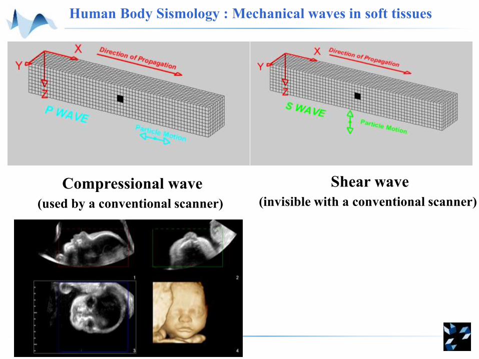
Human Body Sismology : Mechanical waves in soft tissues
Compressional wave
(used by a conventional scanner)
Shear wave
(invisible with a conventional scanner)
Human Body Sismology : Mechanical waves in soft tissues
Compressional wave
(used by a conventional scanner)
Shear wave
(invisible with a conventional scanner)
1500 m.s-1 1-10 m.s-1
3s
Ec
ρ
KcP
E: Young‘s modulus
(elasticity)
K: Compressibility modulus
Compressional wave celerity: Shear wave celerity:
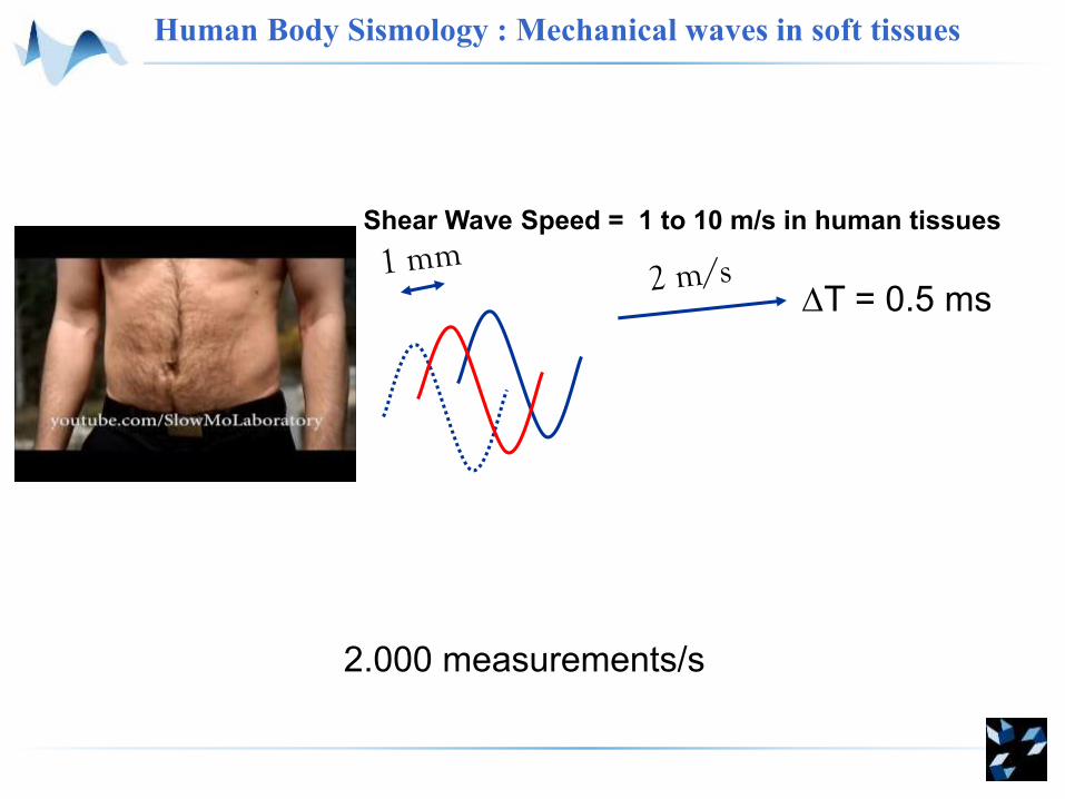
T = 0.5 ms
Shear Wave Speed = 1 to 10 m/s in human tissues
2.000 measurements/s
Human Body Sismology : Mechanical waves in soft tissues
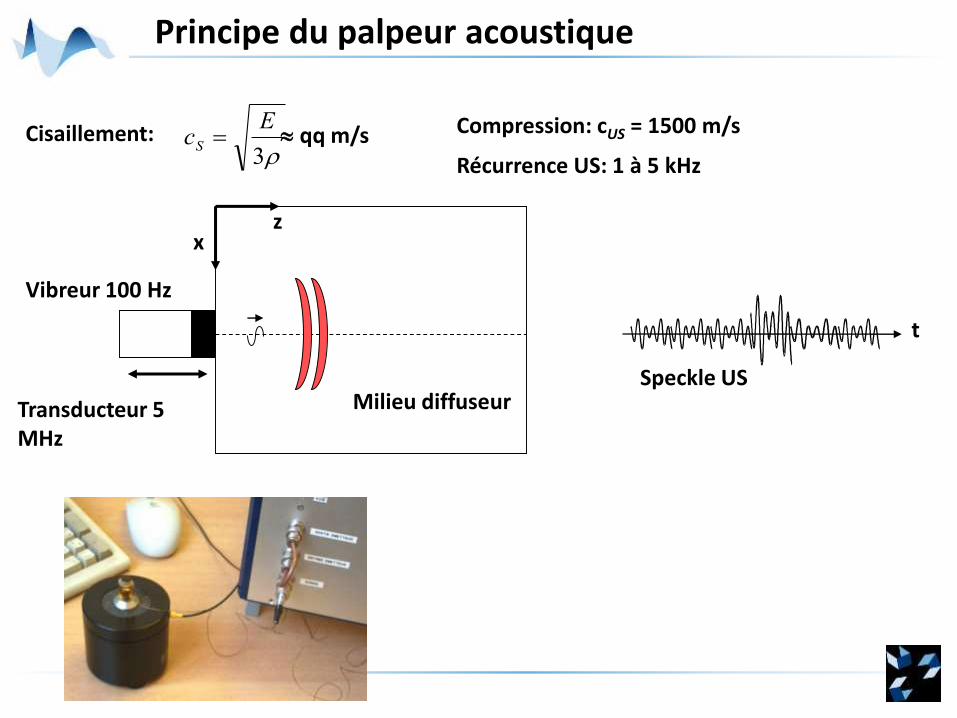
Principe du palpeur acoustique
Milieu diffuseur
xz
Vibreur 100 Hz
Transducteur 5 MHz
Speckle US
t
3
EcS Cisaillement: Compression: cUS = 1500 m/s
Récurrence US: 1 à 5 kHz
qq m/s
Mesure du déplacement axial
Profondeur
layer
A scan at shot n
Recurence time ~ 200 ms1 µm < d < 100 µm
R
d
max
Profondeur
Prof.
Prof.A scan
at shot n+1
Profondeur
Cross-correlation in moving window
Coup basse fréquence : 100 Hz
Mesure de l’élasticité
-20 -10 0 10 20 30 40Retard de phase (ms)
50
40
30
20
10
Pro
fon
de
ur
z (m
m)
cS = 2,84 ± 0,02 m/s
E = 8,07 ± 0,09 kPa
Pro
fon
de
ur
z (m
m)
0 10 20 30 50
10
20
30
40
50
60
70
80
(P)
-50µm
50µm
(S)
Temps (ms)40
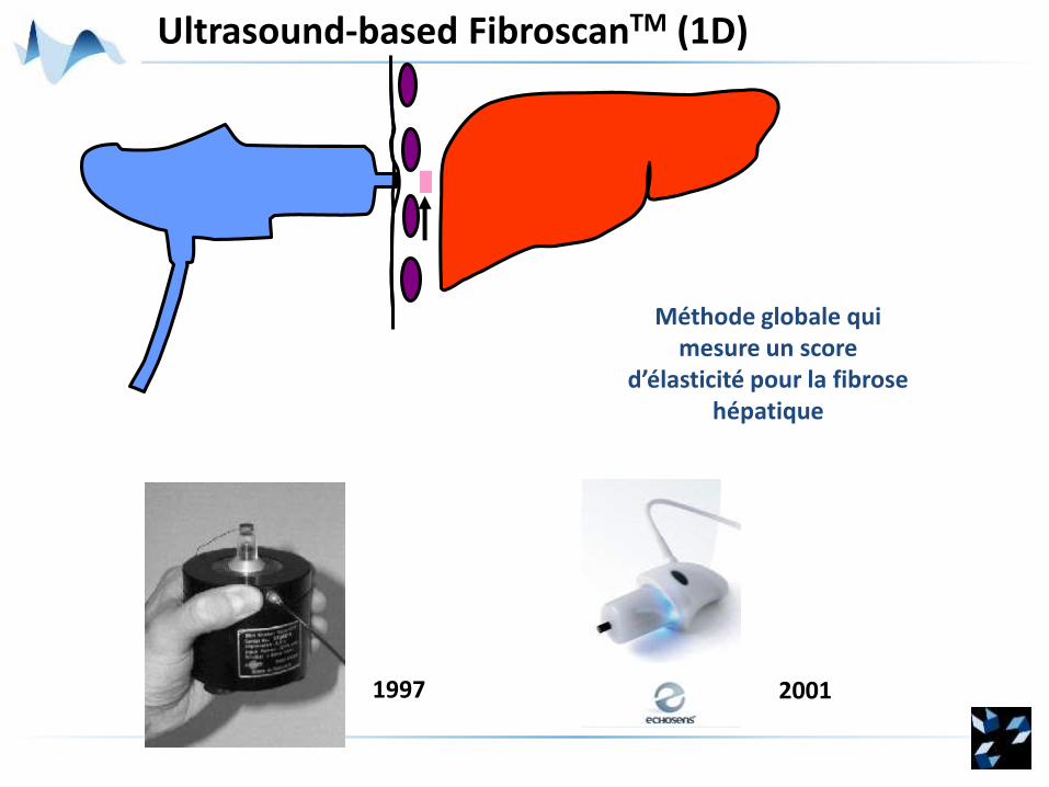
Ultrasound-based FibroscanTM (1D)
Méthode globale qui mesure un score
d’élasticité pour la fibrosehépatique
1997 2001
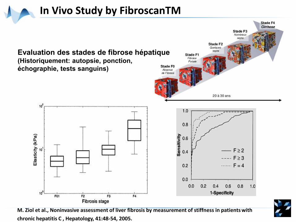
In Vivo Study by FibroscanTM
M. Ziol et al., Noninvasive assessment of liver fibrosis by measurement of stiffness in patients with
chronic hepatitis C , Hepatology, 41:48-54, 2005.
Evaluation des stades de fibrose hépatique(Historiquement: autopsie, ponction,
échographie, tests sanguins)
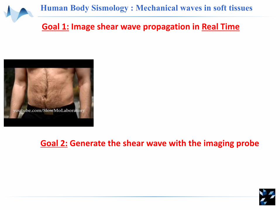
Goal: Image shear wave propagation in Real Time
T = 0.5 ms
2.000 images/s !
Shear Wave Speed = 1 to 10 m/s in human tissues
Conventionnal US imaging50 Hz
US imaging for TE5000 Hz
ULTRAFAST IMAGING
Human Body Sismology : Mechanical waves in soft tissues
D
F
Conventional Imaging Ultrafast Imaging
RAM
Parallel ProcessingProcessing
128 transmits for a full image 1 transmit for a full image
Principles of Ultrafast Ultrasonic Imaging
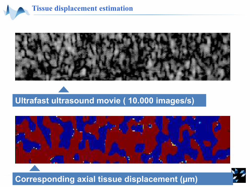
Tissue displacement estimation
Corresponding axial tissue displacement (µm)
Ultrafast ultrasound movie ( 10.000 images/s)
Goal 1: Image shear wave propagation in Real Time
Human Body Sismology : Mechanical waves in soft tissues
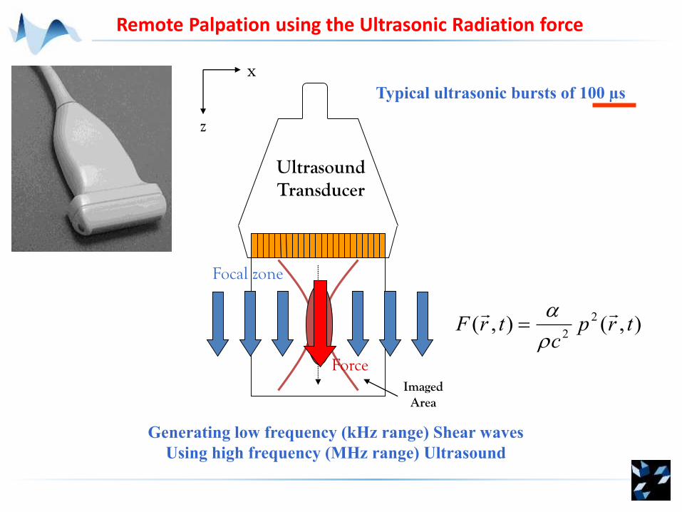
Goal 2: Generate the shear wave with the imaging probe
UltrasoundTransducer
Imaged Area
x
z
Focal zone
Force
),(),(2
2trp
ctrF
Remote Palpation using the Ultrasonic Radiation force
Typical ultrasonic bursts of 100 µs
Generating low frequency (kHz range) Shear waves
Using high frequency (MHz range) Ultrasound
~ 100 µs
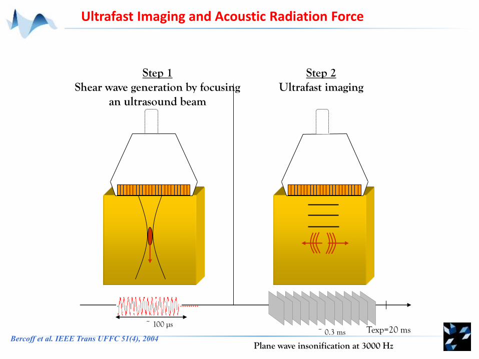
Step 1Shear wave generation by focusing
an ultrasound beam
Plane wave insonification at 3000 Hz
Texp=20 ms~ 0.3 ms
Step 2Ultrafast imaging
Ultrafast Imaging and Acoustic Radiation Force
Bercoff et al. IEEE Trans UFFC 51(4), 2004
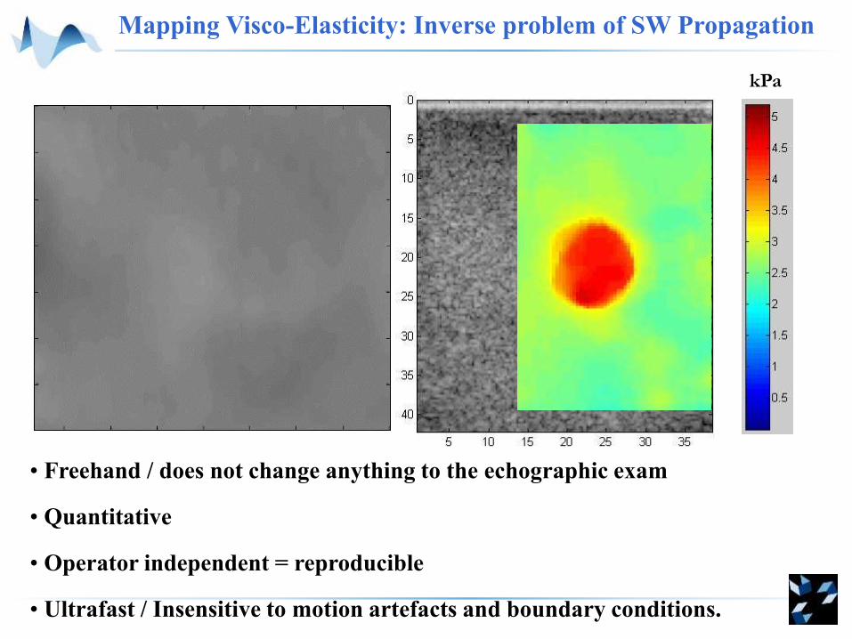
Mapping Visco-Elasticity: Inverse problem of SW Propagation
kPa
• Freehand / does not change anything to the echographic exam
• Quantitative
• Operator independent = reproducible
• Ultrafast / Insensitive to motion artefacts and boundary conditions.
Leveraging this research elastography imaging modality into a product