Guest Editors: Goran N. Kalud ¯ erovic ´, Santiago Gómez-Ruiz, Danijela Maksimovic ´-Ivanic ´, Reinhard Paschke, and Sanja Mijatovic ´ Bioinorganic Chemistry and Applications Metals in Medicine
Transcript
Guest Editors: Goran N. Kalud erovic , Santiago Gómez-Ruiz, Danijela Maksimovic -Ivanic , Reinhard Paschke, and Sanja Mijatovic
Bioinorganic Chemistry and Applications
Metals in Medicine
Metals in Medicine
Bioinorganic Chemistry and Applications
Metals in Medicine
Guest Editors: Goran N. Kaluderovic, Santiago Gomez-Ruiz,Danijela Maksimovic-Ivanic, Reinhard Paschke,and Sanja Mijatovic
This is a special issue published in “Bioinorganic Chemistry and Applications.” All articles are open access articles distributed under theCreative Commons Attribution License, which permits unrestricted use, distribution, and reproduction in any medium, provided theoriginal work is properly cited.
Editorial Board
Triantafillos Albanis, GreecePatrick Bednarski, GermanyIvano Bertini, ItalyViktor Brabec, Czech RepublicIan S. Butler, CanadaLuigi Casella, ItalyZhe-Sheng Chen, USAZheng Dong, USANicholas P. Farrell, USAIgor O. Fritsky, UkraineCl?udio M. Gomes, PortugalNick Hadjiliadis, Greece
Nick Katsaros, GreeceBernhard Klaus Keppler, AustriaAnastasios Keramidas, CyprusDimitris P. Kessissoglou, GreeceConcepci?n L?pez, SpainLuigi Marzilli, USAGuillermo Mendoza-Diaz, MexicoAlbrecht Messerschmidt, GermanyE. R. Milaeva, RussiaVirtudes Moreno, SpainGovindasamy Mugesh, IndiaGiovanni Natile, Italy
Akihiro Nawa, JapanEbbe Nordlander, SwedenLorenzo Pellerito, ItalySpyros P. Perlepes, GreeceClaudio Pettinari, ItalyEnrico Rizzarelli, ItalyTracey A. Rouault, USAImre Sovago, HungaryMarie Stiborova, Czech RepublicKonstantinos Tsipis, GreeceTakeshi Uchiumi, JapanTakao Yagi, USA
Contents
Metals in Medicine, Goran N. Kaluderovic, Santiago Gomez-Ruiz, Danijela Maksimovic-Ivanic,Reinhard Paschke, and Sanja MijatovicVolume 2012, Article ID 705907, 2 pages
On the Discovery, Biological Effects, and Use of Cisplatin and Metallocenes in Anticancer Chemotherapy,Santiago Gomez-Ruiz, Danijela Maksimovic-Ivanic, Sanja Mijatovic, and Goran N. KaluderovicVolume 2012, Article ID 140284, 14 pages
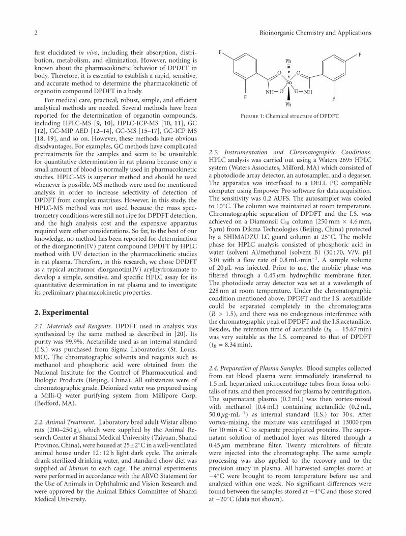
Pharmacokinetic Study of Di-Phenyl-Di-(2,4-Difluobenzohydroxamato)Tin(IV): Novel Metal-BasedComplex with Promising Antitumor Potential, Yunlan Li, Zhuyan Gao, Pu Guo, and Qingshan LiVolume 2012, Article ID 210682, 8 pages
Antifungal and Antioxidant Activities of Pyrrolidone Thiosemicarbazone Complexes,Ahmed A. Al-Amiery, Abdul Amir H. Kadhum, and Abu Bakar MohamadVolume 2012, Article ID 795812, 6 pages
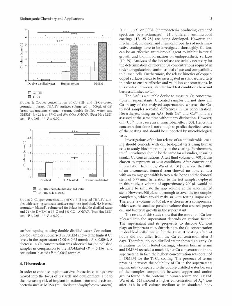
Analysis of the Release Characteristics of Cu-Treated Antimicrobial Implant Surfaces Using AtomicAbsorption Spectrometry, Carmen Zietz, Andreas Fritsche, Birgit Finke, Vitezslav Stranak,Maximilian Haenle, Rainer Hippler, Wolfram Mittelmeier, and Rainer BaderVolume 2012, Article ID 850390, 5 pages
DNA-Platinum Thin Films for Use in Chemoradiation Therapy Studies, Mohammad Rezaee,Elahe Alizadeh, Darel Hunting, and Leon SancheVolume 2012, Article ID 923914, 9 pages
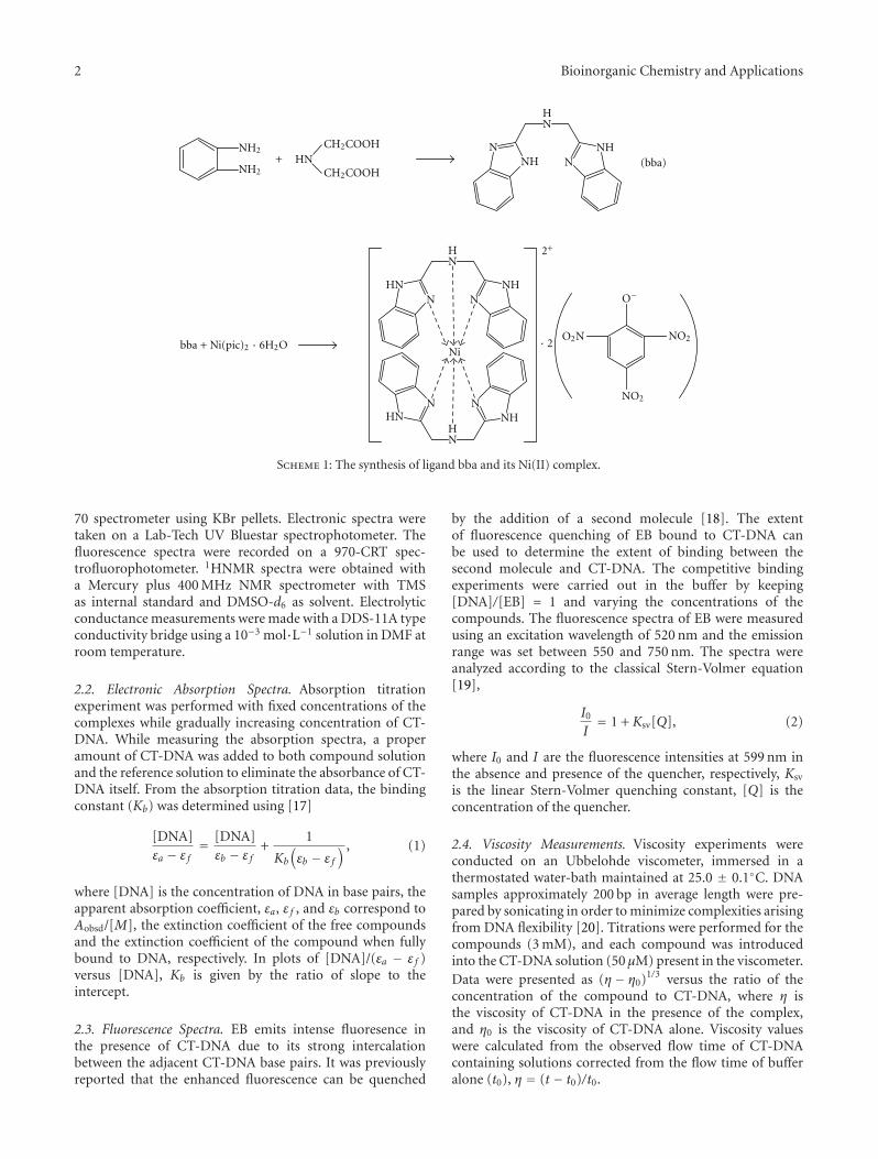
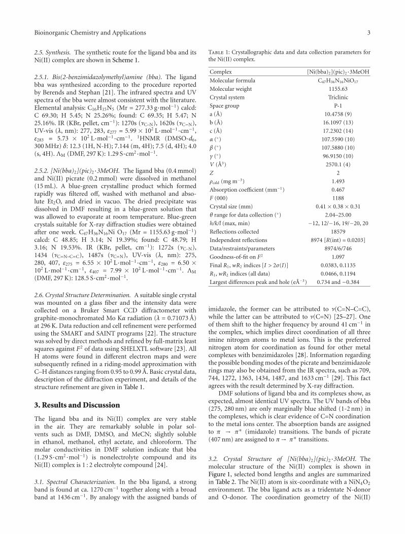
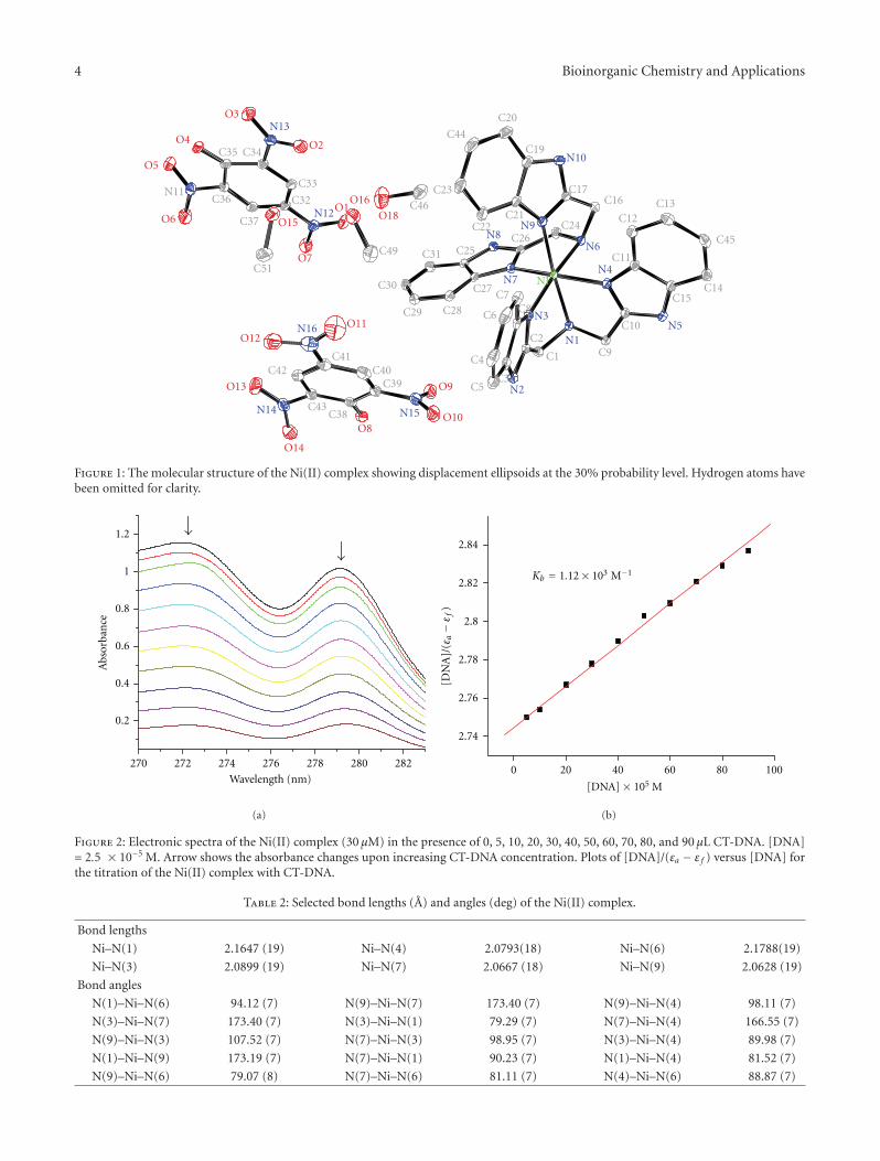
Synthesis, Crystal Structure, and DNA-Binding Studies of a Nickel(II) Complex with theBis(2-benzimidazolymethyl)amine Ligand, Huilu Wu, Tao Sun, Ke Li, Bin Liu, Fan Kou, Fei Jia,Jingkun Yuan, and Ying BaiVolume 2012, Article ID 609796, 7 pages
Hindawi Publishing CorporationBioinorganic Chemistry and ApplicationsVolume 2012, Article ID 705907, 2 pagesdoi:10.1155/2012/705907
Editorial
Metals in Medicine
Goran N. Kalud−erovic,1 Santiago Gomez-Ruiz,2 Danijela Maksimovic-Ivanic,3
Reinhard Paschke,4 and Sanja Mijatovic3
1 Institut fur Chemie, Martin-Luther-Universitat Halle-Wittenberg, Kurt-Mothes-Straße 2, 06120 Halle, Germany2 Departamento de Quımica Inorganicay Analıtica, ESCET, Universidad Rey Juan Carlos, 28933 Mostoles, Spain3 Institute for Biological Research “Sinisa Stankovic,” University of Belgrade, Bulevar despota Stefana 142, 11060 Belgrade, Serbia4 Biozentrum, Martin-Luther-Universitat Halle-Wittenberg, Weinbergweg 22, 06120 Halle, Germany
Correspondence should be addressed to Goran N. Kalud−erovic, [email protected]
Metals in medicine are bridging the areas of inorganic chem-istry and medicine. Metal-based materials, metallodrugs, andagents for treating and detecting diseases, their synthesis,structure, and general properties, as well as biological appli-cations on cellular and living system level, are of great impor-tance. The mechanisms of action and the roles of these metalcompounds in cellular regulation and signaling in health anddiseases are of principal interest. These areas are linked bythe need to involve researchers having a deep understandingof inorganic chemistry in medically relevant research. Thisspecial issue presents a collection of papers dealing with dif-ferent compounds/materials investigated for antitumoral,antimicrobial, and antifungal activity as well as DNA bindingstudy.
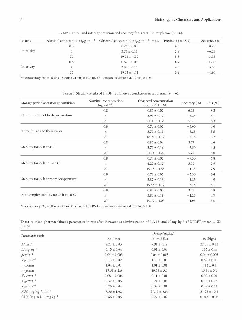
Y. Li et al. reported on the efficient and specific methodfor the determination of diphenyl-di-(2,4-difluobenzohy-droxamato)tin(II), DPDFT, in rat plasma. Their prelimi-nary studies indicated nonlinearity pharmacokinetics in theinvestigated dose ranges in rats and that the concentration-time curves of DPDFT in rat plasma could be fitted totwo-compartment model. Additionally, results hinted thatDPDFT might accumulate in certain organs, thus producingthe toxicity, or could be quickly metabolized in the plasmainto active antitumoral constituents.
The synthesis and characterization of novel salicylalde-hyde-derived ligands and corresponding Cu(II), Co(II),Ni(II), and Zn(II) complexes are described by Kursunlu etal. Ligands bearing chlorine, bromine and –OH substituents
showed moderate inhibition activity against some Gram-positive and Gram-negative bacteria including methicillin-resistant Staphylococcus aureus. Ni(II) and Zn(II) complexeswere generally more effective against tested bacteria thanCu(II) and Co(II) complexes.
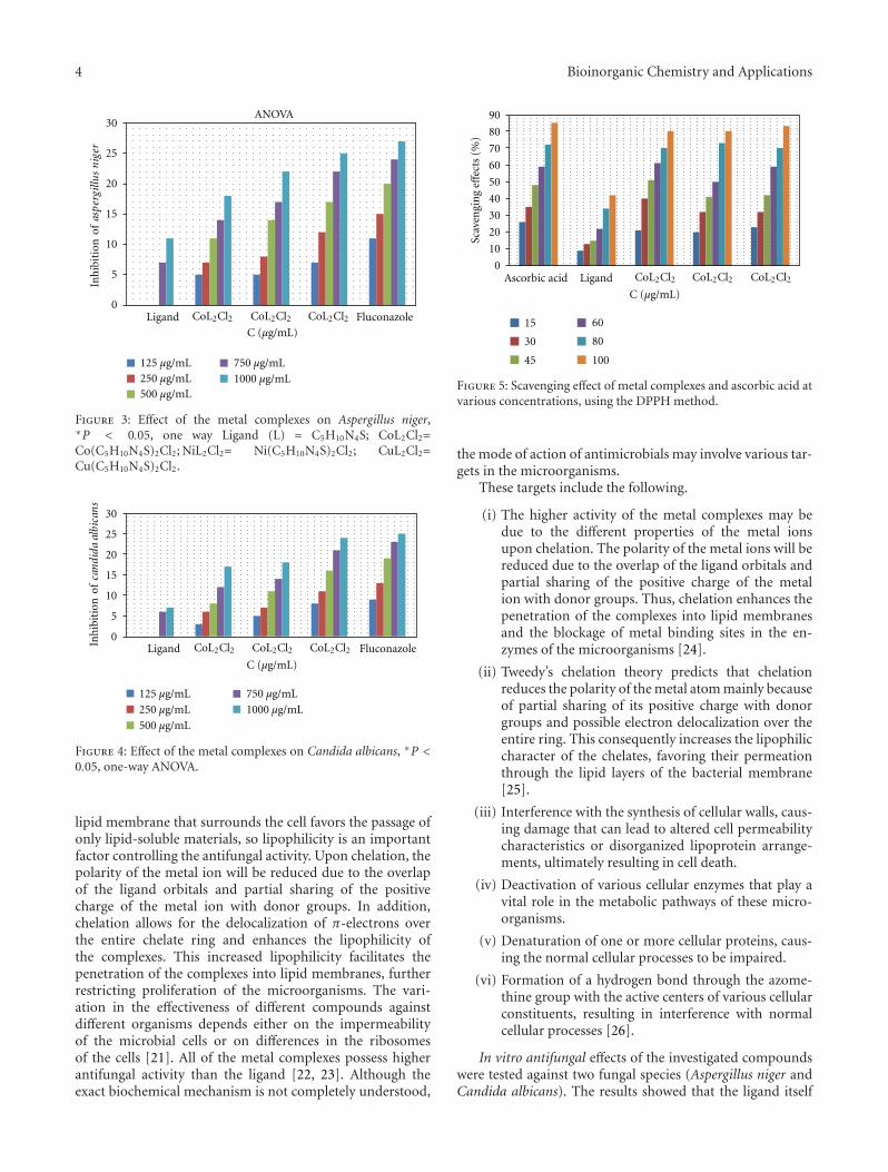
In the work of A. A. Al-Amiery et al., significantantifungal activity of Cu(II), Co(II), and Ni(II) complexeswith (Z)-2-(pyrrolidin-2-ylidene)hydrazinecarbothioamideand chloride ligands is described. The complexes were foundto be superior antioxidants compared to ascorbic acid.
Zietz et al. evaluated Cu release characteristics from Cudoped titanium alloy (Ti6Al4V) of antimicrobial implantsurfaces in vitro according to the storage fluid and surfaceroughness. Plasma immersion ion implantation of Cu (Cu-PIII) and pulsed magnetron sputtering process of a titaniumcopper film (Ti-Cu) were applied to Ti6Al4V sampleswith different surface finishing of the implant material(polished, hydroxyapatite, and corundum blasted). The Cuconcentration in the supernatant was measured using atomicabsorption spectrometry.
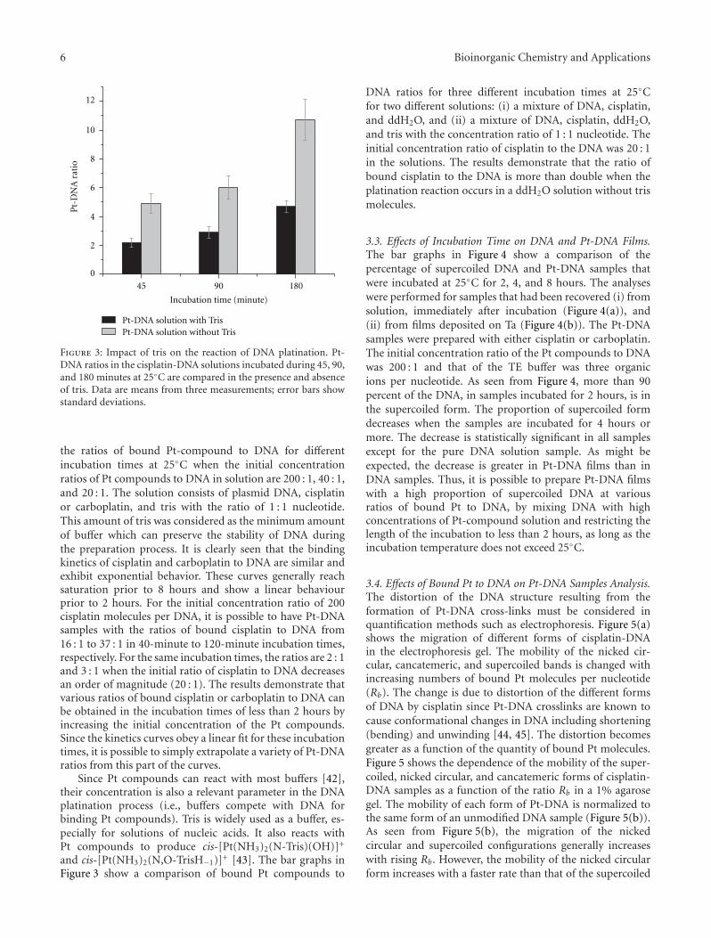
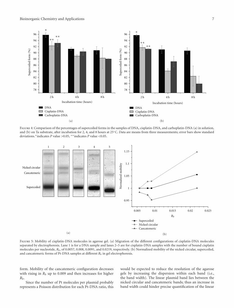
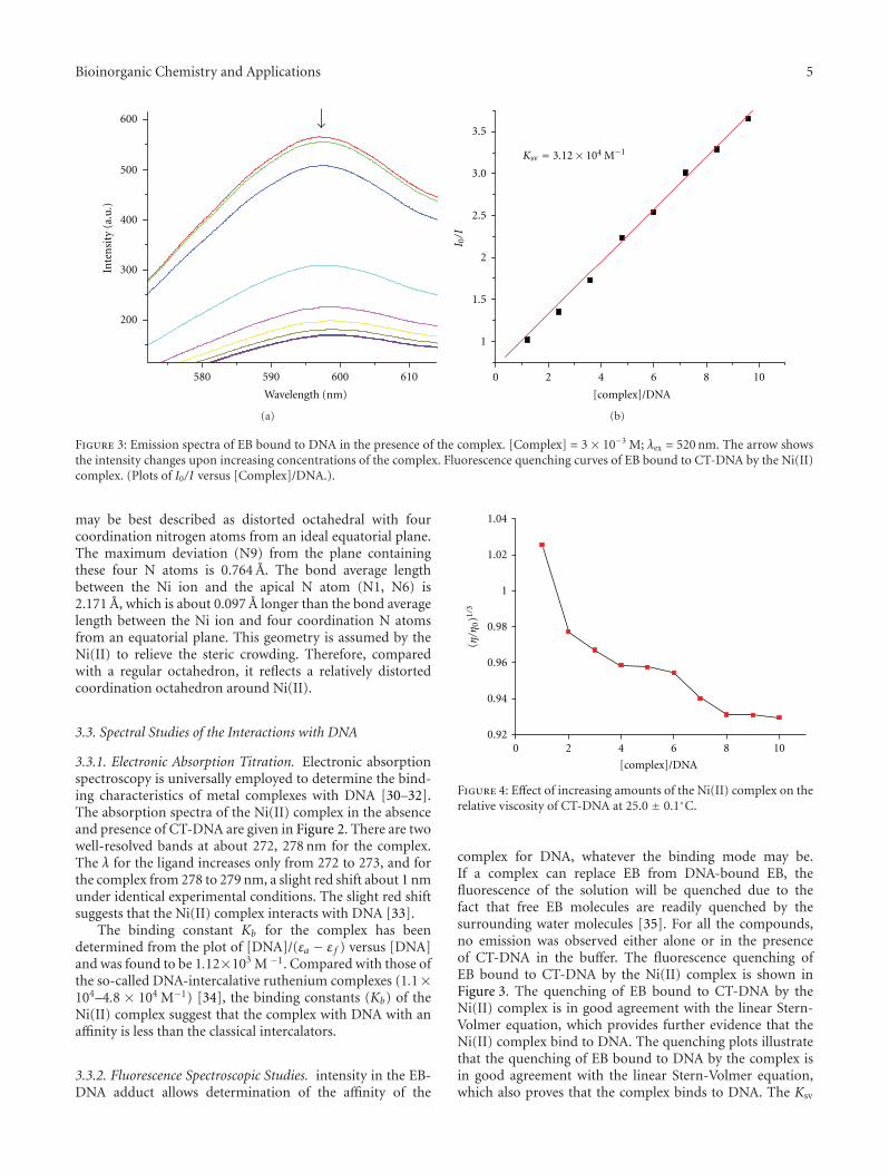
M. Rezaee et al. investigated the optimum experimentalconditions to prepare dry thin films of Pt compounds boundto plasmid DNA on a Ta substrate. Their results show thatused conditions can induce damage to DNA and highlysensitize them to manipulations required to form thin filmsand recover DNA from the Ta substrate. The concentrationof intact DNA increases significantly in the film sampleswhen used lower incubation temperature and shorter incu-bation time. Thus, the optimum condition is obtained from
equilibrium between temperature, time, and Pt-compoundsconcentration during the DNA platination reaction.
In the review by S. Gomez-Ruiz et al., the mode of actionof cisplatin against tumor cells as well as a brief outlook onthe metallocene compounds as antitumor drugs and futuretendencies for the use of the latter in anticancer chemother-apy are summarized. The authors reported on the molecularmechanisms of cisplatin interaction with DNA, DNA repairmechanisms, and cellular proteins. Molecular backgroundof the sensitivity and resistance to cisplatin as well as itsinfluence on the efficacy of the antitumor immune responsewere evaluated. Moreover, the use and mechanism ofsome metallocenes (titanocene, vanadocene, molybdocene,ferrocene and zirconocene) with high antitumor activity arereported.
Acknowledgments
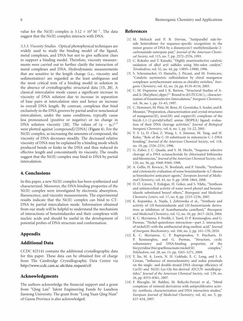
The authors thank the referees who devoted considerabletime and effort for reviewing the papers. Furthermore, theywould like to express their gratitude to Mr. Noran El-Zohearyand other editorial assistants of Bioinorganic Chemistry andApplications for their help in many practical problems andfor great help during the organization of the special issue.
Goran N. Kalud−erovicSantiago Gomez-Ruiz
Danijela Maksimovic-IvanicReinhard Paschke
Sanja Mijatovic
Hindawi Publishing CorporationBioinorganic Chemistry and ApplicationsVolume 2012, Article ID 140284, 14 pagesdoi:10.1155/2012/140284
Review Article
On the Discovery, Biological Effects, andUse of Cisplatin and Metallocenes in Anticancer Chemotherapy
Santiago Gomez-Ruiz,1 Danijela Maksimovic-Ivanic,2
Sanja Mijatovic,2 and Goran N. Kaluderovic3
1 Departamento de Quımica Inorganica y Analıtica, E.S.C.E.T., Universidad Rey Juan Carlos, 28933 Mostoles, Spain2 Institute for Biological Research “Sinisa Stankovic”, University of Belgrade, Boulevard of Despot Stefan 142, 11060 Belgrade, Serbia3 Institut fur Chemie, Martin-Luther-Universitat Halle-Wittenberg, Kurt-Mothes-Straße 2, 06120 Halle, Germany
Correspondence should be addressed to Goran N. Kaluderovic, [email protected]
The purpose of this paper is to summarize mode of action of cisplatin on the tumor cells, a brief outlook on the metallocenecompounds as antitumor drugs as well as the future tendencies for the use of the latter in anticancer chemotherapy. Molecularmechanisms of cisplatin interaction with DNA, DNA repair mechanisms, and cellular proteins are discussed. Molecularbackground of the sensitivity and resistance to cisplatin, as well as its influence on the efficacy of the antitumor immune responsewas evaluated. Furthermore, herein are summarized some metallocenes (titanocene, vanadocene, molybdocene, ferrocene, andzirconocene) with high antitumor activity.
1. Cisplatin
Since 1845, when Italian doctor Peyrone synthesized cis-platin (Figure 1), through Rosenberg’s discovery of cisplatinantiproliferative potential [1], and subsequent approval forclinical usage in 1978, this drug is considered as mostpromising anticancer therapeutic [2, 3]. Cisplatin is highlyeffective against testicular, ovarian, head and neck, bladder,cervical, oesophageal as well as small cell lung cancer [4].
For more than 150 years, first exaltation about this“drug of the 20th century” was replaced with discouragingdata about its toxicity and ineffectiveness got from clinicalpractice. It was found that cisplatin induced serious sideeffects such as nephrotoxicity, neurotoxicity, ototoxicity,nausea, and vomiting [5]. General toxicity and low biologicalavailability restricted its therapeutically application. Inaddition, it is known that some tumors such as colorectaland nonsmall lung cancers are initially resistant to cisplatinwhile other like ovarian and small cell lung cancers easilyacquired resistance to drug [6]. Numerous examples fromin vitro studies confirmed that exposure to cisplatin oftenresulted in development of apoptotic resistant phenotype
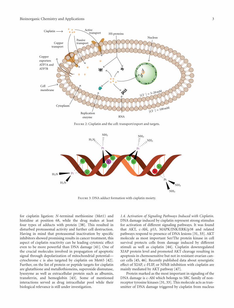
[7–9]. Following this, development of cisplatin resistant celllines is found useful for testing the efficacy of future cisplatinmodified drugs and on the other hand for evaluation ofmechanisms involved in development of resistance. For bet-ter understanding of unresponsiveness to cisplatin, it isnecessary to define the exact molecular targets of drug actionfrom the moment of entering tumor cell. It is proposed theintact cisplatin which avoided bounding to plasma proteinsenter the cell by diffusion or active transport via specificreceptors (Figure 2) [10, 11]. Cisplatin is able to use copper-transporting proteins to reach intracellular compartments[12–14]. In addition, regarding to its chemical reactivity, cis-platin can influence cell physiology even through interactionwith cell membrane molecules such as different receptors.
1.1. Cisplatin and DNA. Although it is known that DNAis a major target for cisplatin, only 5–10% intracellularconcentration of cisplatin is found in DNA fraction while 75–85% binds to nucleophilic sites of intracellular constituentslike thiol containing peptides, proteins, replication enzymes,and RNA [6, 15–17]. This preferential binding to non-DNAtargets offers the explanation for cisplatin resistance but
2 Bioinorganic Chemistry and Applications
Pt
Cl
Cl
H3N
H3N
Figure 1: Cisplatin.
also its high toxicity. Prerequisite of efficient formation ofcisplatin DNA adducts is hydratization of cisplatin enabledby low chloride ions content inside the cells [18]. N7 ofguanine and in less extend adenine nucleotide are targeted byplatinum [19]. Binding of cisplatin to DNA is irreversible andstructurally different adducts are formed. The adducts areclassified as intrastrand crosslinking of two nucleobases ofsingle DNA strand, interstrand crosslinking of two differentstrands of one DNA molecule, chelate formation through N-and O-atoms of one guanine, and DNA-protein crosslinks[20, 21]. Cisplatin forms about 65% pGpG-intrastrandcrosslinks, 25% pApG-intrastrand crosslinks, 13% inter-strand or intrastrand crosslinks on pGpXpG sequences,and less than 1% of monofunctional adducts (Figure 3)[22]. Crucial role of 1,2-intrastrand crosslinks in antitumorpotential of the cisplatin is supported by two facts. First,high mobility group proteins (HMG) specifically recognizethis type of cisplatin-DNA interaction and second, theseadducts are less efficiently removed by repair enzymes [17].In addition, important mediators of cisplatin toxicity areternary DNA-platinum-protein crosslinks (DPCL) whosefrequency is dependent from the cell type as well as thetype of the treatment. DPCLs inhibited DNA polymerizationor their own removal by nucleotide excision repair systemmore potently than other DNA adducts [17]. In fact,cisplatin DNA adducts can be repaired by nucleotide excisionrepair proteins (NER), mismatch repair (MMR), and DNA-dependent protein kinases protein [17].
1.2. DNA Repair Mechanism. Nucleotide excision repair pro-teins are ATP-dependent multiprotein complex able to effi-ciently repair both inter as well as intrastrand DNA-cisplatinadducts. Successful repair of 1,2-d(GpG) and 1,3-d(GpNpG)intrastrand crosslinks has been found in different humanand rodent NER systems [23, 24]. This repair mechanismis able to correct the lesions promoted by chemotherapeuticdrugs, UV radiation as well as oxidative stress [17]. Efficacyof NER proteins varying in different type of tumors and isresponsible for acquirement of cisplatin resistance. Low levelof mentioned proteins is found in testis tumor defining theirhigh sensitivity to cisplatin treatment. Oppositely, ovarian,bladder, prostate, gastric, and cervical cancers are resistant tocisplatin based therapy due to overexpression of several NERgenes [25, 26].
Mismatch repair (MMR) proteins are the post replicationrepair system for correction of mispaired and unpairedbases in DNA caused by DNA Pt adducts. MMR recognizedthe DNA adducts formed by ligation of cisplatin but notoxaliplatin [27–30]. Defective MMR is behind the resistanceof ovarian cancer to cisplatin and responsible for the muta-genicity of cisplatin [31].
DNA dependent protein kinase is a part of eukaryoticDNA double strand repair pathway. This protein is involvedin maintaining of genomic stability as well as in repairof double strand breaks induced by radiation [31]. Inovarian cancer presence of cisplatin DNA adducts inhibitedtranslocation of DNA-PK subunit Ku resulting in inhibitionof this repair protein [32].
Special attention is focused on recognition of cisplatin-modified DNA by HMG proteins (HMG). It is hypothesizedthat HMG proteins protected adducts from recognition andreparation [17, 31]. Moreover, it was postulated that theseproteins modulate cell cycle events and triggered cell deathas a consequence of DNA damage. One of the membersfrom this group marked as HMGB1 is involved in MMR,increased the p53 DNA-binding activity and further stim-ulated binding of different sequence specific transcriptionfactors [33]. Few studies revealed that cisplatin sensitivitywas in correlation with HMGB level, while other studieseliminated its significance in response to cisplatin treatment.Contradictory data about the relevance of HMG proteinsin efficacy of cisplatin therapy indicated that this relation isdefined by cell specificity.
1.3. Cytotoxicity of Cisplatin. Other non-HMG nuclear pro-teins are also involved in cytotoxicity of cisplatin. Presence ofcisplatin DNA adducts is able to significantly change or evendisable the primary function of nuclear proteins essential fortranscription of mammalian genes (TATA binding protein,histon-linker protein H1 or 3-methyladenine DNA glycosy-lase mammalian repair protein) [34–36].
Although cytotoxicity of cisplatin is usually attributed toits reactivity against DNA and subsequent lesions, the factthat more than 80% of internalized drug did not reach DNAindicated the involvement of numerous non-DNA cellulartargets in mediation of cisplatin anticancer action [6]. Asa consequence of exposure to cisplatin, different signalingpathways are affected. There is no general concept applicableto all types of tumor. It is evident that response to cisplatin isdefined by cell specificity. Numerous data revealed changes inactivity of most important signaling pathways involved in cellproliferation, differentiation and cell death such as PI3K/Akt,MAPK as well as signaling pathways involved in realization ofdeath signals dependent or independent of death receptors[33]. It is very important to note that alteration in signaltransduction upon the cisplatin treatment could be theconsequence of both, DNA damage or interaction withexact protein or protein which is relevant for appropriatemolecular response. Some of the interactions between pro-tein and cisplatin are already described. Therefore, it wasfound that cisplatin directly interacts with telomerase, anenzyme that repairs the ends of eukaryotic chromosomes[31, 37]. In parallel, cisplatin-induced damage of telomereswhich are not transcribed and therefore hidden from NER.Other important protein targeted by cisplatin is small,tightly folded molecule known as ubiquitin (Ub) [38]. Ubis implicated in selective degradation of short-lived cellularproteins [39]. It has been hypothesized that direct interactionof cisplatin with this protein presented a strong signal forcell death [40]. Two binding sites were identified as target
Bioinorganic Chemistry and Applications 3
PassivetransportCopper
transport
Cytoplasm
Cell membrane
Nucleus
Copper exporters ATP7A and ATP7B
Active transport HS proteins
Cisplatin
RNAReplication
enzyme
[Cl− ] ≈ 3–20 mM
[Cl− ] ≈ 100 mM
Figure 2: Cisplatin and the cell: transport/export and targets.
Pt
Pt
G
GGG
G
G
G A
Pt
Pt
X
H3N
H3N
H3N
NH3 NH3
NH3
NH3
NH3
Figure 3: DNA adduct formation with cisplatin moiety.
for cisplatin ligation: N-terminal methionine (Met1) andhistidine at position 68, while the drug makes at leastfour types of adducts with protein [38]. This resulted indisturbed proteasomal activity and further cell destruction.Having in mind that proteasomal inactivation by specificinhibitors showed promising results in cancer treatment, thisaspect of cisplatin reactivity can be leading cytotoxic effecteven to be more powerful than DNA damage [41]. One ofthe crucial molecules involved in propagation of apoptoticsignal through depolarization of mitochondrial potential—cytochrome c is also targeted by cisplatin on Met65 [42].Further, on the list of protein or peptide targets for cisplatinare glutathione and metallothioneins, superoxide dismutase,lysozyme as well as extracellular protein such as albumin,transferrin, and hemoglobin [43]. Some of mentionedinteractions served as drug intracellular pool while theirbiological relevance is still under investigation.
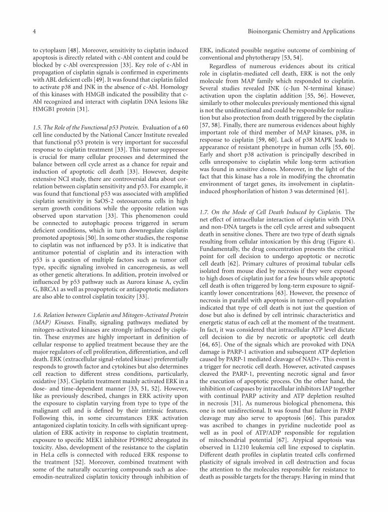
1.4. Activation of Signaling Pathways Induced with Cisplatin.DNA damage induced by cisplatin represent strong stimulusfor activation of different signaling pathways. It was foundthat AKT, c-Abl, p53, MAPK/JNK/ERK/p38 and relatedpathways respond to presence of DNA lesions [31, 33]. AKTmolecule as most important Ser/Thr protein kinase in cellsurvival protects cells from damage induced by differentstimuli as well as cisplatin [44]. Cisplatin downregulatedXIAP protein level and promoted AKT cleavage resulting inapoptosis in chemosensitive but not in resistant ovarian can-cer cells [45, 46]. Recently published data about synergisticeffect of XIAP, c-FLIP, or NFkB inhibition with cisplatin aremainly mediated by AKT pathway [47].
Protein marked as the most important in signaling of theDNA damage is c-Abl which belongs to SRC family of non-receptor tyrosine kinases [31, 33]. This molecule acts as tran-smitter of DNA damage triggered by cisplatin from nucleus
4 Bioinorganic Chemistry and Applications
to cytoplasm [48]. Moreover, sensitivity to cisplatin inducedapoptosis is directly related with c-Abl content and could beblocked by c-Abl overexpression [33]. Key role of c-Abl inpropagation of cisplatin signals is confirmed in experimentswith ABL deficient cells [49]. It was found that cisplatin failedto activate p38 and JNK in the absence of c-Abl. Homologyof this kinases with HMGB indicated the possibility that c-Abl recognized and interact with cisplatin DNA lesions likeHMGB1 protein [31].
1.5. The Role of the Functional p53 Protein. Evaluation of a 60cell line conducted by the National Cancer Institute revealedthat functional p53 protein is very important for successfulresponse to cisplatin treatment [33]. This tumor suppressoris crucial for many cellular processes and determined thebalance between cell cycle arrest as a chance for repair andinduction of apoptotic cell death [33]. However, despiteextensive NCI study, there are controversial data about cor-relation between cisplatin sensitivity and p53. For example, itwas found that functional p53 was associated with amplifiedcisplatin sensitivity in SaOS-2 osteosarcoma cells in highserum growth conditions while the opposite relation wasobserved upon starvation [33]. This phenomenon couldbe connected to autophagic process triggered in serumdeficient conditions, which in turn downregulate cisplatinpromoted apoptosis [50]. In some other studies, the responseto cisplatin was not influenced by p53. It is indicative thatantitumor potential of cisplatin and its interaction withp53 is a question of multiple factors such as tumor celltype, specific signaling involved in cancerogenesis, as wellas other genetic alterations. In addition, protein involved orinfluenced by p53 pathway such as Aurora kinase A, cyclinG, BRCA1 as well as proapoptotic or antiapoptotic mediatorsare also able to control cisplatin toxicity [33].
1.6. Relation between Cisplatin and Mitogen-Activated Protein(MAP) Kinases. Finally, signaling pathways mediated bymitogen-activated kinases are strongly influenced by cispla-tin. These enzymes are highly important in definition ofcellular response to applied treatment because they are themajor regulators of cell proliferation, differentiation, and celldeath. ERK (extracellular signal-related kinase) preferentiallyresponds to growth factor and cytokines but also determinescell reaction to different stress conditions, particularly,oxidative [33]. Cisplatin treatment mainly activated ERK in adose- and time-dependent manner [33, 51, 52]. However,like as previously described, changes in ERK activity uponthe exposure to cisplatin varying from type to type of themalignant cell and is defined by their intrinsic features.Following this, in some circumstances ERK activationantagonized cisplatin toxicity. In cells with significant upreg-ulation of ERK activity in response to cisplatin treatment,exposure to specific MEK1 inhibitor PD98052 abrogated itstoxicity. Also, development of the resistance to the cisplatinin HeLa cells is connected with reduced ERK response tothe treatment [52]. Moreover, combined treatment withsome of the naturally occurring compounds such as aloe-emodin-neutralized cisplatin toxicity through inhibition of
ERK, indicated possible negative outcome of combining ofconventional and phytotherapy [53, 54].
Regardless of numerous evidences about its criticalrole in cisplatin-mediated cell death, ERK is not the onlymolecule from MAP family which responded to cisplatin.Several studies revealed JNK (c-Jun N-terminal kinase)activation upon the cisplatin addition [55, 56]. However,similarly to other molecules previously mentioned this signalis not the unidirectional and could be responsible for realiza-tion but also protection from death triggered by the cisplatin[57, 58]. Finally, there are numerous evidences about highlyimportant role of third member of MAP kinases, p38, inresponse to cisplatin [59, 60]. Lack of p38 MAPK leads toappearance of resistant phenotype in human cells [55, 60].Early and short p38 activation is principally described incells unresponsive to cisplatin while long-term activationwas found in sensitive clones. Moreover, in the light of thefact that this kinase has a role in modifying the chromatinenvironment of target genes, its involvement in cisplatin-induced phosphorilation of histon 3 was determined [61].
1.7. On the Mode of Cell Death Induced by Cisplatin. Thenet effect of intracellular interaction of cisplatin with DNAand non-DNA targets is the cell cycle arrest and subsequentdeath in sensitive clones. There are two type of death signalsresulting from cellular intoxication by this drug (Figure 4).Fundamentally, the drug concentration presents the criticalpoint for cell decision to undergo apoptotic or necroticcell death [62]. Primary cultures of proximal tubular cellsisolated from mouse died by necrosis if they were exposedto high doses of cisplatin just for a few hours while apoptoticcell death is often triggered by long-term exposure to signif-icantly lower concentrations [63]. However, the presence ofnecrosis in parallel with apoptosis in tumor-cell populationindicated that type of cell death is not just the question ofdose but also is defined by cell intrinsic characteristics andenergetic status of each cell at the moment of the treatment.In fact, it was considered that intracellular ATP level dictatecell decision to die by necrotic or apoptotic cell death[64, 65]. One of the signals which are provoked with DNAdamage is PARP-1 activation and subsequent ATP depletioncaused by PARP-1 mediated cleavage of NAD+. This event isa trigger for necrotic cell death. However, activated caspasescleaved the PARP-1, preventing necrotic signal and favorthe execution of apoptotic process. On the other hand, theinhibition of caspases by intracellular inhibitors IAP togetherwith continual PARP activity and ATP depletion resultedin necrosis [31]. As numerous biological phenomena, thisone is not unidirectional. It was found that failure in PARPcleavage may also serve to apoptosis [66]. This paradoxwas ascribed to changes in pyridine nucleotide pool aswell as in pool of ATP/ADP responsible for regulationof mitochondrial potential [67]. Atypical apoptosis wasobserved in L1210 leukemia cell line exposed to cisplatin.Different death profiles in cisplatin treated cells confirmedplasticity of signals involved in cell destruction and focusthe attention to the molecules responsible for resistance todeath as possible targets for the therapy. Having in mind that
Bioinorganic Chemistry and Applications 5
Figure 4: Mode of cell death induced by cisplatin: apoptosis (left) and necrosis (right).
cisplatin is toxic agent against whom the cell can activateautophagy as protective process; the specific inhibition ofautophagy by certain type of molecules could amplify theeffectiveness of cisplatin [50].
1.8. Cisplatin in Immune Senzitization. One of the rarelymentioned but very important aspects of antitumor activityof cisplatin is based on the experimental data about itspotential to amplified the sensitivity of malignant cells toone or the most potent and selective antitumor immuneresponse mediated by TNF-related apoptosis inducing ligandTRAIL [68, 69]. This molecule is produced by almost allimmune cells involved in nonspecific as well as adoptiveimmune response. Unfortunately, in the moment whentumor is diagnosed, its sensitivity to natural immunity isdebatable. In most of the situations, malignant cells becameresistant to TRAIL-mediated cytotoxicity [70]. Moreover,it was confirmed that cisplatin promoted their sensitivityto TRAIL. Nature of its immune sensitizing potential is atleast partly due to upregulation of expression of TRAILreceptors—DR4 and DR5 on the cellular membrane glioma,
colon and prostate cell lines as well as downregulation ofcellular form of caspase 8 inhibitor FLIP [68, 69]. In addition,presence of cysteine rich domen in the structure of TRAILspecific death receptors indicated possibility that cisplatindirectly interact with them.
1.9. Resistance to Cisplatin and How to Surmount It. Resis-tance to cisplatin could be established at multiple levels, fromcellular uptake of the drug through interaction with proteinand DNA and finally activation of signals which lead the cellto death. Disturbed drug uptake, drug scavenging by cellularproteins, upregulation of prosurvival signals together withupregulated expression of antiapoptotic molecules such asBcl-2 and BclXL, overexpressed natural inhibitors of caspaseslike FLIP and XIAP, diminished MAP signaling pathway ordeficiency in proteins involved in signal transferring fromdamaged DNA to cytoplasm, enhanced activity of repairmechanisms and efficient redox system are features mainlyresponsible for unsuccessful treatment with cisplatin [33].Well defined molecular background of the resistance tocisplatin point out the way on how to surmount it. It was
6 Bioinorganic Chemistry and Applications
already known that some of combined treatments of cis-platin with other chemotherapeutics such as 5-fluorouracilimproved therapeutic response rates in patients with headand neck cancer [71, 72]. Furthermore, inhibition of NERDNA repair system, cotreatment with histone deacetylaseinhibitors (HDAC) such as trichostatin A (TSA) or suberoy-lanilide hydeoxamic (SAHA) [73], small molecules inhibitorsof FLIP and XIAP as well as topoisomerase inhibitorsstrongly synergized with cisplatin, elevating its therapeuticpotential.
2. Metallocenes in Anticancer Chemotherapy
Most of the metallodrugs used currently in chemotherapytreatment are based on platinum (cisplatin analogues),although as side effects are the weakest point in the use ofcisplatin-based drugs in chemotherapy are the high numberof side effects, many efforts are focused on the search ofnovel metal complexes with similar antineoplastic activityand less side effects as an alternative for platinum complexes.Transition-metal complexes have shown very useful proper-ties in cancer treatment, and the most important work inchemotherapy with transition metals has been carried outwith Group 4, 5, 6, 8, and 11 metal complexes.
From all the studied metal complexes, a wide variety ofstudies have been carried out for metallocenes which havebecome an alternative to platinum-based drugs.
According to the IUPAC classification metallocenecontains a transition metal and two cyclopentadienyl ligandscoordinated in a sandwich structure. These compounds havecaused a great interest in chemistry due to their versatilitywhich comes from their interesting physical properties,electronic structure, bonding, and their chemical andspectroscopical properties [74]. Academic and industrialresearch on metallocene chemistry has led to the utilizationof these derivatives in many different applications suchas olefin polymerization catalysis, asymmetric catalysis ororganic syntheses, preparation of magnetic materials, use asnonlinear optics or molecular recognizers, flame retardantsor in medicine [74].
Within medicine, metallocene complexes are being nor-mally used as biosensors or as antitumor agents. Regard-ing their anticancer applicability, titanocene, vanadocene,molybdocene, and ferrocene have been traditionally usedwith very good results, however, recently also zirconocenederivatives have pointed towards a future potential applica-bility due to the increase of their cytotoxicity. All the othermetallocene derivatives have been either not tested or havedemonstrated no remarkable applicability in the fight againstcancer.
In this part of the paper, we will briefly discuss separatelythe properties of metallocene derivatives of titanium, zirco-nium, vanadium, molybdenum, and iron.
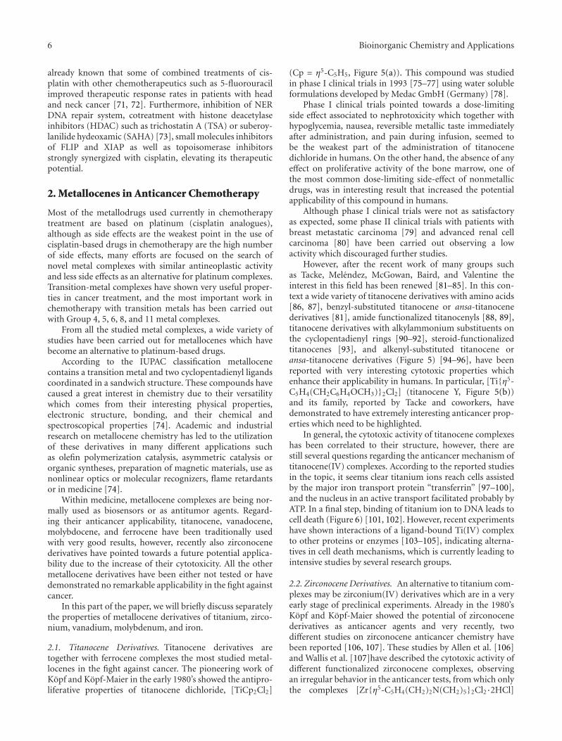
2.1. Titanocene Derivatives. Titanocene derivatives aretogether with ferrocene complexes the most studied metal-locenes in the fight against cancer. The pioneering work ofKopf and Kopf-Maier in the early 1980’s showed the antipro-liferative properties of titanocene dichloride, [TiCp2Cl2]
(Cp = η5-C5H5, Figure 5(a)). This compound was studiedin phase I clinical trials in 1993 [75–77] using water solubleformulations developed by Medac GmbH (Germany) [78].
Phase I clinical trials pointed towards a dose-limitingside effect associated to nephrotoxicity which together withhypoglycemia, nausea, reversible metallic taste immediatelyafter administration, and pain during infusion, seemed tobe the weakest part of the administration of titanocenedichloride in humans. On the other hand, the absence of anyeffect on proliferative activity of the bone marrow, one ofthe most common dose-limiting side-effect of nonmetallicdrugs, was in interesting result that increased the potentialapplicability of this compound in humans.
Although phase I clinical trials were not as satisfactoryas expected, some phase II clinical trials with patients withbreast metastatic carcinoma [79] and advanced renal cellcarcinoma [80] have been carried out observing a lowactivity which discouraged further studies.
However, after the recent work of many groups suchas Tacke, Melendez, McGowan, Baird, and Valentine theinterest in this field has been renewed [81–85]. In this con-text a wide variety of titanocene derivatives with amino acids[86, 87], benzyl-substituted titanocene or ansa-titanocenederivatives [81], amide functionalized titanocenyls [88, 89],titanocene derivatives with alkylammonium substituents onthe cyclopentadienyl rings [90–92], steroid-functionalizedtitanocenes [93], and alkenyl-substituted titanocene oransa-titanocene derivatives (Figure 5) [94–96], have beenreported with very interesting cytotoxic properties whichenhance their applicability in humans. In particular, [Ti{η5-C5H4(CH2C6H4OCH3)}2Cl2] (titanocene Y, Figure 5(b))and its family, reported by Tacke and coworkers, havedemonstrated to have extremely interesting anticancer prop-erties which need to be highlighted.
In general, the cytotoxic activity of titanocene complexeshas been correlated to their structure, however, there arestill several questions regarding the anticancer mechanism oftitanocene(IV) complexes. According to the reported studiesin the topic, it seems clear titanium ions reach cells assistedby the major iron transport protein “transferrin” [97–100],and the nucleus in an active transport facilitated probably byATP. In a final step, binding of titanium ion to DNA leads tocell death (Figure 6) [101, 102]. However, recent experimentshave shown interactions of a ligand-bound Ti(IV) complexto other proteins or enzymes [103–105], indicating alterna-tives in cell death mechanisms, which is currently leading tointensive studies by several research groups.
2.2. Zirconocene Derivatives. An alternative to titanium com-plexes may be zirconium(IV) derivatives which are in a veryearly stage of preclinical experiments. Already in the 1980’sKopf and Kopf-Maier showed the potential of zirconocenederivatives as anticancer agents and very recently, twodifferent studies on zirconocene anticancer chemistry havebeen reported [106, 107]. These studies by Allen et al. [106]and Wallis et al. [107]have described the cytotoxic activity ofdifferent functionalized zirconocene complexes, observingan irregular behavior in the anticancer tests, from which onlythe complexes [Zr{η5-C5H4(CH2)2N(CH2)5}2Cl2·2HCl]
Bioinorganic Chemistry and Applications 7
Ti
Cl
Cl
(a)
OMe
OMe
Ti
Cl
Cl
(b)
Ti
Cl
Cl
(c)
Ti
Cl
O
+
Cl−
OCF3
(d)
Cl−
+
Ti
Cl
Cl
NH
SiMe3
Me3Si
(e)
Ti
Cl
Cl
O
O
H
HH
(f)
Figure 5: Titanocene derivatives used in preclinical and clinical trials: (a) titanocene dichloride; (b) titanocene-Y; (c) alkenyl-substitutedtitanocene derivative; (d) titanocenyl complex; (e) titanocene derivative with alkylammonium substituents; (f) steroid-functionalizedtitanocene derivative.
(Figure 7(a)) and [Zr{η5-C5H4(CH2C6H4OCH3)}2Cl2] (zir-conocene Y, Figure 7(b)) have shown promising activity thatneeds to be improved in order to apply them in anticancerchemotherapy.
In parallel, our research group reported the synthesis,structural characterization, catalytic behavior in the poly-merization of ethylene and copolymerization of ethyleneand 1-octene and the cytotoxic activity on different humancancer cell lines of a novel alkenyl substituted silicon-bridgedansa-zirconocene complex (Figure 7(c)) which proved tobe the most active zirconocene complex on human A2780ovarian cancer cells, reported to date [108].
There is still hard work to do in this field to find a suitablezirconocene complex with increased cytotoxic activity andgood applicability in humans.
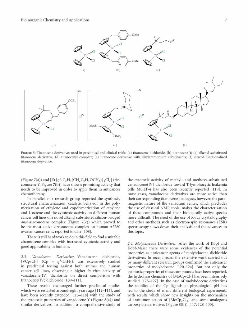
2.3. Vanadocene Derivatives. Vanadocene dichloride,[VCp2Cl2] (Cp = η5-C5H5), was extensively studiedin preclinical testing against both animal and humancancer cell lines, observing a higher in vitro activity ofvanadocene(IV) dichloride on direct comparison withtitanocene(IV) dichloride [109–111].
These results encouraged further preclinical studieswhich were restarted around eight years ago [112–114], andhave been recently extended [115–118] with the study ofthe cytotoxic properties of vanadocene Y (Figure 8(a)) andsimilar derivatives. In addition, a comprehensive study of
the cytotoxic activity of methyl- and methoxy-substitutedvanadocene(IV) dichloride toward T-lymphocytic leukemiacells MOLT-4 has also been recently reported [119]. Inmost cases, vanadocene derivatives are more active thantheir corresponding titanocene analogues, however, the para-magnetic nature of the vanadium center, which precludesthe use of classical NMR tools, makes the characterizationof these compounds and their biologically active speciesmore difficult. The need of the use of X-ray crystallographyand other methods such as electron-spin resonance (ESR)spectroscopy slows down their analysis and the advances inthis topic.
2.4. Molybdocene Derivatives. After the work of Kopf andKopf-Maier there were some evidences of the potentialproperties as anticancer agents of molybdocene dichloridederivatives. In recent years, the extensive work carried outby many different research groups confirmed the anticancerproperties of molybdocene [120–124]. But not only thecytotoxic properties of these compounds have been reported,the hydrolysis chemistry of [MoCp2Cl2] has been intensivelystudied [125–127]. In the case of molybdocene derivativesthe stability of the Cp ligands at physiological pH hasled to the study of many different biological experimentswith results which show new insights on the mechanismof antitumor action of [MoCp2Cl2] and some analogouscarboxylate derivatives (Figure 8(b)) [117, 128–130].
8 Bioinorganic Chemistry and Applications
Ti Ti(IV)Fe(III)
Ti(IV)
Ti(IV)Fe(III)
DNA attack of Ti(IV) ions
Ti(IV)
Ti(IV)Ti(IV)
Ti(IV)Nucleus
Ti(IV)
Ti(IV)
Cl
Cl Transferrin
Transferrin receptors
Extracellular medium
Cellular membrane
Cytoplasm
Nucleus transport,probably facilitated by ATP
Ions liberation
R
R R
H2O
Endosomal pH = 5.5
Figure 6: Proposed mechanism of action of titanocene derivatives (adapted from Abeysinghe and Harding, Dalton Trans. 32 (2007) 3474).
Zr
Cl
Cl
NH
HN
2 Cl
+
−
2
(a)
Zr
Cl
Cl
OMe
OMe
(b)
Zr
Cl
Cl
SiMe
Me
(c)
Figure 7: Zirconocene derivatives with anticancer activity: (a) zirconocene derivative with alkylammonium substituents; (b) zirconocene-Y;(c) alkenyl-substituted ansa-zirconocene complex.
2.5. Ferrocene Derivatives. The discovery of the cytotoxicproperties of ferricinium salts on Ehrlich ascite tumors byKopf and Kopf-Maier [131, 132] were an early breakthroughfor the subsequent development of novel preparations of thisclass of anticancer agents.
There are different groups working in this field, however,to date, the most interesting work in the field of anticancerapplications of ferrocene derivatives is being carried out byJaouen and coworkers.
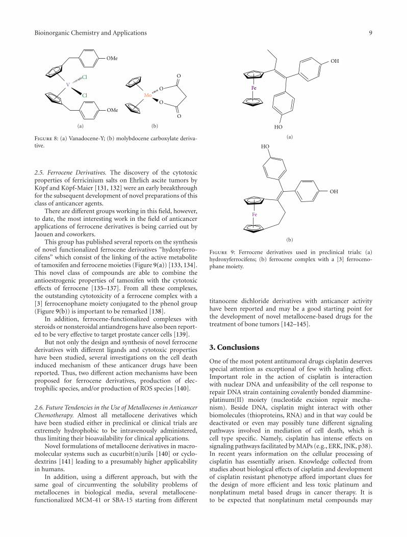
This group has published several reports on the synthesisof novel functionalized ferrocene derivatives “hydoxyferro-cifens” which consist of the linking of the active metaboliteof tamoxifen and ferrocene moieties (Figure 9(a)) [133, 134].This novel class of compounds are able to combine theantioestrogenic properties of tamoxifen with the cytotoxiceffects of ferrocene [135–137]. From all these complexes,the outstanding cytotoxicity of a ferrocene complex with a[3] ferrocenophane moiety conjugated to the phenol group(Figure 9(b)) is important to be remarked [138].
In addition, ferrocene-functionalized complexes withsteroids or nonsteroidal antiandrogens have also been report-ed to be very effective to target prostate cancer cells [139].
But not only the design and synthesis of novel ferrocenederivatives with different ligands and cytotoxic propertieshave been studied, several investigations on the cell deathinduced mechanism of these anticancer drugs have beenreported. Thus, two different action mechanisms have beenproposed for ferrocene derivatives, production of elec-trophilic species, and/or production of ROS species [140].
2.6. Future Tendencies in the Use of Metallocenes in AnticancerChemotherapy. Almost all metallocene derivatives whichhave been studied either in preclinical or clinical trials areextremely hydrophobic to be intravenously administered,thus limiting their bioavailability for clinical applications.
Novel formulations of metallocene derivatives in macro-molecular systems such as cucurbit(n)urils [140] or cyclo-dextrins [141] leading to a presumably higher applicabilityin humans.
In addition, using a different approach, but with thesame goal of circumventing the solubility problems ofmetallocenes in biological media, several metallocene-functionalized MCM-41 or SBA-15 starting from different
Fe
OH
HO
Fe
(a)
Fe
OH
HO
(b)
Figure 9: Ferrocene derivatives used in preclinical trials: (a)hydroxyferrocifens; (b) ferrocene complex with a [3] ferroceno-phane moiety.
titanocene dichloride derivatives with anticancer activityhave been reported and may be a good starting point forthe development of novel metallocene-based drugs for thetreatment of bone tumors [142–145].
3. Conclusions
One of the most potent antitumoral drugs cisplatin deservesspecial attention as exceptional of few with healing effect.Important role in the action of cisplatin is interactionwith nuclear DNA and unfeasibility of the cell response torepair DNA strain containing covalently bonded diammine-platinum(II) moiety (nucleotide excision repair mecha-nism). Beside DNA, cisplatin might interact with otherbiomolecules (thioproteins, RNA) and in that way could bedeactivated or even may possibly tune different signalingpathways involved in mediation of cell death, which iscell type specific. Namely, cisplatin has intense effects onsignaling pathways facilitated by MAPs (e.g., ERK, JNK, p38).In recent years information on the cellular processing ofcisplatin has essentially arisen. Knowledge collected fromstudies about biological effects of cisplatin and developmentof cisplatin resistant phenotype afford important clues forthe design of more efficient and less toxic platinum andnonplatinum metal based drugs in cancer therapy. It isto be expected that nonplatinum metal compounds may
10 Bioinorganic Chemistry and Applications
demonstrate anticancer activity and toxic side effects notice-ably different from that of platinum based drugs. Thustitanocene, vanadocene, molybdocene, ferrocene, and zir-conocene revealed encouraging results in in vitro studies.These compounds might enter by different transport mech-anism through cell membrane and distinctly interact withbiomolecules than cisplatin. Notwithstanding the extensiveapplications of cisplatin in the new investigations willprovide us with powerful facts for finding a novel efficientand nontoxic metallotherapeutics in anticancer treatment.
Acknowledgments
The authors would like to acknowledge financial supportfrom Alexander von Humboldt Foundation (GNK, SGR),from the Ministerio de Educacion y Ciencia, Spain (Grantno. CTQ2011-24346), and from the Ministry of Science andTechnological Development of the Republic of Serbia (Grantno. 173013 DMI, SI).
References
[1] F. Arnesano and G. Natile, “Mechanistic insight into thecellular uptake and processing of cisplatin 30 years after itsapproval by FDA,” Coordination Chemistry Reviews, vol. 253,no. 15-16, pp. 2070–2081, 2009.
[2] D. J. Higby, H. J. Wallace, D. J. Albert, and J. F. Hol-land, “Diaminodichloroplatinum: a phase I study showingresponses in testicular and other tumors,” Cancer, vol. 33, no.5, pp. 1219–1225, 1974.
[3] T. W. Hambley, “Developing new metal-based therapeutics:challenges and opportunities,” Dalton Transactions, vol. 21,no. 43, pp. 4929–4937, 2007.
[4] E. R. Jamieson and S. J. Lippard, “Structure, recognition, andprocessing of cisplatin-DNA adducts,” Chemical Reviews, vol.99, no. 9, pp. 2467–2498, 1999.
[5] G. Giaccone, “Clinical perspectives on platinum resistance,”Drugs, vol. 59, no. 4, pp. 9–17, 2000, discussion 37-38.
[6] M. A. Fuertes, C. Alonso, and J. M. Perez, “Biochemicalmodulation of cisplatin mechanisms of action: enhancementof antitumor activity and circumvention of drug resistance,”Chemical Reviews, vol. 103, no. 3, pp. 645–662, 2003.
[7] B. Koberle, J. R. W. Masters, J. A. Hartley, and R. D. Wood,“Defective repair of cisplatin-induced DNA damage causedby reduced XPA protein in testicular germ cell tumours,”Current Biology, vol. 9, no. 5, pp. 273–276, 1999.
[8] E. L. Mamenta, E. E. Poma, W. K. Kaufmann, D. A. Del-mastro, H. L. Grady, and S. G. Chaney, “Enhanced replica-tive bypass of platinum-DNA adducts in cisplatin-resistanthuman ovarian carcinoma cell lines,” Cancer Research, vol.54, no. 13, pp. 3500–3505, 1994.
[9] A. G. Eliopoulos, D. J. Kerr, J. Herod et al., “The control ofapoptosis and drug resistance in ovarian cancer: influence ofp53 and Bcl-2,” Oncogene, vol. 11, no. 7, pp. 1217–1228, 1995.
[10] A. I. Ivanov, J. Christodoulou, J. A. Parkinson et al., “Cis-platin binding sites on human albumin,” The Journal of Bio-logical Chemistry, vol. 273, no. 24, pp. 14721–14730, 1998.
[11] R. C. DeConti, B. R. Toftness, R. C. Lange, and W. A.Creasey, “Clinical and pharmacological studies with cis dia-mminedichloroplatinum(II),” Cancer Research, vol. 33, no. 6,pp. 1310–1315, 1973.
[12] D. P. Gately and S. B. Howell, “Cellular accumulation of theanticancer agent cisplatin: a review,” British Journal of Cancer,vol. 67, no. 6, pp. 1171–1176, 1993.
[13] S. Ishida, J. Lee, D. J. Thiele, and I. Herskowitz, “Uptake of theanticancer drug cisplatin mediated by the copper transporterCtr1 in yeast and mammals,” Proceedings of the National Aca-demy of Sciences of the United States of America, vol. 99, no.22, pp. 14298–14302, 2002.
[14] R. A. Alderden, M. D. Hall, and T. W. Hambley, “The discov-ery and development of cisplatin,” Journal of Chemical Edu-cation, vol. 83, no. 5, pp. 728–734, 2006.
[15] A. R. Timerbaev, C. G. Hartinger, S. S. Aleksenko, and B.K. Keppler, “Interactions of antitumor metallodrugs withserum proteins: advances in characterization using modernanalytical methodology,” Chemical Reviews, vol. 106, no. 6,pp. 2224–2248, 2006.
[16] E. Volckova, F. Evanics, W. W. Yang, and R. N. Bose, “Un-winding of DNA polymerases by the antitumor drug, cis-diamminedichloroplatinum(II),” Chemical Communications,vol. 9, no. 10, pp. 1128–1129, 2003.
[17] S. Ahmad, “Platinum-DNA interactions and subsequent cel-lular processes controlling sensitivity to anticancer platinumcomplexes,” Chemistry and Biodiversity, vol. 7, no. 3, pp. 543–566, 2010.
[18] R. J. Knox, F. Friedlos, D. A. Lydall, and J. J. Roberts, “Mech-anism of cytotoxicity of anticancer platinum drugs: evidencethat cis-diamminedichloroplatinum(II) and cis-diammine-(1,1-cyclobutanedicarboxylato)platinum(II) differ only inthe kinetics of their interaction with DNA,” Cancer Research,vol. 46, no. 4, pp. 1972–1979, 1986.
[19] L. L. Munchausen and R. O. Rahn, “Physical studies on thebinding of cis dichlorodiamine platinum(II) to DNA andhomopolynucleotides,” Biochimica et Biophysica Acta, vol.414, no. 3, pp. 242–255, 1975.
[20] A. Eastman, “Characterization of the adducts produced inDNA by cis-diamminedichloroplatinum(II) and cis-dichlo-ro(ethylenediamine)platinum(II),” Biochemistry, vol. 22, no.16, pp. 3927–3933, 1983.
[21] A. C. M. Plooy, A. M. J. Fichtinger-Schepman, and H. H.Schutte, “The quantitative detection of various Pt-DNA-adducts in Chinese hamster ovary cells treated with cisplatin:application of immunochemical techniques,” Carcinogenesis,vol. 6, no. 4, pp. 561–566, 1985.
[22] A. E. Egger, C. G. Hartinger, H. B. Hamidane, Y. O. Tsybin,B. K. Keppler, and P. J. Dyson, “High resolution massspectrometry for studying the interactions of cisplatin witholigonucleotides,” Inorganic Chemistry, vol. 47, no. 22, pp.10626–10633, 2008.
[23] D. B. Zamble, D. Mu, J. T. Reardon, A. Sancar, and S. J. Lip-pard, “Repair of cisplatin-DNA adducts by the mammalianexcision nuclease,” Biochemistry, vol. 35, no. 31, pp. 10004–10013, 1996.
[24] J. T. Reardon, A. Vaisman, S. G. Chaney, and A. Sancar, “Effi-cient nucleotide excision repair of cisplatin, oxaliplatin, andbis- acetoammine-dichloro-cyclohexylamine-platinum(IV)(JM216) platinum intrastrand DNA diadducts,” Cancer Re-search, vol. 59, no. 16, pp. 3968–3971, 1999.
[25] S. W. Johnson, R. P. Perez, A. K. Godwin et al., “Role ofplatinum-DNA adduct formation and removal in cisplatinresistance in human ovarian cancer cell lines,” BiochemicalPharmacology, vol. 47, no. 4, pp. 689–697, 1994.
[26] K. V. Ferry, T. C. Hamilton, and S. W. Johnson, “Increasednucleotide excision repair in cisplatin-resistant ovarian
Bioinorganic Chemistry and Applications 11
cancer cells: role of ERCC1-XPF,” Biochemical Pharmacology,vol. 60, no. 9, pp. 1305–1313, 2000.
[27] D. Fink, H. Zheng, S. Nebel et al., “In vitro and in vivoresistance to cisplatin in cells that have lost DNA mismatchrepair,” Cancer Research, vol. 57, no. 10, pp. 1841–1845, 1997.
[28] E. D. Scheeff, J. M. Briggs, and S. B. Howell, “Molecularmodeling of the intrastrand guanine-guanine DNA adductsproduced by cisplatin and oxaliplatin,” Molecular Pharmacol-ogy, vol. 56, no. 3, pp. 633–643, 1999.
[29] A. Vaisman, M. Varchenko, A. Umar et al., “The role ofhMLH1, hMSH3, and hMSH6 defects in cisplatin and oxali-platin resistance: correlation with replicative bypass of plati-num-DNA adducts,” Cancer Research, vol. 58, no. 16, pp.3579–3585, 1998.
[30] S. G. Chaney, S. L. Campbell, E. Bassett, and Y. Wu, “Recog-nition and processing of cisplatin- and oxaliplatin-DNAadducts,” Critical Reviews in Oncology/Hematology, vol. 53,no. 1, pp. 3–11, 2005.
[31] V. Cepeda, M. A. Fuertes, J. Castilla, C. Alonso, C. Quevedo,and J. M. Perez, “Biochemical mechanisms of cisplatincytotoxicity,” Anti-Cancer Agents in Medicinal Chemistry, vol.7, no. 1, pp. 3–18, 2007.
[32] K. M. Henkels and J. J. Turchi, “Induction of apoptosis incisplatin-sensitive and -resistant human ovarian cancer celllines,” Cancer Research, vol. 57, no. 20, pp. 4488–4492, 1997.
[33] D. Wang and S. J. Lippard, “Cellular processing of platinumanticancer drugs,” Nature Reviews Drug Discovery, vol. 4, no.4, pp. 307–320, 2005.
[34] Z. S. Juo, T. K. Chiu, P. M. Leiberman, I. Baikalov, A. J. Berk,and R. E. Dickerson, “How proteins recognize the TATA box,”Journal of Molecular Biology, vol. 261, no. 2, pp. 239–254,1996.
[35] J. Yaneva, S. H. Leuba, K. Van Holde, and J. Zlatanova, “Themajor chromatin protein his tone H1 binds preferentiallyto cis-platinum-damaged DNA,” Proceedings of the NationalAcademy of Sciences of the United States of America, vol. 94,no. 25, pp. 13448–13451, 1997.
[36] M. Kartalou, L. D. Samson, and J. M. Essigmann, “Cisplatinadducts inhibit 1,N6-ethenoadenine repair by interactingwith the human 3-methyladenine DNA glycosylase,” Bio-chemistry, vol. 39, no. 27, pp. 8032–8038, 2000.
[37] C. Soti, A. Racz, and P. Csermely, “A nucleotide-dependentmolecular switch controls ATP binding at the C-terminaldomain of Hsp90. N-terminal nucleotide binding unmasksa C-terminal binding pocket,” The Journal of BiologicalChemistry, vol. 277, no. 9, pp. 7066–7075, 2002.
[38] T. Peleg-Shulman and D. Gibson, “Cisplatin-protein adductsare efficiently removed by glutathione but not by 5′-guanosine monophosphate,” Journal of the American Chemi-cal Society, vol. 123, no. 13, pp. 3171–3172, 2001.
[39] H. Daino, I. Matsumura, K. Takada et al., “Induction ofapoptosis by extracellular ubiquitin in human hematopoieticcells: possible involvement of STAT3 degradation by pro-teasome pathway in interleukin 6-dependent hematopoieticcells,” Blood, vol. 95, no. 8, pp. 2577–2585, 2000.
[40] P. A. Nguewa, M. A. Fuertes, V. Cepeda et al., “Pentamidine isan antiparasitic and apoptotic drug that selectively modifiesubiquitin,” Chemistry and Biodiversity, vol. 2, no. 10, pp.1387–1400, 2005.
[41] D. Maksimovic-Ivanic, S. Mijatovic, D. Miljkovic et al., “Theantitumor properties of a nontoxic, nitric oxide-modifiedversion of saquinavir are independent of Akt,” MolecularCancer Therapeutics, vol. 8, no. 5, pp. 1169–1178, 2009.
[42] A. Casini, C. Gabbiani, G. Mastrobuoni et al., “Insights intothe molecular mechanisms of protein platination from a casestudy: the reaction of anticancer platinum(II) iminoetherswith horse heart cytochrome C,” Biochemistry, vol. 46, no.43, pp. 12220–12230, 2007.
[43] F. Arnesano and G. Natile, ““Platinum on the road”: inter-actions of antitumoral cisplatin with proteins,” Pure and Ap-plied Chemistry, vol. 80, no. 12, pp. 2715–2725, 2008.
[44] S. R. Datta, A. Brunet, and M. E. Greenberg, “Cellular sur-vival: a play in three akts,” Genes and Development, vol. 13,no. 22, pp. 2905–2927, 1999.
[45] M. Fraser, B. M. Leung, X. Yan, H. C. Dan, J. Q. Cheng, andB. K. Tsang, “p53 is a determinant of X-linked inhibitor ofapoptosis protein/Akt-mediated chemoresistance in humanovarian cancer cells,” Cancer Research, vol. 63, no. 21, pp.7081–7088, 2003.
[46] H. C. Dan, M. Sun, S. Kaneko et al., “Akt phosphorylationand stabilization of X-linked inhibitor of apoptosis protein(XIAP),” The Journal of Biological Chemistry, vol. 279, no. 7,pp. 5405–5412, 2004.
[47] J. G. Viniegra, J. H. Losa, V. J. Sanchez-Arevalo et al., “Mod-ulation of PI3K/Akt pathway by E1a mediates sensitivity tocisplatin,” Oncogene, vol. 21, no. 46, pp. 7131–7136, 2002.
[48] Y. Shaul, “c-Abl: activation and nuclear targets,” Cell Deathand Differentiation, vol. 7, no. 1, pp. 10–16, 2000.
[49] J. Gong, A. Costanzo, H. Q. Yang et al., “The tyrosinekinase c-Abl regulates p73 in apoptotic response to cisplatin-induced DNA damage,” Nature, vol. 399, no. 6738, pp. 806–809, 1999.
[50] L. Harhaji-Trajkovic, U. Vilimanovich, T. Kravic-Stevovic, V.Bumbasirevic, and V. Trajkovic, “AMPK-mediated autopha-gy inhibits apoptosis in cisplatin-treated tumour cells,” Jour-nal of Cellular and Molecular Medicine, vol. 13, no. 9, pp.3644–3654, 2009.
[51] X. Wang, J. L. Martindale, and N. J. Holbrook, “Requirementfor ERK activation in cisplatin-induced apoptosis,” The Jour-nal of Biological Chemistry, vol. 275, no. 50, pp. 39435–39443,2000.
[52] W. Cui, E. M. Yazlovitskaya, M. S. Mayo et al., “Cisplatin-induced response of c-jun N-terminal kinase 1 and extracel-lular signal—regulated protein kinases 1 and 2 in a series ofcisplatin-resistant ovarian carcinoma cell lines,” MolecularCarcinogenesis, vol. 29, pp. 219–228, 2000.
[53] S. Mijatovic, D. Maksimovic-Ivanic, J. Radovic et al., “Aloeemodin decreases the ERK-dependent anticancer activity ofcisplatin,” Cellular and Molecular Life Sciences, vol. 62, no. 11,pp. 1275–1282, 2005.
[54] S. Mijatovic, D. Maksimovic-Ivanic, J. Radovic et al., “Anti-glioma action of aloe emodin: the role of ERK inhibition,”Cellular and Molecular Life Sciences, vol. 62, no. 5, pp. 589–598, 2005.
[55] A. Mansouri, L. D. Ridgway, A. L. Korapati et al., “Sustainedactivation of JNK/p38 MAPK pathways in response to cis-platin leads to Fas ligand induction and cell death in ovariancarcinoma cells,” The Journal of Biological Chemistry, vol. 278,no. 21, pp. 19245–19256, 2003.
[56] I. Sanchez-Perez, J. R. Murguıa, and R. Perona, “Cisplatininduces a persistent activation of JNK that is related to celldeath,” Oncogene, vol. 16, no. 4, pp. 533–540, 1998.
[57] J. Hayakawa, M. Ohmichi, H. Kurachi et al., “Inhibition ofextracellular signal-regulated protein kinase or c-Jun N-terminal protein kinase cascade, differentially activated by
12 Bioinorganic Chemistry and Applications
cisplatin, sensitizes human ovarian cancer cell line,” TheJournal of Biological Chemistry, vol. 274, no. 44, pp. 31648–31654, 1999.
[58] R. J. Davis, “Signal transduction by the JNK group of MAPkinases,” Cell, vol. 103, no. 2, pp. 239–252, 2000.
[59] P. Pandey, J. Raingeaud, M. Kaneki et al., “Activation ofp38 mitogen-activated protein kinase by c-Abl-dependentand -independent mechanisms,” The Journal of BiologicalChemistry, vol. 271, no. 39, pp. 23775–23779, 1996.
[60] J. Hernandez Losa, C. Parada Cobo, J. Guinea Viniegra, V. J.Sanchez-Arevalo Lobo, S. Ramon y Cajal, and R. Sanchez-Prieto, “Role of the p38 MAPK pathway in cisplatin-basedtherapy,” Oncogene, vol. 22, no. 26, pp. 3998–4006, 2003.
[61] D. Wang and S. J. Lippard, “Cisplatin-induced post-transla-tional modification of histones H3 and H4,” The Journal ofBiological Chemistry, vol. 279, no. 20, pp. 20622–20625, 2004.
[62] V. M. Gonzalez, M. A. Fuertes, C. Alonso, and J. M. Perez, “Iscisplatin-induced cell death always produced by apoptosis?”Molecular Pharmacology, vol. 59, no. 4, pp. 657–663, 2001.
[63] W. Lieberthal, V. Triaca, and J. Levine, “Mechanisms of deathinduced by cisplatin in proximal tubular epithelial cells: apo-ptosis vs. necrosis,” American Journal of Physiology, vol. 270,no. 4, pp. F700–F708, 1996.
[64] Y. Eguchi, S. Shimizu, and Y. Tsujimoto, “Intracellular ATPlevels determine cell death fate by apoptosis or necrosis,”Cancer Research, vol. 57, no. 10, pp. 1835–1840, 1997.
[65] R. Zhou, M. G. Vander Heiden, and C. M. Rudin, “Genotoxicexposure is associated with alterations in glucose uptake andmetabolism,” Cancer Research, vol. 62, no. 12, pp. 3515–3520,2002.
[66] Z. Herceg and Z. Q. Wang, “Failure of poly(ADP-ribose)polymerase cleavage by caspases leads to induction ofnecrosis and enhanced apoptosis,” Molecular and CellularBiology, vol. 19, no. 7, pp. 5124–5133, 1999.
[67] T. Hirsch, P. Marchetti, S. A. Susin et al., “The apoptosis-necrosis paradox. Apoptogenic proteases activated aftermitochondrial permeability transition determine the modeof cell death,” Oncogene, vol. 15, no. 13, pp. 1573–1581, 1997.
[68] L. Ding, C. Yuan, F. Wei et al., “Cisplatin restoresTRAIL apoptotic pathway in glioblastoma-derived stem cellsthrough up-regulation of DR5 and down-regulation of c-FLIP,” Cancer Investigation, vol. 29, pp. 511–520, 2011.
[69] O. Vondalova Blanarova, I. Jelınkova, A. Szoor et al., “Cispla-tin and a potent platinum(IV) complex-mediated enhance-ment of TRAIL-induced cancer cells killing is associated withmodulation of upstream events in the extrinsic apoptoticpathway,” Carcinogenesis, vol. 32, no. 1, pp. 42–51, 2011.
[70] D. Maksimovic-Ivanic, S. Stosic-Grujicic, F. Nicoletti, and S.Mijatovic, “Resistance to TRAIl and how to surmount it,”Immunology Research, vol. 52, no. 1-2, pp. 157–168, 2012.
[71] M. P. Decatris, S. Sundar, and K. J. O’Byrne, “Platinum-basedchemotherapy in metastatic breast cancer: current status,”Cancer Treatment Reviews, vol. 30, no. 1, pp. 53–81, 2004.
[72] M. D. Shelley, K. Burgon, and M. D. Mason, “Treatmentof testicular germ-cell cancer: a cochrane evidence-basedsystematic review,” Cancer Treatment Reviews, vol. 28, no. 5,pp. 237–253, 2002.
[73] M. S. Kim, M. Blake, J. H. Baek, G. Kohlhagen, Y. Pommier,and F. Carrier, “Inhibition of histone deacetylase increasescytotoxicity to anticancer drugs targeting DNA,” Cancer Re-search, vol. 63, no. 21, pp. 7291–7300, 2003.
[74] N. J. Long, Metallocenes, Blackwell Science, Oxford, UK,1998.
[75] A. Korfel, M. E. Scheulen, H. J. Schmoll et al., “Phase I clinicaland pharmacokinetic study of titanocene dichloride in adultswith advanced solid tumors,” Clinical Cancer Research, vol. 4,no. 11, pp. 2701–2708, 1998.
[76] C. V. Christodoulou, D. R. Ferry, D. W. Fyfe et al., “PhaseI trial of weekly scheduling and pharmacokinetics of titano-cene dichloride in patients with advanced cancer,” Journal ofClinical Oncology, vol. 16, no. 8, pp. 2761–2769, 1998.
[77] K. Mross, P. Robben-Bathe, L. Edler et al., “Phase I clinicaltrial of a day-1, -3, -5 every 3 weeks schedule with titanocenedichloride (MKT 5) in patients with advanced cancer: a studyof the phase I study group of the association for medicaloncology (AIO) of the German Cancer Society,” Onkologie,vol. 23, no. 6, pp. 576–579, 2000.
[78] B. W. Muller, R. Muller, S. Lucks, and W. Mohr, “MedacGesellschaft fur Klinische Spzeilpraparate GmbH,” US Patent5, 296, 237, 1994.
[79] N. Kroger, U. R. Kleeberg, K. Mross, L. Edler, G. Saß, and D.K. Hossfeld, “Phase II clinical trial of titanocene dichloridein patients with metastatic breast cancer,” Onkologie, vol. 23,no. 1, pp. 60–62, 2000.
[80] G. Lummen, H. Sperling, H. Luboldt, T. Otto, and H.Rubben, “Phase II trial of titanocene dichloride in advancedrenal-cell carcinoma,” Cancer Chemotherapy and Pharmacol-ogy, vol. 42, no. 5, pp. 415–417, 1998.
[81] E. Melendez, “Titanium complexes in cancer treatment,”Critical Reviews in Oncology/Hematology, vol. 42, no. 3, pp.309–315, 2002.
[82] F. Caruso and M. Rossi, “Antitumor titanium compoundsand related metallocenes,” in Metal Ions in Biological System,A. Sigel and H. Sigel, Eds., vol. 42 of Metal Complexes inTumor Diagnostics and as Anticancer Agents, Marcel Dekker,New York, NY, USA, 2004.
[83] J. C. Dabrowiak, Metals in Medicine, John Wiley & Sons, WestSussex, UK, 2009.
[84] U. Olszewski and G. Hamilton, “Mechanisms of cytotoxicityof anticancer titanocenes,” Anti-Cancer Agents in MedicinalChemistry, vol. 10, no. 4, pp. 302–311, 2010.
[85] K. Strohfeldt and M. Tacke, “Bioorganometallic fulvene-derived titanocene anti-cancer drugs,” Chemical SocietyReviews, vol. 37, no. 6, pp. 1174–1187, 2008.
[86] R. Hernandez, J. Lamboy, L. M. Gao, J. Matta, F. R. Roman,and E. Melendez, “Structure-activity studies of Ti(IV) com-plexes: aqueous stability and cytotoxic properties in coloncancer HT-29 cells,” Journal of Biological Inorganic Chemistry,vol. 13, no. 5, pp. 685–692, 2008.
[87] R. Hernandez, J. Mendez, J. Lamboy, M. Torres, F. R. Roman,and E. Melendez, “Titanium(IV) complexes: cytotoxicity andcellular uptake of titanium(IV) complexes on caco-2 cellline,” Toxicology in Vitro, vol. 24, no. 1, pp. 178–183, 2010.
[88] L. M. Gao, J. Matta, A. L. Rheingold, and E. Melendez, “Syn-thesis, structure and biological activity of amide-functiona-lized titanocenyls: improving their cytotoxic properties,”Journal of Organometallic Chemistry, vol. 694, no. 26, pp.4134–4139, 2009.
[89] A. Gansauer, I. Winkler, D. Worgull et al., “Carbonyl-sub-stituted titanocenes: a novel class of cytostatic compoundswith high antitumor and antileukemic activity,” Chemistry,vol. 14, no. 14, pp. 4160–4163, 2008.
[90] O. R. Allen, L. Croll, A. L. Gott, R. J. Knox, and P.C. McGowan, “Functionalized cyclopentadienyl titaniumorganometallic compounds as new antitumor drugs,” Orga-nometallics, vol. 23, no. 2, pp. 288–292, 2004.
Bioinorganic Chemistry and Applications 13
[91] O. R. Allen, A. L. Gott, J. A. Hartley, J. M. Hartley, R. J.Knox, and P. C. McGowan, “Functionalised cyclopentadienyltitanium compounds as potential anticancer drugs,” DaltonTransactions, no. 43, pp. 5082–5090, 2007.
[92] G. D. Potter, M. C. Baird, and S. P. C. Cole, “A new seriesof titanocene dichloride derivatives bearing chiral alkylam-monium groups; Assessment of their cytotoxic properties,”Inorganica Chimica Acta, vol. 364, no. 1, pp. 16–22, 2010.
[93] L. M. Gao, J. L. Vera, J. Matta, and E. Melendez, “Synthesisand cytotoxicity studies of steroid-functionalized titanocenesas potential anticancer drugs: sex steroids as potential vectorsfor titanocenes,” Journal of Biological Inorganic Chemistry,vol. 15, no. 6, pp. 851–859, 2010.
[94] S. Gomez-Ruiz, G. N. Kaluderovic, S. Prashar et al., “Cyto-toxic studies of substituted titanocene and ansa-titanoceneanticancer drugs,” Journal of Inorganic Biochemistry, vol. 102,no. 8, pp. 1558–1570, 2008.
[95] S. Gomez-Ruiz, G. N. Kaluderovic, Z. Zizak et al., “Anticancerdrugs based on alkenyl and boryl substituted titanocenecomplexes,” Journal of Organometallic Chemistry, vol. 694,no. 13, pp. 1981–1987, 2009.
[96] G. N. Kaluderovic, V. Tayurskaya, R. Paschke, S. Prashar,M. Fajardo, and S. Gomez-Ruiz, “Synthesis, characterizationand biological studies of alkenyl-substituted titanocene(IV)carboxylate complexes,” Applied Organometallic Chemistry,vol. 24, no. 9, pp. 656–662, 2010.
[97] H. Sun, H. Li, R. A. Weir, and P. J. Sadler, “You have full textaccess to this content the first specific TiIV -protein complex:potential relevance to anticancer activity of titanocenes,”Angewandte Chemie International Edition, vol. 37, no. 11, pp.1577–1579, 1998.
[98] M. Guo and P. J. Sadler, “Competitive binding of the anti-cancer drug titanocene dichloride to N,N′-ethylenebis(o-hydroxyphenylglycine) and adenosine triphosphate: a modelfor TiIV uptake and release by transferrin,” Journal of theChemical Society, Dalton Transactions, vol. 1, pp. 7–9, 2000.
[99] M. Guo, H. Sun, S. Bihari et al., “Stereoselective formationof seven-coordinate titanium(IV) monomer and dimer com-plexes of ethylenebis(o-hydroxyphenyl)glycine,” InorganicChemistry, vol. 39, no. 2, pp. 206–215, 2000.
[100] M. Guo, H. Sun, H. J. McArdle, L. Gambling, and P. J. Sadler,“Ti(IV) uptake and release by human serum transferrinand recognition of Ti(IV)-transferrin by cancer cells: under-standing the mechanism of action of the anticancer drugtitanocene dichloride,” Biochemistry, vol. 39, no. 33, pp.10023–10033, 2000.
[101] P. Kopf-Maier and D. Krahl, “Tumor inhibition by metallo-genes: ultrastructural localization of titanium and vanadiumin treated tumor cells by electron energy loss spectroscopy,”Chemico-Biological Interactions, vol. 44, no. 3, pp. 317–328,1983.
[102] P. Kopf-Maier, “Intracellular localization of titanium withinxenografted sensitive human tumors after treatment with theantitumor agent titanocene dichloride,” Journal of StructuralBiology, vol. 105, no. 1–3, pp. 35–45, 1990.
[103] A. D. Tinoco, C. D. Incarvito, and A. M. Valentine, “Calori-metric, spectroscopic, and model studies provide insight intothe transport of Ti(IV) by human serum transferrin,” Journalof the American Chemical Society, vol. 129, no. 11, pp. 3444–3454, 2007.
[104] A. D. Tinoco, E. V. Eames, and A. M. Valentine, “Reconsid-eration of serum Ti(IV) transport: albumin and transferrintrafficking of Ti(IV) and its complexes,” Journal of the Amer-ican Chemical Society, vol. 130, no. 7, pp. 2262–2270, 2008.
[105] M. Pavlaki, K. Debeli, I. E. Triantaphyllidou, N. Klouras, E.Giannopoulou, and A. J. Aletras, “A proposed mechanismfor the inhibitory effect of the anticancer agent titanocenedichloride on tumour gelatinases and other proteolytic enzy-mes,” Journal of Biological Inorganic Chemistry, vol. 14, no. 6,pp. 947–957, 2009.
[106] O. R. Allen, R. J. Knox, and P. C. McGowan, “Functionalisedcyclopentadienyl zirconium compounds as potential anti-cancer drugs,” Dalton Transactions, no. 39, pp. 5293–5295,2008.
[107] D. Wallis, J. Claffey, B. Gleeson, M. Hogan, H. Muller-Bunz,and M. Tacke, “Novel zirconocene anticancer drugs?” Journalof Organometallic Chemistry, vol. 694, no. 6, pp. 828–833,2009.
[108] S. Gomez-Ruiz, G. N. Kaluderovic, D. Polo-Ceron et al.,“A novel alkenyl-substituted ansa-zirconocene complex withdual application as olefin polymerization catalyst and anti-cancer drug,” Journal of Organometallic Chemistry, vol. 694,no. 18, pp. 3032–3038, 2009.
[109] P. Kopf-Maier, “Antitumor bis(cyclopentadienyl) metal com-plexes,” in Metal Complexes in Cancer Chemotherapy, B.K. Keppler, Ed., pp. 259–296, VCH Verlagsgesellschaft,Weinheim, Germany, 1993.
[110] C. S. Navara, A. Benyumov, A. Vassilev, R. K. Narla, P. Ghosh,and F. M. Uckun, “Vanadocenes as potent anti-proliferativeagents disrupting mitotic spindle formation in cancer cells,”Anti-Cancer Drugs, vol. 12, no. 4, pp. 369–376, 2001.
[111] P. Ghosh, O. J. D’Cruz, R. K. Narla, and F. M. Uckun,“Apoptosis-inducing vanadocene compounds against humantesticular cancer,” Clinical Cancer Research, vol. 6, no. 4, pp.1536–1545, 2000.
[112] H. Palackova, J. Vinklarek, J. Holubova, I. Cısarova, andM. Erben, “The interaction of antitumor active vanadocenedichloride with sulfur-containing amino acids,” Journal ofOrganometallic Chemistry, vol. 692, no. 17, pp. 3758–3764,2007.
[113] J. Vinklarek, J. Honzıcek, and J. Holubova, “Interaction ofthe antitumor agent vanadocene dichloride with phosphatebuffered saline,” Inorganica Chimica Acta, vol. 357, no. 12,pp. 3765–3769, 2004.
[114] J. Vinklarek, H. Palackova, J. Honzıcek, J. Holubova, M.Holcapek, and I. Cısarova, “Investigation of vanadocene(IV)α-amino acid complexes: synthesis, structure, and behaviorin physiological solutions, human plasma, and blood,”Inorganic Chemistry, vol. 45, no. 5, pp. 2156–2162, 2006.
[115] B. Gleeson, J. Claffey, A. Deally et al., “Synthesis andcytotoxicity studies of fluorinated derivatives of vanadoceneY,” European Journal of Inorganic Chemistry, no. 19, pp. 2804–2810, 2009.
[116] B. Gleeson, J. Claffey, M. Hogan, H. Muller-Bunz, D. Wallis,and M. Tacke, “Novel benzyl-substituted vanadocene anti-cancer drugs,” Journal of Organometallic Chemistry, vol. 694,no. 9-10, pp. 1369–1374, 2009.
[117] B. Gleeson, J. Claffey, A. Deally et al., “Novel benzyl-substituted molybdocene anticancer drugs,” InorganicaChimica Acta, vol. 363, no. 8, pp. 1831–1836, 2010.
[118] B. Gleeson, M. Hogan, H. Muller-Bunz, and M. Tacke,“Synthesis and preliminary cytotoxicity studies of indole-substituted vanadocenes,” Transition Metal Chemistry, vol.35, no. 8, pp. 973–983, 2010.
[119] J. Honzıcek, I. Klepalova, J. Vinklarek et al., “Synthesis, char-acterization and cytotoxic effect of ring-substituted and ansa-bridged vanadocene complexes,” Inorganica Chimica Acta,vol. 373, no. 1, pp. 1–7, 2011.
14 Bioinorganic Chemistry and Applications
[120] J. B. Waern and M. M. Harding, “Bioorganometallic chem-istry of molybdocene dichloride,” Journal of OrganometallicChemistry, vol. 689, no. 25, pp. 4655–4668, 2004.
[121] J. B. Waern, C. T. Dillon, and M. M. Harding, “Organometal-lic anticancer agents: cellular uptake and cytotoxicity studieson thiol derivatives of the antitumor agent molybdocenedichloride,” Journal of Medicinal Chemistry, vol. 48, no. 6, pp.2093–2099, 2005.
[122] J. B. Waern, H. H. Harris, B. Lai, Z. Cai, M. M. Harding,and C. T. Dillon, “Intracellular mapping of the distributionof metals derived from the antitumor metallocenes,” Journalof Biological Inorganic Chemistry, vol. 10, no. 5, pp. 443–452,2005.
[123] J. B. Waern, P. Turner, and M. M. Harding, “Synthe-sis and hydrolysis of thiol derivatives of molybdoce-ne dichloride incorporating electron-withdrawing substitu-ents,” Organometallics, vol. 25, no. 14, pp. 3417–3421, 2006.
[124] K. S. Campbell, A. J. Foster, C. T. Dillon, and M. M. Harding,“Genotoxicity and transmission electron microscopy studiesof molybdocene dichloride,” Journal of Inorganic Biochem-istry, vol. 100, no. 7, pp. 1194–1198, 2006.
[125] J. H. Toney and T. J. Marks, “Hydrolysis chemistry ofthe metallocene dichlorides M(η5-C5H5)2Cl2, M=Ti, V, Zr.Aqueous kinetics, equilibria, and mechanistic implicationsfor a new class of antitumor agents,” Journal of the AmericanChemical Society, vol. 107, no. 4, pp. 947–953, 1985.
[126] C. Balzarek, T. J. R. Weakley, L. Y. Kuo, and D. R. Tyler,“Investigation of the monomer-dimer equilibria of molyb-docenes in water,” Organometallics, vol. 19, no. 15, pp. 2927–2931, 2000.
[127] L. Y. Kuo, M. G. Kanatzidis, M. Sabat, A. L. Tipton, andT. J. Marks, “Metallocene antitumor agents. Solution andsolid-state molybdenocene coordination chemistry of DNAconstituents,” Journal of the American Chemical Society, vol.113, no. 24, pp. 9027–9045, 1991.
[128] P. M. Abeysinghe and M. M. Harding, “Antitumour bis(cyclo-pentadienyl) metal complexes: titanocene and molybdocenedichloride and derivatives,” Dalton Transactions, no. 32, pp.3474–3482, 2007.
[129] M. M. Harding and G. Mokdsi, “Antitumour metallocenes:structure-activity studies and interactions with biomolecu-les,” Current Medicinal Chemistry, vol. 7, no. 12, pp. 1289–1303, 2000.
[130] K. S. Campbell, C. T. Dillon, S. V. Smith, and M. M. Harding,“Radiotracer studies of the antitumor metallocene molyb-docene dichloride with biomolecules,” Polyhedron, vol. 26,no. 2, pp. 456–459, 2007.
[131] P. Kopf-Maier, H. Kopf, and E. W. Neuse, “Ferroceniumsalts—the first antineoplastic iron compounds,” AngewandteChemie—International Edition in English, vol. 23, no. 6, pp.456–457, 1984.
[132] P. Kopf-Maier, H. Kopf, and E. W. Neuse, “Ferricenium com-plexes: a new type of water-soluble antitumor agent,” Journalof Cancer Research and Clinical Oncology, vol. 108, no. 3, pp.336–340, 1984.
[133] S. Top, J. Tang, A. Vessieres, D. Carrez, C. Provot, and G.Jaouen, “Ferrocenyl hydroxytamoxifen: a prototype for a newrange of oestradiol receptor site-directed cytotoxics,” Chemi-cal Communications, vol. 8, pp. 955–956, 1996.
[134] S. Top, B. Dauer, J. Vaissermann, and G. Jaouen, “Facile routeto ferrocifen, 1-[4-(2-dimethylaminoethoxy)]-1-(phenyl-2-ferrocenyl-but-1-ene), first organometallic analogue of tam-oxifen, by the McMurry reaction,” Journal of OrganometallicChemistry, vol. 541, no. 1-2, pp. 355–361, 1997.
[135] S. Top, A. Vessieres, C. Cabestaing et al., “Studies on orga-nometallic selective estrogen receptor modulators. (SERMs)Dual activity in the hydroxy-ferrocifen series,” Journal ofOrganometallic Chemistry, vol. 637–639, no. 1, pp. 500–506,2001.
[136] S. Top, A. Vessieres, G. Leclercq et al., “Synthesis, biochemicalproperties and molecular modelling studies of organometal-lic Specific Estrogen Receptor Modulators (SERMs), theferrocifens and hydroxyferrocifens: evidence for an antipro-liferative effect of hydroxyferrocifens on both hormone-dependent and hormone-independent breast cancer celllines,” Chemistry, vol. 9, no. 21, pp. 5223–5236, 2003.
[137] G. Jaouen, S. Top, A. Vessieres, G. Leclercq, and M. J. McGli-nchey, “The first organometallic selective estrogen receptormodulators (SERMs) and their relevance to breast cancer,”Current Medicinal Chemistry, vol. 11, no. 18, pp. 2505–2517,2004.
[138] D. Plazuk, A. Vessieres, E. A. Hillard et al., “A [3]ferroceno-phane polyphenol showing a remarkable antiproliferativeactivity on breast and prostate cancer cell lines,” Journal ofMedicinal Chemistry, vol. 52, no. 15, pp. 4964–4967, 2009.
[139] E. A. Hillard, A. Vessieres, and G. Jaouen, “Ferrocene func-tionalized endocrine modulators as anticancer agents,” Topicsin Organometallic Chemistry, vol. 32, pp. 81–117, 2010.
[140] D. P. Buck, P. M. Abeysinghe, C. Cullinane, A. I. Day, J.G. Collins, and M. M. Harding, “Inclusion complexes ofthe antitumour metallocenes Cp2MCl2 (M=Mo, Ti) withcucurbit[n]urils,” Dalton Transactions, no. 17, pp. 2328–2334, 2008.
[141] C. C. L. Pereira, C. V. Diogo, A. Burgeiro et al., “Complex for-mation between heptakis(2,6-di-O-methyl)-β-cyclodextrinand cyclopentadienyl molybdenum(II) dicarbonyl com-plexes: structural studies and cytotoxicity evaluations,”Organometallics, vol. 27, no. 19, pp. 4948–4956, 2008.
[142] D. Perez-Quintanilla, S. Gomez-Ruiz, Z. Zizak et al., “Anew generation of anticancer drugs: mesoporous materialsmodified with titanocene complexes,” Chemistry, vol. 15, no.22, pp. 5588–5597, 2009.
[143] G. N. Kaluderovic, D. Perez-Quintanilla, I. Sierra et al.,“Study of the influence of the metal complex on the cytotoxicactivity of titanocene-functionalized mesoporous materials,”Journal of Materials Chemistry, vol. 20, no. 4, pp. 806–814,2010.
[144] G. N. Kaluderovic, D. Perez-Quintanilla, Z. Zizak, Z. D.Juranic, and S. Gomez-Ruiz, “Improvement of cytotoxicityof titanocene-functionalized mesoporous materials by theincrease of the titanium content,” Dalton Transactions, vol.39, no. 10, pp. 2597–2608, 2010.
[145] A. Garcıa-Penas, S. Gomez-Ruiz, D. Perez-Quintanilla et al.,“Study of the cytotoxicity and particle action in human can-cer cells of titanocene-functionalized materials,” Journal ofInorganic Biochemistry, vol. 106, no. 2, pp. 100–110, 2012.
Hindawi Publishing CorporationBioinorganic Chemistry and ApplicationsVolume 2012, Article ID 210682, 8 pagesdoi:10.1155/2012/210682
Research Article
Pharmacokinetic Study ofDi-Phenyl-Di-(2,4-Difluobenzohydroxamato)Tin(IV): NovelMetal-Based Complex with Promising Antitumor Potential
Yunlan Li, Zhuyan Gao, Pu Guo, and Qingshan Li
School of Pharmaceutical Science, Shanxi Medical University, Taiyuan 030001, China
Correspondence should be addressed to Qingshan Li, [email protected]
Received 31 August 2011; Revised 31 October 2011; Accepted 17 November 2011
Di-phenyl-di-(2,4-difluobenzohydroxamato)tin(IV)(DPDFT), a new metal-based arylhydroxamate antitumor complex, showedhigh in vivo and in vitro antitumor activity with relative low toxicity, but no data was reported regarding its pharmacokinetics anddependent toxicity. In this paper, a rapid, sensitive, and reproducible HPLC method in vivo using Diamonsil ODS column witha mixture of methanol and phosphoric acid in water (30 : 70, V/V, pH 3.0) as mobile phase was developed and validated for thedetermination of DPDFT. The plasma was deproteinized with methanol that contained acetanilide as the internal standard (I.S.).The photodiode array detector was set at a wavelength of 228 nm at room temperature and a linear curve over the concentrationrange 0.1∼25 μg·mL−1 (r = 0.9993) was obtained. The method was used to determine the concentration-time profiles for DPDFTin the plasma after single intravenous administration with doses of 5, 10, 15 mg·kg−1 to rats. The pharmacokinetics parametercalculations and modeling were carried out using the 3p97 software. The results showed that the concentration-time curves ofDPDFT in rat plasma could be fitted to two-compartment model.
1. Introduction
Metals offer potential advantages over the more commonorganic-based drugs, including a wide range of coordinationnumbers and geometries, accessible redox states, “tuneabil-ity” of the kinetics of ligand substitution, as well as a widestructural diversity. Medicinal inorganic chemistry is a thriv-ing area of research [1, 2], initially fueled by the discovery ofcisplatin, a metal-based antitumor drug about 40 years ago.Since the discovery of cisplatin and its introduction in theclinics, metal compounds have been intensely investigated inview of their possible application in cancer therapy. Platinumanticancer agents, such as cisplatin, have been highly success-ful but there are several disadvantages associated with theirclinical use. What needs to be recognized is that there aremany other nonplatin metal-based antitumor drugs in theperiodic table with therapeutic potential. Diorganotin(IV)complexes are potential antitumor agents mainly activeagainst P388 lymphocytic leukemia and other tumors [3–5].
Lately, these antitumor agents are actually being studiedwidely. Among diorganotin(IV) compounds, dibutyltin(IV)derivatives of hydroxamic acid have received more attentiondue to their structural and biological importance [6–8].
Recently, we reported a series of diorganotin(IV) aryl-hydroxamates which exhibit in vitro antitumor activities(against a series of human tumor cell lines) which, in somecase, are identical to, or even higher than, that of cisplatin[3, 5]. Di-phenyl-di-(2,4- difluobenzohydroxamato)tin(IV)(DPDFT, its structure was shown in Figure 1), a kindof efficient diorganotin(IV) patent compound (number:ZL01135148 and 102826Z) with lower toxicity, is a potentialantitumor candidate for the clinical application due to itshigh in vivo and in vitro activity mainly against hepatoma,gastric cancer, nasopharyngeal carcinoma and other tumors(data not shown here). However, its antitumor molecularaction mechanism is still unclear. In order to study theprecise action mechanism and toxicity of this metal-basedantitumor diorganotin(IV) compound, its fate should be
2 Bioinorganic Chemistry and Applications
first elucidated in vivo, including their absorption, distri-bution, metabolism, and elimination. However, nothing isknown about the pharmacokinetic behavior of DPDFT inbody. Therefore, it is essential to establish a rapid, sensitive,and accurate method to determine the pharmacokinetic oforganotin compound DPDFT in a body.
For medical care, practical, robust, simple, and efficientanalytical methods are needed. Several methods have beenreported for the determination of organotin compounds,including HPLC-MS [9, 10], HPLC-ICP-MS [10, 11], GC[12], GC-MIP AED [12–14], GC-MS [15–17], GC-ICP MS[18, 19], and so on. However, these methods have obviousdisadvantages. For examples, GC methods have complicatedpretreatments for the samples and seem to be unsuitablefor quantitative determination in rat plasma because only asmall amount of blood is normally used in pharmacokineticstudies. HPLC-MS is superior method and should be usedwhenever is possible. MS methods were used for mentionedanalysis in order to increase selectivity of detection ofDPDFT from complex matrixes. However, in this study, theHPLC-MS method was not used because the mass spec-trometry conditions were still not ripe for DPDFT detection,and the high analysis cost and the expensive apparatusrequired were other considerations. So far, to the best of ourknowledge, no method has been reported for determinationof the diorganotin(IV) patent compound DPDFT by HPLCmethod with UV detection in the pharmacokinetic studiesin rat plasma. Therefore, in this research, we chose DPDFTas a typical antitumor diorganotin(IV) arylhydroxamate todevelop a simple, sensitive, and specific HPLC assay for itsquantitative determination in rat plasma and to investigateits preliminary pharmacokinetic properties.
2. Experimental
2.1. Materials and Reagents. DPDFT used in analysis wassynthesized by the same method as described in [20]. Itspurity was 99.9%. Acetanilide used as an internal standard(I.S.) was purchased from Sigma Laboratories (St. Louis,MO). The chromatographic solvents and reagents such asmethanol and phosphoric acid were obtained from theNational Institute for the Control of Pharmaceutical andBiologic Products (Beijing, China). All substances were ofchromatographic grade. Deionized water was prepared usinga Milli-Q water purifying system from Millipore Corp.(Bedford, MA).
2.2. Animal Treatment. Laboratory bred adult Wistar albinorats (200–250 g), which were supplied by the Animal Re-search Center at Shanxi Medical University (Taiyuan, ShanxiProvince, China), were housed at 25±2◦C in a well-ventilatedanimal house under 12 : 12 h light dark cycle. The animalsdrank sterilized drinking water, and standard chow diet wassupplied ad libitum to each cage. The animal experimentswere performed in accordance with the ARVO Statement forthe Use of Animals in Ophthalmic and Vision Research andwere approved by the Animal Ethics Committee of ShanxiMedical University.
F
F
O O
OONH
Sn
NH
F
FPh
Ph
Figure 1: Chemical structure of DPDFT.
2.3. Instrumentation and Chromatographic Conditions.HPLC analysis was carried out using a Waters 2695 HPLCsystem (Waters Associates, Milford, MA) which consisted ofa photodiode array detector, an autosampler, and a degasser.The apparatus was interfaced to a DELL PC compatiblecomputer using Empower Pro software for data acquisition.The sensitivity was 0.2 AUFS. The autosampler was cooledto 10◦C. The column was maintained at room temperature.Chromatographic separation of DPDFT and the I.S. wasachieved on a Diamonsil C18 column (250 mm × 4.6 mm,5 μm) from Dikma Technologies (Beijing, China) protectedby a SHIMADZU LC guard column at 25◦C. The mobilephase for HPLC analysis consisted of phosphoric acid inwater (solvent A)/methanol (solvent B) (30 : 70, V/V, pH3.0) with a flow rate of 0.8 mL·min−1. A sample volumeof 20 μL was injected. Prior to use, the mobile phase wasfiltered through a 0.45 μm hydrophilic membrane filter.The photodiode array detector was set at a wavelength of228 nm at room temperature. Under the chromatographiccondition mentioned above, DPDFT and the I.S. acetanilidecould be separated completely in the chromatograms(R > 1.5), and there was no endogenous interference withthe chromatographic peak of DPDFT and the I.S.acetanilide.Besides, the retention time of acetanilide (tR = 15.67 min)was very suitable as the I.S. compared to that of DPDFT(tR = 8.34 min).
2.4. Preparation of Plasma Samples. Blood samples collectedfrom rat blood plasma were immediately transferred to1.5 mL heparinized microcentrifuge tubes from fossa orbi-talis of rats, and then processed for plasma by centrifugation.The supernatant plasma (0.2 mL) was then vortex-mixedwith methanol (0.4 mL) containing acetanilide (0.2 mL,50.0 μg·mL−1) as internal standard (I.S.) for 30 s. Aftervortex-mixing, the mixture was centrifuged at 13000 rpmfor 10 min 4◦C to separate precipitated proteins. The super-natant solution of methanol layer was filtered through a0.45 μm membrane filter. Twenty microliters of filtratewere injected into the chromatography. The same sampleprocessing was also applied to the recovery and to theprecision study in plasma. All harvested samples stored at−4◦C were brought to room temperature before use andanalyzed within one week. No significant differences werefound between the samples stored at −4◦C and those storedat −20◦C (data not shown).
Bioinorganic Chemistry and Applications 3
2.5. Bioanalytical Method Validation
2.5.1. Preparation of Stocks, Calibration Standards, and Qual-ity Control Samples. A stock solution of DPDFT was pre-pared in methanol at the concentration of 100 mg·mL−1 andwas further diluted in HPLC mobile phase to make workingstandards. The I.S. stock solution was prepared and dilutedto 50.0 μg·mL−1 working solution with HPLC mobile phase.All the stock solutions were maintained at 4◦C until use.
The linearity of HPLC method for the determination ofDPDFT was evaluated by a calibration curve in the rangeof 0.1∼25 μg·mL−1. Calibration standard samples were pre-pared by adding different concentrations of the standards ofDPDFT and 0.2 mL of the I.S. working solution to the blankplasma. The final concentrations of DPDFT standard sam-ples of plasma (0.1, 0.5, 1.0, 2.5, 5.0, 10.0, and 25.0 μg·mL−1,resp.) were prepared by spiking control rat plasma withappropriate amounts of the standard stock solution pre-pared above. The I.S. was added to each standard sampleimmediately before sample processing. For the evaluation ofthe linearity of the standard calibration curve, the analysesof DPDFT in plasma samples were performed on threeindependent days using fresh preparations. Each calibrationcurve consisted of a double blank sample (without internalstandard), a blank sample (with internal standard), andseven calibrator concentrations. Each calibration curve wasconstructed by plotting the analyte to internal standardpeak area ratio (y) against analyte concentrations (x). Thecalibration curves were fitted using a least-square linearregression model y = ax + b. The resulting a, b parameterswere used to determine back-calculated concentrations,which were then statistically evaluated. All calibration curvesof DPDFT were constructed before the experiments withcorrelation coefficient (r2) of 0.99 or better.
2.5.2. Bioanalytical Method Validation. The specificity wasdefined as non-interference when DPDFT was being retainedfrom the endogenous plasma components, and no crossin-terference between DPDFT and the I.S. using the proposedextraction procedure. Six different lots of blank (DPDFT-free plasma) were evaluated both with and without internalstandard to assess the specificity of the method.
Quality control (QC) samples were used to determine theaccuracy and precision of method and were independentlyprepared at low (0.8 μg·mL−1), medium (4 μg·mL−1), andhigh (20 μg·mL−1) concentrations. To evaluate the accuracyand precision, we used at least five QC samples of threedifferent concentrations of DPDFT. The intraday and inter-day accuracies were expressed as the percentage differencebetween the measured concentration and the nominal con-centration in rat plasma. The intraday precision and accuracywere calculated using replicate (n = 6) determinations foreach concentration of the spiked plasma sample during asingle analytical run. The interassay precision and accuracywere calculated using replicate (n = 6) determinationsof each concentration made on three separate days. Thevariability of determination was expressed as the relativestandard deviation (RSD) which should be ≤15%, coveringthe range of actual experimental concentrations.
The extraction efficiency of DPDFT was determinedby analyzing replicate sets (n = 6) of QC samples: 0.4,8, 20 μg·mL−1 for rat plasma, representing low, medium,and high QCs, respectively. The recoveries were calculatedby comparing the peak areas of DPDFT added into blanksamples and extracted using the protein precipitation proce-dure, with those obtained from DPDFT spiked directly intopostprotein precipitation solvent at three QC concentrationlevels.
The stability of DPDFT in rat plasma was assessed byanalyzing replicates (n = 6) of QC samples at concentrationsof 0.8, 4, 20 μg·mL−1, respectively. The investigation coveredthe expected conditions during all of the sample storageand process periods, which included the stability data fromfreeze/thaw cycle and long-term stability tests. The con-centrations obtained from stability studies were comparedwith the freshly prepared QC samples, and the percentageconcentration deviation was calculated. In each freeze–thawcycle, the samples were frozen and stored at −20◦C for 10days, then thawed at room temperature. The stability of thefresh plasma samples was tested after keeping the samplesat −4, −20◦C and room temperature for 72 h. The stabilityof deproteinized samples at 10◦C in the autosampler wasevaluated up to 24 h.
To determine the limit and quantification of detection,we prepared the dilutions of 1, 2.5, 5, 10, 15, 20, and30 ng·mL−1 DPDFT in plasma. The results were evaluatedby analyzing each plasma sample spiked with the analyte ata final concentration at which the signal-to-noise ratio (S/N)was 10 and 3.
2.5.3. Pharmacokinetic Study. To evaluate the suitabilityof the assay for pharmacokinetic studies, 7.5, 15, and30 mg·kg−1 of DPDFT were intravenously administered torats (half males and half females, resp.). Six animals wereused in each dosage by direct injection into a lateral tailvein, with a duration of infusion less than 1 min. Heparinizedblood samples (0.5 mL) were collected at 0, 1, 3, 5, 10, 30,60, and 120 min after injection. Eighteen rats were usedfor each time point. After each sampling, the removedvolume of blood was supplemented with an equal volumeof sodium chloride. The blood samples were immediatelycentrifuged and the resulting plasma was prepared accordingto the procedure given for the calibrators. Pharmacokineticcalculations were performed using the observed data. Phar-macokinetic analysis of DPDFT concentrations in plasmawas performed using two-compartment model methods viathe 3p97 software package (Chinese Pharmacology Society).All values obtained were expressed in mean ± standarddeviation.
3. Results and Discussion
A deproteinized method to detect DPDFT, an typical antitu-mor diorganotin(IV) compound in plasma, was first devel-oped in this paper. Diverse proportional solvents (methanoland acidified water) were selected in the deproteinizingprocess. The plasma deproteinized with double volume of
4 Bioinorganic Chemistry and Applications
methanol could produce the minimal dilution, optimumpeak shape, and an increase of detector sensitivity along withthe satisfactory recovery.
3.1. Wavelength Selection Results. The Waters 2695 HPLC–DAD measurement was performed under the chromato-graphic condition mentioned above. Chromatographic sep-aration of DPDFT was achieved on a Diamonsil C18 column(250 mm × 4.6 mm, 5 μm) protected by a SHIMADZUguard column at 25◦C. The mobile phase for HPLC analysisconsisted of phosphoric acid in water (solvent A)/methanol(solvent B) (30 : 70, V/V, pH 3.0) with a flow rate of0.8 mL·min−1. A sample volume of 20 μL was injected. TheDAD wavelength range was set on 190∼400 nm with a slidwidth of 1 nm and a response time of 2.0 s at room tem-perature. As shown in Figures 2(a), 2(b), and 2(c), thepeaks were detected with good baseline separation. Peakidentification was confirmed by comparison of UV spec-tra. The maximum absorption wavelength of DPDFT was228 nm, and there was no endogenous interference with thechromatographic peak of DPDFT.
3.2. Validation Data of Bioanalytical Method. The methodwas validated using the criteria described above. The datawere found to be linear over a concentration range of0.1∼25 μg·mL−1 in blood samples. The regression equationwas y = 32.001x + 0.31, with the correlation coefficient r =0.9993 (n = 7), where y represented the peak-area ratio ofDPDFT to the I.S. in rat plasma and x was the concentrationof DPDFT. The limit of quantitation (LOQ) was 10 ng, whichcan be determined with a relative error (RE) and precision(RSD) of <15% at a signal to-noise ratio of 10. The limits ofdetection (LOD) were 3.5 ng, based on a signal-to-noise ratioof 3.
Under the chromatographic condition, the number oftheoretical plates was 5000. The degree of interference byendogenous plasma with DPDFT and the I.S. was assessedby inspection of chromatograms derived from a processedblank plasma sample. The results show that there were noendogenous interfering peaks with the I.S. and DPDFT in therat plasma. Typical chromatograms of blank plasma, blankplasma spiked with DPDFT QC sample (3 μg·mL−1) and theI.S., and a rat plasma sample after dosing with 15 mg·kg−1
DPDFT are presented (Figure 3). DPDFT and the I.S. wereeluted at 15.67 and 8.34 min, respectively. The total run timewas less than 30 min. A good separation of the I.S. andDPDFT was obtained under the specified chromatographicconditions.
The recoveries of the assay were assessed by comparingthe peak-area ratios (analyte/the I.S.) obtained from spikedplasma samples of three DPDFT standard concentrations(0.4, 8, 20 mg/mL) with the peak-area ratios (analyte/the I.S.)for the samples containing the equivalent analyte and the I.S.which were directly dissolved in methanol. The recoverieswere approximately 90.0–97.0% in the rat plasma, as shownin Table 1, the mean extraction recovery and the coefficientof variation RSD of DPDFT at three various concentrations
Table 1: Recoveries of the assay for determining DPDFT in ratplasma (n = 6).
Spiked concentration(μg/mL)
Recovery (%, mean ± SD) RSD (%)
0.8 90.8± 7.0 7.7
4 95.7± 12.9 13.5
20 96.2± 10.8 11.3
from the rat plasma were 94.2%± 10.3% and 10.9% (n =18), respectively.
The precision and accuracy of this method were evalu-ated by assaying each low, middle, and high concentrationQC sample. The reproducibility of the method was assessedby examining both intraday and interday variance. Accuracy(%) = [(Cobs − Cnom)/Cnom] × 100. The precision(%RSD) was calculated from the observed concentrationsas follows: RSD = [standard deviation (SD)/Cobs] ×100. Asshown in Table 2, the data showed that the intraday andinterday precisions (% RSD) of the three QC samples in ratplasma were <15%. The RSD values of the intraday and theinterday for rat plasma samples ranged from 3.8%∼9.0%and 4.0% ∼9.0%, respectively. These validations demon-strated the reliability of the assay.
Stability of DPDFT during storage and processing waschecked using quality control samples. The DPDFT andI.S. stock solutions were stable for at least 2 months whenstored at 4◦C. The deviation of the mean test responses waswithin ±10% of appropriate controls in all stability testsof DPDFT in rat plasma. After three freeze-thaw cycles,the concentration changes of DPDFT were less than 7%.The analyte was stable in the matrices at 4, −20◦C androom temperature for 72 h without significant degradation(<8%). The run-time stability study showed that DPDFT indeproteinized rat plasma was stable at 10◦C for up to 24 h(<6%). These results suggested that the rat plasma samplescontaining DPDFT can be handled under normal laboratoryconditions without significant loss of the compound. Allstability results are summarized in Table 3.
3.3. Pharmacokinetic Applicability. The developed RP-HPLCanalytical method has been successfully used for the phar-macokinetic study after a single intravenous administrationof DPDFT within 120 min. The mean plasma concentration-time curves of DPDFT after administration of 7.5, 15, and30 mg·kg−1 in rats are shown in Figure 4, the concentration-time data conformed to a two-compartment model andthe major mean pharmacokinetic parameters (mean ± SD)are summarized in Table 4. The RP-HPLC method satisfiedthe requirement of this study and demonstrated its generalsuitability for pharmacokinetics studies of DPDFT in rats.
3.4. The Pharmacokinetics Features of DPDFT in Rat Plasma.HPLC analysis for amphoteric, polar substances with lowwavelength ultraviolet absorption is always associated withsome difficulties, especially when high sensitivity is required.The results showed that this classic liquid extraction HPLC
Bioinorganic Chemistry and Applications 5
λ (nm)
0.80.60.40.20
200
4002015
t (min)
1052
A
(a)
λ (nm)
0.80.6
A0.40.20
200
400
t (min)
DPDFT
20151052
(b)
20151052
t (min)
0.80.6
A0.40.20
λ (nm)
200
400
DPDFT
(c)
Figure 2: HPLC-DAD profiles of DPDFT in plasma sample. (a) Blank plasma; (b) DPDFT (2 μg·mL−1) standard; (c) blood samplecontaining DPDFT (2.4 μg·mL−1) collected at 3 min after administration of DPDFT (15 mg·kg−1, i.v.).
0
1
2
0 2 4 6 8 10 12 14 16 18 20
t (min)
2 4 6 8 10 12 14 16 18 2
(mA
U)
(a)
0
1
2
0 2 4 6 8 10 12 14 16 18 20
DPDFTThe I.S. acetanilide
2 4 6 8 10 12 14 16 18 20
DPDFTThe I.S. acetanilide
t (min)
(mA
U)
(b)
0
1
2
0 2 4 6 8 10 12 14 16 18 20
DPDFTThe I.S. acetanilide
DPDFTThe I.S. acetanilide
t (min)
(mA
U)
(c)
Figure 3: Chromatograms of DPDFT in plasma sample. Separation was performed using Waters 2695 HPLC system. The mobile phaseconsisted of phosphoric acid in water (solvent A)/methanol (solvent B) (30 : 70, V/V, pH 3.0) using Diamonsil C18 column at 25◦C with aflow rate of 0.8 mL·min−1. (a) Blank plasma; (b) blank plasma spiked with DPDFT (3 μg·mL−1) and the I.S. (8 μg·mL−1); (c) blood samplecontaining DPDFT (2.4 μg·mL−1) collected at 3 min after administration of DPDFT (15 mg·kg−1, i.v.).
with UV detecting method was sensitive (the limit ofdetection was 3.5 ng) enough for pharmacokinetics studiesof DPDFT in rats and did not require any forms of analytederivatization or special columns or instruments. The mostimportant factor for achievement of high sensitivity wasthe clean baseline owing to appropriate sample preparationon the chromatograms. Thus, a high signal/noise ratio wasachieved, which constituted the base for the high sensitivity.The second factor was the high recovery of DPDFT fromplasma during the extraction process. Methanol was usednot only for deproteination, but also as extractive solventfor assay. So, the high recoveries of DFDPT (90.8, 95.7, and96.2% for three determinations, resp.) were obtained. Dur-ing the process of method development, it was discoveredthat DPDFT spiked with aqueous I.S. showed a symmetricsingle peak in the chromatograms at a pH around 3.0.
With high sensitivity, small sample requirement, and simplesample treatment procedures, this method was successfullyapplied to the analysis of rat plasma samples and thepharmacokinetic study of DPDFT in rat.
The mean pharmacokinetic parameters (mean ± SD)are summarized in Table 4. There were no significant differ-ences in all pharmacokinetic parameters between male andfemale rats at dose of 7.5, 15, and 30 mg·kg−1. The resultsshowed that there was significant difference for AUC(0–t),they were calculated to be 7.56, 37.15, 81.25 mg·kg−1·min−1
at doses of 7.5, 15, and 30 mg·kg−1, respectively, and forthe value of Vd after three dosages, 2.13, 1.13, 0.62 L·kg−1,respectively. These results suggested that the pharmacoki-netics of the complex is a nonlinear process from 7.5 to30 mg/kg. Otherwise, the distribution half-life t1/2a (1.04,1.01, 1.12 min, resp.) and elimination half-life t1/2β (17.68,
6 Bioinorganic Chemistry and Applications
Table 2: Intra- and interday precision and accuracy for DPDFT in rat plasma (n = 6).
Figure 4: Mean plasma concentration-time profiles of DPDFT inrats after intravenous administration of 7.5, 15, and 30 mg·kg−1.Each point represents the mean concentration of six rats.
19.38, 16.81 min, resp.) have no significant difference whenthe administration dosage of DPDFT was increased from7.5 to 30 mg/kg, indicating that DPDFT distributed andeliminated very quickly.
4. Conclusions
In this paper, a simple, economical, sensitive, and specificmethod for the determination of DPDFT, a typical antitumordiorganotin(IV) compound in rat plasma, was first reported.The assay was validated for linearity, specificity, accuracy,precision, recovery, and stability, and good results wereobtained. The results of preliminary pharmacokinetic studiesindicated that DPDFT showed nonlinear pharmacokineticsin the studied dose ranges in rats and the concentration-time curves of DPDFT in rat plasma could be fitted totwo-compartment model. These results hinted that DPDFTmight accumulate in certain organs, thus produce thetoxicity or could be quickly metabolized in the plasma intoactive constituent for antitumor. In order to study the precisetoxicity mechanism of this metal-based antitumor diorgan-otin(IV) compounds, we should further elucidate their invivo absorption, distribution, metabolism, and elimination.Meanwhile, based on the structure of DPDFT as a lead com-pound, structure reconstitution and optimization should becarried out to explore the better antitumor diorganotin(IV)compounds with higher activity, relative lower toxicity, andgood pharmacokinetics features in the future.
I.S.: Internal standardHPLC: High performance liquid chromatography
QC: Quality controlS/N: The signal-to-noise ratioLOQ: The limit of quantitationLOD: The limits of detectionRE: Relative errorCobs: Observed concentrationCnom: Nominal concentrationSD: Standard deviationRSD: Relative standard deviationt1/2a: Distribution half-lifet1/2β: Elimination half-lifeAUC: Area under the plasma concentration-time curveK21: First order transfer from compartment 2 to
compartment 1K10: First order elimination from compartment 1K12: First order transfer from compartment 1 to
compartment 2Cl: Clearance.
Acknowledgments
Financial supports from the support of the State “InnovativeDrug Development” Key Science and Technology Projects ofChina (no. 2009ZX09103-104), the National Natural ScienceFoundation of China (nos. 30973603 and 30772682), theShanxi Foundation for overseas returned (no. 2010-54), theProgram for the Top Young and Middle-aged InnovativeTalents of Higher Learning Institutions of Shanxi Provinceand by Shanxi Province Foundation Science for Youths, theFoundation for the Younth Doctor from Shanxi MedicalUniversity in 2008, and Program for the Top Science andTechnology Innovation Teams of Higher Learning Institu-tions of Shanxi province (2011) are gratefully acknowledged.
References
[1] T. W. Hambley, “Chemistry: metal-based therapeutics,” Sci-ence, vol. 318, no. 5855, pp. 1392–1393, 2007.
[2] T. Storr, K. H. Thompson, and C. Orvig, “Design of targetingligands in medicinal inorganic chemistry,” Chemical SocietyReviews, vol. 35, no. 6, pp. 534–544, 2006.
[3] L. Yunlan, L. Jinjie, and L. Qingshan, “Mechanisms by whichthe antitumor compound di-n-butyl-di-(4- chlorobenzohy-droxamato)tin(IV) induces apoptosis and the mitochondrial-mediated signaling pathway in human cancer SGC-7901 cells,”Molecular Carcinogenesis, vol. 49, no. 6, pp. 566–581, 2010.
[4] Q. Li, M. F. C. Guedes Da Silva, and A. J. L. Pombeiro, “Dior-ganotin(IV) derivatives of substituted benzohydroxamic acidswith high antitumor activity,” Chemistry—A European Jour-nal, vol. 10, no. 6, pp. 1456–1462, 2004.
[5] L. Yunlan, L. Yang, N. Xiaoqiang et al., “Synthesis and anti-tumor activity of a new mixed-ligand complex di-n-butyl-(4-chlorobenzohydroxamato)tin(IV) chloride,” Journal of Inor-ganic Biochemistry, vol. 102, no. 9, pp. 1731–1735, 2008.
[6] L. Pellerito, C. Prinzivalli, G. Casella et al., “Diorganotin(IV)N-acetyl-l-cysteinate complexes: synthesis, solid state, solu-tion phase, DFT and biological investigations,” Journal of Inor-ganic Biochemistry, vol. 104, no. 7, pp. 750–758, 2010.
8 Bioinorganic Chemistry and Applications
[7] T. S. Basu Baul, A. Paul, L. Pellerito et al., “Dibutyltin(IV)complexes containing arylazobenzoate ligands: chemistry, invitro cytotoxic effects on human tumor cell lines and modeof interaction with some enzymes,” Investigational New Drugs,vol. 29, no. 2, pp. 285–289, 2011.
[8] W. Li, Z. W. Zhang, S. M. Ren, Y. Sibiril, D. Parent Massin, andT. Jiang, “Synthesis and antitumor activity of novel dibutyltincarboxylates of aminoglucosyl derivatives,” Chemical Biologyand Drug Design, vol. 73, no. 6, pp. 682–686, 2009.
[9] Y. Suzuki, Y. Endo, M. Ogawa, Y. Kim, N. Onda, and K.Yamanaka, “Development of an analytical method to confirmtoxic trimethylated tin in human urine,” Journal of Chromatog-raphy B, vol. 868, no. 1-2, pp. 116–119, 2008.
[10] Z. H. Yu, M. Jing, X. R. Wang, D. Y. Chen, and Y. L. Huang,“Simultaneous determination of multi-organotin compoundsin seawater by liquid-liquid extraction-high performance liq-uid chromatography-inductively coupled plasma mass spec-trometry,” Guang Pu Xue Yu Guang Pu Fen Xi, vol. 29, no. 10,pp. 2855–2859, 2009.
[11] G. Zhai, J. Liu, L. Li et al., “Rapid and direct speciation ofmethyltins in seawater by an on-line coupled high perfor-mance liquid chromatography-hydride generation-ICP/MSsystem,” Talanta, vol. 77, no. 4, pp. 1273–1278, 2009.
[12] G. A. Zachariadis and E. Rosenberg, “Speciation of organotincompounds in urine by GC-MIP-AED and GC-MS afterethylation and liquid-liquid extraction,” Journal of Chromatog-raphy B, vol. 877, no. 11-12, pp. 1140–1144, 2009.
[13] D. Sakati and T. Teddy, “Seperation of organotin and or-ganolead conpounds in drinking water by GC-MIP ADE,”Chemosphere, vol. 32, no. 10, pp. 1983–1992, 1996.
[14] J. Carpinteiro, I. Rodrıguez, and R. Cela, “Applicability ofsolid-phase microextraction combined with gas chromatogra-phy atomic emission detection (GC-MIP AED) for the deter-mination of butyltin compounds in sediment samples,” Ana-lytical and Bioanalytical Chemistry, vol. 380, no. 5-6, pp. 853–857, 2004.
[15] Y. Li, Y. Hu, J. Liu, Y. Guo, and G. Wang, “Determinationof organotin compounds in textile auxiliaries by gas chro-matography-mass spectrometry,” Se Pu, vol. 29, no. 4, pp. 353–357, 2011.
[16] K. Inagaki, A. Takatsu, T. Watanabe et al., “Certification ofbutyltins and phenyltins in marine sediment certified refer-ence material by species-specific isotope-dilution mass spec-trometric analysis using synthesized 118Sn-enriched organ-otin compounds,” Analytical and Bioanalytical Chemistry, vol.387, no. 7, pp. 2325–2334, 2007.
[17] R. B. Rajendran, H. Tao, T. Nakazato, and A. Miyazaki,“A quantitative extraction method for the determination oftrace amounts of both butyl- and phenyltin compounds insediments by gas chromatography-inductively coupled plasmamass spectrometry,” The Analyst, vol. 125, no. 10, pp. 1757–1763, 2000.
[18] H. Nsengimana, E. M. Cukrowska, A. Dinsmore, E. Tessier,and D. Amouroux, “In situ ethylation of organolead, organo-tin and organomercury species by bromomagnesium tetraeth-ylborate prior to GC-ICP-MS analysis,” Journal of SeparationScience, vol. 32, no. 14, pp. 2426–2433, 2009.
[19] M. Vahcic, R. Milacic, and J. Scancar, “Development of ana-lytical procedure for the determination of methyltin, butyltin,phenyltin and octyltin compounds in landfill leachates by gaschromatography-inductively coupled plasma mass spectrom-etry,” Analytica Chimica Acta, vol. 694, no. 1-2, pp. 21–30,2011.
[20] S. Xianmen, C. Jingrong, and L. Qingshan, “The preliminarystructure-activity relationship of aromatic hydroxamic acidorganotin compounds,” Science China Chemistry Series B, vol.38, no. 5, pp. 429–440, 2008.
Hindawi Publishing CorporationBioinorganic Chemistry and ApplicationsVolume 2012, Article ID 795812, 6 pagesdoi:10.1155/2012/795812
Research Article
Antifungal and Antioxidant Activities of PyrrolidoneThiosemicarbazone Complexes
Ahmed A. Al-Amiery,1, 2 Abdul Amir H. Kadhum,1 and Abu Bakar Mohamad1
1 Department of Chemical and Process Engineering, Faculty of Engineering and Built Environment,University of Kebangsaan Malaysia, 43600 Bangi, Selangor, Malaysia
2 Biotechnology Division, Applied Science Department, University of Technology, Baghdad 10066, Iraq
Correspondence should be addressed to Ahmed A. Al-Amiery, [email protected]
Received 14 September 2011; Revised 3 October 2011; Accepted 18 October 2011
Metal complexes of (Z)-2-(pyrrolidin-2-ylidene)hydrazinecarbothioamide (L) with Cu(II), Co(II), and Ni(II) chlorides were testedagainst selected types of fungi and were found to have significant antifungal activities. The free-radical-scavenging ability of themetal complexes was determined by their interaction with the stable free radical 2,2′′-diphenyl-1-picrylhydrazyl, and all thecompounds showed encouraging antioxidant activities. DFT calculations of the Cu complex were performed using molecularstructures with optimized geometries. Molecular orbital calculations provide a detailed description of the orbitals, including spatialcharacteristics, nodal patterns, and the contributions of individual atoms.
1. Introduction
Schiff bases have often been used as chelating ligands in thefield of coordination chemistry, and their metal complex-es have been of great interest to researchers for many years.It is well known that N and S atoms play a key role in the co-ordination of metals at the active sites of many metallo-biomolecules [1]. The importance of metal ions in biologicalsystems is well established. One of the most interesting fea-tures of metal-coordinated systems is the concerted spatialarrangement of the ligands around the metal ion. Amongmetal ions of biological importance, the Cu(II) ion involvedin a large number of distorted complexes [2]. Over the pasttwo decades, considerable attention has been paid to metalcomplexes of Schiff bases containing nitrogen and otherdonor atoms [3, 4]. Bioorganometallic chemistry is dedicat-ed to the study of metallic complexes and their biological ap-plications [5], including the design of new drugs that aremore effective than those already known. The developmentof the field of bioinorganic chemistry has increased the inter-est in Schiff base complexes, because it has been recognizedthat many of these complexes may serve as models for biolog-ically important species [6–9]. Antioxidants are extensivelystudied for their capacity to protect organisms and cells from
damage induced by oxidative stress. Scientists in various dis-ciplines have become more interested in new compounds,either synthesized or obtained from natural sources, thatcould provide active components to prevent or reduce theimpact of oxidative stress on cells [10].
Thiosemicarbazones are well established as an importantclass of sulfur-donor Schiff base ligands that are particularlyuseful for transition metal ions. This is due to the remarkablebiological activities observed for these compounds, whichhave been shown to be related to their metal-complexingability. Thiosemicarbazone Schiff bases are an importantclass of compounds in the medicinal and pharmaceuticalfields [11].
The work discussed herein describes the in vitro antioxi-dant and antifungal activities for metal complexes derivedfrom (Z)-2-(pyrrolidin-2-ylidene)hydrazinecarbothioamide(L) [12].
2. Experimental
2.1. General. All chemicals used in this study were of reagentgrade (supplied either by Sigma-Aldrich or Fluka) and usedwithout further purification.
2 Bioinorganic Chemistry and Applications
The FTIR spectra were recorded in the 4.000–200 cm−1
range on cesium iodide windows using a Shimadzu FTIR8300 spectrophotometer. Proton NMR spectra were recordedon a Bruker-DPX 300 MHz spectrometer using TMS as aninternal standard. The UV-VIS spectra were measured inethanol using the Shimadzu UV-VIS -160A spectrophotome-ter in the range 200–1.000 nm. Magnetic susceptibility mea-surements for the complexes were obtained at room temper-ature using a Magnetic Susceptibility Balance Model MSB-MKI. Flame atomic absorption spectra from the ShimadzuAA-670 elemental analyzer were used for metal determina-tion. Elemental microanalysis was performed using a CHNelemental analyzer model 5500-Carlo Erba. A GallenkampM.F.B.600.010 F melting point apparatus was used to mea-sure the melting points of all the synthesized compounds.
2.2. Chemistry. The ligand and metal complexes were syn-thesized according to reference [12], and the structures of thecompounds were confirmed with elemental analyses, spectralanalyses (IR, UV-VIS, 1H-NMR), conductance experiments,and magnetic measurements.
2.2.1. DFT. The molecular sketches of the reference com-pounds were plotted using Visualization Materials Studio 5.5software. All quantum chemical calculations were performedusing density functional theory (DFT) methodology. TheDMol3 model was employed to obtain quantum chemicalparameters and optimization of the molecular geometry.Molecular atomic charges were calculated by Mulliken pop-ulation analysis [13].
2.3. Pharmacology
2.3.1. Evaluation of Antifungal Assay. All tests with the mi-croorganisms (Aspergillus niger and Candida albicans) wereobtained from the Biotechnology Division, Department ofApplied Science, University of Technology.
Antifungal activity [14–16] was determined based onthe growth inhibition rates of the mycelia of Aspergillusniger and Candida albicans strains grown in potato dextrosebroth medium (PDB). Under aseptic conditions, one mLof spore suspension (5 × 10⁶ cfu/mL) of the fungus beingtested was added to 50 mL of PDB medium in a 100 mLErlenmeyer flask. Appropriate volumes of tested metalcomplexes were added to produce concentrations rangingfrom 10 to 100 μg mL−1. The flasks were incubated at 27 ±1◦C in the dark for 5 days, at which time the mycelia werecollected on filter papers. The filter papers were dried to aconstant weight, and the level of inhibition relative to thecontrol flasks was calculated from the following formula:
percentage of inhibition = C − T
C× 100, (1)
where T is the weight of mycelia from the test flasks and Crepresents the weight of mycelia from the control flasks.
A note on statistical analysis is that significant differencesbetween values were determined by a multiple-range test(P < 0.05) following one-way ANOVA.
Cl
L L
S
SN
N
Cl
M = Co, Ni and Cu
Scheme 1: Proposed structure of the complexes.
2.3.2. Evaluation of Antioxidant Activity. A stock solution(1 mg/mL) was diluted to final concentrations of 20–100 μg/mL. An ethanolic DPPH solution (1 mL, 0.3 mmol)was added to sample solutions in DMSO (3 mL) at vari-ous concentrations (50–300 μg/mL) [17]. The mixture wasshaken vigorously and allowed to stand at room temperaturefor 30 min. The absorbance was then measured at 517 nmusing the UV-VIS. spectrophotometer. Less absorbance bythe reaction mixture indicates higher free-radical-scavengingactivity. Ethanol was used as the solvent and ascorbic acidas the standard. The DPPH radical scavenger effect wascalculated using the following equation:
scavenging effect (%) = A0 − A1
A0× 100, (2)
where A0 is the absorbance of the control reaction and A1 isthe absorbance in the presence of the samples or standards.
3. Results and Discussion
3.1. Chemistry. The ligand was synthesized according to[12]. Reaction could be explained by a Schiff base mecha-nism.
The complexes (Scheme 1) were then synthesized by thereactions of hot ethanolic solutions of the ligand (L) with themetal ions. The ligand behaves as a bidentate ligand via boththe thione sulfur and the azomethine nitrogen [12].
3.1.1. Density Functional Theory (DFT). DFT calculationswere performed for L and CuL2Cl2. The optimized molecularstructure of the most stable form for the Cu complex isshown in Figure 1. Orbital calculations provide a detaileddescription of the orbitals, including spatial characteris-tics, nodal patterns, and individual atomic contributions.Contour plots of the frontier orbitals for the ground stateof the ligand are shown in Figure 2, including the hi-ghest occupied molecular orbital (HOMO) and the lowestunoccupied molecular orbital (LUMO) [18]. It is interestingthat both orbitals are substantially distributed over the planeof conjugation. It can be seen from Figure 2 that HOMOorbitals are located on the substituted molecule where-as the LUMO orbitals resemble those obtained for theunsubstituted molecule. Therefore, the substitution has an
Bioinorganic Chemistry and Applications 3
Figure 1: Optimized 3D structure of CuL2Cl2.
HOMO−0.2042 hart er e
(a)
LUMO−0.1094 hart er e
(b)
HOMO−1−0.2108 hart er e
(c)
LUMO+1−0.0725 hart er e
(d)
Figure 2: HOMO-LUMO energies for ligand (the energy in hartrees).
influence on the electron donation ability but only a smallimpact on the electron acceptance ability. The orbital energylevels of the HOMO and LUMO for the ligand are listed. Itcan be seen that the energy gap between the HOMO andLUMO is 0.0984 Hartrees for the ligand. The low value forthe HOMO-LUMO energy gap explains the eventual chargetransfer interaction taking place within these molecules.
3.1.2. Stereochemistry of the Metal Complexes. A thiosemicar-bazone was first used in this study with the expectation that itwould bind to the metal ion as a bidentate N,S-donor. Fromthe preliminary characterization data, it was evident that thethiosemicarbazone ligand does indeed serve as a bidentateligand, but the coordination mode of the ligand was not clear.The two ligands that are in the coordination sphere aroundthe metal are significantly distorted from the ideal octahedralgeometry [19]. To determine the coordination mode of thethiosemicarbazone ligand in these complexes, the structureshows that the thiosemicarbazone ligand is again coordi-nated to the metal in the same fashion as before. Due to
the restricted rotation around the C=N bond, the ligandmay exist as two different geometric isomers. The structuraldetermination of one representative ligand (Scheme 2) showsthat the free ligand exists in the thione form.
The absence of a thiol group in both the IR and NMRspectra indicates that the ligand exists predominantly as thethione tautomeric form in solution, as shown in Scheme 2.None of the synthesized ligands or metal complexes haveany bands between 2,000 and 2,500 cm−1, suggesting that theligand and metal complexes in the solid state are not in thethiol form, as shown in Scheme 2.
3.2. Pharmacology
3.2.1. Antifungal Activities. Metal ions are adsorbed on thecell walls of the microorganisms, disturbing the respirationprocesses of the cells and thus blocking the protein synthesisthat is required for further growth of the organisms. Hence,metal ions are essential for the growth-inhibitory effects [20].According to Overtone’s concept of cell permeability, the
4 Bioinorganic Chemistry and Applications
ANOVA
Ligand CoL2Cl2 CoL2Cl2 CoL2Cl2 Fluconazole
30
25
20
15
10
5
0
Inh
ibit
ion
ofas
per
gillu
sn
iger
125 μg/mL250 μg/mL500 μg/mL
750 μg/mL
1000 μg/mL
C (μg/mL)
Figure 3: Effect of the metal complexes on Aspergillus niger,∗P < 0.05, one way Ligand (L) = C5H10N4S; CoL2Cl2=Co(C5H10N4S)2Cl2; NiL2Cl2= Ni(C5H10N4S)2Cl2; CuL2Cl2=Cu(C5H10N4S)2Cl2.
30
25
20
15
10
5
0
can
dida
albi
can
s
Ligand CoL2Cl2 CoL2Cl2 CoL2Cl2 Fluconazole
C (μg/mL)
125 μg/mL
250 μg/mL
500 μg/mL
750 μg/mL
1000 μg/mL
Inh
ibit
ion
of
Figure 4: Effect of the metal complexes on Candida albicans, ∗P <0.05, one-way ANOVA.
lipid membrane that surrounds the cell favors the passage ofonly lipid-soluble materials, so lipophilicity is an importantfactor controlling the antifungal activity. Upon chelation, thepolarity of the metal ion will be reduced due to the overlapof the ligand orbitals and partial sharing of the positivecharge of the metal ion with donor groups. In addition,chelation allows for the delocalization of π-electrons overthe entire chelate ring and enhances the lipophilicity ofthe complexes. This increased lipophilicity facilitates thepenetration of the complexes into lipid membranes, furtherrestricting proliferation of the microorganisms. The vari-ation in the effectiveness of different compounds againstdifferent organisms depends either on the impermeabilityof the microbial cells or on differences in the ribosomesof the cells [21]. All of the metal complexes possess higherantifungal activity than the ligand [22, 23]. Although theexact biochemical mechanism is not completely understood,
0
10
20
30
40
50
60
70
80
90
Ascorbic acid Ligand CoL2Cl2 CoL2Cl2 CoL2Cl2
Scav
engi
ng
effec
ts(%
)
C (μg/mL)
15
30
45
60
80
100
Figure 5: Scavenging effect of metal complexes and ascorbic acid atvarious concentrations, using the DPPH method.
the mode of action of antimicrobials may involve various tar-gets in the microorganisms.
These targets include the following.
(i) The higher activity of the metal complexes may bedue to the different properties of the metal ionsupon chelation. The polarity of the metal ions will bereduced due to the overlap of the ligand orbitals andpartial sharing of the positive charge of the metalion with donor groups. Thus, chelation enhances thepenetration of the complexes into lipid membranesand the blockage of metal binding sites in the en-zymes of the microorganisms [24].
(ii) Tweedy’s chelation theory predicts that chelationreduces the polarity of the metal atom mainly becauseof partial sharing of its positive charge with donorgroups and possible electron delocalization over theentire ring. This consequently increases the lipophiliccharacter of the chelates, favoring their permeationthrough the lipid layers of the bacterial membrane[25].
(iii) Interference with the synthesis of cellular walls, caus-ing damage that can lead to altered cell permeabilitycharacteristics or disorganized lipoprotein arrange-ments, ultimately resulting in cell death.
(iv) Deactivation of various cellular enzymes that play avital role in the metabolic pathways of these micro-organisms.
(v) Denaturation of one or more cellular proteins, caus-ing the normal cellular processes to be impaired.
(vi) Formation of a hydrogen bond through the azome-thine group with the active centers of various cellularconstituents, resulting in interference with normalcellular processes [26].
In vitro antifungal effects of the investigated compoundswere tested against two fungal species (Aspergillus niger andCandida albicans). The results showed that the ligand itself
Bioinorganic Chemistry and Applications 5
HN
HN
HN
HN
HN
N N
N
N
S
SNH2
NH2 NH2
SH
Scheme 2
N
N
N
N
N
NNH NH NH
H H
S
S SNH2
NH2 NH2α
α
ββ
Scheme 3: Suggested mechanism for the antioxidant activity of the ligand.
does not exhibit any antifungal activity, but all metal-ligandcomplexes exhibit good activities. The CuL2Cl2 shows moreactivity than NiL2Cl2 and CoL2Cl2 which may be due to thehigher stability of the CuL2Cl2 complex (DFT studies, [12],Figures 3 and 4).
The mode of action of the compounds may involve for-mation of a hydrogen bond through the azomethine group(>C=N–) with the active centers of various cellular constit-uents, resulting in interference with normal cellular processes[27, 28].
3.2.2. Radical-Scavenging Activity. DPPH is a stable freeradical that is often used for detection of the radical-scav-enging activity in chemical analysis [29, 30]. The reductioncapability of DPPH radicals was determined by the decreasein its absorbance at 517 nm which can be induced byantioxidants. [31]. A graph may be plotted with percentagescavenging effects on the y-axis and concentration (μg/mL.)on the x-axis. The metal complexes used in the studyshowed good activities as a radical scavenger compared to thescavenging ability of ascorbic acid, which was used as astandard (Figure 5). These results were in agreement withprevious studies of metallic complexes [32, 33] in which theligand has antioxidant activity and it is expected that themetal moiety will increase its activity.
A postulated mechanism for the antioxidant ability of theligand is shown in Scheme 3. The mechanism depends on thehydrogen atom of the secondary amine, which is influencedby both the allylic double bond and inductive effects. Theallylic stabilization facilitates the release of hydrogen as a freeradical, whereas the inductive effect from sulfur and nitrogenpushes electron density toward the free radical, resulting in arelatively stable molecule.
4. Conclusions
In this study, a ligand and its metal complexes were testedfor antioxidant and antifungal activities. Of the complexesstudied, CuL2Cl2 showed significant antifungal activitiescompared to either CoL2Cl2 or NiL2Cl2. In addition, allcomplexes were found to be superior antioxidants compared
to ascorbic acid. The synthesized compounds were studiedtheoretically, and the atomic charges, heat of formation, andstereochemistry were estimated. Furthermore, it was foundthat the compounds are not planar.
References
[1] K. Singh, M. S. Barwa, and P. Tyagi, “Synthesis, characteriza-tion and biological studies of Co(II), Ni(II), Cu(II) and Zn(II)complexes with bidentate Schiff bases derived by heterocyclicketone,” European Journal of Medicinal Chemistry, vol. 41, no.1, pp. 147–153, 2006.
[2] J. A. Obaleye, J. F. Adediji, and M. A. Adebayo, “Synthesisand biological activities on metal complexes of 2,5-diamino-1,3,4-thiadiazole derived from semicarbazide hydrochloride,”Molecules, vol. 16, no. 7, pp. 5861–5874, 2011.
[3] X. Tai, X. Yin, Q. Chen, and M. Tan, “Synthesis of some tran-sition metal complexes of a novel Schiff base ligand derivedfrom 2,2′-bis(p-methoxyphenylamine) and salicylicaldehyde,”Molecules, vol. 8, no. 5, pp. 439–443, 2003.
[4] A. A. H. Kadhum, A. B. Mohamad, A. A. Al-Amiery, and M.S. Takriff, “Antimicrobial and antioxidant activities of newmetal complexes derived from 3-aminocoumarin,” Molecules,vol. 16, no. 8, pp. 6969–6984, 2011.
[5] A. Corona-Bustamante, J. M. Viveros-Paredes, A. Flores-Parraet al., “Antioxidant activity of butyl- and phenylstannoxanesderived from 2-, 3- and 4-pyridinecarboxylic acids,” Molecules,vol. 15, no. 8, pp. 5445–5459, 2010.
[6] N. H. Al-Sha’alan, “Antimicrobial activity and spectral, mag-netic and thermal studies of some transition metal complexesof a Schiff base hydrazone containing a quinoline moiety,”Molecules, vol. 12, no. 5, pp. 1080–1091, 2007.
[7] S. Chandra, D. Jain, A. K. Sharma, and P. Sharma, “Coor-dination modes of a Schiff base pentadentate derivative of4-aminoantipyrine with cobalt(II), nickel(II) and copper(II)metal ions: synthesis, spectroscopic and antimicrobial stud-ies,” Molecules, vol. 14, no. 1, pp. 174–190, 2009.
[8] K. S. Prasad, L. S. Kumar, M. Prasad, and H. D. Revanasid-dappa, “Novel organotin(IV)-Schiff base complexes: synthesis,characterization, antimicrobial activity, and DNA interactionstudies,” Bioinorganic Chemistry and Applications, vol. 2010,Article ID 854514, 9 pages, 2010.
6 Bioinorganic Chemistry and Applications
[9] H. L. Singh and A. K. Varshney, “Synthetic, structural, andbiochemical studies of organotin(IV) with Schiff bases havingnitrogen and sulphur donor ligands,” Bioinorganic Chemistryand Applications, vol. 2006, Article ID 23245, 7 pages, 2006.
[10] M. Alkan, H. Yuksek, O. Gursoy-Kol, and M. Calapoglu,“Synthesis, acidity and antioxidant properties of some novel3,4-disubstituted-4,5-dihydro-1H-1,2,4-triazol-5-one deriva-tives,” Molecules, vol. 13, no. 1, pp. 107–121, 2008.
[11] K. S. Abou-Melha and H. Faruk, “Bimetallic complexes ofschiff base bis-[4-hydroxycuomarin-3-yl]- 1N,5N-thiocar-bohydrazone as a potentially dibasic pentadentate ligand.Synthesis, spectral, and antimicrobial properties,” Journal ofthe Iranian Chemical Society, vol. 5, no. 1, pp. 122–134, 2008.
[12] A. A. Al-Amiery, Y. K. Al-Majedy, H. Abdulreazak, andH. Abood, “Synthesis, characterization, theoretical crystalstructure, and antibacterial activities of some transitionmetal complexes of the thiosemicarbazone (Z)-2-(pyrrolidin-2-ylidene)hydrazinecarbothioamide,” Bioinorganic Chemistryand Applications, vol. 2011, Article ID 483101, 6 pages, 2011.
[13] A. A. H. Kadhum, A. A. Al-Amiery, A. Y. Musa, and A.B. Mohamad, “The antioxidant activity of new coumarinderivatives,” International Journal of Molecular Sciences, vol.12, no. 9, pp. 5747–5761, 2011.
[14] Z. Y. Daw, G. S. EL-Baroty, and A. E. Mahmoud, “Inhibitionof Aspergillus parasiticus growth and aflatoxin production bysome essential oils,” Chemie, Mikrobiologie, Technologie derLebensmittel, vol. 16, pp. 129–135, 1994.
[15] S. Myiut, W. R. W. Daud, A. B. Mohamed, and A. A. H.Kadhum, “Gas chromatographic determination of eugenol inethanolextract of cloves,” Journal of Chromatography B, vol. 76,pp. 193–195, 1996.
[16] A. A. Al-Amiery, “Antimicrobial and antioxidant activitiesof new metal complexes derived from (E)-3-((5-phenyl-1,3,4-oxadiazol-2-ylimino)methyl)naphthalen-2-ol,” Medici-nal Chemistry Research. In press.
[17] Y. Chen, M. Wang, R. T. Rosen, and C. T. Ho, “2,2-Diphenyl-1-picrylhydrazyl radical-scavenging active components fromPolygonum multiflorum Thunb,” Journal of Agricultural andFood Chemistry, vol. 47, no. 6, pp. 2226–2228, 1999.
[18] A. A. Al-Amiery, R. I. Al-Bayati, K. Y. Saour, and M. F. Radi,“Cytotoxicity, antioxidant and antimicrobial activitiesof novel2-quinolone derivatives derived from coumarin,” Research onChemical Intermediates. In press.
[19] A. B. P. Lever, Lever, Inorganic Electronic Spectroscopy, Elsevier,New York, NY, USA, 1984.
[20] I. Pal, F. Basuli, and S. Bhattacharya, “Thiosemicarbazonecomplexes of the platinum metals. A story of variable coordi-nation modes,” Proceedings of the Indian Academy of Sciences:Chemical Sciences, vol. 114, no. 4, pp. 255–268, 2002.
[21] Y. Anjaneyula and R. P. Rao, “Preparation, characterizationand antimicrobial activity studies on some ternary complexesof Cu(II) with acetylacetone and various salicylic acids,”Synthesis and Reactivity in Inorganic and Metal-Organic Chem-istry, vol. 16, pp. 257–272, 1986.
[22] Z. H. Chohan, M. Arif, M. A. Akhtar, and C. T. Supuran,“Metal-based antibacterial and antifungal agents: synthesis,characterization, and in vitro biological evaluation of Co(II),Cu(II), Ni(II), and Zn(II) complexes with amino acid-derivedcompounds,” Bioinorganic Chemistry and Applications, vol.2006, Article ID 83131, 13 pages, 2006.
[23] Z. H. Chohan, A. Scozzafava, and C. T. Supuran, “Zinccomplexes of benzothiazole-derived Schiff bases with antibac-terial activity,” Journal of Enzyme Inhibition and MedicinalChemistry, vol. 18, no. 3, pp. 259–263, 2003.
[24] K. S. Prasad, L. S. Kumar, S. C. Shekar, M. Prasad, and H. D.Revanasiddappa, “Oxovanadium complexes with bidentate N,O ligands: synthesis, characterization, DNA binding, nucleaseactivity and antimicrobial studies,” Chemical Sciences Journal,vol. 12, pp. 1–10, 2011.
[25] T. D. Thangadurai and K. Natarajan, “Mixed ligand complexesof ruthenium(II) containing α,β-unsaturated-β-ketoaminesand their antibacterial activity,” Transition Metal Chemistry,vol. 26, no. 4-5, pp. 500–504, 2001.
[26] N. Dharmaraj, P. Viswanathamurthi, and K. Natarajan,“Ruthenium(II) complexes containing bidentate Schiff basesand their antifungal activity,” Transition Metal Chemistry, vol.26, no. 1-2, pp. 105–109, 2001.
[27] R. Joseyphus and M. Nair, “Antibacterial and antifungal stud-ies on some schiff base complexes of zinc(II),” Mycobiology,vol. 36, pp. 93–98, 2008.
[28] L. Malhota, S. Kumar, K. S. Dhindsa et al., “Synthesis, charac-terization and microbial activity of Co(II), Ni(II), Cu(II) andZn(II) complexes of aryloxyacetic acid and hydrazides,” IndianJournal of Chemistry Section A, vol. 32, pp. 457–459, 1993.
[29] J. R. Soares, T. C. P. Dinis, A. P. Cunha, and L. M. Almeida,“Antioxidant activities of some extracts of Thymus zygis,” FreeRadical Research, vol. 26, no. 5, pp. 469–478, 1997.
[30] P. D. Duh, Y. Y. Tu, and G. C. Yen, “Antioxidant activityof water extract of Harng Jyur (Chyrsanthemum morifoliumRamat),” Lebensmittel-Wissenschaft und-Technologie, vol. 32,no. 5, pp. 269–277, 1999.
[31] B. Matthaus, “Antioxidant activity of extracts obtained fromresidues of different oilseeds,” Journal of Agricultural and FoodChemistry, vol. 50, no. 12, pp. 3444–3452, 2002.
[32] S. B. Bukhari, S. Memon, M. Mahroof-Tahir, and M. I.Bhanger, “Synthesis, characterization and antioxidant activitycopper-quercetin complex,” Spectrochimica Acta Part A, vol.71, no. 5, pp. 1901–1906, 2009.
[33] J. Gabrielska, M. Soczynska-Kordala, J. Hładyszowski, R.Zyłka, J. Miskiewicz, and S. Przestalski, “Antioxidative effectof quercetin and its equimolar mixtures with phenyltin com-pounds on liposome membranes,” Journal of Agricultural andFood Chemistry, vol. 54, no. 20, pp. 7735–7746, 2006.
Hindawi Publishing CorporationBioinorganic Chemistry and ApplicationsVolume 2012, Article ID 850390, 5 pagesdoi:10.1155/2012/850390
Research Article
Analysis of the Release Characteristics ofCu-Treated Antimicrobial Implant Surfaces UsingAtomic Absorption Spectrometry
Carmen Zietz,1 Andreas Fritsche,1 Birgit Finke,2 Vitezslav Stranak,3 Maximilian Haenle,1
Rainer Hippler,3 Wolfram Mittelmeier,1 and Rainer Bader1
1 Biomechanics and Implant Technology Research Laboratory, Department of Orhtopeadics, University of Rostock,Doberaner Straβe 142, 18057 Rostock, Germany
2 Leibniz Institute for Plasma Science and Technology (INP e.V. Greifswald), Felix-Hausdorff-Straβe 2, 17489 Greifswald, Germany3 Institute of Physics, Ernst-Moritz-Arndt University of Greifswald, Felix-Hausdorff-Straβe 6, 17487 Greifswald, Germany
Correspondence should be addressed to Carmen Zietz, [email protected]
Received 1 September 2011; Accepted 24 October 2011
New developments of antimicrobial implant surfaces doped with copper (Cu) ions may minimize the risk of implant-associatedinfections. However, experimental evaluation of the Cu release is influenced by various test parameters. The aim of our studywas to evaluate the Cu release characteristics in vitro according to the storage fluid and surface roughness. Plasma immersionion implantation of Cu (Cu-PIII) and pulsed magnetron sputtering process of a titanium copper film (Ti-Cu) were applied totitanium alloy (Ti6Al4V) samples with different surface finishing of the implant material (polished, hydroxyapatite and corundumblasted). The samples were submersed into either double-distilled water, human serum, or cell culture medium. Subsequently,the Cu concentration in the supernatant was measured using atomic absorption spectrometry. The test fluid as well as the surfaceroughness can alter the Cu release significantly, whereby the highest Cu release was determined for samples with corundum-blastedsurfaces stored in cell medium.
1. Introduction
Total joint replacement (TJR) meets high quality and safetystandards and has become a frequent surgical procedure inorder to restore joint function [1]. However, implant revisionremains a relevant problem in clinical use. Failure of TJRis mainly due to aseptic loosening caused by inflammatoryreactions due to wear particles [2]. Postoperative implant-associated infections are rare but considered devastatingcomplications after TJR. Although surgical techniques andenvironmental conditions during the surgical interventionhave improved over the years, infections occur with a fre-quency of 0.5–2% with incisive consequences for the patientsand medical costs [3]. Most implant-associated infectionsare caused by Staphylococcus aureus and Staphylococcusepidermidis [4, 5].
Immediately after implantation bacteria, and humanhost cells compete for the implant surface in the so-called
“race for the surface” [6]. If bacteria adhere to the implantsurface prior to human bone cells, biofilm formation mightoccur and osseous integration of the implant is precarious. Interms of biofilms, the treatment of implant-associated infec-tions can be further hindered by the thus increased bacterialresistance against antibiotics [7]. Novel developments of ion-based antimicrobial implant surfaces such as silver (Ag) [8]or copper (Cu) [9] might offer a possible solution to thisproblem. Various in vitro and in vivo studies confirm theantibacterial properties and cytocompatibility of Cu [10–13].Other alternative antibacterial materials and agents are indevelopment or already in use to prevent or treat implant-associated infections [14–18]. In vitro investigations of theantibacterial effects are usually performed on simplifiedsamples and under simplified testing conditions, whereas invivo tests are usually closer to the final application. In vitroconditions are often adjusted according to the respective test,that is, cell biological and microbiological tests are performed
2 Bioinorganic Chemistry and Applications
with regard to their specific test protocols. Test fluids,storage times and fluid volume are essential parametersto characterise the antibacterial behaviour as well as thecytocompatibility and release kinetics of the antibacterialagents. Furthermore, the surface characteristics, such asthe surface roughness, are an important aspect for coatedsurfaces with regard to the ion release properties [19].Moreover, the ion release of ion-based antimicrobial coatingsis of essential interest for the bactericidal activity and thecyto-compatibility of the coating.
The aim of the present study was to evaluate theCu release characteristics of two different plasma surfacetreatments doped with Cu according to the storage fluidand surface roughness of the samples in order to providean approach for possible standardised investigations in thefuture.
2. Materials and Methods
Titanium alloy (Ti6Al4V) discs (11 mm in diameter, 2 mmin height) were used as specimens for the investigationof ion release characteristics of Cu-doped plasma implanttreatments. Two different plasma processes applying Cu wereused: a plasma immersion ion implantation process (Cu-PIII) [20] and a pulsed magnetron sputtering process of aTi-Cu film [21].
To analyse the influence of the composition of the testfluid on the Cu release, the Cu-PIII, and Ti-Cu-coatedtest samples were placed in 24-well plates and coveredwith 700 μL test fluid. Additionally, uncoated Ti6Al4V testsamples were submersed as a reference. The following testfluids were used: double-distilled water (TKA Wasserauf-bereitungssysteme GmbH, Niederelbert, Germany), humanserum (invitrogen, Darmstadt, Germany), and Dulbecco’sModified Eagle’s medium (DMEM, Invitrogen, Carlsbad,USA) with 10% fetal calf serum (FCS Gold, PAA LaboratoriesGmbH, Pasching, Austria) as well as 1% gentamicin (Ratio-pharm GmbH, Ulm, Germany). Subsequently, the sampleswere incubated at 37◦C in a humidified atmosphere with5% CO2 for 24 h to simulate physiological conditions. Allsamples were corundum-blasted before plasma treating andexhibited a surface roughness after plasma treatment of Ra= 3.76 ± 0.7 μm and 2.02 ± 0.1 μm for the Cu-PIII and Ti-Cu coatings, respectively. Three samples of each coating weresubmersed for each test fluid configuration.
In addition, to evaluate a possible influence on the Curelease due to surface topology and roughness, three differentsurface treatments of the Ti6Al4V samples were performedbefore Cu-PIII plasma treatment: polishing (Ra = 0.09 ±0.09 μm), hydroxyapatite (HA) blasting (Ra = 1.17± 0.2 μm),and corundum blasting (Ra = 4.44 ± 0.5 μm). Subsequentlythree Cu-PIII treated samples of each surface roughness, wereimmersed in double-distilled water for 5 days. Furthermore,roughness was investigated after storage in DMEM for 24hours.
After the lapse of submersion time, the supernatantswere removed from the samples and 1% nitric acid (HNO3)was added to stabilize the released Cu ions. In addition,the supernatants were diluted for the following atomic
absorption spectrometry (AAS) analysis. By means of an AASwith electrothermal atomization (ZEEnit 650, Analytik JenaAG, Jena, Germany), the concentration of Cu ions releasedinto the supernatants from the Cu plasma treatments wasdetermined.
Thereby, the solutions of the different samples wereevaporated in a three-step process (90◦C for 20 s, 105◦C for20 s, 110◦C for 10 s) followed by a pyrolysis phase at 850◦C(10 s) in a platform tube. The pyrolysis phase eliminatesresidual organic material and combusts solid particles fromthe solution into ash. Using a rapid heat increase (1500◦C/s),the tube was heated to 2000◦C for 4 s to vaporize and convertsolid particles into free atoms. This step also included theelement analysis using a hollow cathode lamp with a Cucathode radiating at 324.8 nm. Parts of the total emittedintensity were absorbed by the Cu atoms present in the tubefrom the diluted solution samples of the release experiments.The measured intensity was compared with the intensity ofa standard Cu reference allowing the determination of theCu concentration in the supernatants. In a final step theplatform tube was cleaned by heating up to 2300◦C for 4 s.
All data were stored and analyzed using the SPSSstatistical package 15.0 (SPSS Inc. Chicago, Ill, USA).Descriptive statistics were computed for continuous andcategorical variables [22]. The statistical data included meanand standard deviations of continuous variables, frequencies,and relative frequencies of categorical factors. Comparisonswithin the independent groups were achieved using theANOVA test (Post Hoc LSD). All P values resulted from two-sided statistical tests, and values of P < 0.05 were consideredto be significant.
3. Results
The uncoated Ti6Al4V control test samples showed no tracesof copper in the supernatant. However, specific release char-acteristics were found for the analysed Cu-doped Ti6Al4Vsamples in different supernatants (Figure 1). Hereby, DMEMprovoked the highest Cu ion release with a significantincrease compared to double-distilled water (P ≤ 0.001), butno statistical significance was observed when compared tohuman serum (P ≥ 0.068). Higher Cu concentrations werereleased from the Ti-Cu films than from the Cu-PIII-treatedsurfaces in human serum (4.96 ± 0.22 mmol/l versus 1.25 ±0.01 mmol/l) and in DMEM (5.27 ± 0.90 mmol/l versus 2.00± 0.63 mmol/l), respectively. Using double-distilled water,the observed concentration of released Cu was significantlylower (P ≤ 0.019) compared to human serum and DMEMand approximately the same for all samples with differentCu treatments (Ti-Cu: 0.20 ± 0.01 mmol/l, Cu-PIII: 0.25 ±0.02 mmol/l).
The surface roughness did not reveal a significantinfluence on the Cu release in double-distilled water (Fig-ure 2). Polished surfaces resulted in a Cu concentrationin the supernatant of 0.23 ± 0.01 mmol/l. For the HAand corundum-blasted surfaces the Cu concentration is inthe same dimension at 0.16 ± 0.01 mmol/l and 0.20 ±0.01 mmol/l, respectively. In comparison to DMEM, the Cuconcentrations were significantly lower (P ≤ 0.007) for all
Bioinorganic Chemistry and Applications 3
Double-distilled water Human serum DMEM
Cu-PIIITi-Cu
0
1
2
3
4
5
6
7
∗∗∗
∗∗∗∗∗∗
Cu
rele
ase
(mm
ol/L
)
∗
Figure 1: Copper concentration of Cu-PIII- and Ti-Cu-coatedcorundum-blasted Ti6Al4V surfaces submersed in 700 μL of dif-ferent supernatants (human serum, double-distilled water, andDMEM) for 24 h at 37◦C and 5% CO2; ANOVA (Post Hoc LSD)test, ∗P < 0.05, ∗∗∗P ≤ 0.001.
Figure 2: Copper concentration of Cu-PIII-treated Ti6Al4V sam-ples with varying substrate surface roughness (polished, HA blasted,corundum blasted), submersed for 5 days in double-distilled waterand 24 h in DMEM at 37◦C and 5% CO2. ANOVA (Post Hoc LSD)test, ∗P < 0.05, ∗∗∗P ≤ 0.001.
surface topologies using double-distilled water. Corundum-blasted samples submersed in DMEM showed the highest Culevels in the supernatant (2.00 ± 0.63 mmol/l, P ≤ 0.004). Adecrease in Cu concentration was observed for the polishedsamples in comparison to the HA-blasted (P = 0.156) andcorundum-blasted (P ≤ 0.004) samples.
4. Discussion
In order to enhance implant survival, bioactive coatings havemoved into the focus of research and development. Due tothe increasing risk of implant infections from multiresistantbacteria such as MRSA (multiresistant Staphylococcus aureus)
[10, 11, 23] or ESBL (enterobacteria producing extendedspectrum beta-lactamases) [24], different antimicrobialcoatings [17, 25–28] are being developed. However, themechanical, biological and chemical properties of such inno-vative coatings have to be investigated thoroughly. Cu ionscan be an effective antimicrobial agent to inhibit bacterialgrowth and biofilm formation on endoprosthetic surfaces[10, 29]. Analyses of the ion release are strictly necessary forthe determination of relevant Cu concentrations required inorder to regulate both antimicrobial effects and compatibilityto human cells. Furthermore, the release kinetics of copper-doped surfaces needs to be investigated in standardised testsin order to ensure effective and valid ion concentrations. Inthis context, however, standardised test conditions have notbeen established so far.
The AAS is a suitable device to measure Cu concentra-tions in supernatants. Uncoated samples did not show anyCu in any of the analysed supernatants, whereas the Cu-treated samples revealed differences in Cu concentration.Nevertheless, using an AAS, both Cu+ and Cu2+ ions areassessed at the same time without any distinction. However,only Cu2+ ions cause an antimicrobial effect [30]. Hence, theconcentration alone is not enough to predict the effectivenessof the coating and should be supported by microbiologicaltests.
Investigations of the ion release of an antimicrobial coat-ing should coincide with cell biological tests using humancells to study biocompatibility of the coating. Furthermore,test fluid volumes should be the same for all studies, ensuringsimilar Cu concentrations. A test fluid volume of 700 μL waschosen to represent in vivo conditions. After conventionalimplantation technique, Wu et al. [31] observed that 40%of an uncemented femoral stem showed no bone contactwith an average gap width between the bone and the femoralstem of 0.77 mm. In relation to the test samples deployedin this study, a volume of approximately 200 μL would beadequate to simulate the gap volume at the uncementedstem. However, 200 μL is not enough to cover the test samplescompletely, which would make in vitro testing impossible.Therefore, a volume of 700 μL was chosen as a compromise,which was the smallest possible volume that assured propercell and bacterial growth in the supernatant.
The results of this study show that the amount of Cu ionsreleased into the supernatant depends on various factors.The supernatant and its properties to dissolve Cu ionsplays an important role. Surprisingly, the Cu concentrationin double-distilled water for the Cu-PIII coating after 24hours did not differ from the Cu concentration after 5days. Therefore, double-distilled water showed an early Cusaturation for both tested coatings, whereas human serumand DMEM revealed a much higher Cu concentration in thesupernatant. In fact, the highest concentration was obtainedin DMEM for the Ti-Cu coating. The presence of serumproteins increases the solubility of Cu in the supernatantssignificantly compared to the double-distilled water becauseof the complex compounds between copper and aminogroups found in the proteins in human serum and DMEM.Wu et al. [32] showed a higher concentration of Ag+ ionsafter 24 h in cell culture medium as in simulated body
4 Bioinorganic Chemistry and Applications
fluid, which coincides with the Cu concentrations found inour present study. However, based on the current results,it is not possible to appoint a Cu ion saturation level forneither human serum nor DMEM. Repeated and cumulativeinvestigations at different time periods need to be carried outand could provide a precise saturation level.
Surface roughness increases the surface area in contactwith the supernatant. With rising surface roughness, anincrease of the Cu concentration in the supernatant wasobserved. Compared to a polished surface, the corundum-blasted surface released approximately three times as manyCu ions within 24 h. In double-distilled water, the Cu con-centration levels remained constant after 5 days regardlessof the surface roughness, since the Cu saturation has set inwithin the first 24 h.
Temperature may also influence the ion release char-acteristic of an implant material. Therefore, the releasestudies should be carried out at 37◦C body temperature. Atime-dependant Cu release behaviour, cumulative and noncumulative, of the analysed coatings is currently under inves-tigation. First results show that most of the Cu is releasedduring the first 24 h, followed by a highly reduced releaserate during the succeeding days. Furthermore, the effect ofcopper ions on human cells and tissue is currently underinvestigation provided by cell biological and microbiologicaltests as well as animal studies using an infection model.
5. Conclusion
When testing antimicrobial Cu-coated implant surfaces, itis important to apply appropriate test conditions regardingthe ion release into the surrounding tissue. With respect tofuture clinical applications of the coated implants, a suitabletest fluid such as human serum or DMEM should be usedto coincide with cell biological and microbiological studies;otherwise, false conclusions may be drawn. Furthermore,it is important to use test samples with adequate surfaceproperties close to those needed in the final application, sincethe surface roughness can affect the ion release dramatically.
Acknowledgments
This work was supported by the TEAM program ofMecklenburg-Vorpommern and the Helmholtz Associationin Germany (UR 0402210, VH-MV1) and by the BMBF pro-gram Campus PlasmaMed (subproject PlasmaImp 13N9775,13N11188). The authors thank Mr. U. Kellner, U. Lindemann(INP Greifswald), and L. Middelborg for the excellenttechnical assistance as well as Ms. A. Jonitz for the assistancein the statistical analysis.
References
[1] C. A. Jones, L. A. Beaupre, D. W. C. Johnston, and M. E.Suarez-Almazor, “Total joint arthroplasties: current conceptsof patient outcomes after surgery,” Rheumatic Disease Clinicsof North America, vol. 33, no. 1, pp. 71–86, 2007.
[2] T. W. Bauer and J. Schils, “The pathology of total joint arthro-plasty II. Mechanisms of implant failure,” Skeletal Radiology,vol. 28, no. 9, pp. 483–497, 1999.
[3] W. Zimmerli, A. Trampuz, and P. E. Ochsner, “Currentconcepts: prosthetic-joint infections,” New England Journal ofMedicine, vol. 351, no. 16, pp. 1645–1654, 2004.
[4] A. Trampuz and W. Zimmerli, “Prosthetic joint infections:update in diagnosis and treatment,” Swiss Medical Weekly, vol.135, no. 17-18, pp. 243–251, 2005.
[5] D. Campoccia, L. Montanaro, P. Speziale, and C. R. Arciola,“Antibiotic-loaded biomaterials and the risks for the spreadof antibiotic resistance following their prophylactic and ther-apeutic clinical use,” Biomaterials, vol. 31, no. 25, pp. 6363–6377, 2010.
[6] A. Gristina, “Biomaterial-centered infection: microbial adhe-sion versus tissue integration. 1987,” Clinical Orthopaedics andRelated Research, no. 427, pp. 4–12, 2004.
[7] J. W. Costerton, P. S. Stewart, and E. P. Greenberg, “Bacterialbiofilms: a common cause of persistent infections,” Science,vol. 284, no. 5418, pp. 1318–1322, 1999.
[8] H. S. Ryu, I. H. Bae, and K. G. Lee, “Antibacterial effectof silver-platinumcoating for orthodontic appliances,” AngleOrthodontist. In press.
[9] G. Borkow and J. Gabbay, “Copper as a biocidal tool,” CurrentMedicinal Chemistry, vol. 12, no. 18, pp. 2163–2175, 2005.
[10] M. Haenle, A. Fritsche, C. Zietz et al., “An extended spectrumbactericidal titanium dioxide (TiO2) coating for metallicimplants: in vitro effectiveness against MRSA and mechanicalproperties,” Journal of Materials Science: Materials in Medicine,vol. 22, no. 2, pp. 381–387, 2010.
[11] L. Weaver, J. O. Noyce, H. T. Michels, and C. W. Keevil,“Potential action of copper surfaces on meticillin-resistantStaphylococcus aureus,” Journal of Applied Microbiology, vol.109, no. 6, pp. 2200–2205, 2010.
[12] M. Yasuyuki, K. Kunihiro, S. Kurissery, N. Kanavillil, Y. Sato,and Y. Kikuchi, “Antibacterial properties of nine pure metals: alaboratory study using Staphylococcus aureus and Escherichiacoli,” Biofouling, vol. 26, no. 7, pp. 851–858, 2010.
[13] R. Sharan, S. Chhibber, and R. H. Reed, “A murine modelto study the antibacterial effect of copper on infectivityof Salmonella enterica serovar Typhimurium,” InternationalJournal of Environmental Research and Public Health, vol. 8, no.1, pp. 21–36, 2011.
[14] D. Neut, R. J.B. Dijkstra, J. I. Thompson, H. C. van der Mei,and H. J. Busscher, “Antibacterial efficacy of a new gentamicin-coating for cementless prostheses compared to gentamicin-loaded bone cement,” Journal of Orthopaedic Research, vol. 29,no. 11, pp. 1654–1661, 2011.
[15] L. Wang, U. J. Erasquin, M. Zhao et al., “Stability, antimi-crobial activity, and cytotoxicity of poly(amidoamine) den-drimers on titanium substrates,” ACS Applied Materials &Interfaces, vol. 3, no. 8, pp. 2885–2894, 2011.
[16] J. Holt, B. Hertzberg, P. Weinhold, W. Storm, M. Schoenfisch,and L. Dahners, “Decreasing bacterial colonization of externalfixation pins through nitric oxide release coatings,” Journal ofOrthopaedic Trauma, vol. 25, no. 7, pp. 432–437, 2011.
[17] E. M. Hetrick and M. H. Schoenfisch, “Reducing implant-related infections: active release strategies,” Chemical SocietyReviews, vol. 35, no. 9, pp. 780–789, 2006.
[18] S. Chandra, S. Raizada, M. Tyagi, and A. Gautam, “Synthesis,spectroscopic, and antimicrobial studies on bivalent nickeland copper complexes of bis(thiosemicrbazone),” BioinorganicChemistry and Applications, vol. 2007, Article ID 51483, 7page, 2007.
[19] G. D. Christensen, L. Baldassarri, and W. A. Simpson, “Meth-ods for studying microbial colonization of plastics,” Methodsin Enzymology, vol. 253, pp. 477–500, 1995.
Bioinorganic Chemistry and Applications 5
[20] K. Schroder, B. Finke, M. Polak et al., “Gas-discharge plasma-assisted functionalization of titanium implant surfaces,” Mate-rials Science Forum, vol. 638-642, pp. 700–705, 2010.
[21] V. Stranak, H. Wulff, H. Rebl et al., “Deposition of thintitanium-copper films with antimicrobial effect by advancedmagnetron sputtering methods,” Materials Science and Engi-neering C, vol. 31, no. 7, pp. 1512–1519, 2011.
[22] R. Marcus, E. Peritz, and K. R. Gabriel, “On closed testingprocedures with special reference to ordered analysis ofvariance,” Biometrika, vol. 63, no. 3, pp. 655–660, 1976.
[23] D. Ip, S. K. Yam, and C. K. Chen, “Implications of thechanging pattern of bacterial infections following total jointreplacements,” Journal of Orthopaedic Surgery, vol. 13, no. 2,pp. 125–130, 2005.
[24] M. Haenle, A. Podbielski, M. Ellenrieder et al., “Periprostheticinfections following total hip replacement with ESBL-formingbacteria importance for clinical practice,” Orthopade, vol. 40,no. 6, pp. 528–534, 2011.
[25] A. Ewald, S. K. Gluckermann, R. Thull, and U. Gbureck,“Antimicrobial titanium/silver PVD coatings on titanium,”BioMedical Engineering Online, vol. 5, article 22, 2006.
[26] G. Schmidmaier, M. Lucke, B. Wildemann, N. P. Haas, andM. Raschke, “Prophylaxis and treatment of implant-relatedinfections by antibiotic-coated implants: a review,” Injury, vol.37, no. 2, pp. S105–S112, 2006.
[27] N. Y. Pfeufer, K. Hofmann-Peiker, M. Muhle, P. H. Warnke,M. C. Weigel, and M. Kleine, “Bioactive coating of titaniumsurfaces with recombinant human β-defensin-2 (rHuβD2)may prevent bacterial colonization in orthopaedic surgery,”Journal of Bone and Joint Surgery. Series A, vol. 93, no. 9, pp.840–846, 2011.
[28] A. Fritsche, F. Heidenau, H. G. Neumann, W. Mittelmeier,and R. Bader, “Mechanical properties of anti-infectious, bio-active and wear resistant ceramic implant surface coatings,”Key Engineering Materials, vol. 396-398, pp. 357–360, 2009.
[29] F. Heidenau, W. Mittelmeier, R. Detsch et al., “A novelantibacterial titania coating: metal ion toxicity and in vitrosurface colonization,” Journal of Materials Science: Materials inMedicine, vol. 16, no. 10, pp. 883–888, 2005.
[30] I. Iakovidis, I. Delimaris, and S. M. Piperakis, “Copper andits complexes in medicine: a biochemical approach,” MolecularBiology International, vol. 2011, Article ID 594529, 13 pages,2011.
[31] L. D. Wu, H. J. Hahne, and J. Hassenpflug, “The dimensionalaccuracy of preparation of femoral cavity in cementless totalhip arthroplasty,” Journal of Zhejiang University Science, vol. 5,no. 10, pp. 1270–1278, 2004.
[32] X. Wu, J. Li, L. Wang, D. Huang, Y. Zuo, and Y. Li, “Therelease properties of silver ions from Ag-nHA/TiO2/PA66antimicrobial composite scaffolds,” Biomedical Materials, vol.5, no. 4, Article ID 044105, 2010.
Hindawi Publishing CorporationBioinorganic Chemistry and ApplicationsVolume 2012, Article ID 923914, 9 pagesdoi:10.1155/2012/923914
Research Article
DNA-Platinum Thin Films for Use in ChemoradiationTherapy Studies
Mohammad Rezaee, Elahe Alizadeh, Darel Hunting, and Leon Sanche
Groupe en Sciences des Radiations, Department de Medecine Nucleaire et Radiobiologie, Faculte de Medecine et des Sciences de la Sante,Universite de Sherbrooke, Sherbrooke, QC, Canada J1H5N4
Correspondence should be addressed to Mohammad Rezaee, [email protected]
Dry films of platinum chemotherapeutic drugs covalently bound to plasmid DNA (Pt-DNA) represent a useful experimentalmodel to investigate direct effects of radiation on DNA in close proximity to platinum chemotherapeutic agents, a situation ofconsiderable relevance to understand the mechanisms underlying concomitant chemoradiation therapy. In the present paper wedetermine the optimum conditions for preparation of Pt-DNA films for use in irradiation experiments. Incubation conditions forDNA platination reactions have a substantial effect on the structure of Pt-DNA in the films. The quantity of Pt bound to DNA asa function of incubation time and temperature is measured by inductively coupled plasma mass spectroscopy. Our experimentsindicate that chemical instability and damage to DNA in Pt-DNA samples increase when DNA platination occurs at 37◦C for 24hours, the condition which has been extensively used for in vitro studies. Platination of DNA for the formation of Pt-DNA films isoptimal at room temperature for reaction times less than 2 hours. By increasing the concentration of Pt compounds relative to DNAand thus accelerating the rate of their mutual binding, it is possible to prepare Pt-DNA samples containing known concentrationsof Pt while reducing DNA degradation caused by more lengthy procedures.
1. Introduction
Clinical studies have shown that concomitant treatment withchemotherapeutic drugs and radiotherapy often leads to ahigher rate of survival and local tumor control comparedto nonsynchronous treatments [1, 2]. Platinum chemother-apeutic drugs are commonly used in concurrent chemora-diation therapy (CRT) for treatment of solid tumors [3].Although it is clear that platinum drugs and radiation inCRT modalities increase tumor cell killing, improve lo-coregional control of tumors, and enhance patient survival[4, 5], the optimum schedule of the combination andthe underlying mechanisms of their synergistic action havenot been yet defined [6, 7]. Since DNA is the commontarget of both radiation and platinum chemotherapeuticagents, most studies have focused on the structural andfunctional alteration of DNA resulting from the combination[8, 9]. One possible mechanism responsible for the observedsynergy is enhancement in immediate (secondary) species
induced by primary radiation in the vicinity of the bindingsite of the platinum compounds (Pt compounds) to DNA[10, 11]. The most abundant of these secondary speciesare electrons with the most probable energy of 9-10 eV[12]. Studies on the interaction of secondary low energyelectrons (LEEs) with DNA have elucidated some of thefundamental mechanisms leading to DNA damage [13].However, owing to the short range (∼10 nm) of LEE inbiological matters, such studies must be performed on verythin DNA films of similar thickness. Pt-DNA thin films couldprovide an experimental approach to investigate the directeffects of the secondary electrons and other short-rangeparticles (or secondary species) on DNA in the presence of Ptcompounds. Such investigations could disclose mechanismsunderlying the synergistic effect between the radiation andthe drug, which may have implications for the optimizationof protocols in CRT as well as in the design and developmentof new chemotherapeutic and radiosensitizing drugs [14].
2 Bioinorganic Chemistry and Applications
Dry thin films of bacterial plasmid DNA in supercoiledconformation are widely used in low-energy irradiationswith LEEs [15, 16], photons [17], and ions [18]. They pro-vide a simple system to evaluate the direct interaction ofshort-range radiations with DNA, despite the complexity ofthe molecule. Although purified prokaryotic DNA differsfrom eukaryotic DNA in terms of supercoiling and thepresence of N6-methyladenine [19, 20], supercoiled plasmidDNA offers the advantage of very high sensitivity for thedetection of single- and double-strand breaks. One of themain concerns with plasmid DNA films is maintenance ofthe DNA integrity during film preparation [21]. When theirradiation target is supercoiled DNA, the proportion of thesupercoiled configuration is often used as a measure of DNAintegrity. The DNA molecule is very sensitive to conditionssuch as temperature, humidity, and pH, hence, the DNAfilms must be prepared under well-controlled conditions tominimize damage. The concentration of ions in the solutionof DNA has also a considerable influence in maintainingthe DNA during film preparation [21, 22]. Furthermore,the type of substrate on which DNA is deposited affectsthe integrity of the molecule. Among the various substratestested including tantalum (Ta), gold and graphite, Ta inducesthe least damage to DNA [23].
Pt compounds such as cisplatin and carboplatin bindto the N7 atom of purine bases and produce the Pt-DNAadducts including mainly intrastrand cross-links, interstrandcross-links, and monofunctional binding to guanine [24].The adducts distort the DNA conformation and reduce thestructural stability of DNA [24, 25]. Moreover, DNA musttolerate the incubation conditions required to react withPt compounds. In most in vitro studies, a DNA solutionis mixed with a solution of the Pt compounds at 37◦Cfor 24 or 48 hours [26–30]. These conditions affect theintegrity of the DNA as a result of depurination andoxidation processes [31]. To maximize the amount of the Ptcompounds bound to DNA while keeping the DNA intact, allparameters involved in the preparation of the films must beknown and carefully controlled. In particular, experimentalconditions for the reaction of Pt compounds with DNA mustbe determined as well as the effect of chemical binding of Ptcompounds on the stability of DNA.
In the present study, we investigate the parameters of thePt compounds and platination reactions on DNA integrityin the preparation of cisplatin/DNA and carboplatin/DNAfilms. Optimum experimental conditions are determined toretain a high proportion of the supercoiled form of plasmidDNA in Pt-DNA films.
2. Experimental Section
2.1. Preparation of Plasmid DNA. Plasmid DNA (pGEM-3Zf(-), 3197 base pairs, ca. 1968966 amu per plasmid) wasextracted from Escherichia coli JM109 and purified witha HiSpeed plasmid Maxi kit (QIAGEN) [32]. The purifiedplasmid DNA consisted of 96% supercoiled, 2% cancate-meric, and 2% nicked circular forms. The concentration ofDNA and the relative quantity of proteins in the plasmid
DNA solution was then calculated by measuring the ratioof ultraviolet (UV) absorption of DNA and protein at260 nm and 280 nm, respectively, with a Synergy HT-I spec-trophotometer. The ratio was 1.98 which corresponds to apurity greater than 85% [33]. The TE buffer (Tris-EDTA:10 mM–1 mM) was separated from DNA by gel filtrationwith a Sephadex G-50 medium [34]. Thus the final solutionconsisted of DNA and ddH2O after the filtration. To evaluatethe effect of Tris on the binding of Pt compounds to DNA,two different groups of the DNA solutions were prepared. Inthe first group, Tris buffer was added to the DNA solution atthe ratio of the one tris molecule per nucleotide, and in thesecond group, the DNA solution was prepared with ddH2Oalone. The DNA concentration was the same in both groups.In each group, control samples were kept in the temperatureof −20◦C and quantified for the analysis of temperatureeffect on DNA.
2.2. Platination of Plasmid DNA. The Pt compounds, cis-platin [cis-diamminedichloroplatinum(II)] and carboplatin[cis-diammine(1,1-cyclobutanedicarboxylato)platinum(II)],were purchased from Sigma-Aldrich with a stated purity of99.9% and ≥98%, respectively, and used without furtherpurification. Their solutions were prepared in ddH2O in dif-ferent concentrations based on their molar solubility. Reac-tions of cisplatin and carboplatin with the DNA solutionswere performed under diverse experimental conditions.These consisted of (1) two different incubation temperatures,that is, 37◦C and 25◦C, (2) incubation times varying from40 minutes to 24 hours, and (3) molar ratios between Ptcompounds and DNA varying from ratios 2 : 1 up to 200 : 1.DNA platination reactions were performed in the dark toinhibit photoaquation processes as aqueous solutions ofcisplatin and carboplatin are degraded via illumination, es-pecially at wavelengths below 500 nm [35, 36]. To terminatethe reactions after a given incubation time, the solutionswere passed through a gel filtration medium packed into acolumn. By the filtration, the unbound Pt compounds, trismolecules, and complexes of tris with Pt compounds wereseparated from the Pt-DNA solutions. The solutions passedthrough the homemade column packed with Sephadex-G50gel on a glass bead bed. Sephadex G-50 is a suitable mediumfor separation of the molecules having a molecular weightlarger than 3× 10⁴ g mol−1 from molecules with a molecularweight smaller than 1500 g mol−1. Such filtration is expectedto produce clean solutions of Pt-DNA in ddH2O becausethe molecular weights of most undesired compounds andcomplexes found in the solutions during platination havethe molecular weight smaller than 1500 g mol−1.
2.3. Analysis of Platinum-DNA Binding. The concentrationof platinum in the solutions was measured by Elan DRCII inductively coupled plasma mass spectroscopy (ICPMS,from Perkin Elmer) which has been used as a suitable methodfor measurement of platinum in many biomedical appli-cations [37, 38]. Additionally, three control samples con-sisting of the Pt compounds dissolved in ddH2O at knownconcentrations were also prepared to calibrate the ICPMS
Bioinorganic Chemistry and Applications 3
measurements of Pt-DNA samples. The DNA concentrationwas measured by spectrophotometry. It was determinedfrom the optical density of DNA in solution measured by UVabsorption at a wavelength of 260 nm. The concentration ofDNA was calculated from the reference optical density.
2.4. Preparation of Substrate, DNA, and Pt-DNA Films.The DNA and Pt-DNA samples were deposited on a Tasubstrate. As shown in previous studies, the stability ofsupercoiled plasmid DNA on Ta substrate is acceptable forvacuum experiments on LEE-induced damage [23, 39]. TheTa substrates in the current work consist of a thin film ofTa of thickness 450± 50 nm evaporated onto a 0.4 mm thicksilicon wafer. The surface of Ta was cleaned in pure ethanoland ddH2O and dried with a flow of dry nitrogen. Beforedeposition of DNA and Pt-DNA samples onto the substrate,the TE buffer was added to the DNA and Pt-DNA solutionsin the ratio of 3 : 1 (three organic ions per nucleotide). Ithas been shown that this ratio protects the supercoiled formof DNA during the process of DNA film preparation [22].The volumes of 7 μL of the latter solutions of DNA andPt-DNA consisting of 250 ng of each complex (DNA andTE molecules as well as Pt-DNA and TE molecules) weredeposited onto the cleaned Ta surface. These quantities werecalculated to allow formation of a five-monolayer film (about10 nm thickness) on the Ta substrate. Such a thickness hasbeen widely used in DNA-LEE experiments because it issmaller than the effective range of these electrons (12–14 nm)for damaging DNA [40]. After freezing at −65◦C for 10minutes in a glove box, the samples were lyophilized (freeze-dried) under a pressure of 7 mTorr by a hydrocarbon-freeturbomolecular pump for 2 hours.
2.5. Quantification of the DNA and Pt-DNA Films. The DNAand Pt-DNA films were recovered from the Ta substrates with10 μL of TE buffer. Comparison of the amount of recoveredDNA with the original DNA solution used for depositionshowed that approximately 98% of DNA was recoveredby the TE buffer. Quantification of the different structuralforms (e.g., supercoiled, nicked circular, linear, etc.) in theDNA and Pt-DNA samples was performed by agarose gelelectrophoresis. The DNA samples and the agarose gels werestained with SYBR Green I in the concentration of 100xand 10000x, respectively. The samples were run on 1%agarose gel in 1x TAE buffer at 100 volts for 7 minutesfollowing by 75 volts for 68 minutes (5 V cm−1). The gelswere then scanned by Typhoon-Trio laser scanner (fromGE Healthcare) adjusted for the blue fluorescent mode atan excitation wavelength of 488 nm and filter type 520 nm-bandpass (520 BP 40) in the normal sensitivity mode. Var-ious forms of the DNA such as supercoiled, nicked circular,etc. were analyzed by ImageQuant 5.0 (Molecular Dynamics)software. To accurately quantify, the binding efficiencies ofSYBR Green I for the same amount (75 ng) of supercoiledand linear DNA were measured, and then the correctionfactor was determined. This factor arises from the weakerbinding of SYBR Green I to supercoiled DNA than to the
nicked circular and linear forms. A correction factor of 1.2was obtained and applied to the quantification of plasmidDNA.
2.6. Statistical Analysis. OriginPro 8.1 SR1 (OriginLab Cor-poration) software was used for statistical and mathematicalanalysis. Paired t-test was the statistical test in which aprobability of 0.05 (5%) has been considered significant.
3. Results and Discussion
3.1. Effects of Incubation Temperature on DNA and Pt-DNASamples. Figure 1 (panels a and b) shows a comparison ofthe percentage of supercoiled and nicked circular forms ofthe DNA in the samples that had been incubated for 24 hoursat three different temperatures: −20◦C, 25◦C, and 37◦C. Foreach incubation temperature, DNA analysis was performedfor two types of samples: (i) “DNA solutions”, that is, samplesobtained directly from the incubated solutions, and (ii)“DNA films”, that is, samples that had, after incubation, beendeposited and recovered from a Ta substrate. TE buffer wasadded to the samples at a concentration corresponding tothree organic ions per nucleotide. Increasing the incubationtemperature resulted in a reduction of the supercoiled formof DNA in both the solution and the film samples. Thedecrease is relatively small for the DNA solution samples;the samples incubated at 25◦C and 37◦C show a decreaseof 3.8% and 9.5%, respectively, relative to that seen in thesample maintained at −20◦C. At each temperature, the DNAsamples recovered from Ta show a greater loss of supercoiledDNA than do the samples analyzed directly from solution. Afraction of the supercoiled loss in the film samples is relatedto the damages which were induced during the incubationin solution. Consequently, for DNA recovered from Ta, adecrease in the supercoiled form with increasing temperatureis also observed, and the decrease is very large for the samplesincubated at 37◦C. The decreases in the supercoiled form arenot statistically significant among the DNA solution sampleswith different incubation temperatures (P value: 0.314,0.106). However, the difference is statistically significantbetween the DNA film samples incubated at 37◦C and theDNA films from samples incubated at 25◦C and −20◦C (Pvalue: 0.012 and 0.009). Additionally, there is no significantdifference between the DNA films incubated at 25◦C and−20◦C (P value: 0.136).
As expected, there are enhancements in the formationof the nicked circular form with increasing incubationtemperature. The increase is small except for the DNA filmsamples which were incubated at 37◦C. In these samplesthe nicked circular form increases by factors of 3.7 and 3.4compared to those kept at −20◦C and 25◦C, respectively.These differences are statistically significant (P value: 0.02and 0.011). The high proportion of the nicked circular formin the DNA recovered from films introduces considerableinaccuracy in the evaluation of radiation-induced DNAdamage.
In vitro studies have shown that heat can induce varioustypes of DNA damage such as depurination and guanine
4 Bioinorganic Chemistry and Applications
65
70
75
80
85
90
95
∗∗
∗∗
∗Su
perc
oile
dD
NA
(%)
Supercoiled DNA in solutionSupercoiled DNA on Ta
−20 25 37
Incubation temperature (◦C)
(a)
0
5
10
15
20
25
30
∗
∗
∗
Nicked circular DNA in solutionNicked circular DNA on Ta
∗∗
−20 25 37
Incubation temperature (◦C)
Nic
ked
circ
ula
rD
NA
(%)
(b)
∗
∗∗
50
55
60
65
70
75
80
85
90
95
Supercoiled DNA in Pt-DNA solutionSupercoiled DNA in Pt-DNA film on Ta
25 37Incubation temperature (◦C)
Supe
rcoi
led
DN
A(%
)
(c)
Figure 1: Comparison of the percentages of DNA supercoiled (a), DNA nicked circular (b), and Pt-DNA supercoiled (c) forms in thesolution and film samples after incubation at −20◦C, 25◦C, and 37◦C for 24 hours. Data in (a)–(c) are means from three independentexperiments; three samples at each temperature are analyzed in each experiment; error bars show standard deviations. ∗indicates P value>0.05, ∗∗indicates P value <0.05.
oxidation mediated by reactive oxygen species (ROS) [31,41]. Reaction rate constants for formation of 8-oxoguanineand guanine depurination at 37◦C are 4.7 × 10⁻¹⁰ s−1 and1.3 × 10−9 s−1 in DNA solutions, respectively [41]. In ourexperiment, each plasmid sample contained 0.065 pmole ofDNA bases in a volume of 7 μL. After a 24-hour incubationof the plasmid DNA at 37◦C, we can estimate that approx-imately 7% and 18% of the plasmid contain 8-oxoguaninemolecules or have undergone guanine depurination, respec-tively. Such DNA molecules are more susceptible to strandbreakage than the original DNA. Furthermore, evacuation
and lyophilisation during film preparation induce physicalstress and can damage DNA [21]. Therefore, the DNA mol-ecules, which have been kept at 37◦C for 24 hours or more, donot have sufficient structural stability to tolerate the processof film preparation. Our results suggest that the samplesincubated at 37◦C are more sensitive and vulnerable to thefilm preparation and recovery processes than DNA samplesincubated at 25◦C and −20◦C.
Figure 1(c) shows the comparison of the percentageconcentration of supercoiled forms in samples of cisplatin-DNA complexes incubated at 25◦C and 37◦C for 24 hours.
Bioinorganic Chemistry and Applications 5
0 100 200 300 400 500
0
1
2
3
4
5
6
7
8Q
uan
tity
ofpl
atin
um
com
pou
nds
per
plas
mid
DN
A
Incubation time (minutes)
−1
Initial ratio of cisplatin to DNA-20 : 1
(a)
0 100 200 300 400 500
0
20
40
60
80
Qu
anti
tyof
plat
inu
mco
mpo
un
dspe
rpl
asm
idD
NA
Incubation time (minutes)
Initial ratio of cisplatin to DNA-200 : 1
(b)
0 100 200 300 400 500
0
2
4
6
8
Qu
anti
tyof
plat
inu
mco
mpo
un
dspe
rpl
asm
idD
NA
Incubation time (minutes)
Initial ratio of carboplatin to DNA-40 : 1
(c)
Qu
anti
tyof
plat
inu
mco
mpo
un
dspe
rpl
asm
idD
NA
Incubation time (minutes)
0 100 200 300 400 500
0
20
40
60
80 Initial ratio of carboplatin to DNA-200 : 1
(d)
Figure 2: Kinetics of binding of Pt compounds to plasmid DNA. The Pt compounds are: (a) cisplatin with the initial ratios in the solutionof 20 : 1, (b) 200 : 1, and (c) carboplatin with the initial ratios of 40 : 1 and (d) 200 : 1. The curves show the quantity of bound Pt compoundsper DNA molecule at different incubation times at 25◦C. Data in (a)–(d) are means from three measurements; error bars show standarddeviations. The continuous black lines are exponential fits to the data.
Again, the analyses were performed for two groups ofsamples: (i) Pt-DNA solutions and (ii) Pt-DNA films on a Tasubstrate. In the solution and film samples, the proportionof the supercoiled form of Pt-DNA is less than those forDNA alone. The molar ratio of cisplatin to DNA in thesolutions was 2 : 1. TE buffer was added to the samplesin the concentration of three organic ions per nucleotide.Predictably, in both samples the supercoiled form of DNAdecreased when the incubation temperature increased. Thedecrease is small (4.2%) in the samples of Pt-DNA solution.In contrast, there is a large decrease in the supercoiled formof the Pt-DNA film samples (20.5%). Statistical analysisalso showed that the decrease is significantly different forthe Pt-DNA films with different incubation temperatures(P value: 0.0049). According to our results, the incubation
temperature during preparation of the Pt-DNA solution isa substantial factor in determining the composition of Pt-DNA films on Ta substrate for use in irradiation experiments.Moreover, the results suggest that a film composed ofcisplatin-DNA complexes with a high proportion of intactDNA molecules (supercoiled form) on a Ta substrate can beobtained when DNA platination occurs at 25◦C.
3.2. Kinetics of Binding Pt Compounds to DNA. Followingplatination at 25◦C, DNA has much less damage during theprocess of deposition and recovery from the Ta substrate.However, the DNA platination reaction proceeds with aslower rate. Increasing the concentration of the Pt com-pounds can compensate for this lower rate. Figure 2 shows
6 Bioinorganic Chemistry and Applications
0
2
4
6
8
10
12
Pt-
DN
Ara
tio
Incubation time (minute)
Pt-DNA solution with TrisPt-DNA solution without Tris
45 90 180
Figure 3: Impact of tris on the reaction of DNA platination. Pt-DNA ratios in the cisplatin-DNA solutions incubated during 45, 90,and 180 minutes at 25◦C are compared in the presence and absenceof tris. Data are means from three measurements; error bars showstandard deviations.
the ratios of bound Pt-compound to DNA for differentincubation times at 25◦C when the initial concentrationratios of Pt compounds to DNA in solution are 200 : 1, 40 : 1,and 20 : 1. The solution consists of plasmid DNA, cisplatinor carboplatin, and tris with the ratio of 1 : 1 nucleotide.This amount of tris was considered as the minimum amountof buffer which can preserve the stability of DNA duringthe preparation process. It is clearly seen that the bindingkinetics of cisplatin and carboplatin to DNA are similar andexhibit exponential behavior. These curves generally reachsaturation prior to 8 hours and show a linear behaviourprior to 2 hours. For the initial concentration ratio of 200cisplatin molecules per DNA, it is possible to have Pt-DNAsamples with the ratios of bound cisplatin to DNA from16 : 1 to 37 : 1 in 40-minute to 120-minute incubation times,respectively. For the same incubation times, the ratios are 2 : 1and 3 : 1 when the initial ratio of cisplatin to DNA decreasesan order of magnitude (20 : 1). The results demonstrate thatvarious ratios of bound cisplatin or carboplatin to DNA canbe obtained in the incubation times of less than 2 hours byincreasing the initial concentration of the Pt compounds.Since the kinetics curves obey a linear fit for these incubationtimes, it is possible to simply extrapolate a variety of Pt-DNAratios from this part of the curves.
Since Pt compounds can react with most buffers [42],their concentration is also a relevant parameter in the DNAplatination process (i.e., buffers compete with DNA forbinding Pt compounds). Tris is widely used as a buffer, es-pecially for solutions of nucleic acids. It also reacts withPt compounds to produce cis-[Pt(NH3)2(N-Tris)(OH)]+
and cis-[Pt(NH3)2(N,O-TrisH−1)]+ [43]. The bar graphs inFigure 3 show a comparison of bound Pt compounds to
DNA ratios for three different incubation times at 25◦Cfor two different solutions: (i) a mixture of DNA, cisplatin,and ddH2O, and (ii) a mixture of DNA, cisplatin, ddH2O,and tris with the concentration ratio of 1 : 1 nucleotide. Theinitial concentration ratio of cisplatin to the DNA was 20 : 1in the solutions. The results demonstrate that the ratio ofbound cisplatin to the DNA is more than double when theplatination reaction occurs in a ddH2O solution without trismolecules.
3.3. Effects of Incubation Time on DNA and Pt-DNA Films.The bar graphs in Figure 4 show a comparison of thepercentage of supercoiled DNA and Pt-DNA samples thatwere incubated at 25◦C for 2, 4, and 8 hours. The analyseswere performed for samples that had been recovered (i) fromsolution, immediately after incubation (Figure 4(a)), and(ii) from films deposited on Ta (Figure 4(b)). The Pt-DNAsamples were prepared with either cisplatin or carboplatin.The initial concentration ratio of the Pt compounds to DNAwas 200 : 1 and that of the TE buffer was three organicions per nucleotide. As seen from Figure 4, more than 90percent of the DNA, in samples incubated for 2 hours, is inthe supercoiled form. The proportion of supercoiled formdecreases when the samples are incubated for 4 hours ormore. The decrease is statistically significant in all samplesexcept for the pure DNA solution sample. As might beexpected, the decrease is greater in Pt-DNA films than inDNA samples. Thus, it is possible to prepare Pt-DNA filmswith a high proportion of supercoiled DNA at variousratios of bound Pt to DNA, by mixing DNA with highconcentrations of Pt-compound solution and restricting thelength of the incubation to less than 2 hours, as long as theincubation temperature does not exceed 25◦C.
3.4. Effects of Bound Pt to DNA on Pt-DNA Samples Analysis.The distortion of the DNA structure resulting from theformation of Pt-DNA cross-links must be considered inquantification methods such as electrophoresis. Figure 5(a)shows the migration of different forms of cisplatin-DNAin the electrophoresis gel. The mobility of the nicked cir-cular, cancatemeric, and supercoiled bands is changed withincreasing numbers of bound Pt molecules per nucleotide(Rb). The change is due to distortion of the different formsof DNA by cisplatin since Pt-DNA crosslinks are known tocause conformational changes in DNA including shortening(bending) and unwinding [44, 45]. The distortion becomesgreater as a function of the quantity of bound Pt molecules.Figure 5 shows the dependence of the mobility of the super-coiled, nicked circular, and cancatemeric forms of cisplatin-DNA samples as a function of the ratio Rb in a 1% agarosegel. The mobility of each form of Pt-DNA is normalized tothe same form of an unmodified DNA sample (Figure 5(b)).As seen from Figure 5(b), the migration of the nickedcircular and supercoiled configurations generally increaseswith rising Rb. However, the mobility of the nicked circularform increases with a faster rate than that of the supercoiled
Bioinorganic Chemistry and Applications 7
78
80
82
84
86
88
90
92
94
96 ∗∗∗∗
Incubation time (hours)
DNACisplatin-DNACarboplatin-DNA
2 h 4 h 8 h
∗Su
per
coile
dfo
rm(%
)
(a)
∗∗∗∗
∗
78
80
82
84
86
88
90
92
94
96
Incubation time (hours)
DNACisplatin-DNACarboplatin-DNA
2 h 4 h 8 h
Supe
rcoi
led
form
(%)
(b)
Figure 4: Comparison of the percentages of supercoiled forms in the samples of DNA, cisplatin-DNA, and carboplatin-DNA (a) in solution,and (b) on Ta substrate, after incubation for 2, 4, and 8 hours at 25◦C. Data are means from three measurements; error bars show standarddeviations.∗indicates P value >0.05, ∗∗indicates P value <0.05.
Supercoiled
Nicked circular
Cancatemeric
1 2 3 4 5
(a)
0.005 0.01 0.015 0.02 0.025
0.95
1
1.05
1.1
1.15
Nor
mal
ized
mob
ility
Rb
SupercoiledNicked circularCancatemeric
(b)
Figure 5: Mobility of cisplatin-DNA molecules in agarose gel. (a) Migration of the different configurations of cisplatin-DNA moleculesseparated by electrophoresis. Lane 1 is for a DNA sample and lanes 2–5 are for cisplatin-DNA samples with the number of bound cisplatinmolecules per nucleotide, Rb, of 0.0057, 0.008, 0.0091, and 0.0219, respectively. (b) Normalized mobility of the nicked circular, supercoiled,and cancatemeric forms of Pt-DNA samples at different Rb in gel electrophoresis.
form. Mobility of the cancatemeric configuration decreaseswith rising in Rb up to 0.009 and then increases for higherRb.
Since the number of Pt molecules per plasmid probablyrepresents a Poisson distribution for each Pt-DNA ratio, this
would be expected to reduce the resolution of the agarosegels by increasing the dispersion within each band (i.e.,the band width). The linear plasmid band lies between thenicked circular and cancatemeric bands; thus an increase inband width could hinder precise quantification of the linear
8 Bioinorganic Chemistry and Applications
band which usually is weaker than the others. Furthermore,the nicked circular and cancatemeric bands merge owing toincreased band width and form one band at Rb = 0.022. Ourresults show that the mobility changes are substantial for Rb
greater than 0.005.
4. Conclusion
Thin films of platinum-DNA adducts can be consideredas useful models in irradiation experiments to study themolecular mechanisms of radiosensitization which underlieconcomitant chemoradiation therapy. We have investigatedthe optimum experimental conditions to prepare dry thinfilms of Pt compounds bound to plasmid DNA on a Tasubstrate. Incubation conditions in DNA platination re-actions have substantial effects on the stability of Pt-DNA,particularly in the thin film samples preparation. In mostin vitro experiments, reaction of Pt compounds with DNAsolutions has been performed at 37◦C for incubation timesvarying from 24 to 48 hours. However, our results showthat these conditions can induce damage to the DNAand highly sensitize them to manipulations required toform thin films and recover DNA from the Ta substrate.The concentration of intact DNA increases significantly inthe film samples when the incubation temperature duringreaction with the Pt is reduced to 25◦C and the time ofincubation is 2 hours. By increasing the concentration ofthe Pt compounds, it is possible to compensate for thereduced reaction rate at lower temperature. High levels ofplasmid platination however affect the quantification of Pt-DNA samples in agarose gel electrophoresis, because Pt-DNA adducts distort the conformation of DNA molecules.Therefore, the optimum condition is obtained from anequilibrium between temperature, time, and Pt compoundsconcentration during the DNA platination reaction.
By recording the kinetics of binding Pt compounds toDNA, it is possible to extrapolate different Pt-DNA ratiosfrom the kinetics curves. We have found that the proportionof supercoiled DNA is more than 90% in the Pt-DNA filmwhen the DNA platination reaction is performed at 25◦C forless than 2 hours in solutions containing the Pt compoundwith quantities of less than 3 × 10⁻² Pt molecules pernucleotide and the minimum concentration of Tris buffer(one tris molecule per nucleotide). Under these conditions,agarose gel electrophoresis is an accurate method for quan-tification of DNA damage. We have also determined that themaximum number of bound Pt-compound per nucleotideis about 5 × 10⁻³ under our optimum conditions. Thisratio is an order of magnitude higher than those found inbiological studies and clinical applications [46]. These highratios, however, are useful for in vitro mechanistic studies inwhich substantial quantities of product are required. Hence,we have found that by adjusting the initial concentration ofPt compounds in solution, Pt-DNA films having a knowncontrolled ratio of platinum chemotherapeutic agents toDNA can be obtained while maintaining DNA integrity.
Acknowledgments
Financial support for this work was provided by the Ca-nadian Institute of Health Research (CIHR) and the MarieCurie international incoming fellowship program. Theauthors wish to thank Dr. Andrew D. Bass, Mr. Pierre Clou-tier, and Ms. Sonia Girouard for their helpful comments andsuggestions.
References
[1] H. Choy, Chemoradiation in Cancer Therapy, Humana Press,Totowa, NJ, USA, 2003.
[2] V. T. Devita Jr., S. Hellman, and S. A. Resenberg, Cancer:Principle and Practice of Oncology, Lippincott Williams andWilkins, New York, NY, USA, 2001.
[3] T. Y. Seiwert, J. K. Salama, and E. E. Vokes, “The concurrentchemoradiation paradigm—general principles,” Nature Clini-cal Practice Oncology, vol. 4, no. 2, pp. 86–100, 2007.
[4] C. Schaake-Koning, W. Van den Bogaert, O. Dalesio et al.,“Effects of concomitant cisplatin and radiotherapy on inop-erable non-small- cell lung cancer,” The New England Journalof Medicine, vol. 326, no. 8, pp. 524–530, 1992.
[5] G. S. Montana, G. M. Thomas, D. H. Moore et al., “Preoper-ative chemo-radiation for carcinoma of the vulva with N2/N3nodes: a gynecologic oncology group study,” InternationalJournal of Radiation Oncology Biology Physics, vol. 48, no. 4,pp. 1007–1013, 2000.
[6] J. K. Salma, T. Y. Siewert, and E. E. Vokes, “Chemoradiother-apy for locally advanced head and neck cancer,” Journal ofClinical Oncology, vol. 25, pp. 4118–4126, 2007.
[7] C. C. E. Koning, J. S. A. Belderbos, and A. L. J. Uitterhoeve,“From cisplatin-containing sequential radiochemotherapytowards concurrent treatment for patients with inoperable lo-coregional non-small cell lung cancer: still unanswered ques-tions,” Chemotherapy Research and Practice, vol. 2010, ArticleID 506047, 5 pages, 2010.
[8] J. W. J. Bergs, N. A. P. Franken, R. Ten Cate, C. Van Bree,and J. Haveman, “Effects of cisplatin and γ-irradiation oncell survival, the induction of chromosomal aberrations andapoptosis in SW-1573 cells,” Mutation Research, vol. 594, no.1-2, pp. 148–154, 2006.
[9] G. P. Raaphorst, J. M. Leblanc, and L. F. Li, “A comparison ofresponse to cisplatin, radiation and combined treatment forcells deficient in recombination repair pathways,” AnticancerResearch, vol. 25, no. 1, pp. 53–58, 2005.
[10] G. D. Wilson, S. M. Bentzen, and P. M. Harari, “Biologic basisfor combining drugs with radiation,” Seminars in RadiationOncology, vol. 16, no. 1, pp. 2–9, 2006.
[11] L. Dewit, “Combined treatment of radiation and cisdiammin-edichloroplatinum (II): a review of experimental and clinicaldata,” International Journal of Radiation Oncology BiologyPhysics, vol. 13, no. 3, pp. 403–426, 1987.
[12] S. M. Pimblott and J. A. LaVerne, “Production of low-energy electrons by ionizing radiation,” Radiation Physics andChemistry, vol. 76, no. 8-9, pp. 1244–1247, 2007.
[13] L. Sanche, “Low-energy electron interaction with DNA: bonddissociation and formation of transient anions, radicals, andradical anions,” in Radiacal and Radical Ion Reactivity inNucleic Acid Chemistry, M. M. Greenberg, Ed., pp. 239–293,John Wiley & Sons, Hoboken, NJ, USA, 2009.
[14] L. Sanche, “Role of secondary low energy electrons in radiobi-ology and chemoradiation therapy of cancer,” Chemical Phys-ics Letters, vol. 474, no. 1–3, pp. 1–6, 2009.
Bioinorganic Chemistry and Applications 9
[15] B. Boudaıffa, P. Cloutier, D. Hunting, M. A. Huels, and L.Sanche, “Resonant formation of DNA strand breaks by low-energy (3 to 20 eV) electrons,” Science, vol. 287, no. 5458, pp.1658–1660, 2000.
[16] E. Brun, P. Cloutier, C. Sicard-Roselli, M. Fromm, and L.Sanche, “Damage induced to DNA by low-energy (0-30 eV)electrons under vacuum and atmospheric conditions,” Journalof Physical Chemistry B, vol. 113, no. 29, pp. 10008–10013,2009.
[17] K. M. Prise, M. Folkard, B. D. Michael et al., “Critical energiesfor SSB and DSB induction in plasmid DNA by low-energyphotons: action spectra for strand-break induction in plasmidDNA irradiated in vacuum,” International Journal of RadiationBiology, vol. 76, no. 7, pp. 881–890, 2000.
[18] C. A. Hunniford, R. W. McCullough, R. J. H. Davies, and D.J. Timson, “DNA damage by low-energy ions,” BiochemicalSociety Transactions, vol. 37, no. 4, pp. 893–896, 2009.
[19] G. N. Giaever, L. Snyder, and J. C. Wang, “DNA supercoiling invivo,” Biophysical Chemistry, vol. 29, no. 1-2, pp. 7–15, 1988.
[20] D. M. Heithoff, R. L. Sinsheimer, D. A. Low, and M. J. Mahan,“An essential role for DNA adenine methylation in bacterialvirulence,” Science, vol. 284, no. 5416, pp. 967–970, 1999.
[21] M. A. Smialek, R. Balog, N. C. Jones, D. Field, and N. J.Mason, “Preparation of DNA films for studies under vacuumconditions: the influence of cations in buffer solutions,”European Physical Journal D, vol. 60, no. 1, pp. 31–36, 2010.
[22] A. Dumont, Y. Zheng, D. Hunting, and L. Sanche, “Protectionby organic ions against DNA damage induced by low energyelectrons,” Journal of Chemical Physics, vol. 132, no. 4, ArticleID 045102, 2010.
[23] L. Sanche, “Nanoscopic aspects of radiobiological damage:fragmentation induced by secondary low-energy electrons,”Mass Spectrometry Reviews, vol. 21, no. 5, pp. 349–369, 2002.
[24] E. R. Jamieson and S. J. Lippard, “Structure, recognition, andprocessing of cisplatin-DNA adducts,” Chemical Reviews, vol.99, no. 9, pp. 2467–2498, 1999.
[25] S. J. Lippard, “Chemistry and molecular biology of platinumanticancer drugs,” Pure and Applied Chemistry, vol. 59, no. 6,pp. 731–742, 1987.
[26] C. P. Saris, P. J. M. Van de Vaart, R. C. Rietbroek, and F. A.Blommaert, “In vitro formation of DNA adducts by cisplatin,lobaplatin and oxaliplatin in calf thymus DNA in solution andin cultured human cells,” Carcinogenesis, vol. 17, no. 12, pp.2763–2769, 1996.
[27] J. Malina, O. Vrana, and V. Brabec, “Mechanistic studies of themodulation of cleavage activity of topoisomerase I by DNAadducts of mono- and bi-functional PtII complexes,” NucleicAcids Research, vol. 37, no. 16, Article ID gkp580, pp. 5432–5442, 2009.
[28] R. J. Knox, F. Friedlos, D. A. Lydall, and J. J. Roberts, “Mech-anism of cytotoxicity of anticancer platinum drugs: evidencethat cis-diamminedichloroplatinum(II) and cis-diammine-(1,1-cyclobutanedicarboxylato)platinum(II) differ only in thekinetics of their interaction with DNA,” Cancer Research, vol.46, no. 4, pp. 1972–1979, 1986.
[29] G. B. Onoa and V. Moreno, “Study of the modifications causedby cisplatin, transplatin, and Pd(II) and Pt(II) mepirizolederivatives on pBR322 DNA by atomic force microscopy,”International Journal of Pharmaceutics, vol. 245, no. 1-2, pp.55–65, 2002.
[30] G. Natarajan, R. Malathi, and E. Holler, “Increased DNA-binding activity of cis-1,1-cyclobutanedicarboxylatodiam-mineplatinum(II) (carboplatin) in the presence of nucle-ophiles and human breast cancer MCF-7 cell cytoplasmic
extracts: activation theory revisited,” Biochemical Pharmacol-ogy, vol. 58, no. 10, pp. 1625–1629, 1999.
[31] A. V. Chernikov, S. V. Gudkov, I. N. Shtarkman, and V.I. Bruskov, “Oxygen effect in heat-induced DNA damage,”Biophysics, vol. 52, no. 2, pp. 185–190, 2007.
[32] HiSpeed, Plasmid Purification Handbook, QIAGEN, 2005.[33] J. A. Glasel, “Validity of nucleic acid purities monitored by
[34] Gel Filtration- Principles and Methods Handbook, GE Health-care, 2007.
[35] M. Macka, J. Borak, L. Semenkova, and F. Kiss, “Decompo-sition of cisplatin in aqueous solutions containing chloridesby ultrasonic energy and light,” Journal of PharmaceuticalSciences, vol. 83, no. 6, pp. 815–818, 1994.
[36] M. Pujol, J. Part, M. Trillas, and X. Domenech, “Stability ofaqueous carboplatin solutions under illumination,” Monat-shefte fur Chemie Chemical Monthly, vol. 124, no. 11-12, pp.1077–1081, 1993.
[37] E. E. M. Brouwers, M. Tibben, H. Rosing, J. H. M. Schellen,and J. H. Beijnen, “The application of inductively coupledplasma mass spectrometry in clinical pharmacological oncol-ogy research,” Mass Spectrometry Reviews, vol. 27, no. 2, pp.67–100, 2008.
[38] J. Huang, X. Hu, J. Zhang, K. Li, Y. Yan, and X. Xu, “Theapplication of inductively coupled plasma mass spectrometryin pharmaceutical and biomedical analysis,” Journal of Phar-maceutical and Biomedical Analysis, vol. 40, no. 2, pp. 227–234,2006.
[39] M. A. Huels, B. Boudaıffa, P. Cloutier, D. Hunting, and L.Sanche, “Single, double, and multiple double strand breaks in-duced in DNA by 3-100 eV electrons,” Journal of the AmericanChemical Society, vol. 125, no. 15, pp. 4467–4477, 2003.
[40] B. Boudaıffa, P. Cloutier, D. Hunting, M. A. Huels, and L.Sanche, “Cross sections for low-energy (10-50 eV) electrondamage to DNA,” Radiation Research, vol. 157, no. 3, pp. 227–234, 2002.
[41] V. I. Bruskov, L. V. Malakhova, Z. K. Masalimov, and A.V. Chernikov, “Heat-induced formation of reactive oxygenspecies and 8-oxoguanine, a biomarker of damage to DNA,”Nucleic Acids Research, vol. 30, no. 6, pp. 1354–1363, 2002.
[42] S. J. Berners-Price and T. G. Appleton, “The chemistry ofcisplatin in aqueous solution,” in Platinum-Based Drugs inCancer Therapy, L. R. Kelland and N. Farell, Eds., pp. 3–35,Humana Press, Totowa, NJ, USA, 2000.
[43] P. D. Prenxler and W. D. McFadyen, “Reactions of cisplatinand the cis-diamminediaqua platinum(II) cation with tris andhepes,” Journal of Inorganic Biochemistry, vol. 68, no. 4, pp.279–282, 1997.
[44] S. Kobayashi, M. Furukawa, C. Dohi, H. Hamashima, T.Arai, and A. Tanaka, “Topology effect for DNA structure ofcisplatin: topological transformation of cisplatin-closed cir-cular DNA adducts by DNA topoisomerase,” Chemical andPharmaceutical Bulletin, vol. 47, no. 6, pp. 783–790, 1999.
[45] G. B. Onoa, G. Cervantes, V. Moreno, and M. J. Prieto, “Studyof the interaction of DNA with cisplatin and other Pd(II) andPt(II) complexes by atomic force microscopy,” Nucleic AcidsResearch, vol. 26, no. 6, pp. 1473–1480, 1998.
[46] T. Boulikas, G. P. Stathopoulos, N. Volakakis, and M. Vou-giouka, “Systemic lipoplatin infusion results in preferentialtumor uptake in human studies,” Anticancer Research, vol. 25,no. 4, pp. 3031–3040, 2005.
Hindawi Publishing CorporationBioinorganic Chemistry and ApplicationsVolume 2012, Article ID 609796, 7 pagesdoi:10.1155/2012/609796
Research Article
Synthesis, Crystal Structure, and DNA-BindingStudies of a Nickel(II) Complex withthe Bis(2-benzimidazolymethyl)amine Ligand
Huilu Wu, Tao Sun, Ke Li, Bin Liu, Fan Kou, Fei Jia, Jingkun Yuan, and Ying Bai
School of Chemical and Biological Engineering, Lanzhou Jiaotong University, Lanzhou 730070, China
A V-shaped ligand Bis(2-benzimidazolymethyl)amine (bba) and its nickel(II) picrate (pic) complex, with composition[Ni(bba)2](pic)2·3MeOH, have been synthesized and characterized on the basis of elemental analyses, molar conductivities, IRspectra, and UV/vis measurements. In the complex, the Ni(II) ion is six-coordinated with a N2O4 ligand set, resulting in a distortedoctahedron coordination geometry. In addition, the DNA-binding properties of the Ni(II) complex have been investigated byelectronic absorption, fluorescence, and viscosity measurements. The experimental results suggest that the nickel(II) complexbinds to DNA by partial intercalation binding mode.
1. Introduction
Binding studies of small molecules to DNA are very impor-tant in the development of DNA molecular probes and newtherapeutic reagents [1]. Transition metal complexes haveattracted considerable attention as catalytic systems for use inthe oxidation of organic compounds [2], probes in electron-transfer reactions involving metalloproteins [3], and interca-lators with DNA [4]. Numerous biological experiments havedemonstrated that DNA is the primary intracellular target ofanticancer drugs; interaction between small molecules andDNA can cause damage in cancer cells, blocking the divisionand resulting in cell death [5–7].
Since the benzimidazole unit is the key-building blockfor a variety of compounds which have crucial roles inthe functions of biologically important molecules, there isa constant and growing interest over the past few yearsfor the synthesis and biological studies of benzimidazolederivatives [8–10]. Since the characterization of urease as anickel enzyme in 1975, the knowledge of the role of nickelin bioinorganic chemistry has been rapidly expanding [11].The interaction of Ni(II) complexes with DNA appears to bemainly dependent on the structure of the ligand exhibitingintercalative behavior [12–14].
In this context, we synthesized and characterized a novelNi(II) complex. Moreover, we describe the interaction of thenovel Ni(II) complex with DNA using electronic absorptionand fluorescence spectroscopy and viscosity measurements.
2. Experimental
2.1. Materials and Methods. Calf thymus DNA (CT-DNA)and Ethidium bromide (EB) were purchased from SigmaChemicals Co. (USA). All chemicals used were of analyticalgrade. All the experiments involving interaction of the ligandand the complexes with CT-DNA were carried out in doublydistilled water buffer containing 5 mM Tris and 50 mM NaCland adjusted to pH 7.2 with hydrochloric acid. A solutionof CT-DNA gave a ratio of UV absorbance at 260 and280 nm of about 1.8–1.9, indicating that the CT-DNA wassufficiently free of protein [15]. The CT-DNA concentrationper nucleotide was determined spectrophotometrically byemploying an extinction coefficient of 6600 M−1 cm−1 at260 nm [16].
Elemental analyses were performed on Carlo Erba1106 elemental analyzer. The IR spectra were recordedin the 4000–400 cm−1 region with a Nicolet FT-VERTEX
2 Bioinorganic Chemistry and Applications
NH2
NH2
+ (bba)
NHHNNN
NH
NHHNN N
NH
Ni
HNCH2COOH
CH2COOH
NHNH N
N
NH
2+
O2N
O−
NO2
NO2· 2bba + Ni(pic)2 · 6H2O
Scheme 1: The synthesis of ligand bba and its Ni(II) complex.
70 spectrometer using KBr pellets. Electronic spectra weretaken on a Lab-Tech UV Bluestar spectrophotometer. Thefluorescence spectra were recorded on a 970-CRT spec-trofluorophotometer. 1HNMR spectra were obtained witha Mercury plus 400 MHz NMR spectrometer with TMSas internal standard and DMSO-d6 as solvent. Electrolyticconductance measurements were made with a DDS-11A typeconductivity bridge using a 10−3 mol·L−1 solution in DMF atroom temperature.
2.2. Electronic Absorption Spectra. Absorption titrationexperiment was performed with fixed concentrations of thecomplexes while gradually increasing concentration of CT-DNA. While measuring the absorption spectra, a properamount of CT-DNA was added to both compound solutionand the reference solution to eliminate the absorbance of CT-DNA itself. From the absorption titration data, the bindingconstant (Kb) was determined using [17]
[DNA]εa − ε f
= [DNA]εb − ε f
+1
Kb
(εb − ε f
) , (1)
where [DNA] is the concentration of DNA in base pairs, theapparent absorption coefficient, εa, ε f , and εb correspond toAobsd/[M], the extinction coefficient of the free compoundsand the extinction coefficient of the compound when fullybound to DNA, respectively. In plots of [DNA]/(εa − ε f )versus [DNA], Kb is given by the ratio of slope to theintercept.
2.3. Fluorescence Spectra. EB emits intense fluoresence inthe presence of CT-DNA due to its strong intercalationbetween the adjacent CT-DNA base pairs. It was previouslyreported that the enhanced fluorescence can be quenched
by the addition of a second molecule [18]. The extentof fluorescence quenching of EB bound to CT-DNA canbe used to determine the extent of binding between thesecond molecule and CT-DNA. The competitive bindingexperiments were carried out in the buffer by keeping[DNA]/[EB] = 1 and varying the concentrations of thecompounds. The fluorescence spectra of EB were measuredusing an excitation wavelength of 520 nm and the emissionrange was set between 550 and 750 nm. The spectra wereanalyzed according to the classical Stern-Volmer equation[19],
I0
I= 1 + Ksv[Q], (2)
where I0 and I are the fluorescence intensities at 599 nm inthe absence and presence of the quencher, respectively, Ksv
is the linear Stern-Volmer quenching constant, [Q] is theconcentration of the quencher.
2.4. Viscosity Measurements. Viscosity experiments wereconducted on an Ubbelohde viscometer, immersed in athermostated water-bath maintained at 25.0 ± 0.1◦C. DNAsamples approximately 200 bp in average length were pre-pared by sonicating in order to minimize complexities arisingfrom DNA flexibility [20]. Titrations were performed for thecompounds (3 mM), and each compound was introducedinto the CT-DNA solution (50 μM) present in the viscometer.Data were presented as (η − η0)1/3 versus the ratio of theconcentration of the compound to CT-DNA, where η isthe viscosity of CT-DNA in the presence of the complex,and η0 is the viscosity of CT-DNA alone. Viscosity valueswere calculated from the observed flow time of CT-DNAcontaining solutions corrected from the flow time of bufferalone (t0), η = (t − t0)/t0.
Bioinorganic Chemistry and Applications 3
2.5. Synthesis. The synthetic route for the ligand bba and itsNi(II) complex are shown in Scheme 1.
2.5.1. Bis(2-benzimidazolymethyl)amine (bba). The ligandbba was synthesized according to the procedure reportedby Berends and Stephan [21]. The infrared spectra and UVspectra of the bba were almost consistent with the literature.Elemental analysis: C16H15N5 (Mr = 277.33 g·mol−1) calcd:C 69.30; H 5.45; N 25.26%; found: C 69.35; H 5.47; N25.16%. IR (KBr, pellet, cm−1): 1270s (νC–N), 1620s (νC=N),UV-vis (λ, nm): 277, 283, ε277 = 5.99 × 10² L·mol−1·cm−1,ε283 = 5.73 × 10² L·mol−1·cm−1. 1HNMR (DMSO-d6,300 MHz) δ: 12.3 (1H, N-H); 7.144 (m, 4H); 7.5 (d, 4H); 4.0(s, 4H). ΛM (DMF, 297 K): 1.29 S·cm2·mol−1.
2.5.2. [Ni(bba)2](pic)2·3MeOH. The ligand bba (0.4 mmol)and Ni(II) picrate (0.2 mmol) were dissolved in methanol(15 mL). A blue-green crystalline product which formedrapidly was filtered off, washed with methanol and abso-lute Et2O, and dried in vacuo. The dried precipitate wasdissolved in DMF resulting in a blue-green solution thatwas allowed to evaporate at room temperature. Blue-greencrystals suitable for X-ray diffraction studies were obtainedafter one week. C47H36N16Ni O17 (Mr = 1155.63 g·mol−1)calcd: C 48.85; H 3.14; N 19.39%; found: C 48.79; H3.16; N 19.53%. IR (KBr, pellet, cm−1): 1272s (νC–N),1434 (νC=N–C=C), 1487s (νC=N), UV-vis (λ, nm): 275,280, 407, ε275 = 6.55 × 10² L·mol−1·cm−1, ε280 = 6.50 ×10² L·mol−1·cm−1, ε407 = 7.99 × 10² L·mol−1·cm−1. ΛM
(DMF, 297 K): 128.5 S·cm2·mol−1.
2.6. Crystal Structure Determination. A suitable single crystalwas mounted on a glass fiber and the intensity data werecollected on a Bruker Smart CCD diffractometer withgraphite-monochromated Mo Kα radiation (λ = 0.71073 A)at 296 K. Data reduction and cell refinement were performedusing the SMART and SAINT programs [22]. The structurewas solved by direct methods and refined by full-matrix leastsquares against F2 of data using SHELXTL software [23]. AllH atoms were found in different electron maps and weresubsequently refined in a riding-model approximation withC–H distances ranging from 0.95 to 0.99 A. Basic crystal data,description of the diffraction experiment, and details of thestructure refinement are given in Table 1.
3. Results and Discussion
The ligand bba and its Ni(II) complex are very stablein the air. They are remarkably soluble in polar sol-vents such as DMF, DMSO, and MeCN; slightly solublein ethanol, methanol, ethyl acetate, and chloroform. Themolar conductivities in DMF solution indicate that bba(1.29 S·cm2·mol−1) is nonelectrolyte compound and itsNi(II) complex is 1 : 2 electrolyte compound [24].
3.1. Spectral Characterization. In the bba ligand, a strongband is found at ca. 1270 cm−1 together along with a broadband at 1436 cm−1. By analogy with the assigned bands of
Table 1: Crystallographic data and data collection parameters forthe Ni(II) complex.
Complex [Ni(bba)2](pic)2·3MeOH
Molecular formula C47H36N16NiO17
Molecular weight 1155.63
Crystal system Triclinic
Space group P-1
a (A) 10.4758 (9)
b (A) 16.1097 (13)
c (A) 17.2302 (14)
α (◦) 107.5590 (10)
β (◦) 107.5880 (10)
γ (◦) 96.9150 (10)
V (A3) 2570.1 (4)
Z 2
ρcald (mg m−3) 1.493
Absorption coefficient (mm−1) 0.467
F (000) 1188
Crystal size (mm) 0.41 × 0.38 × 0.31
θ range for data collection (◦) 2.04–25.00
h/k/l (max, min) −12, 12/−16, 19/−20, 20
Reflections collected 18579
Independent reflections 8974 [R(int) = 0.0203]
Data/restraints/parameters 8974/6/746
Goodness-of-fit on F2 1.097
Final R1, wR2 indices [I > 2σ(I)] 0.0383, 0.1135
R1, wR2 indices (all data) 0.0466, 0.1194
Largest differences peak and hole (eA−3) 0.734 and −0.384
imidazole, the former can be attributed to ν(C=N–C=C),while the latter can be attributed to ν(C=N) [25–27]. Oneof them shift to the higher frequency by around 41 cm−1 inthe complex, which implies direct coordination of all threeimine nitrogen atoms to metal ions. This is the preferrednitrogen atom for coordination as found for other metalcomplexes with benzimidazoles [28]. Information regardingthe possible bonding modes of the picrate and benzimidazolerings may also be obtained from the IR spectra, such as 709,744, 1272, 1363, 1434, 1487, and 1633 cm−1 [29]. This factagrees with the result determined by X-ray diffraction.
DMF solutions of ligand bba and its complexes show, asexpected, almost identical UV spectra. The UV bands of bba(275, 280 nm) are only marginally blue shifted (1-2 nm) inthe complexes, which is clear evidence of C=N coordinationto the metal ions center. The absorption bands are assignedto π → π∗ (imidazole) transitions. The bands of picrate(407 nm) are assigned to π→ π∗ transitions.
3.2. Crystal Structure of [Ni(bba)2](pic)2·3MeOH. Themolecular structure of the Ni(II) complex is shown inFigure 1, selected bond lengths and angles are summarizedin Table 2. The Ni(II) atom is six-coordinate with a NiN4O2
environment. The bba ligand acts as a tridentate N-donorand O-donor. The coordination geometry of the Ni(II)
4 Bioinorganic Chemistry and Applications
C51
C37
C36
C34C35
C33
C32 C46
C44C19
C20
C21C22
C23
C24
C25C26
C27
C28C29
C30
C31
C1
C2
C3
C4
C5
C6
C7C8
C9
C10
C11
C12C13
C14C15
C16C17
C49C45
C38
C39C40
C41C42
C43
N11
O1
O2
O3
O4
O5
O6
O7
O8
O9
O10
O11O12
O13
O14
O15
O16
O18
NI
N1
N2
N3
N4
N5
N6
N7
N8N9
N10
N12
N13
N14 N15
N16
Figure 1: The molecular structure of the Ni(II) complex showing displacement ellipsoids at the 30% probability level. Hydrogen atoms havebeen omitted for clarity.
Abs
orba
nce
Wavelength (nm)
0.2
0.4
0.6
0.8
1
1.2
270 272 274 276 278 280 282
(a)
[DN
A]/
(εa−ε f
)
2.74
2.76
2.78
2.8
2.82
2.84
0 20 40 60 80 100
Kb = 1.12× 10³ M−1
[DNA] × 105 M
(b)
Figure 2: Electronic spectra of the Ni(II) complex (30 μM) in the presence of 0, 5, 10, 20, 30, 40, 50, 60, 70, 80, and 90 μL CT-DNA. [DNA]= 2.5 × 10⁻⁵ M. Arrow shows the absorbance changes upon increasing CT-DNA concentration. Plots of [DNA]/(εa − ε f ) versus [DNA] forthe titration of the Ni(II) complex with CT-DNA.
Table 2: Selected bond lengths (A) and angles (deg) of the Ni(II) complex.
Figure 3: Emission spectra of EB bound to DNA in the presence of the complex. [Complex] = 3× 10⁻³ M; λex = 520 nm. The arrow showsthe intensity changes upon increasing concentrations of the complex. Fluorescence quenching curves of EB bound to CT-DNA by the Ni(II)complex. (Plots of I0/I versus [Complex]/DNA.).
may be best described as distorted octahedral with fourcoordination nitrogen atoms from an ideal equatorial plane.The maximum deviation (N9) from the plane containingthese four N atoms is 0.764 A. The bond average lengthbetween the Ni ion and the apical N atom (N1, N6) is2.171 A, which is about 0.097 A longer than the bond averagelength between the Ni ion and four coordination N atomsfrom an equatorial plane. This geometry is assumed by theNi(II) to relieve the steric crowding. Therefore, comparedwith a regular octahedron, it reflects a relatively distortedcoordination octahedron around Ni(II).
3.3. Spectral Studies of the Interactions with DNA
3.3.1. Electronic Absorption Titration. Electronic absorptionspectroscopy is universally employed to determine the bind-ing characteristics of metal complexes with DNA [30–32].The absorption spectra of the Ni(II) complex in the absenceand presence of CT-DNA are given in Figure 2. There are twowell-resolved bands at about 272, 278 nm for the complex.The λ for the ligand increases only from 272 to 273, and forthe complex from 278 to 279 nm, a slight red shift about 1 nmunder identical experimental conditions. The slight red shiftsuggests that the Ni(II) complex interacts with DNA [33].
The binding constant Kb for the complex has beendetermined from the plot of [DNA]/(εa − ε f ) versus [DNA]and was found to be 1.12×10³ M −1. Compared with those ofthe so-called DNA-intercalative ruthenium complexes (1.1×10⁴–4.8 × 10⁴ M−1) [34], the binding constants (Kb) of theNi(II) complex suggest that the complex with DNA with anaffinity is less than the classical intercalators.
3.3.2. Fluorescence Spectroscopic Studies. intensity in the EB-DNA adduct allows determination of the affinity of the
0.92
0.94
0.96
0.98
1
1.02
1.04
[complex]/DNA
0 2 4 6 8 10
(η/η
0)1/
3
Figure 4: Effect of increasing amounts of the Ni(II) complex on therelative viscosity of CT-DNA at 25.0 ± 0.1◦C.
complex for DNA, whatever the binding mode may be.If a complex can replace EB from DNA-bound EB, thefluorescence of the solution will be quenched due to thefact that free EB molecules are readily quenched by thesurrounding water molecules [35]. For all the compounds,no emission was observed either alone or in the presenceof CT-DNA in the buffer. The fluorescence quenching ofEB bound to CT-DNA by the Ni(II) complex is shown inFigure 3. The quenching of EB bound to CT-DNA by theNi(II) complex is in good agreement with the linear Stern-Volmer equation, which provides further evidence that theNi(II) complex bind to DNA. The quenching plots illustratethat the quenching of EB bound to DNA by the complex isin good agreement with the linear Stern-Volmer equation,which also proves that the complex binds to DNA. The Ksv
6 Bioinorganic Chemistry and Applications
value for the Ni(II) complex is 3.12 × 10⁴ M−1. The datasuggest that the Ni(II) complex interacts with DNA.
3.3.3. Viscosity Studies. Optical photophysical techniques arewidely used to study the binding model of the ligand,metal complexes, and DNA but not to give sufficient cluesto support a binding model. Therefore, viscosity measure-ments were carried out to further clarify the interaction ofmetal complexes and DNA. Hydrodynamic measurementsthat are sensitive to the length change (i.e., viscosity andsedimentation) are regarded as the least ambiguous andthe most critical tests of a binding model in solution inthe absence of crystallographic structural data [15, 20]. Aclassical intercalative mode causes a significant increase inviscosity of DNA solution due to increase in separationof base pairs at intercalation sites and hence an increasein overall DNA length. By contrast, complexes that bindexclusively in the DNA grooves by partial and/or nonclassicalintercalation, under the same conditions, typically causeless pronounced (positive or negative) or no change inDNA solution viscosity [20]. The values of (η − η0)1/3
were plotted against [compound]/[DNA] (Figure 4). For theNi(II) complex, as increasing the amounts of compound, theviscosity of DNA decreases steadily. The decreased relativeviscosity of DNA may be explained by a binding mode whichproduced bends or kinks in the DNA and thus reduced itseffective length and concomitantly its viscosity. The resultssuggest that the Ni(II) complex may bind to DNA by partialintercalation.
4. Conclusions
In this paper, a new Ni(II) complex has been synthesized andcharacterized. Moreover, the DNA-binding properties of theNi(II) complex were investigated by electronic absorption,fluorescence, and viscosity measurements. The experimentalresults indicate that the Ni(II) complex can bind to CT-DNA by partial intercalation mode. Information obtainedfrom our study will be helpful to understand the mechanismof interactions of benzimidazoles and their complexes withnucleic acids and should be useful in the development ofpotential probes of DNA structure and conformation.
Appendix
Additional Data
CCDC 825141 contains the additional crystallographic datafor this paper. These data can be obtained free of chargefrom The Cambridge Crystallographic Data Centre viahttp://www.ccdc.cam.ac.uk/data request/cif.
Acknowledgments
The authors acknowledge the financial support and a grantfrom “Qing Lan” Talent Engineering Funds by LanzhouJiaotong University. The grant from “Long Yuan Qing Nian”of Gansu Province is also acknowledged.
References
[1] M. Mrksich and P. B. Dervan, “Antiparallel side-by-side heterodimer for sequence-specific recognition in theminor groove of DNA by a distamycin/1-methylimidazole-2-carboxamide-netropsin pair,” Journal of the American Chemi-cal Society, vol. 115, no. 7, pp. 2572–2576, 1993.
[2] C. Kokubo and T. Katsuki, “Highly enantioselective catalyticoxidation of alkyl aryl sulfides using Mn-salen catalyst,”Tetrahedron, vol. 52, no. 44, pp. 13895–13900, 1996.
[3] S. Schoumacker, O. Hamelin, J. Pecaut, and M. Fontecave,“Catalytic asymmetric sulfoxidation by chiral manganesecomplexes: acetylacetonate anions as chirality switches,” Inor-ganic Chemistry, vol. 42, no. 24, pp. 8110–8116, 2003.
[4] C. M. Dupureur and J. K. Barton, “Structural Studies of Λ-and Δ-[Ru(phen)2dppz]2+ Bound to d(GTCGAC)2: character-ization of Enantioselective Intercalation,” Inorganic Chemistry,vol. 36, no. 1, pp. 33–43, 1997.
[5] C. Hemmert, M. Pitie, M. Renz, H. Gornitzka, S. Soulet, and B.Meunier, “Preparation, characterization and crystal structuresof manganese(II), iron(III) and copper(II) complexes of thebis[di-1,1-(2-pyridyl)ethyl] amine (BDPEA) ligand; evalua-tion of their DNA cleavage activities,” Journal of BiologicalInorganic Chemistry, vol. 6, no. 1, pp. 14–22, 2001.
[6] V. S. Li, D. Choi, Z. Wang, L. S. Jimenez, M. Tang, and H.Kohn, “Role of the C-10 substituent in mitomycin C-1-DNAbonding,” Journal of the American Chemical Society, vol. 118,no. 10, pp. 2326–2331, 1996.
[7] G. Zuber, J. C. Quada, and S. M. Hecht, “Sequence selectivecleavage of a DNA octanucleotide by chlorinated bithiazolesand bleomycins,” Journal of the American Chemical Society, vol.120, no. 36, pp. 9368–9369, 1998.
[8] A. Gellis, H. Kovacic, N. Boufatah, and P. Vanelle, “Synthesisand cytotoxicity evaluation of some benzimidazole-4,7-dionesas bioreductive anticancer agents,” European Journal of Medic-inal Chemistry, vol. 43, no. 9, pp. 1858–1864, 2008.
[9] O. O. Guven, T. Erdogan, H. Goker, and S. Yildiz, “Synthesisand antimicrobial activity of some novel phenyl and benzim-idazole substituted benzyl ethers,” Bioorganic and MedicinalChemistry Letters, vol. 17, no. 8, pp. 2233–2236, 2007.
[10] K. Kopanska, A. Najda, J. Zebrowska et al., “Synthesis andactivity of 1H-benzimidazole and 1H-benzotriazole deriva-tives as inhibitors of Acanthamoeba castellanii,” Bioorganicand Medicinal Chemistry, vol. 12, no. 10, pp. 2617–2624, 2004.
[11] K. C. Skyrianou, F. Perdih, I. Turel, D. P. Kessissoglou, and G.Psomas, “Nickel-quinolones interaction—part 2: interactionof nickel(II) with the antibacterial drug oxolinic acid,” Journalof Inorganic Biochemistry, vol. 104, no. 2, pp. 161–170, 2010.
[12] K. C. Skyrianou, C. P. Raptopoulou, V. Psycharis, D.P. Kessissoglou, and G. Psomas, “Structure, cyclicvoltammetry and DNA-binding properties of thebis(pyridine)bis(sparfloxacinato)nickel(II) complex,”Polyhedron, vol. 28, no. 15, pp. 3265–3271, 2009.
[13] Y. Jin, M. A. Lewis, N. H. Gokhale, E. C. Long, and J. A.Cowan, “Influence of stereochemistry and redox potentialson the single- and double-strand DNA cleavage efficiency ofCu(II)-and Ni(II)·Lys-Gly-his-derived ATCUN metallopep-tides,” Journal of the American Chemical Society, vol. 129, no.26, pp. 8353–8361, 2007.
[14] F. Bisceglie, M. Baldini, M. Belicchi-Ferrari et al., “Metalcomplexes of retinoid derivatives with antiproliferative activ-ity: synthesis, characterization and DNA interaction studies,”European Journal of Medicinal Chemistry, vol. 42, no. 5, pp.627–634, 2007.
Bioinorganic Chemistry and Applications 7
[15] J. B. Chaires, “Tris(phenanthroline)ruthenium(II) enantiomerinteractions with DNA: mode and specificity of binding,”Biochemistry, vol. 32, no. 10, pp. 2573–2584, 1993.
[16] J. Marmur, “A procedure for the isolation of deoxyribonucleicacid from microorganisms,” Methods in Enzymology, vol. 6, pp.726–738, 1963.
[17] A. Wolfe, G. H. Shimer, and T. Meehan, “Polycyclic aromatichydrocarbons physically intercalate into duplex regions ofdenatured DNA,” Biochemistry, vol. 26, no. 20, pp. 6392–6396,1987.
[18] M. Chauhan, K. Banerjee, and F. Arjmand, “DNA bind-ing studies of novel copper(II) complexes containing L-tryptophan as chiral auxiliary: in vitro antitumor activity ofCu-Sn2 complex in human neuroblastoma cells,” InorganicChemistry, vol. 46, no. 8, pp. 3072–3082, 2007.
[19] J. R. Lakowicz and G. Weber, “Quenching of fluorescenceby oxygen. A probe for structural fluctuations in macro-molecules,” Biochemistry, vol. 12, no. 21, pp. 4161–4170, 1973.
[20] S. Satyanarayana, J. C. Dabrowiak, and J. B. Chaires, “NeitherΔ- norΛ-tris(phenanthroline)ruthenium(II) binds to DNA byclassical intercalation,” Biochemistry, vol. 31, no. 39, pp. 9319–9324, 1992.
[21] H. P. Berends and D. W. Stephan, “Copper(I) and copper(II)complexes of biologically relevant tridentate ligands,” Inorgan-ica Chimica Acta, vol. 93, no. 4, pp. 173–178, 1984.
[22] Bruker, Smart Saint and Sadabs, Bruker Axs, Inc., Madison,Wisc, USA, 2000.
[23] G. M. Sheldrick, SHELXTL, Siemmens Analytical X-RayInstruments, Inc., Madison, Wisc, USA, 1996.
[24] W. J. Geary, “The use of conductivity measurements in organicsolvents for the characterisation of coordination compounds,”Coordination Chemistry Reviews, vol. 7, no. 1, pp. 81–122,1971.
[25] C. Y. Su, B. S. Kang, C. X. Du, Q. C. Yang, and T. C. W.Mak, “Formation of mono-, bi-, tri-, and tetranuclear Ag(I)complexes of C3-symmetric tripodal benzimidaxole ligands,”Inorganic Chemistry, vol. 39, no. 21, pp. 4843–4849, 2000.
[26] R. J. Sundberg and R. B. Martin, “Interactions of histidineand other imidazole derivatives with transition metal ions inchemical and biological systems,” Chemical Reviews, vol. 74,no. 4, pp. 471–517, 1974.
[27] V. McKee, M. Zvagulis, and C. A. Reed, “Further insight intomagnetostructural correlations in binuclear copper(II) speciesrelated to methemocyanin: X-ray crystal structure of a 1,2-μ-nitrito complex,” Inorganic Chemistry, vol. 24, no. 19, pp.2914–2919, 1985.
[28] T. J. Lane, I. Nakagawa, J. L. Walter, and A. J. Kandathil,“Infrared investigation of certain imidazole derivatives andtheir metal chelates,” Inorganic Chemistry, vol. 1, pp. 267–276,1962.
[29] H. Wu, R. Yun, K. Li, K. Wang, X. Huang, and T. Sun,“Synthesis, crystal structure and spectra properties of thenickel (II) complex with 1,3-bis(1-benzylbenzimidazol2-yl)-2-oxopropane,” Synthesis and Reactivity in Inorganic, Metal-Organic and Nano-Metal Chemistry, vol. 39, no. 9, pp. 614–617, 2009.
[30] H. Li, X. Y. Le, D. W. Pang, H. Deng, Z. H. Xu, andZ. H. Lin, “DNA-binding and cleavage studies of novelcopper(II) complex with L-phenylalaninate and 1,4,8,9-tetra-aza-triphenylene ligands,” Journal of Inorganic Biochemistry,vol. 99, no. 11, pp. 2240–2247, 2005.
[31] V. G. Vaidyanathan and B. U. Nair, “Synthesis, characteriza-tion, and DNA binding studies of a chromium(III) complex
containing a tridentate ligand,” European Journal of InorganicChemistry, no. 19, pp. 3633–3638, 2003.
[32] V. G. Vaidyanathan and B. U. Nair, “Nucleobase oxidationof DNA by (terpyridyl)chromium(III) derivatives,” EuropeanJournal of Inorganic Chemistry, no. 9, pp. 1840–1846, 2004.
[33] J. Liu, T. Zhang, T. Lu et al., “DNA-binding and cleavagestudies of macrocyclic copper(II) complexes,” Journal ofInorganic Biochemistry, vol. 91, no. 1, pp. 269–276, 2002.
[34] A. M. Pyle, J. P. Rehmann, R. Meshoyrer, C. V. Kumar,N. J. Turro, and J. K. Barton, “Mixed-ligand complexes ofruthenium(II): factors governing binding to DNA,” Journal ofthe American Chemical Society, vol. 111, no. 8, pp. 3051–3058,1989.
[35] B. C. Baguley and M. Le Bret, “Quenching of DNA-ethidiumfluorescence by amsacrine and other antitumor agents: apossible electron-transfer effect,” Biochemistry, vol. 23, no. 5,pp. 937–943, 1984.