+ All Categories
Home > Documents > Methodology for quantifying the effect of ionizing ...

Methodology for quantifying the effect of ionizing ...

Date post: 03-Dec-2021
Category:
Upload: others
View: 2 times
Download: 0 times
Share this document with a friend
1
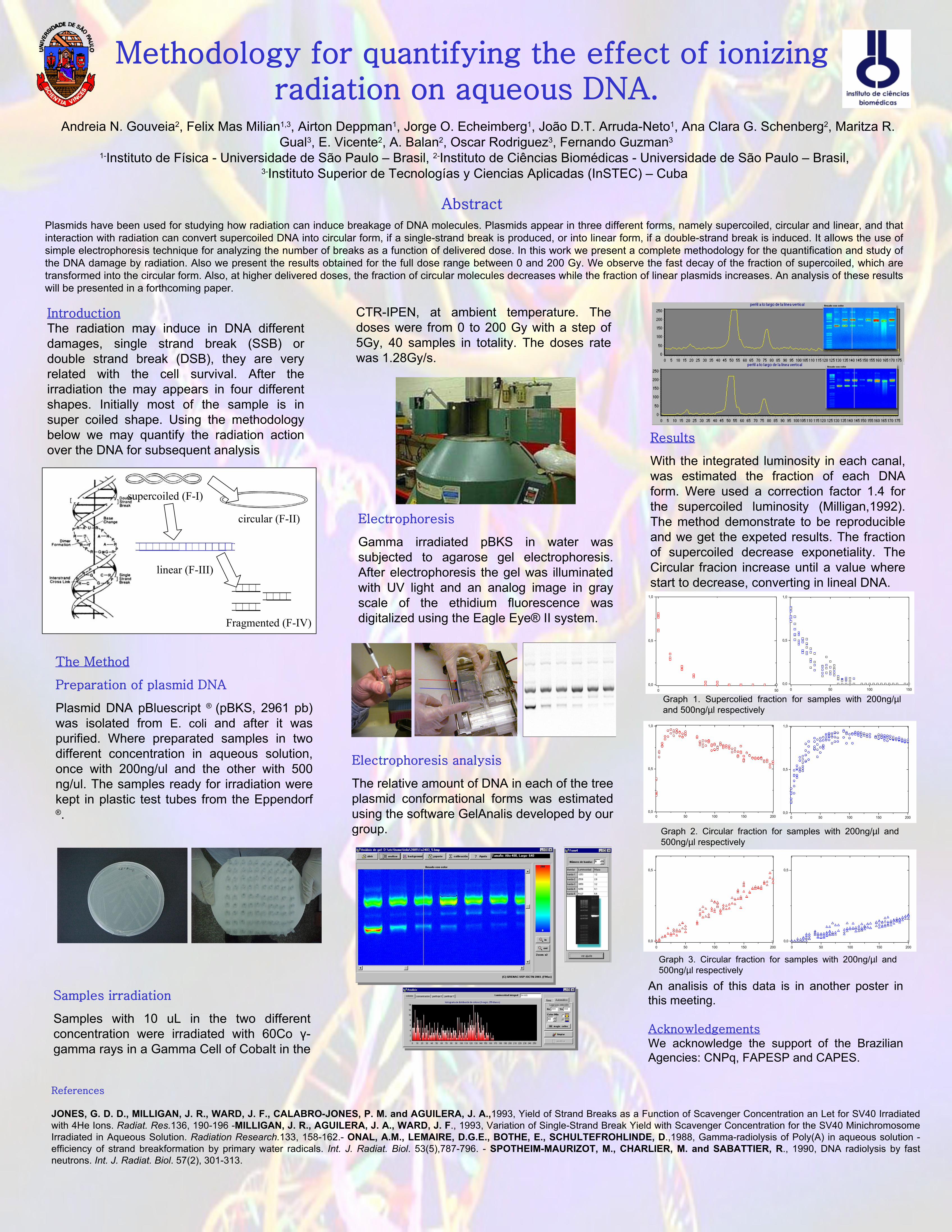
Methodology for quantifying the effect of ionizing radiation on aqueous DNA. Andreia N. Gouveia 2 , Felix Mas Milian 1,3 , Airton Deppman 1 , Jorge O. Echeimberg 1 , João D.T. Arruda-Neto 1 , Ana Clara G. Schenberg 2 , Maritza R. Gual 3 , E. Vicente 2 , A. Balan 2 , Oscar Rodriguez 3 , Fernando Guzman 3 1- Instituto de Física - Universidade de São Paulo – Brasil, 2- Instituto de Ciências Biomédicas - Universidade de São Paulo – Brasil, 3- Instituto Superior de Tecnologías y Ciencias Aplicadas (InSTEC) – Cuba Abstract Plasmids have been used for studying how radiation can induce breakage of DNA molecules. Plasmids appear in three different forms, namely supercoiled, circular and linear, and that interaction with radiation can convert supercoiled DNA into circular form, if a single-strand break is produced, or into linear form, if a double-strand break is induced. It allows the use of simple electrophoresis technique for analyzing the number of breaks as a function of delivered dose. In this work we present a complete methodology for the quantification and study of the DNA damage by radiation. Also we present the results obtained for the full dose range between 0 and 200 Gy. We observe the fast decay of the fraction of supercoiled, which are transformed into the circular form. Also, at higher delivered doses, the fraction of circular molecules decreases while the fraction of linear plasmids increases. An analysis of these results will be presented in a forthcoming paper. Introduction The radiation may induce in DNA different damages, single strand break (SSB) or double strand break (DSB), they are very related with the cell survival. After the irradiation the may appears in four different shapes. Initially most of the sample is in super coiled shape. Using the methodology below we may quantify the radiation action over the DNA for subsequent analysis supercoiled (F-I) circular (F-II) linear (F-III) Fragmented (F-IV) The Method Preparation of plasmid DNA Plasmid DNA pBluescript ® (pBKS, 2961 pb) was isolated from E. coli and after it was purified. Where preparated samples in two different concentration in aqueous solution, once with 200ng/ul and the other with 500 ng/ul. The samples ready for irradiation were kept in plastic test tubes from the Eppendorf ® . Electrophoresis Gamma irradiated pBKS in water was subjected to agarose gel electrophoresis. After electrophoresis the gel was illuminated with UV light and an analog image in gray scale of the ethidium fluorescence was digitalized using the Eagle Eye® II system. Electrophoresis analysis The relative amount of DNA in each of the tree plasmid conformational forms was estimated using the software GelAnalis developed by our group. 0 50 0,0 0,5 1,0 0 50 100 150 0,0 0,5 1,0 0 50 100 150 200 0,0 0,5 1,0 0 50 100 150 200 0,0 0,5 1,0 0 50 100 150 200 0,0 0,5 0 50 100 150 200 0,0 0,5 References JONES, G. D. D., MILLIGAN, J. R., WARD, J. F., CALABRO-JONES, P. M. and AGUILERA, J. A.,1993, Yield of Strand Breaks as a Function of Scavenger Concentration an Let for SV40 Irradiated with 4He Ions. Radiat. Res.136, 190-196 -MILLIGAN, J. R., AGUILERA, J. A., WARD, J. F., 1993, Variation of Single-Strand Break Yield with Scavenger Concentration for the SV40 Minichromosome Irradiated in Aqueous Solution. Radiation Research.133, 158-162.- ONAL, A.M., LEMAIRE, D.G.E., BOTHE, E., SCHULTEFROHLINDE, D.,1988, Gamma-radiolysis of Poly(A) in aqueous solution - efficiency of strand breakformation by primary water radicals. Int. J. Radiat. Biol. 53(5),787-796. - SPOTHEIM-MAURIZOT, M., CHARLIER, M. and SABATTIER, R., 1990, DNA radiolysis by fast neutrons. Int. J. Radiat. Biol. 57(2), 301-313. An analisis of this data is in another poster in this meeting. Acknowledgements We acknowledge the support of the Brazilian Agencies: CNPq, FAPESP and CAPES. Samples irradiation Samples with 10 uL in the two different concentration were irradiated with 60Co γ- gamma rays in a Gamma Cell of Cobalt in the CTR-IPEN, at ambient temperature. The doses were from 0 to 200 Gy with a step of 5Gy, 40 samples in totality. The doses rate was 1.28Gy/s. Results With the integrated luminosity in each canal, was estimated the fraction of each DNA form. Were used a correction factor 1.4 for the supercoiled luminosity (Milligan,1992). The method demonstrate to be reproducible and we get the expeted results. The fraction of supercoiled decrease exponetiality. The Circular fracion increase until a value where start to decrease, converting in lineal DNA. Graph 1. Supercolied fraction for samples with 200ng/μl and 500ng/μl respectively Graph 2. Circular fraction for samples with 200ng/μl and 500ng/μl respectively Graph 3. Circular fraction for samples with 200ng/μl and 500ng/μl respectively
Transcript

Methodology for quantifying the effect of ionizing radiation on aqueous DNA.

 Andreia N. Gouveia2, Felix Mas Milian1,3, Airton Deppman1, Jorge O. Echeimberg1, João D.T. Arruda-Neto1, Ana Clara G. Schenberg2, Maritza R. Gual3, E. Vicente2, A. Balan2, Oscar Rodriguez3, Fernando Guzman3

1-Instituto de Física - Universidade de São Paulo – Brasil, 2-Instituto de Ciências Biomédicas - Universidade de São Paulo – Brasil, 3-Instituto Superior de Tecnologías y Ciencias Aplicadas (InSTEC) – Cuba

Abstract Plasmids have been used for studying how radiation can induce breakage of DNA molecules. Plasmids appear in three different forms, namely supercoiled, circular and linear, and that interaction with radiation can convert supercoiled DNA into circular form, if a single-strand break is produced, or into linear form, if a double-strand break is induced. It allows the use of simple electrophoresis technique for analyzing the number of breaks as a function of delivered dose. In this work we present a complete methodology for the quantification and study of the DNA damage by radiation. Also we present the results obtained for the full dose range between 0 and 200 Gy. We observe the fast decay of the fraction of supercoiled, which are transformed into the circular form. Also, at higher delivered doses, the fraction of circular molecules decreases while the fraction of linear plasmids increases. An analysis of these results will be presented in a forthcoming paper.

IntroductionThe radiation may induce in DNA different damages, single strand break (SSB) or double strand break (DSB), they are very related with the cell survival. After the irradiation the may appears in four different shapes. Initially most of the sample is in super coiled shape. Using the methodology below we may quantify the radiation action over the DNA for subsequent analysis

supercoiled (F-I)

circular (F-II)

linear (F-III)

Fragmented (F-IV)

The Method

Preparation of plasmid DNA

Plasmid DNA pBluescript ® (pBKS, 2961 pb) was isolated from E. coli and after it was purified. Where preparated samples in two different concentration in aqueous solution, once with 200ng/ul and the other with 500 ng/ul. The samples ready for irradiation were kept in plastic test tubes from the Eppendorf ®.

Electrophoresis

Gamma irradiated pBKS in water was subjected to agarose gel electrophoresis. After electrophoresis the gel was illuminated with UV light and an analog image in gray scale of the ethidium fluorescence was digitalized using the Eagle Eye® II system.

Electrophoresis analysis

The relative amount of DNA in each of the tree plasmid conformational forms was estimated using the software GelAnalis developed by our group.

0 500,0

0,5

1,0

0 50 100 1500,0

0,5

1,0

0 50 100 150 2000,0

0,5

1,0

0 50 100 150 2000,0

0,5

1,0

0 50 100 150 200

0,0

0,5

0 50 100 150 200

0,0

0,5

References

JONES, G. D. D., MILLIGAN, J. R., WARD, J. F., CALABRO-JONES, P. M. and AGUILERA, J. A.,1993, Yield of Strand Breaks as a Function of Scavenger Concentration an Let for SV40 Irradiated with 4He Ions. Radiat. Res.136, 190-196 -MILLIGAN, J. R., AGUILERA, J. A., WARD, J. F., 1993, Variation of Single-Strand Break Yield with Scavenger Concentration for the SV40 Minichromosome Irradiated in Aqueous Solution. Radiation Research.133, 158-162.- ONAL, A.M., LEMAIRE, D.G.E., BOTHE, E., SCHULTEFROHLINDE, D.,1988, Gamma-radiolysis of Poly(A) in aqueous solution - efficiency of strand breakformation by primary water radicals. Int. J. Radiat. Biol. 53(5),787-796. - SPOTHEIM-MAURIZOT, M., CHARLIER, M.  and SABATTIER, R., 1990, DNA radiolysis by fast neutrons. Int. J. Radiat. Biol. 57(2), 301-313.

An analisis of this data is in another poster in this meeting.

AcknowledgementsWe acknowledge the support of the Brazilian Agencies: CNPq, FAPESP and CAPES.

Samples irradiation

Samples with 10 uL in the two different concentration were irradiated with 60Co γ-gamma rays in a Gamma Cell of Cobalt in the

CTR-IPEN, at ambient temperature. The doses were from 0 to 200 Gy with a step of 5Gy, 40 samples in totality. The doses rate was 1.28Gy/s.

Results

With the integrated luminosity in each canal, was estimated the fraction of each DNA form. Were used a correction factor 1.4 for the supercoiled luminosity (Milligan,1992). The method demonstrate to be reproducibleand we get the expeted results. The fraction of supercoiled decrease exponetiality. The Circular fracion increase until a value where start to decrease, converting in lineal DNA.

Graph 1. Supercolied fraction for samples with 200ng/µl and 500ng/µl respectively

Graph 2. Circular fraction for samples with 200ng/µl and 500ng/µl respectively

Graph 3. Circular fraction for samples with 200ng/µl and 500ng/µl respectively

Recommended