Bentz et al. Reproductive Biology and Endocrinology 2010, 8:38http://www.rbej.com/content/8/1/38
Open AccessM E T H O D O L O G Y
MethodologyOCT-4 expression in follicular and luteal phase endometrium: a pilot studyEva-Katrin Bentz1, Marina Kenning1, Christian Schneeberger1, Andrea Kolbus1, Johannes C Huber1, Lukas A Hefler2 and Clemens B Tempfer*2
AbstractBackground: The stem cell marker Octamer-4 (OCT-4) is expressed in human endometrium. Menstrual cycle-dependency of OCT-4 expression has not been investigated to date.
Methods: In a prospective, single center cohort study of 98 women undergoing hysteroscopy during the follicular (n = 49) and the luteal (n = 40) phases of the menstrual cycle, we obtained endometrial samples. Specimens were investigated for OCT-4 expression on the mRNA and protein levels using reverse transcriptase polymerase chain reaction (RT-PCR) and immunohistochemistry. Expression of OCT-4 was correlated to menstrual cycle phase.
Results: Of 89 women sampled, 49 were in the follicular phase and 40 were in the luteal phase. OCT-4 mRNA was detected in all samples. Increased OCT-4 mRNA levels in the follicular and luteal phases was found in 35/49 (71%) and 27/40 (68%) of women, respectively (p = 0.9). Increased expression of OCT-4 protein was identified in 56/89 (63%) samples. Increased expression of OCT-4 protein in the follicular and luteal phases was found in 33/49 (67%) and 23/40 (58%) of women, respectively (p = 0.5).
Conclusions: On the mRNA and protein levels, OCT-4 is not differentially expressed during the menstrual cycle. Endometrial OCT-4 is not involved in or modulated by hormone-induced cyclical changes of the endometrium.
BackgroundOctamer-4 (OCT-4) is a homeodomain transcription fac-tor of the Pit-Oct-Unc transcription factor family [1-3]. Atranscription factor is defined as a protein binding to spe-cific DNA binding domains and subsequently regulatingthe transcription from DNA to RNA by activation orrepression of RNA polymerase [4]. Specifically, OCT-4regulates tissue- and cell-specific transcription via theconsensus motif ATGCAAAT and its expression isrestricted to pluripotent cells. Loss of OCT-4 expressionmay be associated with the loss of pluripotentiality [5].During embryogenesis, OCT-4 is initially active as amaternal factor in the oocyte and remains active inembryos throughout the preimplantation period. OCT-4is involved in the self-renewal of undifferentiated embry-onic stem cells and is therefore used as a marker ofembryonic stem cells [5-8].
OCT-4 has also been found to be expressed in malig-nant tissues such as germ cell tumors, embryonic carci-noma cells [9]. Furthermore, breast cancer cells expressOCT-4 [10] and OCT-4 is - among other embryonic geneproducts - re-expressed in cancer cells [11]. Based onthese data and the fact that stem cells are undifferenti-ated, immortal, and invasive, an etiologic role for stemcells as clonogenic origin of various forms of cancer hasbeen proposed [11,12].
Cho et al. described the presence of stem cells in thestroma of the basal layer of human endometrium basedon the presence of C-kit/CD 117, CD34, bcl-2, and Ki67[13]. In a previous study, we demonstrated that OCT-4 isalso expressed in the human endometrium, lending fur-ther support to the hypothesis of endometrial regenera-tion by local stem cells in endometrial tissue [12]. Thisconcept is also supported by the presence of clonogenicepithelial and stromal cells in human endometrium act-ing as putative stem cells [14,15]. It has been speculatedthat local tissue-specific stem cells are involved in theregeneration and maintenance of the endometrial lining
* Correspondence: [email protected] Department of Obstetrics and Gynecology, Medical University of Vienna, Vienna, AustriaFull list of author information is available at the end of the article
Bentz et al. Reproductive Biology and Endocrinology 2010, 8:38http://www.rbej.com/content/8/1/38
Page 2 of 7
during the follicular phase and the menstruation. Thisconcept, however, has been challenged by evidence thatOCT-4 genetic ablation did not result in abnormalities inhomeostasis and regenerative capacity in rodent studies[16].
To further investigate the role of OCT-4 in humanendometrial physiology, we performed a prospectivestudy to assess the mRNA and protein expression ofOCT-4 in follicular and luteal phase endometrium. Wehypothesized that OCT-4 is differentially regulated in thefollicular and luteal phases of the menstrual cycle. Specif-ically, we aimed to answer the question whether or notOCT-4 is overexpressed during endometrial proliferationin the follicular phase and downregulated during thesecretory transformation of the endometrium in theluteal phase.
MethodsPatientsWe performed a prospective, single center cohort studybetween September 2006 and March 2007 in a popula-tion of 89 consecutive patients undergoing hysteroscopyand endometrial sampling at the Endoscopy Unit of theDepartment of Gynecologic Endocrinology and Repro-ductive Medicine at Vienna Medical University, Vienna,Austria. The mean age of the patients was 33.9 ± 5.2years. All women had regular menstrual cycles during thelast 6 months (menstrual cycle length 25-35 days) and didnot take any hormone therapy. Menstrual phase assess-ment was based on the date of the last menstrual periodwith the luteal and follicular phases determined by half-ing the median number of cycle days of the last threemenstrual cycles of the patient and confirmed by histo-pathological analysis of the endometrial specimen. Indi-cations for surgery were primary sterility (n = 32),secondary sterility (n = 34), endometriosis and/or dys-menorrhea (n = 10), recurrent pregnancy loss (n = 4),chronic pelvic pain (n = 4), and others (n = 5). Writteninformed consent was obtained by all patients.
Reverse transcriptase polymerase chain reaction (RT-PCR)For RNA extraction frozen tissue samples were trituratedand total RNA was extracted using the TRI REAGENTmethod (Molecular Research Centre, Inc., OH, USA).RNA concentration was determined by measuring theoptical density at 260 nm. 1 μg RNA was reversely tran-scribed into first strand complementary DNA (cDNA)using Superscript (Invitrogen Ltd., Paisley, UK). Theresulting cDNA was amplified by polymerase chain reac-tion (PCR) using primers specific for OCT-4 [11]. Thefollowing primers were used for RT-PCR reactions: OCT4 forward 5'-GAC AAC AAT GAA AAT CTT CAG GAGA-3' and OCT reverse 5'-TTC TGG CGC CGG TTACAG AAC CA-3'. The PCR was started with a denaturing
step at 94°C for 5 min and the amplification of the 218 bpproduct was performed for 35 cycles at 94°C for 30 sec,61°C for 30 sec and 72°C for 30 sec, and a final extensionat 72°C for 10 min. As control for genomic DNA we usedextracted RNA only (no cDNA). As positive control forthe expression of OCT -4 we used RNA from embryoniccarcinoma of testis. Human glyceraldehyd-3-phosphatedehydrogenase (GAPDH) was amplified in parallel reac-tions as a housekeeping reference gene serving as aninternal control for the quantity and quality of the cDNA.Primers for GAPDH were: forward 5'-TCT GGT AAAGTG GAT ATT GTT G-3' and reverse 5'-GAT GGT GATGGG ATT TCC-3'. The amplified product for GAPDHhas a size of 156 base pairs. Amplified samples were sepa-rated on 1.5% agarose gels in the presence of ethidiumbromide and visualized by the Eagle Eye System (Strata-gene, Amsterdam, The Netherlands). Quantitative analy-sis was performed by densitometric scanning of the gelsusing the AlphaDigiDoc 1000 software (Alpha InnotechCorp., San Leonardo, CA). OCT-4 mRNA levels weredetermined by calculating the OCT-4/GADPH ratios(Figure 1). We optimized all PCR steps with respect tosensitivity, reproducibility, and linearity: different tem-plate, enzyme, and primer concentrations, reaction times,and temperatures were tested. The resulting optimal con-ditions are given above. Since the technician and thescoring person were both blinded to the menstrual phaseof the specific samples, the samples have been placed inthe order of sampling and not arranged according tomenstrual phase.
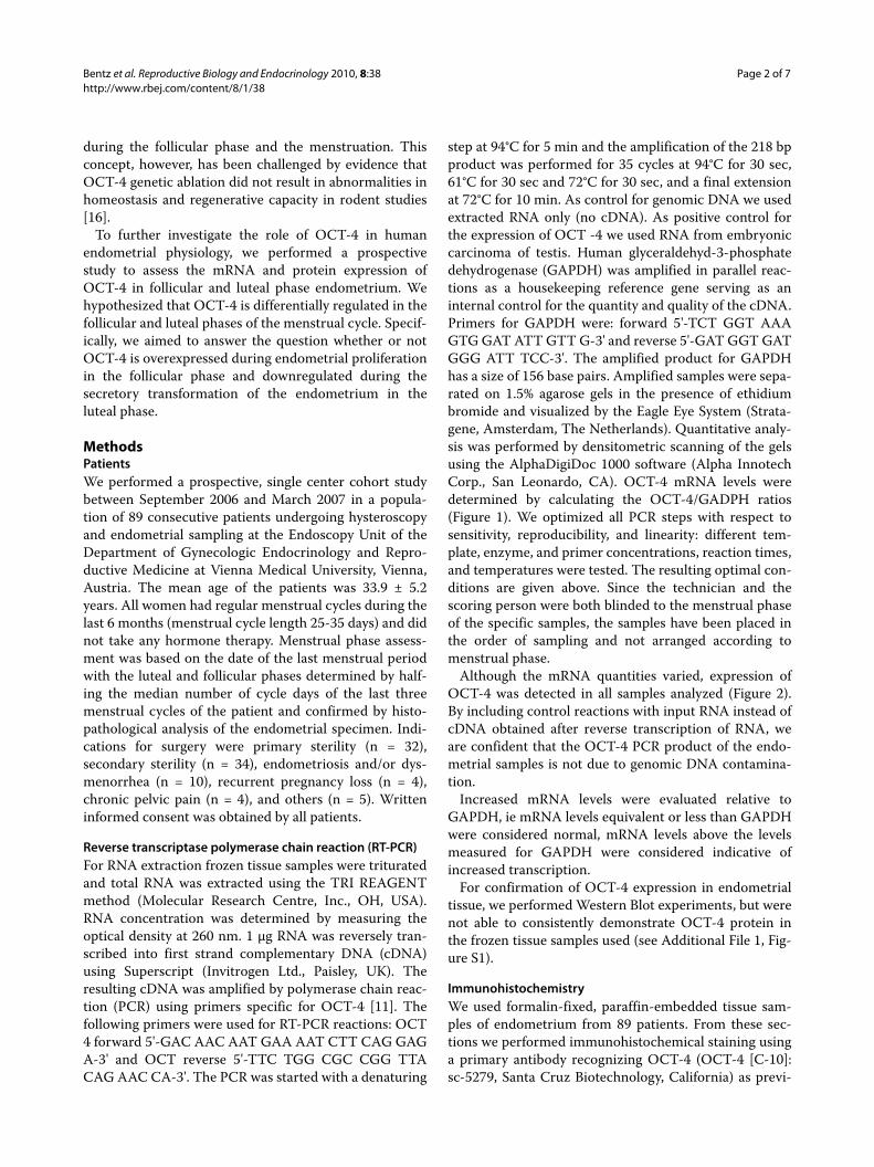
Although the mRNA quantities varied, expression ofOCT-4 was detected in all samples analyzed (Figure 2).By including control reactions with input RNA instead ofcDNA obtained after reverse transcription of RNA, weare confident that the OCT-4 PCR product of the endo-metrial samples is not due to genomic DNA contamina-tion.
Increased mRNA levels were evaluated relative toGAPDH, ie mRNA levels equivalent or less than GAPDHwere considered normal, mRNA levels above the levelsmeasured for GAPDH were considered indicative ofincreased transcription.
For confirmation of OCT-4 expression in endometrialtissue, we performed Western Blot experiments, but werenot able to consistently demonstrate OCT-4 protein inthe frozen tissue samples used (see Additional File 1, Fig-ure S1).
ImmunohistochemistryWe used formalin-fixed, paraffin-embedded tissue sam-ples of endometrium from 89 patients. From these sec-tions we performed immunohistochemical staining usinga primary antibody recognizing OCT-4 (OCT-4 [C-10]:sc-5279, Santa Cruz Biotechnology, California) as previ-
Bentz et al. Reproductive Biology and Endocrinology 2010, 8:38http://www.rbej.com/content/8/1/38
Page 3 of 7
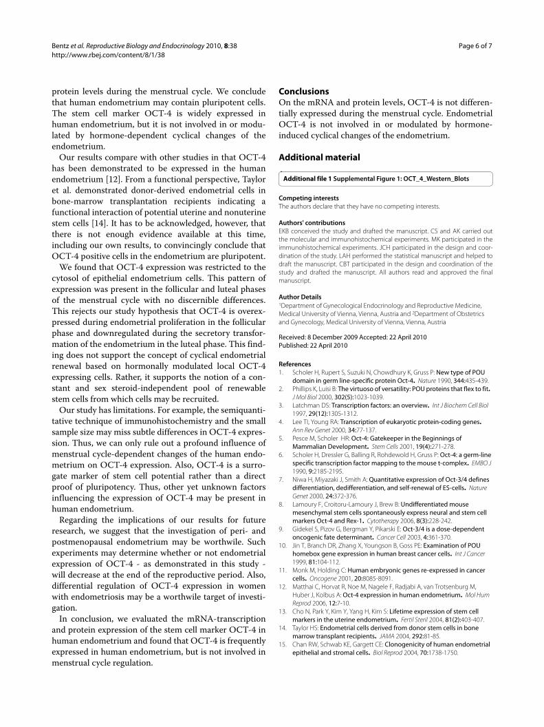
ously described [12]. Stained sections were scoredaccording to the immunoreactive score (IRS) describedby Remmele and Stegner [17]. The IRS results from themultiplication of a staining intensity score (negative = 0;weak = 1; moderate = 2; strong = 3) and the percentagescore of immunopositive cells (no staining = 0, 1-10% ofstained cells = 1; 11-50% of stained cells = 2, 51-80% ofstained cells = 3; 81-100% of stained cells = 4). Each sam-ple was scored according to these IRS criteria. An IRS of≥6 was considered to indicate 'increased expression' ofOCT-4. Figures 3A, B, C, and 3D show examples with astrong (A, B, C) and an absent (D) staining intensity,respectively.
StatisticsCategorical variables were analyzed by x2-test. Multiplecomparisons were corrected using Bonferroni's correc-tion. OCT-4 mRNA and protein expression were corre-lated using Pearson's correlation coefficient. A univariateregression model was used to assess the influence of dayof the menstrual cycle, using 3-day intervals, on OCT-4protein expression, comparing an IRS <6 vs. ≥ 6. All p-
values were two-tailed and 95% CI were calculated. A p-value < 0.05 was considered statistically significant. Weused the statistical software SPSS 11.0 for Windows(SPSS Inc., Chicago, IL) for statistical analyses.
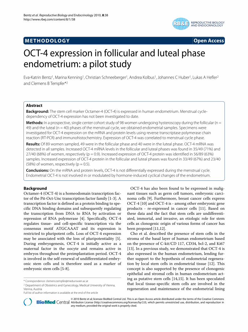
ResultsWe included 89 patient samples. The mean age of thepatients was 33.9 ± 5.2 years. Of these, 49 were in the fol-licular phase and 40 were in the luteal phase. All patientsunderwent hysteroscopy and endometrial sampling. Indi-cations for surgery were primary sterility (n = 32), sec-ondary sterility (n = 34), endometriosis and/ordysmenorrhea (n = 10), recurrent pregnancy loss (n = 4),chronic pelvic pain (n = 4), others (n = 5). OCT-4 mRNAwas detected in all samples (Figure 1). Increased OCT-4mRNA levels were observed in 62/89 (70%) samples.Comparing the number of samples with increased OCT-4mRNA levels in the follicular and luteal phases, we foundno statistically significant difference (35/49 [71%] vs. 27/40 [68%], respectively (p = 0.9). Figure 2 demonstratesOCT-4 cDNA expression in endometrial samples of thefollicular and luteal phases.
Figure 1 OCT-4 cDNA expression after RT-PCR relative to GADPH in 89 patients. OCT-4 = Octamer-4; cDNA = complementary deoxyribonucleic acid; RT-PCR = reverse transcriptase polymerase chain reaction; GADPH = glyceraldehyde-3-phosphatedehydrogenase. Each bar represents one pa-tient; values are given for each patient representing OCT-4 cDNA expression relative to the expression of GADPH with 1 denominating equivalent expression. The lowest (0.4) and highest (2.6) values are shown.
2,6
0,4
0,0
0,5
1,0
1,5
2,0
2,5
3,0
Bentz et al. Reproductive Biology and Endocrinology 2010, 8:38http://www.rbej.com/content/8/1/38
Page 4 of 7
OCT-4 protein overexpression was identified in 56/89(63%) samples. OCT-4 protein overexpression in the folli-cular and luteal phase was found in 33/49 (67%) and 23/40 (58%) of women, respectively (p = 0.5). In a univariateregression model, the day of the menstrual cycle, using 3-day intervals, did not influence OCT-4 protein expres-sion, when comparing an IRS <6 vs. ≥ 6 (p = n.s).Increased mRNA levels and increased OCT-4 proteinexpression were significantly correlated (Pearson's corre-lation coefficient 0.8). Figure 3 shows follicular and lutealphase endometrial samples with and without OCT-4overexpression. We found that OCT-4 expression was
restricted to the plasma of epithelial endometrium cells.This pattern of expression was present in the follicularand luteal phases of the menstrual cycle with no discern-ible differences.
DiscussionIn the present study we assessed the mRNA levels andprotein expression of OCT-4 in human endometrium.We found that human endometrium contains potentiallypluripotent OCT-4-expressing cells with increased OCT-4 mRNA levels observed in 70% of samples. OCT-4, how-ever, is not differentially expressed on the mRNA and
Figure 2 OCT-4 cDNA expression in endometrial samples using RT-PCR. OCT-4 = Octamer-4; cDNA = complementary deoxyribonucleic acid; RT-PCR = reverse transcriptase polymerase chain reaction; GADPH = glyceraldehyde-3-phosphatedehydrogenase. (A) lanes 1,2,5,6,7,8,10,11,12, and 16: patient samples from the luteal phase; lanes 3,4,9,13,14,15,17, and 18: patient samples from the follicular phase. (B) lane 1 = positive control (embry-onic carcinoma); lane 2 = negative control (water); lanes 3,4, and 7: patient samples from the luteal phase: lanes 5 and 6: patient samples from the follicular phase.
Bentz et al. Reproductive Biology and Endocrinology 2010, 8:38http://www.rbej.com/content/8/1/38
Page 5 of 7
Figure 3 Immunohistochemical staining for OCT-4. OCT-4 = Octamer-4; (A) OCT-4 overexpression in a sample of follicular phase endometrium. The picture demonstrates selected tissue compartments, ie three endometrial glands and surrounding endometrial stroma The brown staining shows OCT-4 expression in the cytosol of glandular cells. Magnification ×100. (B) OCT-4 overexpression in a sample of luteal phase endometrium. The picture demonstrates selected tissue compartments, ie two secretory endometrial glands and surrounding endometrial stroma The brown staining shows OCT-4 expression in the cytosol of glandular cells. Magnification ×200. (C) OCT-4 overexpression in a sample of embryonic carcinoma of the testis serving as positive control. Magnification ×200. (D) Section of embryonic carcinoma of the testis stained with IgG1 as described in Materials and Meth-ods serving as negative control. Magnification ×200.
A
B
C
D
Bentz et al. Reproductive Biology and Endocrinology 2010, 8:38http://www.rbej.com/content/8/1/38
Page 6 of 7
protein levels during the menstrual cycle. We concludethat human endometrium may contain pluripotent cells.The stem cell marker OCT-4 is widely expressed inhuman endometrium, but it is not involved in or modu-lated by hormone-dependent cyclical changes of theendometrium.
Our results compare with other studies in that OCT-4has been demonstrated to be expressed in the humanendometrium [12]. From a functional perspective, Tayloret al. demonstrated donor-derived endometrial cells inbone-marrow transplantation recipients indicating afunctional interaction of potential uterine and nonuterinestem cells [14]. It has to be acknowledged, however, thatthere is not enough evidence available at this time,including our own results, to convincingly conclude thatOCT-4 positive cells in the endometrium are pluripotent.
We found that OCT-4 expression was restricted to thecytosol of epithelial endometrium cells. This pattern ofexpression was present in the follicular and luteal phasesof the menstrual cycle with no discernible differences.This rejects our study hypothesis that OCT-4 is overex-pressed during endometrial proliferation in the follicularphase and downregulated during the secretory transfor-mation of the endometrium in the luteal phase. This find-ing does not support the concept of cyclical endometrialrenewal based on hormonally modulated local OCT-4expressing cells. Rather, it supports the notion of a con-stant and sex steroid-independent pool of renewablestem cells from which cells may be recruited.
Our study has limitations. For example, the semiquanti-tative technique of immunohistochemistry and the smallsample size may miss subtle differences in OCT-4 expres-sion. Thus, we can only rule out a profound influence ofmenstrual cycle-dependent changes of the human endo-metrium on OCT-4 expression. Also, OCT-4 is a surro-gate marker of stem cell potential rather than a directproof of pluripotency. Thus, other yet unknown factorsinfluencing the expression of OCT-4 may be present inhuman endometrium.
Regarding the implications of our results for futureresearch, we suggest that the investigation of peri- andpostmenopausal endometrium may be worthwile. Suchexperiments may determine whether or not endometrialexpression of OCT-4 - as demonstrated in this study -will decrease at the end of the reproductive period. Also,differential regulation of OCT-4 expression in womenwith endometriosis may be a worthwile target of investi-gation.
In conclusion, we evaluated the mRNA-transcriptionand protein expression of the stem cell marker OCT-4 inhuman endometrium and found that OCT-4 is frequentlyexpressed in human endometrium, but is not involved inmenstrual cycle regulation.
ConclusionsOn the mRNA and protein levels, OCT-4 is not differen-tially expressed during the menstrual cycle. EndometrialOCT-4 is not involved in or modulated by hormone-induced cyclical changes of the endometrium.
Additional material
Competing interestsThe authors declare that they have no competing interests.
Authors' contributionsEKB conceived the study and drafted the manuscript. CS and AK carried outthe molecular and immunohistochemical experiments. MK participated in theimmunohistochemical experiments. JCH participated in the design and coor-dination of the study. LAH performed the statistical manuscript and helped todraft the manuscript. CBT participated in the design and coordination of thestudy and drafted the manuscript. All authors read and approved the finalmanuscript.
Author Details1Department of Gynecological Endocrinology and Reproductive Medicine, Medical University of Vienna, Vienna, Austria and 2Department of Obstetrics and Gynecology, Medical University of Vienna, Vienna, Austria
References1. Scholer H, Rupert S, Suzuki N, Chowdhury K, Gruss P: New type of POU
domain in germ line-specific protein Oct-4. Nature 1990, 344:435-439.2. Phillips K, Luisi B: The virtuoso of versatility: POU proteins that flex to fit.
J Mol Biol 2000, 302(5):1023-1039.3. Latchman DS: Transcription factors: an overview. Int J Biochem Cell Biol
1997, 29(12):1305-1312.4. Lee TI, Young RA: Transcription of eukaryotic protein-coding genes.
Ann Rev Genet 2000, 34:77-137.5. Pesce M, Scholer HR: Oct-4: Gatekeeper in the Beginnings of
9. Gidekel S, Pizov G, Bergman Y, Pikarski E: Oct-3/4 is a dose-dependent oncogenic fate determinant. Cancer Cell 2003, 4:361-370.
10. Jin T, Branch DR, Zhang X, Youngson B, Goss PE: Examination of POU homebox gene expression in human breast cancer cells. Int J Cancer 1999, 81:104-112.
11. Monk M, Holding C: Human embryonic genes re-expressed in cancer cells. Oncogene 2001, 20:8085-8091.
12. Matthai C, Horvat R, Noe M, Nagele F, Radjabi A, van Trotsenburg M, Huber J, Kolbus A: Oct-4 expression in human endometrium. Mol Hum Reprod 2006, 12:7-10.
13. Cho N, Park Y, Kim Y, Yang H, Kim S: Lifetime expression of stem cell markers in the uterine endometrium. Fertil Steril 2004, 81(2):403-407.
14. Taylor HS: Endometrial cells derived from donor stem cells in bone marrow transplant recipients. JAMA 2004, 292:81-85.
15. Chan RW, Schwab KE, Gargett CE: Clonogenicity of human endometrial epithelial and stromal cells. Biol Reprod 2004, 70:1738-1750.
Bentz et al. Reproductive Biology and Endocrinology 2010, 8:38http://www.rbej.com/content/8/1/38
Page 7 of 7
16. Lengner CJ, Camargo FD, Hochedlinger K, Welstead GG, Zaidi S, Gokhale S: Oct4 expression is not required for mouse somatic stem cell self-renewal. Cell Stem Cell 2007, 1(4):401-415.
17. Remmele W, Stegner HE: Recommendation for uniform definition of an immunoreactive score (IRS) for immunohistochemical estrogen receptor detection (ER-ICA) in breast cancer tissue. Pathologe 1987, 8(3):138-140.
doi: 10.1186/1477-7827-8-38Cite this article as: Bentz et al., OCT-4 expression in follicular and luteal phase endometrium: a pilot study Reproductive Biology and Endocrinology 2010, 8:38