When asked to describe the treatment of any condition, it is a good principle and a safe answer to reply that treatment may be conservative or operative. This is particularly true in orthopaedic surgery, where there are more things to offer the patient than operation. Before selecting the right treatment for the individual patient, the physical requirements of work, home circumstances and motivation, as well as the patient’s likely cooperation with treatment and rehabilitation, all have to be con- sidered. It must also be remembered that many conditions get better on their own and there is a great temptation for doctors to ‘ascribe to their own skill the kindly work of time’ (John of Salisbury 1180). Physiotherapy An injured limb can be made to work again by a programme of graded exercises to increase the range of joint movement and muscle power using weights, springs and other devices in the ward or gymnasium. Rehabilitation and the supervision of day-to-day progress are essential parts of orthopaedic treatment, but only part of the physiotherapist’s role; a good physiotherapist will also build up the patient’s morale to achieve goals previously thought impossible. Physiotherapists can also reduce inflammation and swelling in injured areas by ultrasound, electrotherapy and the cautious application of ice and heat. Electrotherapy includes pulsed electromagnetic fields (PEMF) and interferential treatment, which depends on two slightly different electrical waveforms crossing within the area to be treated and raising the temperature at their point of intersection. Short wave diathermy was once in general use but is now becoming less popular. Muscles can be made to contract by applying an intermittent current (faradic stimulation or ‘faradism’) if the patient is unable to contract the muscle voluntarily. Faradism will not work if muscle is denervated, although interrupted direct current and other types of electrotherapy are effective. Physiotherapists also manipulate the spine and other joints when necessary. Remedial gymnastics, which once required a separate qualification from physiotherapy, takes the physical rehabilitation of the patient further than routine physio- therapy and is helpful to the young patient with physical or sporting ambitions. Occupational therapy The popular concept of occupational therapy is centred on handicrafts such as raffia and wickerwork but this has little in common with modern occupational therapy, which concentrates on rehabilitating the patient through tasks relevant to work and everyday activities. Occupational therapy departments include a small kitchen, a bath and a lavatory so that problems can be overcome before the patient leaves hospital, rather than after discharge when there is no help available. The department may also include a small printing press, so 67 Part 1 Background knowledge 6 Methods of treatment Physical therapy
Transcript
When asked to describe the treatment of any condition,it is a good principle and a safe answer to reply thattreatment may be conservative or operative. This isparticularly true in orthopaedic surgery, where there aremore things to offer the patient than operation. Beforeselecting the right treatment for the individual patient,the physical requirements of work, home circumstancesand motivation, as well as the patient’s likely cooperationwith treatment and rehabilitation, all have to be con-sidered.
It must also be remembered that many conditions getbetter on their own and there is a great temptation fordoctors to ‘ascribe to their own skill the kindly work oftime’ (John of Salisbury 1180).
Physiotherapy
An injured limb can be made to work again by aprogramme of graded exercises to increase the range ofjoint movement and muscle power using weights,springs and other devices in the ward or gymnasium.Rehabilitation and the supervision of day-to-day progressare essential parts of orthopaedic treatment, but onlypart of the physiotherapist’s role; a good physiotherapistwill also build up the patient’s morale to achieve goalspreviously thought impossible.
Physiotherapists can also reduce inflammation andswelling in injured areas by ultrasound, electrotherapyand the cautious application of ice and heat. Electrotherapy
includes pulsed electromagnetic fields (PEMF) andinterferential treatment, which depends on two slightlydifferent electrical waveforms crossing within the area tobe treated and raising the temperature at their point ofintersection. Short wave diathermy was once in generaluse but is now becoming less popular.
Muscles can be made to contract by applying anintermittent current (faradic stimulation or ‘faradism’) ifthe patient is unable to contract the muscle voluntarily.Faradism will not work if muscle is denervated, althoughinterrupted direct current and other types of electrotherapyare effective.
Physiotherapists also manipulate the spine and otherjoints when necessary.
Remedial gymnastics, which once required a separatequalification from physiotherapy, takes the physicalrehabilitation of the patient further than routine physio-therapy and is helpful to the young patient with physicalor sporting ambitions.
Occupational therapy
The popular concept of occupational therapy is centredon handicrafts such as raffia and wickerwork but this haslittle in common with modern occupational therapy,which concentrates on rehabilitating the patient throughtasks relevant to work and everyday activities.
Occupational therapy departments include a smallkitchen, a bath and a lavatory so that problems can beovercome before the patient leaves hospital, rather thanafter discharge when there is no help available. Thedepartment may also include a small printing press, so
67
Part 1B
ackground knowledge
6
Methods of treatment
Physical therapy
that patients can regain fine finger movement while theyset type, and a treadle-operated woodworking machineto improve the coordination and strength of the legs, aswell as the manual skills of carpentry. Apart from en-couraging physical coordination, to produce somethinguseful is tangible evidence of recovery and excellent formorale.
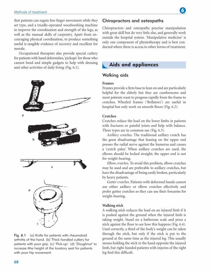
Occupational therapists also provide special cutleryfor patients with hand deformities, ‘pickups’ for those whocannot bend and simple gadgets to help with dressingand other activities of daily living (Fig. 6.1).
Chiropractors and osteopaths
Chiropractors and osteopaths practise manipulationwith great skill but do very little else, and generally workoutside the hospital system. ‘Manipulative medicine’ isonly one component of physiotherapy and is best con-ducted where there is access to other forms of treatment.
Walking aids
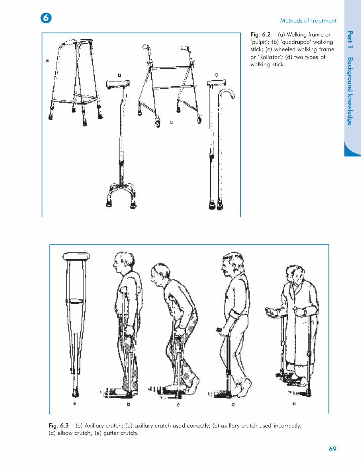
FramesFrames provide a firm base to lean on and are particularlyhelpful for the elderly but they are cumbersome andmost patients want to progress rapidly from the frame tocrutches. Wheeled frames (‘Rollators’) are useful inhospital but only work on smooth floors (Fig. 6.2).
CrutchesCrutches reduce the load on the lower limbs in patientswith fractures or painful joints and help with balance.Three types are in common use (Fig. 6.3).
Axillary crutches. The traditional axillary crutch hasthe great disadvantage that leaning on the upper endpresses the radial nerve against the humerus and causesa ‘crutch palsy’. When axillary crutches are used, theelbows should be locked straight; the upper end is notfor weight-bearing.
Elbow crutches. To avoid this problem, elbow crutchesmay be used and are preferable to axillary crutches, buthave the disadvantage of being easily broken, particularlyby heavy patients.
Gutter crutches. Patients with deformed hands cannotuse either axillary or elbow crutches effectively andprefer gutter crutches so they can use their forearms forweight-bearing.
Walking stickA walking stick reduces the load on an injured limb if itis pushed against the ground when the injured limb istaking weight. Stand on a bathroom scale and press astick against the floor to see how this happens (Fig. 6.4).Used correctly, a third of the body’s weight can be takenthrough the stick, but only if the stick is put to theground at the same time as the injured leg. This usuallymeans holding the stick in the hand opposite the injuredlimb, but right-handed patients with injuries of the rightleg find this difficult.
Methods of treatment 6
68
Aids and appliances
Fig. 6.1 (a) Knife for patients with rheumatoidarthritis of the hand. (b) Thick handled cutlery forpatients with poor grip. (c) ‘Pick-up’. (d) ‘Doughnut’ toincrease tthe height of the lavatory seat for patientswith poor hip movement.
Surgical appliances include splints and braces to supportlimbs, prostheses to replace absent parts of the body,surgical shoes and spinal supports. Appliances are ex-pensive, although economical by comparison with hospitaladmission.
OrthosesOrthoses, or braces, are used to support limbs (Fig. 6.5).A leg with no active dorsiflexors of the ankle is helped bya device to lift the foot, and an unstable knee is helped by a simple exoskeleton or caliper. The design anddevelopment of orthoses has advanced enormously inrecent times and many heavy and unsightly devices ofthe past (Fig. 6.6) can be replaced with lightweightcosmetic orthoses. Close cooperation with the fitter, ororthotist, is essential if the patient is to receive the bestappliance for his or her own requirements.
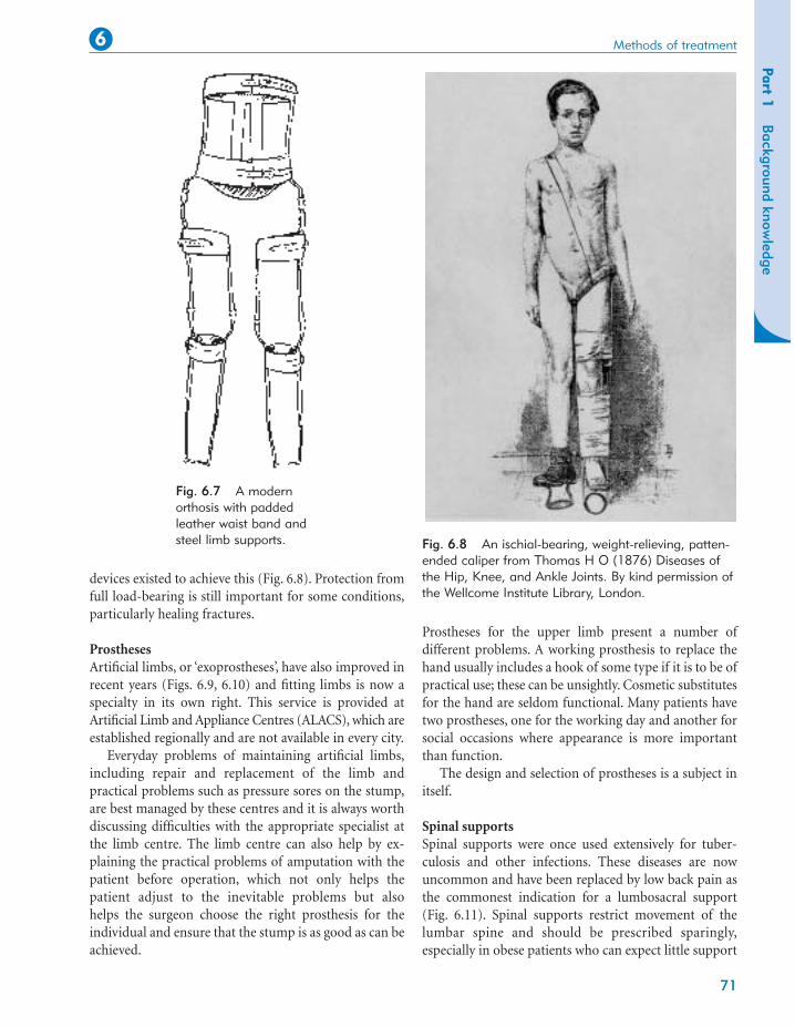
For patients with paraplegia, complex braces can alsobe made that support the lower limbs well enough tostand, and in some cases walk, unaided (Fig. 6.7).
Weight reductionIn the past, it was considered important to relieve theweight taken by an injured or diseased limb and many
Methods of treatment 6
70
Fig. 6.4 Mechanism of action of a walking stick.When the stick is pushed against the floor, thepatient’s weight on the limb is reduced, relievingpressure on joints.
Fig. 6.5 Modern types of orthoses: (a) weightrelieving caliper; (b) below-knee caliper with T strap;(c) cosmetic drop foot splint.
Fig. 6.6 An early orthosis. From Fabricius abAquapendente, Opera chirurgica (1647), Padua. Bykind permission of the Wellcome Institute Library,London.
devices existed to achieve this (Fig. 6.8). Protection fromfull load-bearing is still important for some conditions,particularly healing fractures.
ProsthesesArtificial limbs, or ‘exoprostheses’, have also improved inrecent years (Figs. 6.9, 6.10) and fitting limbs is now aspecialty in its own right. This service is provided atArtificial Limb and Appliance Centres (ALACS), which areestablished regionally and are not available in every city.
Everyday problems of maintaining artificial limbs,including repair and replacement of the limb andpractical problems such as pressure sores on the stump,are best managed by these centres and it is always worthdiscussing difficulties with the appropriate specialist atthe limb centre. The limb centre can also help by ex-plaining the practical problems of amputation with thepatient before operation, which not only helps thepatient adjust to the inevitable problems but also helps the surgeon choose the right prosthesis for theindividual and ensure that the stump is as good as can beachieved.
Prostheses for the upper limb present a number ofdifferent problems. A working prosthesis to replace thehand usually includes a hook of some type if it is to be ofpractical use; these can be unsightly. Cosmetic substitutesfor the hand are seldom functional. Many patients havetwo prostheses, one for the working day and another forsocial occasions where appearance is more importantthan function.
The design and selection of prostheses is a subject initself.
Spinal supportsSpinal supports were once used extensively for tuber-culosis and other infections. These diseases are nowuncommon and have been replaced by low back pain asthe commonest indication for a lumbosacral support(Fig. 6.11). Spinal supports restrict movement of thelumbar spine and should be prescribed sparingly,especially in obese patients who can expect little support
71
Part 1B
ackground knowledge
Methods of treatment6
Fig. 6.7 A modernorthosis with paddedleather waist band andsteel limb supports. Fig. 6.8 An ischial-bearing, weight-relieving, patten-
ended caliper from Thomas H O (1876) Diseases ofthe Hip, Knee, and Ankle Joints. By kind permission ofthe Wellcome Institute Library, London.
These simple devices may be enough to make operationunnecessary.
Social services
Many patients have been helped more by social servicesor modifications to their home than by hospital treat-ment. Elderly patients unable to fend for themselves mayneed a home help to assist with housework and shoppingrather than a total hip replacement, and the insuperableproblem of climbing stairs can be solved by fitting a stouthandrail.
Methods of treatment 6
72
Fig. 6.9 An early artificial limb. From ADiscourse of the Whole Art of Chirurgerie(1612). By kind permission of theWellcome Institute Library, London.
from a lumbosacral corset that cannot get near the pelvisbecause of fat.
CollarsCollars to support the neck are used after trauma and forpatients with acutely painful necks. Do not use the term‘cervical collar’: a collar cannot be worn anywhere else.
FootwearSurgical shoes or boots are prescribed for patients withfoot deformities. Normal shoes are designed for normalfeet and patients with abnormal feet are sometimes dis-abled only because they cannot find shoes that fit. Manypatients with quite severe deformities can cope in softshoes such as trainers, but specially made shoes will beneeded if these are unsatisfactory. There are several typesof appliance (Fig. 6.12):
◗ Special shoes made by taking a cast of thefoot and building the shoe around it
◗ Boots with a firm ankle support for patientswith unstable ankles
◗ Floats to the sole and heel for feet that tendto roll over.
Community services
◗ Insoles: soft insoles are helpful for patientswith prominent metatarsal heads; firmmoulded insoles support flat feet.
◗ Raises to the sole or heel to compensate forlimb inequality.
Fig. 6.10 A modern artificial limb with waist bandand thigh socket.
heavy manual work and unable to lift after a back injurymust find another job; a builder’s labourer with a stifffoot needs work that does not involve walking overrough ground. These problems commonly occur inpatients with little academic aptitude and the selection ofa new job can be difficult.
Advising a patient to give up his or her work and beretrained involves a great deal of thought and sympatheticassessment to determine the patient’s own particularskills (Fig. 6.13). This is best done in a resettlement orretraining centre, contacted through the local Disable-ment Resettlement Officer. It is not uncommon to findthat a resettled patient can earn more in the new job andenjoys it better. On the other hand, ‘outdoor people’ arealways unhappy at the prospect of working indoors,seated at a bench doing repetitive manual tasks, and needmuch encouragement.
Rehabilitation centres
Rehabilitation of the severely injured patient requires acoordinated approach and a sense of direction, which ismissing when the patient is an outpatient receiving alittle physiotherapy here and some occupational therapythere while they wait their turn for industrial retraining.Combining all these facilities under one roof, wherepatients receive more concentrated and continuoustreatment than an ordinary hospital can offer, makes thetask of rehabilitation a full-time occupation. This can
73
Part 1B
ackground knowledge
Methods of treatment6
Fig. 6.11 A lumbosacral support.
Fig. 6.12 Modifications to shoes and footwear: (a)raised sole and heel; (b) boot to accommodate fixedequinus deformity: (c) float to heels; (d) wedge to heel;(e) an insole; (f) surgical shoe for foot deformity.
These aspects of patient care cannot be ignored justbecause they happen outside hospital.
Warden-controlled accommodation is suitable formany elderly or infirm patients and allows them to livean independent existence in a home of their own. As theage of the population increases, small developments ofsingle-storey accommodation designed specifically forthe elderly are seen more often.
Those who cannot cope with warden-controlledaccommodation need long-term residential care. Suchaccommodation may be provided by local authorities orprivate individuals but is under great pressure and thelevel of care offered is very variable.
If a patient is to be cared for by the family it isimportant to consider before discharge the impact sucharrangements will have upon the rest of the family.Home visits from social workers, occupational therapistsand the local health visitor are very helpful.
Resettlement
Patients who cannot be restored to their former workneed to be resettled or retrained. A man engaged in
achieve results not possible if patients are at homethinking about their problems.
Non-steroidal anti-inflammatory drugs(NSAIDs)
Non-steroidal anti-inflammatory drugs are used for theconservative management of joint disease. Many suchdrugs are available and there is little to choose betweenthem, but patients do not react predictably. One patientmay be completely relieved by a drug that has no effecton a patient with the same condition and several mayhave to be tried before finding one that ‘suits’ the individual.
NSAIDs are the first line of treatment for most jointpains but they are not a panacea. They may not be effectiveand they have complications, of which the commonest isgastrointestinal irritation. They may impair renalfunction if given for long periods. Urea and electrolytesshould be measured before operation in patients whohave been taking NSAIDs for more than a few months.
Steroids
Steroids, gold, hydroxychloroquine, sulfasalazine,methotrexate and penicillamine are all used in the
management of rheumatoid arthritis. Patients requiringsuch treatment are better treated by rheumatologiststhan orthopaedic surgeons.
Colchicine
Colchicine is effective in acute attacks of gout but alarger dose of a non-steroidal anti-inflammatory is alsoeffective, and causes fewer side-effects.
Antibiotics
Antibiotics are used prophylactically before implantprocedures, such as joint replacement, and in thetreatment of joint infections. They are also used to treatacute and chronic infections, including osteomyelitis.
Prophylactic antibioticsAn appropriate prophylactic regimen for joint replacementis flucloxacillin 500 mg, given either intravenously in theanaesthetic room or intramuscularly with the premedi-cation and continued for three doses. It is also acceptableto give 1 g 6-hourly for 24 h. It is important that the anti-biotic is given before the incision is made so that there isa high blood level at the wound site.
If the patient is sensitive to penicillin, cephalosporinsmay be adequate but 10% of patients with penicillinsensitivity are also sensitive to cephalosporins. In those
Methods of treatment 6
74
Fig. 6.13 A skill centre for retraining injured patients. By kind permission of SkillsTraining Agency, Sheffield.
Drugs
patients over the age of 60 there is high incidence of Clostridium difficile infection with the use ofcephalosporins. A single intravenous dose of vancomycinis an alternative but the levels of this should bemonitored.
Therapeutic antibioticsIn acute infections the correct choice of antibiotics variesfrom place to place. Flucloxacillin 500 mg four timesdaily with ampicillin 500 mg four times daily intra-venously or orally is a good ‘best guess’ for treatment inchildren before the laboratory results are known. Thedose can be increased to 1 g of each drug in adults.
Anticoagulants and deep veinthrombosis
Deep vein thrombosis of some degree occurs in about60% of patients over the age of 40 after orthopaedicoperations. The majority are ‘silent’, i.e. they are notapparent clinically.
Death from pulmonary embolus occurs in about 1%of patients following hip or knee replacement and is aconstant anxiety. The risks following orthopaedicoperations are far greater than those after other pro-cedures, partly because of the trauma to the tissues sur-rounding the deep veins and partly because of difficultiesin moving injured limbs in order to maintain venousflow.
Prophylactic anticoagulation
There is no agreement on prophylactic measures foranticoagulants after orthopaedic procedures. There areseveral reasons for this:
1. Prophylactic anticoagulants may reduce theincidence of deep vein thrombosis afteroperation but this is not the same as reducing theincidence of fatal pulmonary embolus.
2. Anticoagulants can cause their owncomplications. These include increased blood loss both before and after operation. Woundhaematomas predispose to infection and delayed wound healing and the complications of the additional blood replacement followingincreased blood loss may outweigh its benefits.
3. The incidence of pulmonary embolus is so lowthat huge numbers of patients must be studiedbefore a statistically valid conclusion can bedrawn.
Various methods of prophylaxis have been used,including elastic support stockings, faradic stimulationof the opposite limb during surgery, intermittentcompression of the calf during operation, low dosewarfarin and subcutaneous heparin. None has beenshown to be completely effective. Low molecular weightheparin may decrease the risk of complications asso-ciated with anticoagulation.
If a deep vein thrombosis is suspected, an ultrasoundof the superficial and deep veins is done initially. Avenogram may be used when in doubt but this isinvasive and often painful for the patient. Patients withlarge thrombi in proximal veins usually require fullanticoagulation.
Established deep vein thrombosis
Anticoagulants are required in patients with establisheddeep vein thrombosis. Full therapeutic anticoagulation isbest conducted by the haematologist and should becontinued after discharge.
Intra-articular steroids
Intra-articular steroids should only be given when adiagnosis has been confirmed. They should not be givenwithout careful thought and are more generally used byrheumatologists than orthopaedic surgeons. They aremost effective in patients whose disease is well controlledby drugs.
Hydrocortisone acetate 25 mg is a useful preparationfor injection into painful areas, particularly at the bone–muscle or ligament–bone interface. Other preparations,e.g. triamcinolone, are used for intra-articular injection,but when injected subcutaneously they can cause fatnecrosis and therefore skin atrophy. They should not beused for subcutaneous injection.
There are more different operations in orthopaedicsurgery than any other surgical speciality.
75
Part 1B
ackground knowledge
Methods of treatment6
Operative management
Operations on tendons
TenotomyDividing a tendon is a simple way to stop a muscleworking and can be done either percutaneously, througha short stab incision, or by open operation.
Example: the adductor spasm of cerebral palsy can berelieved by subcutaneous adductor tenotomy.
Tendon lengtheningTendons can be lengthened either by making a Z-shapedcut and joining the two ends together or by cutting thetwo halves of the tendon and allowing them to slide overeach other inside the tendon sheath. Tendon lengtheningrelieves a fixed deformity without defunctioning themuscle altogether, as a tenotomy does.
Example: the Achilles tendon can be lengthened toreduce the equinus deformity of talipes (Fig. 6.14).
Tendon transpositionA tendon can be transposed from its normal insertion toanother so that the line of action of the muscle is altered,or power restored to a denervated muscle group.
Example: drop foot from common peroneal palsy canbe relieved by transposing the tibialis posterior from theback of the leg to the front so that it acts as a dorsiflexor.
Tendon release (tenolysis)Tendons which run through fibrous sheaths may becomeinflamed where they enter the fibrous tunnel or adherentto the sheath after trauma.
Example: thickened nodules within the digital flexortendons as they enter the flexor tunnel in the palm cause‘locking’ of the finger in flexion, relieved by dividing thesheath (p. 362).
TenodesisTendons can be converted to ligaments and used tostabilize unstable joints by attaching the tendon to boneimmediately above the joint upon which it acts.
Example: a fixed flexion deformity of the big toecaused by unopposed action of the flexor hallucis longus
can be controlled by fixing the tendon of extensorhallucis to the neck of the first metatarsal to create a‘dorsal ligament’ (Fig. 6.14).
Tendon repairTendons can be repaired if they are torn or divided.
Example: repair of a ruptured Achilles tendon.
Operations on bones
OsteotomyAn osteotomy is done either to correct deformity or toalter the stresses on a joint (Fig. 6.15). An osteotomy is asurgical fracture and must unite like a fracture, but thesite of the ‘fracture’ is carefully selected and the operation
Methods of treatment 6
76
Tendons can be (Fig. 6.14):
◗ Cut – tenotomy◗ Made longer – tendon lengthening◗ Moved – tendon transposition (transfer)◗ Released – tenolysis◗ Fixed to bone to stabilize joints – tenodesis◗ Repaired.
done so that the bone has the best chance of unitinguneventfully.
Example: tibial osteotomy to relieve the pain of osteo-arthritis at the knee by correcting the varus deformitythat follows wear of the medial compartment (p. 385).
OsteosynthesisFractured or osteotomized bones can be joined togetherusing plates, screws or nails (Figs 6.16, 6.17) (p. 130).Fixation devices do not make bones unite; they only hold
the bones together in the correct position while naturalbony union occurs. If this does not happen, even thestrongest metal plates and screws will eventually break orpull out of the bone (Figs 6.18, 6.19).
Example: internal fixation of unstable fractures (p. 130).
77
Part 1B
ackground knowledge
Methods of treatment6
Fig. 6.15 (a) Osteotomy to correct malalignment; (b) exostectomy to remove bony prominences; (c) osteosynthesis with plate and screws; (d) bonegrafting; (e) decompression of bone abscess.
Fig. 6.16 Different types of screw. From left to right:ASIF cancellous lag screw; ordinary wood screw withtapered shank; chipboard screw with parallel-sidedshank; self-tapping bone screw with fluted self-tappingtip; ASIF cancellous screw; ASIF cortical screw; ASIFsmall fragment screw (compare with Fig. 9.22).
Fig. 6.17 Different types of plate. (a) Very old plates.Note the pitting and corrosion and the very flimsydesign. These plates were not strong enough for rigidfixation. (b) A modern ASIF plate.
a
b
Bone grafting
Autografts. Autografts are used to encourage bone unionor replace lost bone. In the past, a strip of cortical bonewas taken from a site such as the tibia and screwed acrossa fracture site as a ‘cortical inlay graft’ to hold the endstogether. Although a neat surgical exercise, the bone wasbeing used as an internal fixation device and not as livingtissue. Modern metal plates are far stronger thandevitalized cortical bone and have largely replaced thecortical bone graft.
Instead, slivers or chips of cancellous bone, whichcontain osteoblasts and bone marrow, are taken from asite such as the iliac crest and laid around the bone,without disturbing the fracture site, to induce ossification.This technique uses bone as a biological agent and doesnot have an analogy in other branches of surgery.
Example: cancellous bone from the iliac crest isplaced between the transverse processes and the sacrumin an intertransverse lumbosacral fusion.
Allografts. Allograft bone is used to replace lost bonewhen insufficient autograft is available or when wholesegments of structural bone are required. The revision ofjoint replacements in which there has been extensivebone loss is a common use for allografts.
Allograft bone is likely to be used more frequently aspractical problems are overcome. The allograft must bestored sterile and has a limited shelf life that variesaccording to the method of preservation. When wholebone segments are used a large range of sizes for both leftand right sides must be kept.
Methods of treatment 6
78
Fig. 6.18 A tibial plate which has broken; an intactplate is shown below. Note the distorted ends of thebroken plate.
There are three types of grafted tissue:
1. Autograft – from elsewhere in the patient2. Allograft – from another human3. Xenograft – from another species.
Fig. 6.19 (a), (b) Fractured internal fixation. Thescrew used to fix a fractured olecranon has itselffractured because the fragments did not unite.
a
b
Achilles tendon, patellar tendon and tibialis posteriortendon can be used to replace the anterior cruciateligament and other ruptured ligaments.
A particular anxiety, common to all donor materials,is the possible transmission of infections, e.g. thehepatitis virus and HIV. This is a justifiable fear becausepatients have contracted AIDS in this way. Freeze-dryingand irradiation have been claimed to reduce the risk ofdisease transmission but this may be at the expense ofthe mechanical properties of the graft.
Xenografts. Xenografts (tissue from another speicies)are treated to remove protein and fat, leaving only themineralized structure intact. These grafts are essentiallyforeign bodies which act as a skeleton for creepingsubstitution rather than a true osteogenic bone graft.They have little, if any, osteogenic potential but are usefulto fill spaces and hold osteotomies open.
Other materials. Inert sponges of many materialscontaining calcium have been used as scaffolds forcreeping substitution. Among the most recent to beoffered for general use is coral, which has been machinedto exact sizes, sterilized and packed ready for insertion.(‘Full fathom five thy father lies; Of his bones are coralmade;’ The Tempest, Act I, Scene 2.)
Lengthening bonesBones can be lengthened in a number of ways. A varietyof techniques have been used in the past, includingperiosteal stripping, implantation of a foreign substance,alteration in the blood supply, sympathectomy, bio-
electrical stimulation, hormone therapy and surgery.Unfortunately many of these techniques were unreliableand the use of growth hormone in particular led totragedy. Some of the hormone was obtained fromhuman pituitary glands and was contaminated with theCreutzfeldt–Jakob disease (CJD) agent: a few of thepatients treated have contracted this disease.
Operations are more reliable. Simple division of thebone in a step cut fashion with subsequent distraction ofthe two bone ends and refixation at the desired length ispossible but only small segments can be lengthened at atime. The lengthened segment of bone is also weakerthan normal and recovery may be prolonged.
In the immature skeleton a bone can be lengthenedby stretching the growth plate (physeal distraction). Thisis achieved by the application of an external fixator acrossthe growth plate and slowly distracting the physis. Theamount of length achieved is variable and the techniqueis limited only to those patients with an open physis andpotential for growth. Complications are frequent and theresults are often unpredictable.
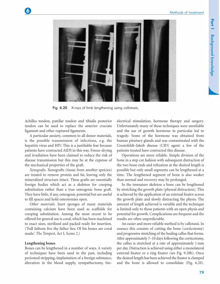
An easier and more reliable method is by callotasis. Inessence this consists of cutting the bone (corticotomy)and progressive stretching of the healing callus that forms.After approximately 7–10 days following the corticotomy,the callus is stretched at a rate of approximately 1 mmper day. Distraction is achieved using either a monolateralexternal fixator or a ring fixator (see Fig. 9.20b). Oncethe desired length has been achieved the fixator is clampedand the bone is allowed to consolidate (Fig. 6.20).
79
Part 1B
ackground knowledge
Methods of treatment6
Fig. 6.20 X-rays of limb lengthening using callotasis.
Patients can walk on the limb with the fixator in situuntil the bone has remodelled and strengthened. There isalways concern for the soft tissues, nerves and bloodvessels while the distraction of the bone is in progress.Meticulous care of the pin sites is needed and patientsshould be prepared for a prolonged period of treatment.Patients should also be advised not to smoke whiletreatment is in progress because smoking interferes withossification.
ExostectomyExostoses and other lumps can be levelled, preferablymaking a slight depression at the site of the originalbump so that the fibrous tissue which forms at the site ofoperation lies in the depression.
Example: levelling an exostosis on the dorsum of thefoot.
Draining infectionIn days gone by, bone infection was a common problemand orthopaedic surgeons spent much of their timeletting pus out of abscesses and removing sequestra.These operations are, thankfully, seldom required nowbut it is still occasionally necessary to perform asequestrectomy and unroof infected bone cavities (p. 300).
Example: drilling acute osteomyelitis (p. 300).
Operations on joints
ArthrodesisStiffening a joint, or arthrodesis, is achieved byremoving the articular surfaces of the joint and holdingthe bone ends together so that they unite like a fracture.The operation is indicated for irreparable damage orinstability of a joint and will produce a sound and painlesslimb that lasts a lifetime, but the loss of movement is aserious disadvantage in large joints such as the hip andknee (Figs 6.22, 6.23).
The effect of arthrodesis is usually irreversible but canbe predicted by immobilizing the joint in plaster, afterwhich the patient may decide that a painful joint thatmoves a little is preferable to a painless limb that will notmove at all.
Arthrodesis should not be performed if there is achance of other joints becoming stiff. A patient canmanage with one arthrodesed hip but to manage withtwo is extremely difficult. If both hip and knee arearthrodesed in the same limb, problems are inevitable:hold your own hip and knee stiff and try to imagine thedifficulties yourself.
Example: arthrodesis of the proximal interphalangealjoint of the second toe for hammer toe (p. 417).
ArthrotomyAny operation that opens a joint is an arthrotomy butthe term is usually applied to an exploratory arthrotomy.
Methods of treatment 6
80
Fig. 6.21 (a) Compressionarthrodesis; (b) adhesions within jointscan be released by arthrolysis.
Joints can be (Figs 6.21, 6.26)
◗ Stiffened – arthrodesis◗ Opened – arthrotomy◗ Refashioned – arthroplasty
◗ Subjected to excision of the synovium –synovectomy
◗ Mobilized – arthrolysis◗ Looked into – arthroscopy◗ Aspirated◗ Manipulated.
With arthroscopy and other modern investigativetechniques, exploratory arthrotomy is seldom performedalone and is largely obsolete.
Example: exploration of the hip for unexplained pain.
ArthroplastyAny operation that creates or reshapes a joint is anarthroplasty. There are several types (Fig. 6.24).
Excision arthroplasty. Excision arthroplasty, in whichthe bone surfaces are removed and the space betweenthem is allowed to fill with fibrous tissues, is the simplestand sometimes the most satisfactory arthroplasty, butleaves an unstable joint.
Example: excision arthroplasty of the hip as a salvageprocedure for failed total hip replacement (p. 382).
Interposition arthroplasty. Rather than leave the twobone surfaces of an excision arthroplasty exposed, aprosthetic or organic material can be laid between thetwo. A stainless steel cup or mould was inserted in thehip joint before the advent of total hip replacement anda similar operation was performed at the knee.
Example: silastic spacers inserted in the metacarpo-phalangeal joints in rheumatoid arthritis.
Replacement hemiarthroplasty. If one surface of a jointis replaced with an artificial material such as metal, theoperation becomes a replacement hemiarthroplasty, orsimply a hemiarthroplasty.
Example: fractures of the femoral neck may be treatedby replacing the femoral head with a metal prosthesis.This is satisfactory until the hard metal of the prosthesiserodes the acetabulum.
Total joint replacement. A metal prosthesis will erodebone affected by osteoarthritis or other disease. Becauseof this, it is usual to replace both joint surfaces in osteo-arthritis and perform a total joint replacement, one ofthe most successful operations in orthopaedic surgery(Fig. 6.25).
Problems arise if the hip becomes infected or thecomponents loosen, when it may be necessary to removethe artificial material and convert the total hip replace-ment to an excision arthroplasty.
Example: total hip replacement for osteoarthritis (p. 375).
SynovectomySynovium affected by rheumatoid arthritis or any otherchronic inflammation may need to be removed. Withoutsynovium, the joint develops a new lining which differsslightly from the original.
Example: synovectomy of the elbow for rheumatoidarthritis.
81
Part 1B
ackground knowledge
Methods of treatment6
Fig. 6.22 Anteroposterior (a) and lateral (b) views ofan arthrodesis of the wrist using ASIF internal fixation.The radiograph was taken immediately after operationand the drain is still in position.
a
b
ArthrolysisFor a joint to function normally the bone surfaces mustbe free to slide over each other and the synovial cavitymust be free of adhesions. If there has been bleeding or infection in the knee, fibrous adhesions will formbetween the two synovial surfaces, and between articularcartilage and synovium, restricting movement severely.Division of the adhesions restores joint movement butthey can reform.
Example: mobilization of the knee following trauma.
ArthroscopyJoints can be looked into with a telescope, an invasiveprocedure usually done under general anaesthetic (Fig.6.26).
AspirationJoints can be aspirated, i.e. fluid drawn out of them, (1)to relieve the tension of a haemarthrosis, (2) to removefluid for culture, (3) to remove synovial fluid forexamination.
Although aspiration requires little more skill than anyordinary injection it must be done with full sterileprecautions to prevent infection. The aspiration can bedone under local anaesthetic, provided that joint capsule,synovium and skin are infiltrated.
Example: diagnostic aspiration of a painless joint effusion.
Manipulation under anaestheticJoints can be manipulated under anaesthetic to breakdown adhesions or assess movement. Manipulation is
Methods of treatment 6
82
Fig. 6.23 An arthrodesed hip.
Fig. 6.24 Different types of arthroplasty using the hip as an example: (a) excisionarthroplasty; (b) interposition arthroplasty; (c) hemiarthroplasty; (d) total hip replacement.
helpful in restoring movement after haemarthrosis orjoint replacement but overenthusiastic manipulation ofa joint can tear ligaments or break the bone.
Example: manipulation of a stiff joint after a fracture.
Operations on ligaments
RepairLigaments are strong and complex structures. If aligament is even a millimetre too long, the joint that itcontrols may be unstable; if it is a millimetre too short,joint movement will be restricted. Even if the ligamentcan be accurately resutured so that it is a perfect lengthand in perfect position, the chances that the repaired
ligament will have the same ‘stretchiness’ as the originalare slight. Because of this, repairing ruptured ligamentsis not generally successful.
Example: repair of the ulnar collateral ligament of thefirst metacarpophalangeal joint (p. 223).
Replacement or reconstruction
Because of the difficulty of repair, ligaments aresometimes replaced with a length of tendon (p. 214) orprosthetic material, but none of these procedures isentirely satisfactory.
Example: reconstruction of the anterior cruciateligament of the knee for instability.
Plication and capsulorrhaphyLigaments can be tightened by advancing the attachmentto bone or plicating the joint capsule to restrict movement.
Examples: distal advancement of the medial ligamentof the knee for instability (p. 253); the Putti–Platt operationfor recurrent dislocation of the shoulder (p. 349).
Operations on nerves
83
Part 1B
ackground knowledge
Methods of treatment6
Fig. 6.25 Immediate postoperative film of a total hipreplacement with suction drains in position.
Ligaments can be (Fig. 6.27):
◗ Repaired when torn◗ Replaced or reconstructed◗ Shortened – plication or capsulorrhaphy.
DecompressionThe most commonly performed operation on nerves isdecompression for dysfunction caused by outsidepressure.
Example: decompression of the median nerve at thewrist for carpal tunnel syndrome (p. 370).
RepairNerves divided by injury can be repaired (p. 213).
Example: repair of the median nerve at the wristfollowing a laceration.
NeurolysisNerves can become involved in dense scar tissue, whichinterferes with function.
Example: mobilization of the median or ulnar nervesfollowing a laceration of the wrist.
GraftingLarge gaps in a nerve can be replaced with a cable graftmade from cutaneous nerves. These operations areunreliable but are sometimes an attractive alternative toaccepting a serious disability.
Example: replacement of the upper cord of thebrachial plexus with a graft made from the sural nerve.
Fig. 6.28 Operations on nerves: (a) decompression of compressed nerve; (b) repair by suture of the perineurium; (c) cable grafting of large defects; (d) neurolysis. Tethering of the nerve to boneor other tissues can be released by operation.
Skin can be
◗ Repaired◗ Grafted◗ Changed in shape – plastic surgery.
Plastic operationsSmall areas of skin can be changed in shape to releasetension, but complex plastic procedures are better doneby plastic surgeons. If you find these procedures difficult tounderstand, try cutting out the patterns in Figure 6.29 ona separate piece of paper and move the flaps to see the effect.
Example: Z-plasty for Dupuytren’s contracture (p. 366).
The way in which the different services are necessary forthe management of orthopaedic patients is illustrated by
comparing the fate of three imaginary 78-year-oldwomen of identical size and weight who had identicalfractures treated by identical operations on the same day.
Patient A (Fig. 6.30a)
This patient is a bright and active woman without relativeswho fended well for herself before operation and livedalone in a bungalow (1). Her operation was uneventfuland the next day, rather against her will, she sat out ofbed. The following day, again under protest, she began towalk with a walking frame (3) and within 2 weeks had
85
Part 1B
ackground knowledge
Methods of treatment6
Case reports
Fig. 6.29 Z-plasty. A Z-plasty canchange the shape of a piece of skin andthe direction of the contracture.
Fig. 6.30A.
discarded the frame and was using elbow crutches. Whilein hospital, the occupational therapist made certain thatthe patient was able to wash and dress herself, use thelavatory, and prepare a meal while still using crutches(4). A few simple aids were necessary to help her dressbut with practice she was well able to cope.
The social worker visited the patient in hospital andarranged that after discharge a home help would assistwith housework, and that ‘meals on wheels’ would call sothat she would have at least one hot meal every few dayseven if she could not cook her own. The communitynurse visited her and arranged for the health visitor tocall after discharge. The patient’s family doctor was notifiedof the time and date of her discharge and a telephone callto the patient’s neighbours ensured that the house washeated and that fresh food was available when shearrived home (5). She was visited at home by the healthvisitor later on the day of discharge and by her familydoctor the following day.
The patient discarded her crutches 6 weeks afteroperation (6) and a few weeks later was again lookingafter herself without assistance.
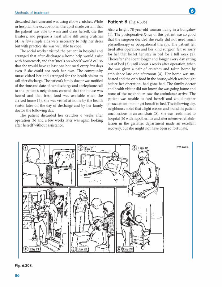
Patient B (Fig. 6.30b)
Also a bright 78-year-old woman living in a bungalow(1). The postoperative X-ray of this patient was so goodthat the surgeon decided she really did not need muchphysiotherapy or occupational therapy. The patient felttired after operation and her kind surgeon felt so sorryfor her that he let her stay in bed for a full week (2).Thereafter she spent longer and longer every day sittingout of bed (3) until about 3 weeks after operation, whenshe was given a pair of crutches and taken home byambulance late one afternoon (4). Her home was un-heated and the only food in the house, which was boughtbefore her operation, had gone bad. The family doctorand health visitor did not know she was going home andnone of the neighbours saw the ambulance arrive. Thepatient was unable to feed herself and could neitherattract attention nor get herself to bed. The following day,neighbours noted that a light was on and found the patientunconscious in an armchair (5). She was readmitted tohospital (6) with hypothermia and after intensive rehabili-tation in the geriatric department made an excellentrecovery, but she might not have been so fortunate.
Methods of treatment 6
86
Fig. 6.30B.
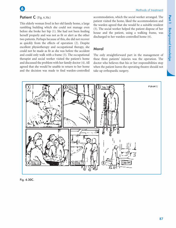
Patient C (Fig. 6.30c)
This elderly woman lived in her old family home, a largerambling building which she could not manage evenbefore she broke her hip (1). She had not been feedingherself properly and was not as fit or alert as the othertwo patients. Perhaps because of this, she did not recoveras quickly from the effects of operation (2). Despiteexcellent physiotherapy and occupational therapy, shecould not be made as fit as she was before the accidentand could only walk with a frame (3). The occupationaltherapist and social worker visited the patient’s homeand discussed the problem with her family doctor (4). Allagreed that she would be unable to return to her homeand the decision was made to find warden-controlled
accommodation, which the social worker arranged. Thepatient visited the home, liked the accommodation andthe warden agreed that she would be a suitable resident(5). The social worker helped the patient dispose of herhouse and the patient, using a walking frame, wasdischarged to her warden-controlled home (6).
Moral
The only straightforward part in the management ofthese three patients’ injuries was the operation. Thedoctor who believes that his or her responsibilities stopwhen the patient leaves the operating theatre should nottake up orthopaedic surgery.