27
GRAM POSITIVE COCCI STAPHYLOCOCCI BY OYOM P. ANTHONY 2013-BMLS-FT-009
| Date post: | 14-Jul-2015 |
| Category: |
Health & Medicine |
| Upload: | mwinek99 |
| View: | 128 times |
| Download: | 1 times |
GRAM POSITIVE COCCISTAPHYLOCOCCI
BY OYOM P. ANTHONY
2013-BMLS-FT-009
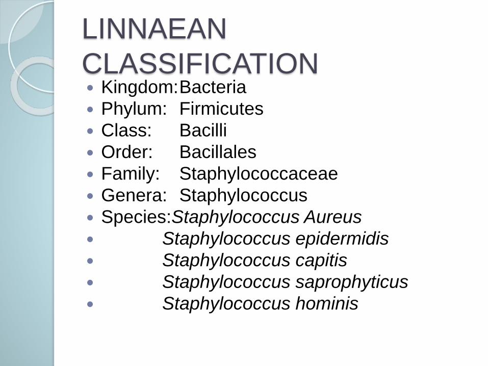
LINNAEAN
CLASSIFICATION Kingdom:Bacteria
Phylum: Firmicutes
Class: Bacilli
Order: Bacillales
Family: Staphylococcaceae
Genera: Staphylococcus
Species:Staphylococcus Aureus
Staphylococcus epidermidis
Staphylococcus capitis
Staphylococcus saprophyticus
Staphylococcus hominis
STAPHYLOCOCCI
Gram positive aerobic organisms Reproduce asexually by binary fission Common microorganism in the
environment; present in air, water and dust
Common strains are S. aureus, S. epidermidis and S. saprophyticus
S. aureus commonly inhabits nasal passages and axillae.
S. epidermidis is a normal flora on the skin.
S. saprophyticus rare but may inhabit the female genital tract
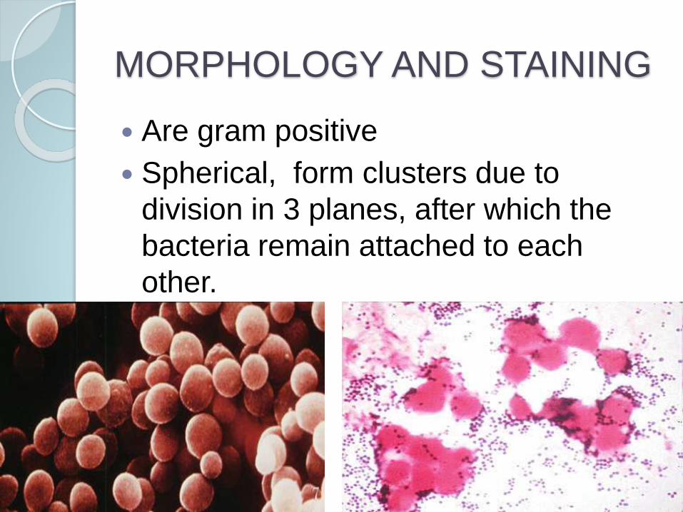
MORPHOLOGY AND STAINING
Are gram positive
Spherical, form clusters due to
division in 3 planes, after which the
bacteria remain attached to each
other.
MORPHOLOGY AND STAINING
Are non-motile
Non spore forming
Are non-capsulated except in young
cultures; capsulation is lost with
prolonged culturing
CULTURE
CHARACTERISTICS Grow aerobically and are facultative
anaerobes.
Capable of growing at temperature s between 220C – 440C (Ideal temp 350C -370C)
Grows on ordinary media; nutrient agar & blood agar
Colonies are 1 – 3 mm diameter, smooth, low convex, glittering and opaque on nutrient agar.
S. aureus will yield large yellow low convex colonies with β-haemolysis (esp. fresh isolates) on blood agar
S. epidermidis will yield small creamy/ white colonies with no haemolysis on blood agar
CULTURE
CHARACTERISTICS Pigmentation in S. aureus may be
enhanced by: aerobic incubation, use
of fatty media (e.g. tween agar), or
prolonged incubation of the plate.
MacConkey Agar will yield small pink
colonies 0.5 – 1mm diameter (for
lactose fermenting strains)
Modified MacConkey Agar yields no
growth due to inhibition by crystal
violet

CULTURE
CHARACTERISTICSStaphylococcus species are salt
tolerant, will grow in selective media
such as:
Cooked meat broth with 10% NaCl –
enrichment of S. aureus
Milk Agar with 7 – 10 % NaCl – for
primary plating and pigmented
colonies
Mannitol Salt Agar – for isolation of S.
aureus
PATHOGENESIS
Staphylococcal infections are
common in hospitals & communities.
S. aureus is the most pathogenic, but
S. epidermidis increasingly associated
with nosocomial infections.
S. saprophyticus associated with
urinary tract infections, especially in
sexually active young females.
PATHOGENESIS: Risk
Factors Neonates and breastfeeding mothers Chronic skin disorders Patients on immunosuppressants Chronic broncho-pulmonary disorders
e.g. emphysema Patients with implants or prosthetics Patients with indwelling catheters Patients with surgical incisions Patients with diabetes mellitus Patients with burns
PATHOGENESIS
Diseases resulting from tissue invasion include:
Skin infections (cutaneous abscesses, mastitis, wound and burn infections)
Neonatal infections (pneumonia, meningitis, skin lesions)
Pneumonia (Not common in community setting)
Endocarditis (particularly in IV drug users and patients with prosthetic heart valves)
Osteomyelitis (especially in children)
PATHOGENESIS
Toxin mediated diseases include:
Toxic Shock Syndrome (via vaginal
tampons, wound and burn infections)
Scalded skin syndrome (common in
infants)
Staphylococcal Food Poisoning
(commonly from canned foods,
pastries and salads)
PATHOGENESIS: Virulence
Factors Protein A: binds to IgG, inhibits phagocytosis
Leukocidins: specifically acts against PMN leucocytes, damages membranes
Catalase: neutralizes some super-oxides in phagosomes
Coagulase: causes localized clotting
Haemolysins: causes destruction of erythrocytes
Exotoxins: e.g. TSST-1, Enterotoxin, Exfolatins
Resistance to antimicrobial agents (inherent or acquired)
Carotenoids (Antioxidant, resists action of super-oxides)
Biofilms: especially S. epidermidis, resist phagocytosis
DIAGNOSIS
Samples collected for diagnosis will depend on type of staphylococcal infection e.g. ;
Scalded skin syndrome – from abnormal skin, blood or urine
Food poisoning – stool or suspect food
Osteomyelitis – bone biopsy (since X-ray might not show changes for 10-14 days after infection)
Endocarditis – blood for culture
Other specimens include pus, sputum
DIAGNOSTIC TESTS
Gram Stain: Will reveal purple cocci
occurring in clusters.
Culture: MacConkey Agar, Blood
Agar, Mannitol Salt Agar can be used
Biochemical Tests: Catalase,
Coagulase, DNAse
NOTE: Susceptibility testing
recommended due to increased
incidence of MRSA.
DIAGNOSTIC TESTS:
CULTURE Blood Agar: S. aureus forms large
cream or yellow low convex colonies with β-haemolysis, S. epidermidis forms
small white colonies with no
haemolysis.
MacConkey Agar: small pale pink
colonies observed.
Mannitol Salt Agar: Large yellow
colonies
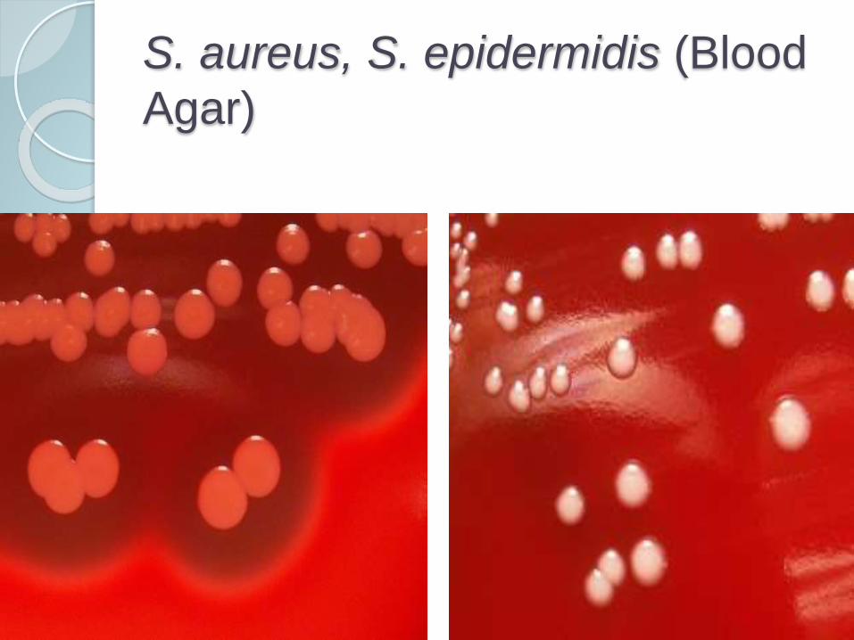
S. aureus, S. epidermidis (Blood
Agar)
DIAGNOSTIC TESTS:
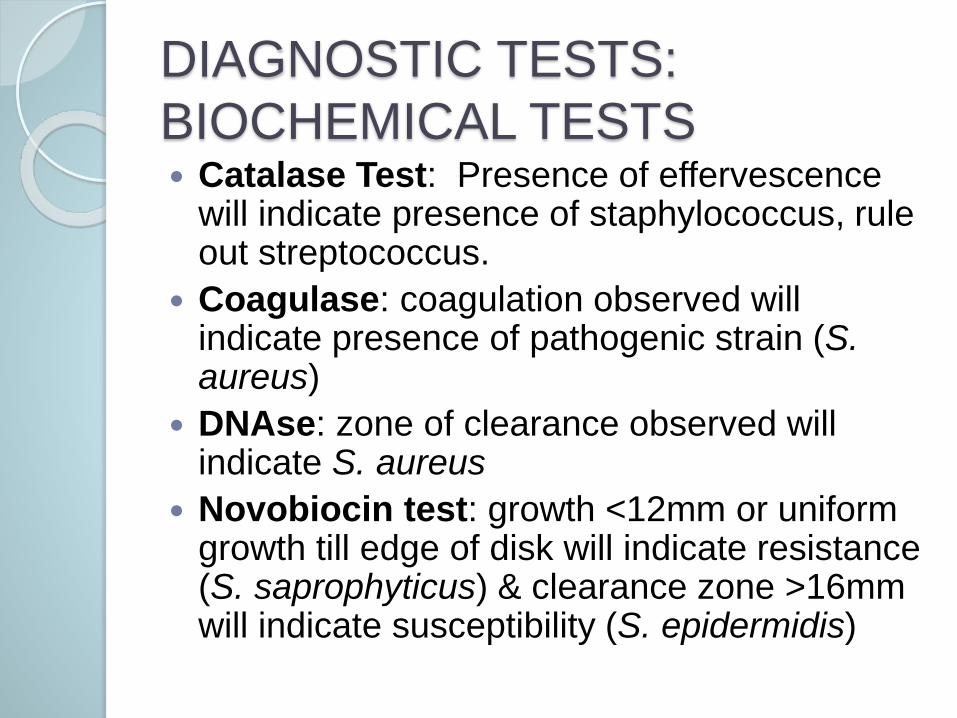
BIOCHEMICAL TESTS Catalase Test: Presence of effervescence
will indicate presence of staphylococcus, rule out streptococcus.
Coagulase: coagulation observed will indicate presence of pathogenic strain (S. aureus)
DNAse: zone of clearance observed will indicate S. aureus
Novobiocin test: growth <12mm or uniform growth till edge of disk will indicate resistance (S. saprophyticus) & clearance zone >16mm will indicate susceptibility (S. epidermidis)
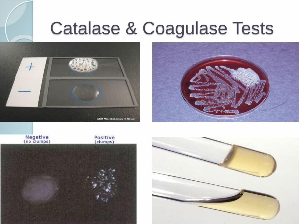
Catalase & Coagulase Tests
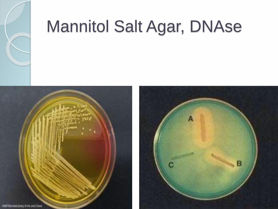
Mannitol Salt Agar, DNAse
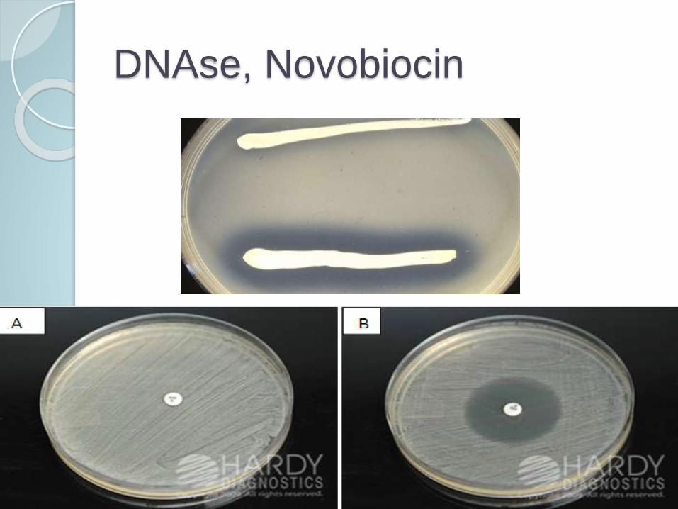
DNAse, Novobiocin

MANAGEMENT/TREATMEN
T Drainage of abscesses
Removal of catheters
Fluid replacement and electrolyte
balancing
Patients with MRSA should be
isolated
Administration of antimicrobials
TREATMENT
Choice and dosage of antibiotics depend on:
Infection site Illness severity Probability that resistant strains are
presentMany strains produce β-lactamase &
therefore resist Penicillin G & V.These can be treated with Methicillin,
Nafcillin or oxacillins.MRSA strains are resistant to the above,
can be treated with Vancomysin.VRSA strains have also emerged,
however.
PREVENTION AND
CONTROL Thoroughly sterilize reusable equipment;
organism is vulnerable to alcohol based sterilizers and moist heat at 600C.
Disinfection of hands between patient examinations
Isolation of MRSA patients
Maintenance of proper sanitation and body hygiene when handling food
Immediately cleaning and treating skin scratches, abrasions or puncture wounds
Use of protective gear when handling patients
EPIDEMIOLOGY
Staphylococcus, especially S. aureus is a major cause of nosocomial and community acquired infections.
Humans are a natural reservoir for S. aureusand S. epidermidis.
Young children will have higher colonization rates due to frequent contact with respiratory secretions & other exposures.
Spread is rapid in crowded areas with poor sanitation.
MRSA, originally associated with hospitals is increasingly acquired in communities.

THANKS FOR YOUR
ATTENTION


