Micro/nanofluidic devices for drug delivery Navid Kashaninejad a, ∗, Ehsanollah Moradi b , and Hajar Moghadas c a Queensland Micro- and Nanotechnology Centre, Nathan Campus, Griffith University, Brisbane, QLD, Australia b Department of Biomedical Engineering, Amirkabir University of Technology, Tehran, Iran c Department of Mechanical Engineering, Gas and Petroleum Faculty, Yasouj University, Yasuj, Iran ∗ Corresponding author: e-mail address: n.kashaninejad@griffith.edu.au Contents 1. Microfluidic in vitro drug delivery at the cellular level 2 1.1 Microfluidic concentration gradient generators (MCGGs) 2 1.2 Gradient generators for high-throughput drug screening 8 1.3 Challenges and possible solutions for microfluidic drug delivery at cellular level 11 2. Microfluidic in vitro drug delivery at tissue level 12 3. Microfluidic in situ drug delivery at organ level 19 3.1 Solid microneedles 22 3.2 Coated microneedles 22 3.3 Dissolvable microneedles 23 3.4 Hollow microneedles 24 3.5 Swollen microneedles 24 3.6 Challenge of microneedle drug delivery systems 24 4. Conclusion and future perspective 25 References 26 Abstract Micro/nanofluidic drug delivery systems have attracted significant attention as they offer unique advantages in targeted and controlled drug delivery. Based on the desired application, these systems can be categorized into three different groups: in vitro, in situ and in vivo microfluidic drug delivery platforms. In vitro microfluidic drug delivery platforms are closely linked with the emerging concept of lab-on-a-chip for cell culture studies. These systems can be used to administer drugs or therapeutic agents, mostly at the cellular or tissue level, to find the therapeutic index and can potentially be used for personalized medicine. In situ and in vivo microfluidic drug delivery platforms are still at the developmental stage and can be used for drug delivery at tissue or organ levels. A famous example of these systems are microneedles that can be used for painless Progress in Molecular Biology and Translational Science Copyright # 2021 Elsevier Inc. ISSN 1877-1173 All rights reserved. https://doi.org/10.1016/bs.pmbts.2021.07.018 1 ARTICLE IN PRESS
Transcript
Micro/nanofluidic devicesfor drug deliveryNavid Kashaninejada,∗, Ehsanollah Moradib, and Hajar MoghadascaQueensland Micro- and Nanotechnology Centre, Nathan Campus, Griffith University, Brisbane,QLD, AustraliabDepartment of Biomedical Engineering, Amirkabir University of Technology, Tehran, IrancDepartment of Mechanical Engineering, Gas and Petroleum Faculty, Yasouj University, Yasuj, Iran∗Corresponding author: e-mail address: [email protected]
Contents
1. Microfluidic in vitro drug delivery at the cellular level 21.1 Microfluidic concentration gradient generators (MCGGs) 21.2 Gradient generators for high-throughput drug screening 81.3 Challenges and possible solutions for microfluidic drug delivery at
cellular level 112. Microfluidic in vitro drug delivery at tissue level 123. Microfluidic in situ drug delivery at organ level 19
3.1 Solid microneedles 223.2 Coated microneedles 223.3 Dissolvable microneedles 233.4 Hollow microneedles 243.5 Swollen microneedles 243.6 Challenge of microneedle drug delivery systems 24
4. Conclusion and future perspective 25References 26
Abstract
Micro/nanofluidic drug delivery systems have attracted significant attention as theyoffer unique advantages in targeted and controlled drug delivery. Based on the desiredapplication, these systems can be categorized into three different groups: in vitro, in situand in vivo microfluidic drug delivery platforms. In vitro microfluidic drug deliveryplatforms are closely linked with the emerging concept of lab-on-a-chip for cell culturestudies. These systems can be used to administer drugs or therapeutic agents, mostly atthe cellular or tissue level, to find the therapeutic index and can potentially be used forpersonalized medicine. In situ and in vivomicrofluidic drug delivery platforms are still atthe developmental stage and can be used for drug delivery at tissue or organ levels.A famous example of these systems are microneedles that can be used for painless
Progress in Molecular Biology and Translational Science Copyright # 2021 Elsevier Inc.ISSN 1877-1173 All rights reserved.https://doi.org/10.1016/bs.pmbts.2021.07.018
and controllable delivery of drugs or vaccines through human skin. This chapter pre-sents the cutting edge advances in the design and fabrication of in vitro microfluidicdrug delivery systems that can be used for both cellular and tissue drug delivery. It alsobriefly discusses the in situ drug delivery platforms using microneedles.
1. Microfluidic in vitro drug delivery at the cellular level
Drug delivery has different stages in the body, and dissolvable drug
molecules go through a process that finally ends up with delivering the drug
molecules to the site of interest and taking up the drug by the targeted cells;
therefore, drug delivery experiments at the cellular level are of great impor-
tance and commonly applied for the study of cytotoxicity testing, drug
screening and drug discovery.1 In the last decade, various in vitro cell culture
platforms have been developed for drug screening and development prac-
tices. Although these conventional methods are cost-efficient, robust, and
easy to manipulate, they lack a physiologically-relevant microenvironment,
sustained cell viability, and functional stability. Hence, there is a dire need in
the drug development pipeline for developing predictive in vitromodels with
high preclinical efficacy that faithfully recapitulate cell-cell interactions and
cellular phenotype.2–6
Microfluidic systems offer a high surface-to-volume ratio that gives rise
to unique surface-dominating phenomena such as slip flow.7 Surface-
dominated forces in microfluidic systems offer significant advantages for
drug delivery application. For instance, the laminar and diffusion-based flow
in microchannels can precisely control the mechanism of drug release.
Recently, microtechnologies and microfluidic cell culture systems have
paved the way for examining complex biologic processes, and with control-
ling spatiotemporal parameters, they can mimic the intricate cellular micro-
environment properties like cell morphology, cell-drug interactions, and
chemical concentration gradients.6,8 Microfluidic devices can offer chemical
concentration gradients with great accuracy and sensitivity, ideal for produc-
ing controllable drug profiles and evaluating concentration-dependent
possess extraordinary characteristics like cost-efficient operation, the ability
to generate accurate and flexible drug profiles, allow real-time cellular
2 Navid Kashaninejad et al.
ARTICLE IN PRESS
response monitoring, and offer high-throughputs.1,12 Microfluidic concen-
tration gradient generators are generally divided into diffusion-based and
flow-based platforms and have become an essential tool for studying biolog-
ical phenomena like cell migration, proliferation, chemotaxis, drug screen-
ing, and personalized medicine.1,13,14 The following section overview
different cell-based MCGGs for drug testing experiments.
1.1.1 Diffusion-based gradient generatorsThe diffusion base method’s gradient generators mostly consist of a mem-
brane or hydrogel to create an environment suitable for diffusive transport.12,15
There are two compartments with high and low reagent concentrations in
most cases, and the targeted cells are cultured in the bridging channel between
the source and the sink.1 In one study, a shear stress-free MCGGwas devel-
oped for studying cytoskeleton dynamics molecular pathways and the effect
of the Cytochalasin D gradient onMG-63 cellular response.16 In this study, a
profile of Cytochalasin D concentration ranging between 0 and 5 μg/mL
was produced in five independent cell chambers, and the concentration-
dependent cell responses were examined by measuring cell area and cell
eccentricity.16
Different biological phenomena like chemotaxis and morphogenesis are
driven by essential cell signaling cues that are often delivered by biochemical
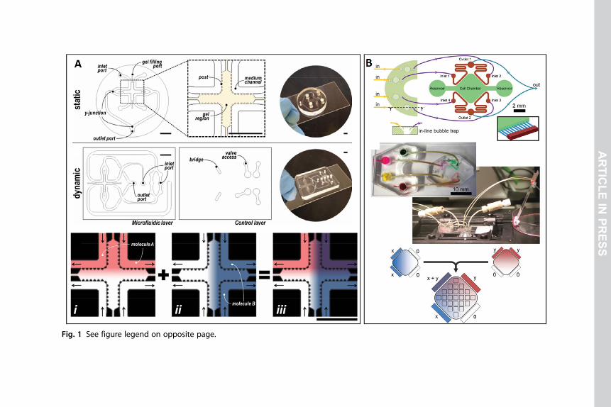
concentration gradients.17,18 A microfluidic biochip was developed in one
study for generating diffusion-based orthogonal linear concentration gradi-
ents in a 3D hydrogel-based scaffold in two different designs: Simultaneous
and Sequential (Fig. 1A).18 It was demonstrated that the two orthogonal
gradients of retinoic acid and smoothened agonist emulated the localized dif-
ferentiation of stem cells, and the neural tube was preferentially developed
in the high morphogens concentration regions. Moreover, HT1080, as a
highly metastatic fibrosarcoma cell line, was exposed to a rotating gradient
of Fetal Bovine Serum (FBS) for probing chemotactic response time and
adaptation in the tumor microenvironment. The direction of cell migration
and cell’s migratory behavior was evaluated by comparing average cell dis-
placements, distribution, and migration speed.18
In work done by Kilinc et al., a diffusion-based microchip was con-
structed to establish multiple compounds linear concentration gradient over
different cells for cell-based drug combination assays.19 The microfluidic
tool consisted of a cell culture chamber and parallel flow channels for cre-
ating a linear concentration gradient across the chamber. Producing
orthogonally-aligned gradients of two compounds, flowing concentrated
3Micro/nanofluidic devices for drug delivery
ARTICLE IN PRESS
Fig. 1 See figure legend on opposite page.
ARTICLE
INPRESS
solutions in two perpendicular directions is needed (Fig. 1B). A431 epider-
moid carcinoma cell line responds dissimilarly to EGF concentrations, and
while low concentration of EGF induces cell proliferation, higher concen-
trations result in cell apoptosis. This device generates orthogonal linear
gradients of EGFR andMEK inhibitors, and the resultant cell apoptosis gra-
dient in the MCGG demonstrated the ability of the device to determine the
combinatorial cell response to specific signaling pathways.19
1.1.2 Flow-based gradient generatorsIn the last decade, microfluidic flow-based gradient generators have revolu-
tionized a wide range of chemical and biological practices and produced
biochemical concentration gradients for cellular monitoring, and drug
screening applications have been one of them.1,10,20 MCGGs based on lam-
inar flow can provide desired ranges of biochemical concentration gradients
with high precision and controllability by manipulating convective
flow.12,21 In one study, a microfluidic gradient device was utilized for drug
screening and disease modeling with evaluating iPSC-derived motoneurons
responses to the riluzole concentration gradient.22 TheMicrofluidic biochip
was constructed with micropillar arrays for trapping uniform-sized neuro-
spheres and promoting neurite network formation. The peripheral channels
were connected to the central flow channel through microgrooves that gen-
erated a stable concentration gradient with a combination of convection and
diffusion. To examine the effect of drug concentration gradient on neuro-
spheres, cells were treated with thapsigargin, a neurotoxin that promotes
neuronal death and is a model reagent for neurodegenerative diseases.
A concentration gradient of riluzole as a drug to alleviate thapsigargin’s
neurotoxicity was produced by injecting drugged and drug-free culture
Fig. 1 Design and operation principle of diffusion-based microfluidic orthogonal gradi-ent generators. (A) Static and dynamic versions of the microfluidic gradient generatorfor producing a combinatorial concentration gradient ofmorphogens to simulatemotorneurons localized differentiation in the process of neural tube development and prob-ing chemotactic response time and adaptation in the tumor microenvironment. (B) Amicrofluidic device for generating linear orthogonally-aligned gradients of MEK andEGFR inhibitors and analyzing the A431 epidermoid carcinoma cells apoptosis pathway.Panel (A) reproduced with permission from Uzel SG, Amadi OC, Pearl TM, Lee RT, So PT,Kamm RD. Simultaneous or sequential orthogonal gradient formation in a 3D cell culturemicrofluidic platform. Small (Weinheim an der Bergstrasse, Germany). 2016;12(5):612–622.Panel (B) reproduced with permission from Kilinc D, Schwab J, Rampini S, et al. A micro-fluidic dual gradient generator for conducting cell-based drug combination assays.Integr. Biol. 2015;8(1):39–49.
5Micro/nanofluidic devices for drug delivery
ARTICLE IN PRESS
mediums into different inlets. It was shown that riluzole promoted neuro-
rescue or neuroprotection in the regions with higher drug concentration,
while thapsigargin induced considerable damage to neurospheres in the dead
zone.22
Christmas tree-like gradient generators have been among the most com-
monly used tools for creating biochemical concentration gradients. In these
flow-based microdevices, two reagents with different concentrations com-
bine at repeated T-junctions, and the desired concentration gradient is
generated in a direction perpendicular to the fluid flow by diffusion across
the laminar flow interface.10,23,24 In one study by Tang et al., a bone-on-
a-chip microdevice was developed for recapitulating bone tissue
microenvironment.25 A hydroxyapatite-PDMS microfluidic concentration
gradient chip was fabricated by the stereolithographic printing process, and it
contained a ceramic layer and a PDMS cover with Christmas tree-like struc-
ture. In high media flow rates, the reagents had inadequate time to diffuse
and mix uniformly and could cause excessive shear stress damage to cells,
while in low flow rates, there was a lack of sufficient drive to push the fluid
forward. To determine the ideal flow rate to generate the appropriate concen-
tration range, a COMSOL flow analysis was applied (Fig. 2A).25 To examine
the device’s functionality for bone-related drug studies, mouse osteosarcoma
cells (UMR-106) were cultured in the device, and a concentration gradient of
doxorubicin as a primary antitumor drug for the treatment of osteosarcoma
was produced by the Christmas tree gradient generator. The confocal micros-
copy examinations showed that the number of UMR-106 cells in the region
with high drug concentration decreased significantly. Moreover, the DOX
concentration in the zone with half of the control group cell population
was measured to determine the IC50 of DOX, and the results were validated
with the conventional culture plate model.25
Shimizu et al. created an ECM-based MCGG from transglutaminase
enzymatically cross-linked gelatin with a porous nature that permitted inter-
stitial flow and generated a culture surface with a controlled biochemical gra-
dient on the surface.26 To evaluate the applicability of the device for drug
screening purposes, the response of HUVECs to histamine concentration gra-
dient induced by interstitial flow was examined (Fig. 2B). Endothelial cells
have a contracting response to histamine introduction to increase the barrier
permeability of the vessel. In this study, the cellular area of the endothelial cells
before and after the histamine treatment was measured as the indication of cel-
lular shrinkage, and HUVECs cultured in the high histamine concentration
region shrunk significantly greater than the histamine-free region.26
6 Navid Kashaninejad et al.
ARTICLE IN PRESS
Fig. 2 Design and applications of flow-based microfluidic drug concentration gradientgenerators. (A) A christmas tree-like HA-PDMS-basedMCGG for creating a concentrationgradient of doxorubicin. (B) An ECM-based MCGG for evaluating HUVECs response tohistamine concentration. (C) The Metabolic Patterning on a Chip platform for recapitu-lating liver zonation through concentration gradients of glucagon and insulin to inducethe carbohydrate/nitrogen metabolism pattern and examine the drug-induced hepato-toxicity via Cytochrome P450 activity-dependent response to acetaminophen. Panel(A) reproduced from Tang Q, Li X, Lai C, et al. Fabrication of a hydroxyapatite-PDMS micro-fluidic chip for bone-related cell culture and drug screening. Bioact. Mater. 2021;6(1):169–178, under a Creative Commons License. Panel (B) reproduced with permissionfrom Shimizu A, GohWH, Itai S, Karyappa R, Hashimoto M, Onoe H. ECM-based microfluidicgradient generator for tunable surface environment by interstitial flow. Biomicrofluidics2020;14(4):044106. Panel (C) reproduced from Kang YB, Eo J, Mert S, Yarmush ML, UstaOB. Metabolic patterning on a chip: towards in vitro liver zonation of primary rat andhuman hepatocytes. Sci. Rep. 2018;8(1):8951, under a Creative Common Attribution 4.0International License.
7Micro/nanofluidic devices for drug delivery
ARTICLE IN PRESS
In another organ-on-chip, a metabolic patterning on a chip (MPOC)
was constructed to mimic the liver zonation.27 One key physiological fea-
ture of the liver sinusoid is hepatic zonation so that cells display various activ-
ities and characteristics based on their location in the liver sinusoid. In this
MPOC device, tree-like shape concentration gradients of hormones and
enzymatic inducers were produced to create a dynamic metabolic pattern
across the cell microchamber and mimic in vivo-like liver zonation and zonal
toxic response (Fig. 2C).27 The hepatocytes’ cell viability and mitochondrial
activity gradually decreased across the cell chamber’s width from zone 1-like
to zone 3-like region due to the higher CYP1A2 activity of the hepatocytes
in zone 3.27
1.2 Gradient generators for high-throughput drug screeningIn the last years, fast-paced progress in developing microfluidic tools for cell
studies and integrating robotics, automated analyzing devices, and biosensors
in biomedical microdevices have made the microfluidic biochips promising
tools for high-throughput drug screening; therefore, microfluidic platforms
like MCGGs and microarrays have been the gold standard in drug testing
experiments.28–32 A microfluidic device was established to cultivate human
high-grade glioma cells (UVW) spheroids obtained from human prostate
biopsies and test the anticancer drugs by an MCGG tool.33 In this device,
drug solution and culture media were injected into two separate reservoirs
on two sides of the cell culture channel to create a symmetrical hydrostatic
pressure-driven flow through the cell microarray that concurrently trans-
ported drug and media from each side of the channel to the central channels
reservoirs (Fig. 3A). This structure produced a self-generated concentration
gradient tool for cancer drug screening. UVW spheroids were treated with a
concentration gradient of a chemotherapeutic agent called cisplatin, and the
cell viability was assessed with PI and FDA double staining. The results
showed that the drug concentration was proportional to the spheroid’s shape
factor parameter, which is a sign of spheroids disaggregation and outline
roughness and is a readout for spheroids’ health.33
A disk-like microfluidic gradient generator with compact double spiral
mixers was developed in one research for producing three stable, accurate,
and controllable drug gradients.34 Two cancer cell lines (MCF-7 and
HepG2) were cultured in the device and treated with three different
concentration profiles of anticancer drugs (doxorubicin and cisplatin).
Doxorubicin, cisplatin, and drug-free mediumwere injected into the device
8 Navid Kashaninejad et al.
ARTICLE IN PRESS
Fig. 3 See figure legend on next page.
ARTICLE
INPRESS
via inlet 1,2 and 3, respectively. Consequently, three concentration gradients
of doxorubicin, cisplatin, and the combination of the drugs were generated
in 12 separate chambers (Fig. 3B). Cells that were cultured in the chambers
with high drug concentration for a prolonged time had significantly lower
live cell populations, and the most effective concentration profile with the
highest cell apoptosis induction was the combination of doxorubicin (75%)
and cisplatin (25%) in Chamber 12. The results demonstrated that this device
is a powerful tool to generate multidrug concentration gradients for evalu-
ating dose- and time-dependent cell-based studies and high-throughput
drug screenings.34
A microfluidic spheroid culture device with a concentration gradient
generator was developed for examining cancer drug efficacy (Fig. 3C).35
The colon cancer cell line (HCT116) and glioma cell line (U87) were cul-
tured in the microdevice with concave microwells to form the cancer spher-
oids with uniform size and distribution. As an anticancer drug, Irinotecanwas
injected into the chip to produce a concentration gradient, and the function-
ality of the device was evaluated by monitoring the shape and viability of the
cells. Spheroids in themicrowells with high irinotecan concentration lost their
roundness, collapsed severely, and live/dead staining images indicated that cell
viability declined continuously with elevated drug concentration.35
A paper-based microfluidic biochip was constructed to generate a
drug concentration gradient and assess cell response to drugs based on
high-throughput screening.28 For creating the drug concentration gradient,
two solutions containing doxorubicin and buffer were injected in two
Fig. 3 Layout and functions of drug concentration gradient generators forhigh-throughput drug screening. (A) A microfluidic device for culturing humanhigh-grade glioma cells (UVW) spheroids obtained from human prostate biopsiesand producing a self-generated concentration gradient for cancer drug screening.(B) Disc-like microfluidic three concentration gradient generator for analyzing differentcombinations of doxorubicin and cisplatin on MCF-7 and HepG2 tumor cell lines. (C) Amicrochip with concave microwells for culturing colon cancer cell line (HCT116) and gli-oma cell line (U87) spheroids and generating Irinotecan concentration gradient. Panel(A) reproduced from Mulholland T, McAllister M, Patek S, et al. Drug screening ofbiopsy-derived spheroids using a self-generated microfluidic concentration gradient. Sci.Rep. 2018;8(1):14672, under a Creative Common Attribution 4.0 International License.Panel (B) reproduced with permission from Shen S, Zhang X, Zhang F, Wang D, Long D,Niu Y. Three-gradient constructions in a flow-rate insensitive microfluidic system fordrug screening towards personalized treatment. Talanta 2020;208:120477. Panel(C) reproduce from Lim W, Park S. A microfluidic spheroid culture device with a concentra-tion gradient generator for high-throughput screening of drug efficacy.Molecules 2018;23(12):3355, under an open access Creative Common CC BY license.
10 Navid Kashaninejad et al.
ARTICLE IN PRESS
separate inlets, and HeLa cells encapsulated in the collagen type I matrix
were cultured in a microwells array, and a contrary trend of cell viability
compared with drug concentration was shown. In paper-based micro-
devices, fluid flow is mainly derived by capillary force in porous paper chan-
nels, and there is no need for additional equipment like pumps and valves.
Moreover, in flow-based MCGGs, operating the device for a prolonged
time would be quite costly due to the excessive reagent requirement, while
in paper-based tools, the solution consumption is significantly reduced.28
1.3 Challenges and possible solutions for microfluidic drugdelivery at cellular level
Although various microfluidic platforms have been developed so far for
recapitulating drug–cell interactions and mimic in vivo cellular microenviron-
ment of different tissues, there are still some limitations in the micro-
physiological complexity and drug-induced cellular pathways resemblance.11
Microfluidic systems for dynamic fluid manipulation and cell culture have rev-
olutionized the research in concentration gradient generators by introducing
state-of-the-art designs and configurations with extraordinary precision in flow
control geometric constraints to form gradients with high stability and accu-
racy. These systems, based on the type of the microdevice, could rely on only
molecular diffusion or diffusion-convection phenomena for generating
gradients.1,12
Diffusion-based MCGGs compared to flow-based platforms, have a more
cost-effective setup, are needless of expensive pumping systems, andminimize
the cell damage due to a shear stress-free environment. However, these
flow-free gradient generators are commonly much slower in producing the
concentration gradient and require improvements in gradient stability and
significant reduction of source-sink replenishment frequency.1,12,18
Convection-based tools are able to sustain a drug concentration gradient
within a fluid-flow environment and are suitable for the maintenance of a bio-
chemical gradient within a specific tissue.21 Moreover, these flow-based sys-
tems can create concentration gradients rapidly in a small area because of their
short relaxation time and large P�eclet (Pe) number, making them appropriate
for applications where quick response is required. Though the significant hur-
dle in these devices is that, the gradient at a fast flow rate can damage the cells
and ultimately trigger adverse efficiency in the cell signaling system.19,20 To
alleviate the problem, one recommended solution has been applying inter-
connected microgrooves and separate chambers for minimizing the effect
of shear stress and generating long-term stable concentration gradients.22
11Micro/nanofluidic devices for drug delivery
ARTICLE IN PRESS
In traditional tree-like microdevices, cells cultivated on the surface of the
microchambers are exposed to direct biochemical factors from the fluid
flow. In contrast, in the physiological microenvironment, cells are exposed
to a complex and controlled concentration gradient of soluble factors
through an interstitial flow.26 Biomimetic hydrogels like collagen and aga-
rose can increase the system’s physiological relevance, generate a 3D scaffold
environment for embedded cells, and recapitulate in vivo-like concentration
gradients. These 3D ECM-like constructs could mimic tissue physiological
functions such as microvasculature formation and axon guidance by chemo-
taxis due to their porous nature, albeit hydrogel-based systems usually
are limited in throughput and spatiotemporal gradient conditions due to
slow diffusion rate and also add experimental complexity and variability
to the system.15,16,18,32 The gradient in most common MCGGs is limited
to one or multiple fixed perpendicular directions that restrict the positioning
and orientation of drug gradients for cell re-orientation and migration
assays. An omnidirectional radially symmetric MCGG was developed to
overcome this concern for orienting laminar flow in 360° freely and manip-
ulating chemical profiles in time and space.36
There is currently an urgent need to execute accurate and
high-throughput predictive in vitro platforms in the pharmaceutical industry
that resembles human biology like biochemical concentration gradients at a
higher level and cut the time-consuming and costly drug procedures discov-
ery pipeline.11,28,30 Microfluidic systems have displayed remarkable benefits
for drug screening assays due to their capacity for miniaturization and para-
llelization. Repeatable and robust results could be generated for an extended
period with a high level of organotypicalness and in vivo microenviron-
ment resemblance by employing scaled-down biosensors and automated
features. The balance between the operational simplicity and biological
complexity may end up with the commercialization of MCGGs and
improve the user-friendliness of these microdevices.11,29,32
2. Microfluidic in vitro drug delivery at tissue level
There is an urgent necessity in the drug screening development pipe-
line for emerging predictive laboratory tissue models that truly orchestrate
in vivo microenvironment and drug–cell interactions.37,38 Conventional
2D or 3D in vitro platforms lack physiologically-relevant tissue microstruc-
ture, sustained cell viability, functional stability, and unchanged cellular
morphology and phenotype.39,40 In the past years, microfluidic tissue on
12 Navid Kashaninejad et al.
ARTICLE IN PRESS
chips have transformed the study of complex biologic practices like drug
metabolism/toxicity and cancer research, and with monitoring spatiotem-
poral factors, these microdevices are capable of recapitulating the in vivo-like
microenvironment properties like cell–cell and cell–ECM interactions, cel-
lular composition, and tissue 3D microarchitecture.2–4,41
Novel patient-specific drug-testing tools are needed in cancer research
and personalized medicine due to the current oncology drug development
pipeline.41,42 In one research, microdissected tissues (MDT) were obtained
from live tumor tissue and were segmented reproducibly to submillimeter
sections and cultured on an MDT-on-chip (Fig. 4A). The objective was
to obtain tissue sections large enough to resemble biologically relevant
gradients of nutrients, waste, and signaling molecules while also being small
enough to preserve high viability all over the tissue without oxygen defi-
ciency in the center. The microfluidic platform was able to trap the
microtissues in a low shear stress environment, and the chemosensitivity
of patient-derived ovarian cancer MDTs to carboplatin was evaluated by
loading the drug into the microchannels and measuring the chemoresponse
of treated and nontreated cell populations.41
In a study by Yin et al., a Microfluidic Kidney Chip was constructed for
coculturing renal proximal tubular epithelial cells (RPTECs) and peritubular
capillary endothelial cells (PCECs) and nephrotoxicity assessment.43 The
kidney-on-chip consisted of three parts: drug concentration gradient gener-
ator, three-layer microfluidic organ chip, and flow-temperature control
device (Fig. 4B). The two groups of drug concentrations were produced
on the gradient chip, then RPTECs and PCECs were seeded on opposite
sides ofmembranes, and after the drug treatment,RPTECs’ viabilitywas eval-
uated to determine the influence of that drug. Finally, a flow-temperature
control device was activated to stabilize the environments for prolonged cell
culture. The results demonstrated a significant reduction in cisplatin-induced
nephrotoxicity caused by the intervention of cimetidine in the microchip.
This controllable biomimetic kidney model replicated critical aspects of kid-
ney biological features, drug response, resistance, and toxicity could speed up
the drug discovery and screening practices.43
In the drug delivery at the cellular level section, we described theMCGGs
for drug delivery and toxicity testing; however, these platforms can generate
concentration gradients in a micron-sized level (e.g., cells).20,28,44 Therefore,
there is a dire need to introduce anMCGG that can produce continuous con-
centration gradients of multi mixtures (e.g., drugs) in a millimeter-sized sam-
ple (e.g., tissue, spheroids). Rismanian et al. proposed a new class of MCGG
13Micro/nanofluidic devices for drug delivery
ARTICLE IN PRESS
Fig. 4 See figure legend on opposite page.
ARTICLE
INPRESS
by linking amodified tree-likeCGGwith amicromixer to develop a so-called
Millimeter-sized sample /Multireagents/ Continuous CG with a diffusion-
convection-based approach (Fig. 4C).20 This microdevice introduced two
reagents concurrently with a continuous concentration gradient of two inlets
to deliver the reagents to a millimeter-sized sample (e.g., microtissue). This
microchip with measurable concentration relaxation time and low effect of
variability in the sample’s porosity and permeability and diffusivity of various
drugs could be an ideal tool for ex vivo drug chemosensitivity testing in per-
sonalized medicine.20
The high complexity and heterogeneity of the tumor microenvironment
play a substantial part in drug resistance and chemoresponse.45–47 Moreover,
the endothelium is essential in systemic drug delivery, provides nutrients and
oxygen to the tumor, and plays a crucial role in promoting new blood vessel
formation via angiogenesis. Thus, a simple capillary force-based microchip
for 3D tumor tissue-2D endothelium interfaces and drug testing was devel-
oped in a study. In this work, breast tumor cells were cultured in 3D micro-
wells, and the endothelial barrier was modeled on top of the 3D culture by
seeding HUVEC cells as an endothelium.47 The endothelium was efficient
and displayed physiological features, like the preferential proliferation of cells
with higher access to nutrients and oxygen. Next, the drug penetration and
toxicity assays were conducted in the tumor-endothelium model, and death
ligand TRAIL (TNF-related apoptosis-inducing ligand) conjugates with a
sizeable unilamellar vesicle (LUV) was tested. The results demonstrated
that LUV-TRAIL prompted cell death in a higher number and was more
effective than TRAIL.47
Fig. 4 Microfluidic systems for drug delivery and assessment at the tissue level.(A) Microdissected Tumor on-chip for ex vivo drug testing and personalized medicine.(B) A Microfluidic Kidney Chip for coculturing kidney cells and efficient drug screeningand cisplatin-induced nephrotoxicity assessment. (C) A Millimeter sized sample/multireagents/continuous concentration generator with functionality in drugchemosensitivity testing for personalizedmedicine. Panel (A) reproduced with permissionfrom Astolfi M, P�eant B, Lateef MA, et al. Micro-dissected tumor tissues on chip: an ex vivomethod for drug testing and personalized therapy. Lab Chip 2016;16(2):312–325. Panel(B) reproduced from Yin L, Du G, Zhang B, et al. Efficient drug screening and nephrotoxicityassessment on co-culture microfluidic kidney chip. Sci. Rep. 2020;10(1):6568, under aCreative Common Attribution 4.0 International License. Panel (C) reproduced with permis-sion from Rismanian M, Saidi MS, Kashaninejad N. A microfluidic concentration gradientgenerator for simultaneous delivery of two reagents on a millimeter-sized sample.J. Flow Chem. 2020;10:615–625.
15Micro/nanofluidic devices for drug delivery
ARTICLE IN PRESS
According to the reports analyzing different compounds’ clinical develop-
ments, oncology drug development’s success rate from phase 1, clinical trials
to FDA approval is less than 4%.48 This low success rate in clinical trials dem-
onstrates the deficiencies in existing prescreening methods. In another study, a
Tumor Chip system was introduced to deliver cancer–stromal interaction,
dynamic perfusion, replicate microvasculature, and high-throughput, as
shown in Fig. 5A.49 The L-TumorChipwas a three-layered biochip with spa-
tial control over the reconstruction of microvessels and tumor compartments,
with continuous media perfusion. The tumor compartment consisted of
human microvascular endothelial cells (HMVEC) and Matrigel-encapsulated
breast cancer cells culture layers, separated by a thin membrane to mimic the
in vivo tumormicroenvironment. The tumor biochip was capable of real-time
drug response monitoring for high-throughput drug kinetics studies. The
drug responses to doxorubicin treatment showed that the presence of
cancer-associated fibroblasts affects drug pharmacokinetics, while apoptotic
reactions specified by caspase-3 activities are higher in the coculture of cancer
cells with normal fibroblasts.49
In another research, a chip-based breast cancer equivalent with a
tumor-replicating microvasculature was developed to recapitulate the breast
tumors’ physiology and pathophysiology and assess anticancer drug effec-
tiveness.50 For resembling high and low flow perfusion regions in the tumor
microenvironment, two specific microvascular designs from mouse vascular
networks were applied for tumor-mimetic chip production. The two
regions with different degrees of fluidic exchange between the microvascu-
lar channels and the central tumor chamber represent well-perfused and
poorly-perfused breast tumors. Breast cancer cells were cocultured for a pro-
longed time in 3D hydrogel-based structure with stromal fibroblasts, and the
interstitial space between the chambers and endothelium with porous vas-
culature mimicked the physiological cancer-endothelial cell interactions.
Before activating their cytotoxic activity, Provided anticancer drugs must
overcome particular rate-controlling steps like the ability to perfuse through
the microvasculature and transport to the site of interest. In this work, doxo-
rubicin and paclitaxel were introduced to the breast tumor-on-chip, and the
cell viability was assessed. A remarkable reduction in viable tumor areas was
observed across all tested conditions, specifically in high perfusion chips,
because of higher drug penetration levels in the central tumor chambers.50
Glioblastoma multiforme (GBM), an extremely aggressive and high-
grade type of brain cancer, has a poor prognosis, with an average survival
time of 12–18 months—only 25% of glioblastoma patients survive more
16 Navid Kashaninejad et al.
ARTICLE IN PRESS
Fig. 5 Chip-based platforms for investigating tumor microenvironment and drug phar-macokinetics. (A) A high-throughput tumor-on-a-chip platform for emulating certaintumor-stroma and tumor–endothelium interactions and the stromal effects on drug
(Continued)
17Micro/nanofluidic devices for drug delivery
ARTICLE IN PRESS
than 1 year, and only 5% of patients survive more than 5 years—with stan-
dard GBM therapies.51,52 To address this concern, a brain cancer chip for
assessing tumor cell drug responses was administered to analyze drugs more
efficiently in preclinical trials and improve patient survival.53 In this work,
3D spheroids obtained from three patient GBM tumor cells were cultured
on a hydrogel layer with a concentration gradient generating design to pro-
duce two drug concentrations (FDA-approved temozolomide and
bevacizumab) simultaneously. The result of drug analysis indicated a com-
mon tendency toward higher cytotoxicity of two drugs combined with
single-drug treatment and a more robust efficacy of temozolomide than
bevacizumab.53
Colorectal cancer is now the third major cause of cancer-related death in
women and the second in men, primarily due to the high rate of cancer
metastasis and chemotherapeutic treatment ineffectiveness.54,55 There is a
dire need for more reliable and predictable screening approaches for colo-
rectal cancer drug development. In one research, a biomimetic tumor-on-
a-chip platform was developed to emulate in vivo relevant colorectal tumor
microenvironment and reconstruct functional microvasculature for preci-
sion nanomedicine delivery.56 Colorectal cancer cells were seeded in
matrigel-supported 3D structure, while endothelial cells were cultured in
a 3D vessel like architecture. To examine the concentration-dependent
response of cells inside the biochip to the antitumor drug, gemcitabine
(GEM) was loaded in CMCht/PAMAM dendrimer nanoparticles for deliv-
ering the drug to HCT-116 cancer cell line.With introducing GEM-loaded
dendrimer nanoparticles to the microchannels, cell death occurred in a gra-
dient manner, which demonstrated that GEM could be efficiently released
from the nanoparticles and taken up by the CRCs as a useful anticancer
therapeutic.56
As a preclinical method, tumor organoids are advantageous for
patient-specific treatment thanks to their ability to sustain critical biological
tissues’ physiological and structural features.57,58 Jung et al. developed a
microfluidic-based 3D lung cancer organoid culture platform enabling drug
Fig. 5—Cont’d responses to doxorubicin treatment. (B) Microfluidic biochip forthree-dimensional lung cancer organoid culturing and cisplatin and etoposideapoptosis tests. Panel (A) reproduced with permission from Chi C-W, Lao Y-H, AhmedAHR, et al. 2020 High-throughput tumor-on-a-chip platform to study tumor–stroma inter-actions and drug pharmacokinetics. Adv. Healthc. Mater. n/a(n/a):2000880. Panel(B) reproduced with permission from Jung DJ, Shin TH, Kim M, Sung CO, Jang SJ, JeongGS. A one-stop microfluidic-based lung cancer organoid culture platform for testing drugsensitivity. Lab Chip 2019;19(17):2854–2865.
18 Navid Kashaninejad et al.
ARTICLE IN PRESS
sensitivity tests directly on a microphysiological system, as shown in
Fig. 5B.59 This platform had high control over the size of lung cancer
organoids (LCO) and validated the production of LCOs derived from pri-
mary small-cell lung cancer tumors with rapid proliferation and exhibition
of disease-specific characteristics. The first-line chemotherapeutic for Lung
cancer (cisplatin and etoposide) was chosen as the test drugs in this work.
While the LCO cores and cells located away from blood vessels were more
resistant to chemotherapy and remained viable, apoptosis was shown in cells
around the organoid outer region. The drug concentration was associated
with reduced rates of cell viability.59
Hepatocellular carcinoma has become the second major cause of
cancer-related death, with an occurrence rate of about 850,000 new patients
each year. As a result of 2D cell culture models’ incapability in mimicking
complex liver pathophysiology and predicting the toxicity of the com-
pounds, few new anticancer drugs are available now.60,61 In one research,
a biomimetic 3D liver cancer-on-chip was developed, and decellularized
liver matrix (DLM) was obtained from rat liver tissue to integrate the
ECM proteins and GelMa into the liver chip.61 This system had an
improved competence to maintain hepatocytes viability and functions under
perfusion conditions and may be accredited to establish biochemical factors,
the maintenance of scaffold proteins, and the restoration of biophysical and
biomechanical cues enhanced resemblance of the 3D liver tumor microen-
vironment. For assessing the platform’s drug analyzing capability, acetamin-
ophen and sorafenib were tested for dose-dependent drug toxicity, and
linear dose-dependent hepatotoxicity was detected for both drugs in terms
of HepG2 cells viability, urea secretion, and albumin production.61
3. Microfluidic in situ drug delivery at organ level
Recent advances in microfabrication technology revolutionized the
drug delivery system. Microneedle (MN) is a noninvasive and painless local-
ized drug delivery system (DDS) that prevents gastrointestinal degradation and
bypasses hepatic metabolism. MNs have been used for drug delivery to differ-
ent organs, including skin, oral mucosa, vaginal mucosa, ocular tissue, nail, anal
sphincter, dermal papilla, and cardiac muscle.62–65 Fig. 6A and B demonstrate
the MNs DDS used for a drug-eluting balloon of vascular tissue66 and intra-
corneal drug injection,67 respectively. Various drugs such as insulin,68,69
calcein,69 DNA vaccine and nucleic acids,69 nanomedicine,69,70 ocular and
chronic wounds treated drug,69,71 peptide or subunit vaccination, and cancer
therapeutics72 have been delivered by MN drug delivery system. MNs are
19Micro/nanofluidic devices for drug delivery
ARTICLE IN PRESS
Fig. 6 See figure legend on opposite page.
ARTICLE
INPRESS
structures of the arrowhead, bullet, conical, cylindrical, pyramidal, obelisk,
octagonal cone shape with the height average between super-short
(<100 μm) to super-long (>1000 μm), the width of 50–250 μm, and the
tip thickness of 1–25 μm.73–75 Both sizes and shapes of MNs affect the
mechanical strength of the MN and the skin penetration’s depth.76 MNs have
been fabricated through various microfabrication techniques such as casting,
dry etching, wet etching, micromolding, 3D–printing, electroplating, laserablation, magnetorheological, and drawing lithography.75,77 The most-
reported microfabrication technique for MNs is the casting method, a
low-cost, massproductive, and easy to operation method shown in
Fig. 6D. Depending on their applications, MNs are made from different mate-
rials, including silicone,metals, polymers,78 and ceramics.79 Thematerial that is
used for MN fabrication must be safe, nontoxic, and strong enough to pene-
trate the skin.80,81 Based on the drug delivery route, MNs can be categorized
Fig. 6 MNs DDS. (A) MNs integrated with eluting balloon for drug delivery to vasculartissue, (a–c) conformal transfer molding steps consisting of premolding with UV curableadhesive resin, (d) alignment the drug eluting balloons (DEB) with flexible planar PDMSmold, (e) UV curing process and (f ) completed MNDEB, (g) Stereo- and optical micro-scopic images of MNDEB and MNs MN onto MNDEB surface (scale bar¼300 μm) (h) thescanning electron microscopic image of the constructed MN in the MNDEB surface,(scale bar¼100 μm). (B) Schematic of sustained drug releasing system using a detach-able hybrid microneedle pen system for intracorneal drug injection. (C) Schematic rep-resentation of the MNs DDS with five different drug delivery mechanisms, (a) Solid MNsthat are used as puncher to create micropores in the skin to increase the permeability,(b) Coated MNs that contain drug on the surface which released after penetration,(c) Dissolvable MNs that released the drug due to dissolution once inserted into thetissue, (d) Hollow MNs that injected the drug into the tissue from the external reservoirby pressure-driven flow or diffusion mechanism. (e) Swollen MNs that absorb the ISFafter insertion and administrate the drug. (D) The casting fabrication process of twolayers MNs consists of 2 components. (a) pouring component 1 into the mold,(b) vacuumed or centrifuged to release air bubbles, (c) solidification component 1,(d) pouring component 2 as the second layer, (e) vacuumed or centrifuged,(f ) Peeled away solidified two-layer MNs. Panel (A) reproduced with permission fromLee K, Lee J, Lee SG, et al. Microneedle drug eluting balloon for enhanced drug deliveryto vascular tissue. J. Control. Release. 2020;321:174–183. Panel (B) reproduced with permis-sion from Lee K, Song HB, Cho W, Kim JH, Kim JH, Ryu W. Intracorneal injection of a detach-able hybrid microneedle for sustained drug delivery. Acta Biomater. 2018;80:48–57. Panel(C) reproduced with some changes from Rzhevskiy AS, Singh TRR, Donnelly RF, AnissimovYG. Microneedles as the technique of drug delivery enhancement in diverse organs andtissues. J. Control. Release. 2018;270:184–202. Panel (D) reproduce with permission fromZhuang J, Rao F, Wu D, et al. Study on the fabrication and characterization oftip-loaded dissolving microneedles for transdermal drug delivery. Eur. J. Pharm.Biopharm. 2020;157:66–73.
21Micro/nanofluidic devices for drug delivery
ARTICLE IN PRESS
into five groups that are solid MN, coated MNs, hollow MN, dissolvable
MNs, and swollen MNs that are shown in Fig. 6C. In the following, each
group will be discussed in more detail to address the operation mechanism,
material, fabrication method, and challenges.
3.1 Solid microneedlesThe first generation of MNs is solid MNs. Solid MNs were applied as micro
punchers to produce small pores in the skin and increase skin penetration.
After solid MN insertion, the drug in the form of liquid, cream, gel, or lotion
is applied in created pores, as shown in Fig. 6C(a). Different types of materials,
including silicone, metals (stainless steel, titanium), polymers,78 and
ceramics,79 have been used for solid MNs preparation. Solid MNs have been
fabricated through various techniques such as dry etching, wet etching,
polycaprolactone, sucrose, and (2-hydroxypropyl)-β-cyclodextrin.73 The
key parameters of the coated material are hydrophilicity, biocompatibility,
and biodegradability. Some stabilizers were usually added to the matrix to
decrease the damage to the therapeutics, such as dextran, glucose, inulin,
sucrose, and trehalose.73 There are various coating methods such as dip coat-
ing, gas-jet drying, rolling coating, spray coating, electrohydrodynamic
atomization coating, and layer-by-layer coating.73,84 The biggest drawback
of the coated MNs is the low drug loading capacity because, in such type of
MN, a very small dose of drug material is only coated on the surface of the
22 Navid Kashaninejad et al.
ARTICLE IN PRESS
MN. For example, for polylactic acid MNs that were coated with a solution
of sulforhodamine B, the drug loadings were up 18 ng per needle for 750 μmMNs. It was shown that by increasing the viscosity of the coating solutions,
the drug loading could be increased. However, the drug delivery efficiency
was reported as 90%.85
3.3 Dissolvable microneedlesDissolvableMNs are made from dissolvable material in aqueous media. After
insertion, MNs absorb the interstitial fluid (ISF) and eventually dissolve,
leading to the sequential release of the loaded drug, Fig. 6C(c).
Dissolvable MNs can be made from a quick or long-time dissolvable mate-
rial, making them good candidates for controlled-release delivery.86–88
Biopolymers and sugars material similar to coated MNs were commonly
used to prepared dissolvable MNs through micromolding,75 casting,89
drawing lithography, droplet-born air blowing methods.90 As a significant
advantage, there is no limitation for the size of therapeutic molecules for
applying in dissolvable MNs. Similar to the coated MNs, the efficiency of
drug delivery of the dissolvedMNs depends on the dissolution of the matrix.
However, the agent dosing is much higher for dissolvedMNs. This group of
MNs is economical and effective, thus attracting many researchers’ atten-
tion, and promising good tools for MNs DDS.88,91,92 One of the major
problems with the dissolvable MNs were poor mechanical strength and
weakness to penetrate the skin, however, recently acceptable strong MNs
were reported.93–96 For example, dissolvable polymeric MNs with a small
amount of graphene oxide (GO) was used for the transdermal delivery of
the chemotherapeutic, HA15, to melanoma-bearing mouse models. MNs
demonstrated good mechanical strength (10–17 times at 500 mg/mL
GO), increased moisture resistance, self-sterilization, antibacterial and
anti-inflammatory properties, and controlled drug release by near-infrared
light-activated.94 There are two categories of dissolvable MNs, hole body
and tip loaded systems. In the first category, the hole MN body was made
of dissolvable material, while in the second one, only the tip of the MNs
consists of dissolvable drug-loaded material.67,97,98 An MN with a
drug-loaded biodegradable tip and a supporting base was developed to
sustained drug release into the cornea of an Acanthamoeba keratitis mouse
model. Follow-up administration for 7 days confirmed the therapeutic effi-
cacy of tip-loaded MN.67 Although the tip-loaded MNs facilitate drug pen-
etration into the deeper region, the dose of the loaded drug is small as the
small volume tip.
23Micro/nanofluidic devices for drug delivery
ARTICLE IN PRESS
3.4 Hollow microneedlesHollow MNs are a miniature version of hypodermic needles. After hollow
MNs insertion, the drug is injected under the tissue by pressure-driven flow
or diffusion, Fig. 6C(d). Agent dosing is more for hollow MNs in compar-
ison with the other kind of MNs. For example, a single hollow microneedle
was integrated with a magnetic polymer composites device and inductive
sensing instrument to controlled drug release as a wireless pumping system.
The fabricated systemwas able to dosage sensing.99 HollowMNswere made
in various configurations with out-off-center pore and side hole. They have
been mainly prepared from similar materials for solid MNs, such as silicon
metals, glass, polymers,81 using the multistep process of deep reactive ion
etching, wet chemical etching, plasma etching, isotropic etching, laser
micromachining, digital light processing-based projection stereo-
lithography, micropipette pulling, and sacrificial micromolding.79 Due to
the complex geometry of the hollow MNs, the manufacturing process of
this type ofMNs aremore complicated and time-consuming than the others.
3.5 Swollen microneedlesA new generation of theMNs is swollenMNs fabricated from swollenmate-
rials such as hydrogels. After the insertion, the MNs will swoll due to ISF
absorption and subsequently, the loaded agent released, Fig. 6C(e).
Swollen MNs were prepared from the combination of the swollen polymers
such as methyl vinyl ether and maleic anhydride (PMVE/MAH) and poly-
ethyleneglycol (PEG), PMVE and maleic acid and PEG, 2-hydroxyethyl
methacrylate and ethylene glycol dimethacrylate, polyvinyl alcohol, ethyl
acrylate, and methacrylate.73,100 The most common fabrication method
of the swollen MNs is the casting method. For example, silk fibroin scaffold
was used with poly(ethylene glycol) diacrylate and sucrose as the needle
matrix to fabricate swollen MNs with three formulations of drugs, including
Rhodamine B, doxorubicin, and indocyanine green.101 Like hollow MNs,
swollen MNs create conduct for drug delivery that can continuously insert
the agent from the external reservoir. However, the administration time for
the swollen MNs is greater than hollow MNs, which makes the drug deliv-
ery time-consuming.
3.6 Challenge of microneedle drug delivery systemsMNs drug delivery has many benefits: MNs realize noninvasive drug deliv-
ery, have better patient compliance because of reducing dose frequency,
24 Navid Kashaninejad et al.
ARTICLE IN PRESS
realize the possibility of self-administration, prevent first-pass metabolism
and gastrointestinal incompatibility, have minimal or no side effects, can
maintain the plasma drug level, have the possibility of large molecules
administration, and are suitable for the drug with short biological half-life
and narrow therapeutic index.102 However, there are some limitations
and challenges for using MNs for drug delivery, including less accuracy of
dose compared to conventional hypodermic needles, variation in delivery
due to skin condition, skin-dependent penetration mechanism, the
possibility of tissue damage and venues collapse due to repetitive injection,
and risk of breaking and remaining under the skin in case of using
nonbiodegradable MNs.103
4. Conclusion and future perspective
Miniaturization offers significant advantages in drug delivery applica-
tion that can be realized using microfluidic technology. In this chapter, the
focus was given to in vitro and in situ microfluidic systems for drug delivery.
To this aim, first, the latest advances in developing microfluidic concentra-
tion gradient generators (MCGGs) that can deliver the drug at cellular level
were discussed. In particular, both diffusion-based and flow-based MCGGs
as well as gradient generators for high-throughput drug screening, were
thoroughly evaluated. These systems have become an essential tool for
studying biological phenomena like cell migration, proliferation, chemo-
taxis, drug screening, and personalized medicine. Next, we evaluated the
state-of-the-art lab-on-a-chip systems for in vitro drug delivery at tissue level
was investigated. Finally, the design and fabrication of various in situ
microneedle-based drug delivery systems, including solid microneedles,
coated microneedles, hollow microneedles, microneedles dissolvable and
swollen microneedles, were explained. We concluded each part by briefly
identifying the present challenges and presenting the possible solutions.
Although tremendous efforts have been made in the design, fabrication
and characterization of microfluidic devices for drug delivery applications,
this field, especially in vivo microfluidic drug delivery platforms, is still at a
preliminary stage. There are still many challenges that need be addressed
in order to realize an efficient implantable microfluidic device for controlled
drug delivery. Besides biocompatibility and biodegradability, one of the
major challenges of the in vivo microfluidic systems for drug delivery is
the integration of an efficient mechanism for continuous pumping the drug
or the therapeutic agent in the body. It should be done autonomously using
25Micro/nanofluidic devices for drug delivery
ARTICLE IN PRESS
mainly through capillary action or other self-powered microfluidic
pump.104 Efficient integration of such a self-powered microfluidic pump
with microneedles can open up a great avenue in this field, leading to rev-
olutionary drug delivery and vaccine administration platforms. Moreover,
droplet-based microfluidics,105 flow-focusing droplet generators106 and
magnetofluidics107 are other highly efficient platforms that can potentially
be used for both drug synthesis and controlled delivery. There are still
plenty of rooms in improving such platforms for in vivo drug delivery and
personalized medicine.
References1. Nguyen N-T, Shaegh SAM, Kashaninejad N, Phan D-T. Design, fabrication and char-
acterization of drug delivery systems based on lab-on-a-chip technology. Adv DrugDeliv Rev. 2013;65(11):1403–1419.
2. Bhatia SN, Ingber DE. Microfluidic organs-on-chips. Nat Biotechnol. 2014;32:760.3. Esch EW, Bahinski A, Huh D. Organs-on-chips at the frontiers of drug discovery. Nat
Rev Drug Discov. 2015;14(4):248–260.4. Zhang B, Korolj A, Lai BFL, Radisic M. Advances in organ-on-a-chip engineering.
Nat Rev Mater. 2018;3(8):257–278.5. Shintu L, Baudoin R, Navratil V, et al. Metabolomics-on-a-chip and predictive
systems toxicology in microfluidic bioartificial organs. Anal Chem. 2012;84(4):1840–1848.
6. Haddrick M, Simpson PB. Organ-on-a-chip technology: turning its potential for clin-ical benefit into reality. Drug Discov Today. 2019;24(5):1217–1223.
7. Kashaninejad N, Chan WK, Nguyen N-T. Fluid mechanics of flow through rectan-gular hydrophobic microchannels. In: Paper Presented at: International Conference onNanochannels, Microchannels, and Minichannels; 2011.
8. Wu Q, Liu J, Wang X, et al. Organ-on-a-chip: recent breakthroughs and future pros-pects. Biomed Eng Online. 2020;19(1):9.
9. Lin JM. Cell Analysis on Microfluidics. Singapore: Springer; 2017.10. Rismanian M, Saidi MS, Kashaninejad N. A new non-dimensional parameter to obtain
the minimum mixing length in tree-like concentration gradient generators. Chem EngSci. 2019;195:120–126.
11. Moradi E, Jalili-Firoozinezhad S, Solati-Hashjin M. Microfluidic organ-on-a-chipmodels of human liver tissue. Acta Biomater. 2020;116:67–83.
12. Dravid A, Raos B, Aqrawe Z, Parittotokkaporn S, O’Carroll SJ, Svirskis D. A macro-scopic diffusion-based gradient generator to establish concentration gradients of solublemolecules within hydrogel scaffolds for cell culture. Front Chem. 2019;7:638.
13. Shi H, Nie K, Dong B, LongM, XuH, Liu Z. Recent progress of microfluidic reactorsfor biomedical applications. Chem Eng J. 2019;361:635–650.
14. Cabaleiro JM. Flowrate independent 3D printed microfluidic concentration gradientgenerator. Chem Eng J. 2020;382:122742.
15. Ahrens L, Tanaka S, Vonwil D, Christensen J, Iber D, Shastri VP. Generation of 3Dsoluble signal gradients in cell-laden hydrogels using passive diffusion. Adv Biosyst.2019;3(1):1800237.
16. Barata D, Spennati G, Correia C, et al. Development of a shear stress-free microfluidicgradient generator capable of quantitatively analyzing single-cell morphology. BiomedMicrodevices. 2017;19(4):81.
17. Jin T, Xu X, Hereld D. Chemotaxis, chemokine receptors and human disease.Cytokine. 2008;44(1):1–8.
18. Uzel SG, Amadi OC, Pearl TM, Lee RT, So PT, KammRD. Simultaneous or sequen-tial orthogonal gradient formation in a 3D cell culture microfluidic platform. Small(Weinheim an der Bergstrasse, Germany). 2016;12(5):612–622.
19. Kilinc D, Schwab J, Rampini S, et al. A microfluidic dual gradient generator for con-ducting cell-based drug combination assays. Integr Biol. 2015;8(1):39–49.
20. Rismanian M, Saidi MS, Kashaninejad N. A microfluidic concentration gradient gen-erator for simultaneous delivery of two reagents on a millimeter-sized sample. J FlowChem. 2020;10:615–625.
21. Parittotokkaporn S, Dravid A, BansalM, et al. Make it simple: long-term stable gradientgeneration in a microfluidic microdevice. Biomed Microdevices. 2019;21(3):77.
22. Mo SJ, Lee JH, Kye HG, et al. A microfluidic gradient device for drug screening withhuman iPSC-derived motoneurons. Analyst. 2020;145(8):3081–3089.
23. Wolfram CJ, Rubloff GW, Luo X. Perspectives in flow-based microfluidic gradientgenerators for characterizing bacterial chemotaxis. Biomicrofluidics. 2016;10(6):061301.
24. Ebadi M, Moshksayan K, Kashaninejad N, Saidi MS, Nguyen N-T. A tool for design-ing tree-like concentration gradient generators for lab-on-a-chip applications. ChemEng Sci. 2020;212:115339.
25. Tang Q, Li X, Lai C, et al. Fabrication of a hydroxyapatite-PDMSmicrofluidic chip forbone-related cell culture and drug screening. Bioact Mater. 2021;6(1):169–178.
26. Shimizu A, GohWH, Itai S, Karyappa R, HashimotoM, Onoe H. ECM-based micro-fluidic gradient generator for tunable surface environment by interstitial flow.Biomicrofluidics. 2020;14(4):044106.
27. Kang YB, Eo J, Mert S, Yarmush ML, Usta OB. Metabolic patterning on a chip:towards in vitro liver zonation of primary rat and human hepatocytes. Sci Rep.2018;8(1):8951.
28. Hong B, Xue P, Wu Y, Bao J, Chuah YJ, Kang Y. A concentration gradient generatoron a paper-based microfluidic chip coupled with cell culture microarray forhigh-throughput drug screening. Biomed Microdevices. 2016;18(1):21.
29. Wei Y, Zhu Y, Fang Q. Nanoliter quantitative high-throughput screening withlarge-scale tunable gradients based on a microfluidic droplet robot under unilateral dis-persion mode. Anal Chem. 2019;91(8):4995–5003.
30. Xu Z, Fang P, Xu B, et al. High-throughput three-dimensional chemotactic assaysreveal steepness-dependent complexity in neuronal sensation to molecular gradients.Nat Commun. 2018;9(1):4745.
31. Zhang Y, Xiao RR, Yin T, et al. Generation of gradients on a microfluidic device:toward a high-throughput investigation of spermatozoa chemotaxis. PLoS One.2015;10(11):e0142555.
32. Ezra Tsur E, Zimerman M,Maor I, Elrich A, Nahmias Y. Microfluidic concentric gra-dient generator design for high-throughput cell-based studies. Front Bioeng Biotechnol.2017;5:21.
33. Mulholland T,McAllisterM, Patek S, et al. Drug screening of biopsy-derived spheroidsusing a self-generated microfluidic concentration gradient. Sci Rep. 2018;8(1):14672.
34. Shen S, Zhang X, Zhang F, Wang D, Long D, Niu Y. Three-gradient constructions ina flow-rate insensitive microfluidic system for drug screening towards personalizedtreatment. Talanta. 2020;208:120477.
35. Lim W, Park S. A microfluidic spheroid culture device with a concentrationgradient generator for high-throughput screening of drug efficacy. Molecules.2018;23(12):3355.
36. Nakajima A, Ishida M, Fujimori T, Wakamoto Y, Sawai S. The microfluidic light-house: an omnidirectional gradient generator. Lab Chip. 2016;16(22):4382–4394.
37. Esch MB, Prot J-M, Wang YI, et al. Multi-cellular 3D human primary liver cellcultures elevate metabolic activity under fluidic flow. Lab Chip. 2015;15(10):2269–2277.
38. Bhushan A, Senutovitch N, Bale SS, et al. Towards a three-dimensional microfluidicliver platform for predicting drug efficacy and toxicity in humans. Stem Cell Res Ther.2013;4(Suppl 1):S16.
39. Lin R-Z, Chang H-Y. Recent advances in three-dimensional multicellular spheroidculture for biomedical research. Biotechnol J. 2008;3(9–10):1172–1184.
40. Ziołkowska K, Kwapiszewski R, Brzozka Z. Microfluidic devices as tools for mimick-ing the in vivo environment. New J Chem. 2011;35(5):979–990.
41. Astolfi M, P�eant B, Lateef MA, et al. Micro-dissected tumor tissues on chip: an ex vivomethod for drug testing and personalized therapy. Lab Chip. 2016;16(2):312–325.
42. Hay M, Thomas DW, Craighead JL, Economides C, Rosenthal J. Clinical develop-ment success rates for investigational drugs. Nat Biotechnol. 2014;32(1):40–51.
43. Yin L, Du G, Zhang B, et al. Efficient drug screening and nephrotoxicity assessment onco-culture microfluidic kidney chip. Sci Rep. 2020;10(1):6568.
44. Mitxelena-Iribarren O, Zabalo J, Arana S, Mujika M. Improved microfluidic platformfor simultaneous multiple drug screening towards personalized treatment. BiosensBioelectron. 2019;123:237–243.
45. Quail DF, Joyce JA. Microenvironmental regulation of tumor progression and metas-tasis. Nat Med. 2013;19(11):1423–1437.
46. Floor SL, Dumont JE,Maenhaut C, Raspe E. Hallmarks of cancer: of all cancer cells, allthe time? Trends Mol Med. 2012;18(9):509–515.
47. Virumbrales-Munoz M, Ayuso JM, Olave M, et al. Multiwell capillarity-based micro-fluidic device for the study of 3D tumour tissue-2D endothelium interactions and drugscreening in co-culture models. Sci Rep. 2017;7(1), 11998.
48. Wong CH, Siah KW, Lo AW. Estimation of clinical trial success rates and relatedparameters. Biostatistics (Oxford, England). 2019;20(2):273–286.
49. Chi C-W, Lao Y-H, Ahmed AHR, et al. High-throughput tumor-on-a-chip platformto study tumor–stroma interactions and drug pharmacokinetics. Adv Healthc Mater.2020. n/a(n/a):2000880.
50. Pradhan S, Smith AM, Garson CJ, et al. A microvascularized tumor-mimetic platformfor assessing anti-cancer drug efficacy. Sci Rep. 2018;8(1):3171.
51. Siegel RL, Miller KD, Jemal A. Cancer statistics, 2017. CA Cancer J Clin. 2017;67(1):7–30.
52. Zhu P, Du XL, Lu G, Zhu J-J. Survival benefit of glioblastoma patients after FDAapproval of temozolomide concomitant with radiation and bevacizumab: apopulation-based study. Oncotarget. 2017;8(27):44015–44031.
53. Akay M, Hite J, Avci NG, et al. Drug screening of human GBM spheroids in braincancer chip. Sci Rep. 2018;8(1):15423.
54. Vatandoust S, Price TJ, Karapetis CS. Colorectal cancer: metastases to a single organ.World J Gastroenterol. 2015;21(41):11767–11776.
55. Carvalho MR, Lima D, Reis RL, Oliveira JM, Correlo VM. Anti-cancer drug valida-tion: the contribution of tissue engineered models. Stem Cell Rev Rep. 2017;13(3):347–363.
56. Carvalho MR, Barata D, Teixeira LM, et al. Colorectal tumor-on-a-chip system: a 3Dtool for precision onco-nanomedicine. Sci Adv. 2019;5(5):eaaw1317.
57. Weeber F, Ooft SN, Dijkstra KK, Voest EE. Tumor organoids as a pre-clinical cancermodel for drug discovery. Cell Chem Biol. 2017;24(9):1092–1100.
58. Sabhachandani P, Motwani V, Cohen N, Sarkar S, Torchilin V, Konry T. Generationand functional assessment of 3D multicellular spheroids in droplet based microfluidicsplatform. Lab Chip. 2016;16(3):497–505.
59. Jung DJ, Shin TH, Kim M, Sung CO, Jang SJ, Jeong GS. A one-stop microfluidic-based lung cancer organoid culture platform for testing drug sensitivity. Lab Chip.2019;19(17):2854–2865.
60. Llovet JM, Zucman-Rossi J, Pikarsky E, et al. Hepatocellular carcinoma. Nat Rev DisPrimers. 2016;2(1):16018.
61. Lu S, Cuzzucoli F, Jiang J, et al. Development of a biomimetic liver tumor-on-a-chipmodel based on decellularized liver matrix for toxicity testing. Lab Chip. 2018;18(22):3379–3392.
62. Bilal M, Mehmood S, Raza A, Hayat U, Rasheed T, Iqbal H. Microneedles in smartdrug delivery. Adv Wound Care (New Rochelle). 2020;10:204–219 [ja].
63. Yang J, Liu X, Fu Y, Song Y. Recent advances of microneedles for biomedical appli-cations: drug delivery and beyond. Acta Pharm Sin B. 2019;9(3):469–483.
64. Lee K, Goudie MJ, Tebon P, et al. Non-transdermal microneedles for advanced drugdelivery. Adv Drug Deliv Rev. 2019;165–166:41–59.
65. Rzhevskiy AS, Singh TRR, Donnelly RF, Anissimov YG. Microneedles as the tech-nique of drug delivery enhancement in diverse organs and tissues. J Control Release.2018;270:184–202.
66. Lee K, Lee J, Lee SG, et al. Microneedle drug eluting balloon for enhanced drug deliv-ery to vascular tissue. J Control Release. 2020;321:174–183.
67. Lee K, SongHB, ChoW, Kim JH, Kim JH, RyuW. Intracorneal injection of a detach-able hybrid microneedle for sustained drug delivery. Acta Biomater. 2018;80:48–57.
68. Ng LC, Gupta M. Transdermal drug delivery systems in diabetes management: areview. Asian J Pharm Sci. 2020;15(1):13–25.
69. Azmana M, Mahmood S, Hilles AR, Mandal UK, Al-Japairai KAS, Raman S.Transdermal drug delivery system through polymeric microneedle: a recent update.J Drug Deliv Sci Technol. 2020;60, 101877.
70. Chen M, Quan G, Sun Y, Yang D, Pan X, Wu C. Nanoparticles-encapsulated poly-meric microneedles for transdermal drug delivery. J Control Release. 2020;325:163–175.
71. Barnum L, Samandari M, Schmidt TA, Tamayol A. Microneedle arrays for the treat-ment of chronic wounds. Expert Opin Drug Deliv. 2020;17:1–14.
72. Moreira AF, Rodrigues CF, Jacinto TA, Miguel SP, Costa EC, Correia IJ.Microneedle-based delivery devices for cancer therapy: a review. Pharmacol Res.2019;148:104438.
73. Kang N-W, Kim S, Lee J-Y, et al. Microneedles for drug delivery: recent advances inmaterials and geometry for preclinical and clinical studies. Expert Opin Drug Deliv.2020;18:929–947.
74. Waghule T, Singhvi G, Dubey SK, et al. Microneedles: a smart approach and increasingpotential for transdermal drug delivery system. Biomed Pharmacother. 2019;109:1249–1258.
75. Larraneta E, Lutton RE, Woolfson AD, Donnelly RF. Microneedle arrays as transder-mal and intradermal drug delivery systems: materials science, manufacture and com-mercial development. Mater Sci Eng: R: Rep. 2016;104:1–32.
76. Sabri AH, Kim Y, Marlow M, et al. Intradermal and transdermal drug delivery usingmicroneedles–fabrication, performance evaluation and application to lymphatic deliv-ery. Adv Drug Deliv Rev. 2020;153:195–215.
77. Kashaninejad N, Munaz A, Moghadas H, Yadav S, Umer M, Nguyen N-T.Microneedle arrays for sampling and sensing skin interstitial fluid. Chemosensors.2021;9(4):83.
78. Al-Japairai KAS, Mahmood S, Almurisi SH, et al. Current trends in polymer micro-needle for transdermal drug delivery. Int J Pharm. 2020;587:119673.
79. Ita K. Ceramic microneedles and hollow microneedles for transdermal drug delivery:two decades of research. J Drug Deliv Sci Technol. 2018;44:314–322.
80. Nagarkar R, Singh M, Nguyen HX, Jonnalagadda S. A review of recent advances inmicroneedle technology for transdermal drug delivery. J Drug Deliv Sci Technol.2020;59:101923.
81. Ali R, Mehta P, Arshad M, Kucuk I, Chang M, Ahmad Z. Transdermalmicroneedles—a materials perspective. AAPS PharmSciTech. 2020;21(1):12.
82. Yang H, Wu X, Zhou Z, Chen X, Kong M. Enhanced transdermal lymphatic deliveryof doxorubicin via hyaluronic acid based transfersomes/microneedle complex fortumor metastasis therapy. Int J Biol Macromol. 2019;125:9–16.
83. Parhi R, Divya Supriya N. Review of microneedle based transdermal drug deliverysystems. Int J Pharm Sci Nanotech. 2019;12(3):4511–4523. https://doi.org/10.37285/ijpsn.2019.12.3.1.
84. Ingrole RS, Gill HS. Microneedle coating methods: a review with a perspective.J Pharmacol Exp Ther. 2019;370(3):555–569.
85. Li L, Eyckmans J, Chen CS. Designer biomaterials for mechanobiology. Nat Mater.2017;16(12):1164–1168.
86. Chandrasekharan A, Hwang YJ, Seong K-Y, Park S, Kim S, Yang SY. Acid-treatedwater-soluble chitosan suitable for microneedle-assisted intracutaneous drug delivery.Pharmaceutics. 2019;11(5):209.
87. Lopez-Ramirez MA, Soto F, Wang C, et al. Built-in active microneedle patch withenhanced autonomous drug delivery. Adv Mater. 2020;32(1), 1905740.
88. Thakur RRS, Tekko IA, Al-Shammari F, Ali AA, McCarthy H, Donnelly RF.Rapidly dissolving polymeric microneedles for minimally invasive intraocular drugdelivery. Drug Deliv Transl Res. 2016;6(6):800–815.
89. Chen C-H, Shyu VB-H, Chen C-T. Dissolving microneedle patches for transdermalinsulin delivery in diabetic mice: potential for clinical applications. Materials (Basel).2018;11(9):1625.
90. Leone M, M€onk€are J, Bouwstra J, Kersten G. Dissolving microneedle patches for der-mal vaccination. Pharm Res. 2017;34(11):2223–2240.
91. GhavamiNejad A, Li J, Lu B, et al. Glucose-responsive composite microneedle patchfor hypoglycemia-triggered delivery of native glucagon. Adv Mater. 2019;31(30):1901051.
92. Ita K. Dissolving microneedles for transdermal drug delivery: advances and challenges.Biomed Pharmacother. 2017;93:1116–1127.
93. Nguyen HX, Bozorg BD, Kim Y, et al. Poly (vinyl alcohol) microneedles: fabrication,characterization, and application for transdermal drug delivery of doxorubicin. Eur JPharm Biopharm. 2018;129:88–103.
94. Chen Y, Yang Y, Xian Y, et al. Multifunctional graphene-oxide-reinforced dissolvablepolymeric microneedles for transdermal drug delivery. ACS Appl Mater Interfaces.2019;12(1):352–360.
95. Luo Z, SunW, Fang J, et al. Biodegradable gelatin methacryloyl microneedles for trans-dermal drug delivery. Adv Healthc Mater. 2019;8(3):1801054.
97. Zhu DD, Wang QL, Liu XB, Guo XD. Rapidly separating microneedles for transder-mal drug delivery. Acta Biomater. 2016;41:312–319.
98. Zhuang J, Rao F, Wu D, et al. Study on the fabrication and characterization oftip-loaded dissolving microneedles for transdermal drug delivery. Eur J PharmBiopharm. 2020;157:66–73.
99. Jayaneththi V, AwK, SharmaM,Wen J, Svirskis D,McDaid A. Controlled transdermaldrug delivery using a wireless magnetic microneedle patch: preclinical device develop-ment. Sensors Actuators B Chem. 2019;297:126708.
100. Queiroz MLB, Shanmugam S, Santos LNS, et al. Microneedles as an alternative tech-nology for transdermal drug delivery systems: a patent review. Expert Opin Ther Pat.2020;30(6):433–452.
101. Gao Y, Hou M, Yang R, et al. Highly porous silk fibroin scaffold packed in PEGDA/sucrose microneedles for controllable transdermal drug delivery. Biomacromolecules.2019;20(3):1334–1345.
102. Prausnitz MR. Engineering microneedle patches for vaccination and drug delivery toskin. Ann Rev Chem Biomol Eng. 2017;8:177–200.
103. Sharma S, Hatware K, Bhadane P, Sindhikar S, Mishra DK. Recent advances in micro-needle composites for biomedical applications: advanced drug delivery technologies.Mater Sci Eng C. 2019;103:109717.
104. Dal Dosso F, Kokalj T, Belotserkovsky J, Spasic D, Lammertyn J. Self-powered infu-sion microfluidic pump for ex vivo drug delivery. Biomed Microdevices. 2018;20(2):44.
105. Maleki MA, Soltani M, Kashaninejad N, Nguyen N-T. Effects of magneticnanoparticles on mixing in droplet-based microfluidics. Phys Fluids. 2019;31(3):032001.
106. Yaghoobi M, Saidi MS, Ghadami S, Kashaninejad N. An interface–particle interactionapproach for evaluation of the co-encapsulation efficiency of cells in a flow-focusingdroplet generator. Sensors. 2020;20(13):3774.
107. Maleki MA, Zhang J, Kashaninejad N, Soltani M, Nguyen N-T. Magnetofluidicspreading in circular chambers under a uniform magnetic field. Microfluid Nanofluid.2020;24(10):80.