Page 1
MITIGATING DISUSE BONE LOSS: ROLE OF RESISTANCE EXERCISE AND
BETA-ADRENERGIC SIGNALING
A Dissertation
by
JOSHUA MICHAEL SWIFT
Submitted to the Office of Graduate Studies of
Texas A&M University
in partial fulfillment of the requirements for the degree of
DOCTOR OF PHILOSOPHY
May 2010
Major Subject: Kinesiology
Page 2
MITIGATING DISUSE BONE LOSS: ROLE OF RESISTANCE EXERCISE AND
BETA-ADRENERGIC SIGNALING
A Dissertation
by
JOSHUA MICHAEL SWIFT
Submitted to the Office of Graduate Studies of
Texas A&M University
in partial fulfillment of the requirements for the degree of
DOCTOR OF PHILOSOPHY
Approved by:
Co-Chairs of Committee, Susan A. Bloomfield
Harry A. Hogan
Committee Members, Christopher R. Woodman
James D. Fluckey
Head of Department, Richard Kreider
May 2010
Major Subject: Kinesiology
Page 3
iii
ABSTRACT
Mitigating Disuse Bone Loss: Role of Resistance Exercise and Beta-Adrenergic
Signaling. (May 2010)
Joshua Michael Swift, B.S., The Pennsylvania State University
Chair of Advisory Committee: Dr. Susan A. Bloomfield
Mechanical loading is an integral component to maintaining bone mass during
periods of disuse (i.e. bedrest or casting) or reduced weightbearing activity. Recent data
has shown a direct relation between the sympathetic nervous system (SNS) and bone
metabolism, however the underlying mechanisms responsible for this relationship are
unknown. Furthermore, the role that beta adrenergic stimulation during disuse has on
cancellous bone mass and microarchitecture have yet to be defined. The central
hypothesis of this research is that resistance exercise and beta-1 adrenergic (Adrb1)
receptor agonist administration attenuate disuse-associated reductions in metaphyseal
bone during 28 days of rodent hindlimb unloading (HU).
Study one determined whether an eccentric- (ECC) or combined
isometric+eccentric- (ISO+ECC) based contraction paradigm, engaged during hindlimb
unloading (HU), mitigates losses in musculoskeletal mass and strength. Both simulated
resistance training (SRT) protocols inhibited reductions in disuse-sensitive cancellous
bone mass and maintained plantarflexor muscle strength.
Page 4
iv
Study two determined whether combining the anabolic effects of SRT with the
anti-resorptive effects of alendronate (ALEN) during HU positively impacts cancellous
bone in an additive or synergistic fashion. ALEN significantly inhibited the anabolic
response of cancellous bone to SRT during HU.
Study three determined whether an Adrb1 receptor agonist (dobutamine; DOB)
mitigates disuse-associated losses in bone mass and formation rate (BFR) during HU.
DOB administration significantly blunted reductions in bone mineral density (vBMD) by
maintaining cancellous BFR.
Study four determined if Adrb1 receptor agonist administration during HU
results in an attenuation of osteocyte apoptosis within cancellous bone and whether this
relates to a decrease in Bax/Bcl-2 mRNA content ratio (pro- and anti-apoptotic proteins).
HU significantly increased cancellous bone osteocyte apoptosis and Bax/Bcl-2 mRNA
content ratio, which was reduced by the administration of DOB.
Collectively, these are the first studies to assess the role of beta-1 adrenergic
signaling and resistance exercise in mitigating disuse-induced loss of cancellous bone
mass in rodents. The long term goals of this research are to understand the exact
molecular mechanisms by which both Adrb1 signaling and high intensity resistance
exercise provide beneficial bone effects during prolonged periods of disuse and to apply
these findings to current osteoporosis research.
Page 5
v
DEDICATION
To my wife and son, who give meaning to everything I do.
Page 6
vi
ACKNOWLEDGEMENTS
I want to extend my deepest gratitude to Sue, my mentor who believed in my
abilities from the beginning of my graduate career. She stood by and supported my
research when others may not have been willing to do so. Her training has provided me
with many valuable resources that I will undoubtedly call upon throughout my career.
She treated me more as a colleague than a student, and always looked out for my best
interest. She helped to open doors for me that I never dreamed possible, and provided
me the opportunity to spend my life doing what I love, research. For all this and more, I
will eternally be thankful.
I also want to thank Harry, the hardest working man I’ve ever met. His tireless
work for his students’ benefit is unmatched by any other mentor. He has inspired me in
many ways beyond science. His humor always kept me in good spirits and I will forever
appreciate his help and support.
My sincerest thanks to Matt Allen, who has selflessly offered his advice and
guidance over the past 3 years. I would be thankful if I became even half the scientist
that Matt is. He is a model researcher that I will always admire and strive to be like.
Additionally, I want to thank my committee members Jim Fluckey and Chris
Woodman for their support and insight.
To Joanne Lupton and Nancy Turner, thank you for giving me the opportunity to
be an NSBRI fellow and renewing my childhood dreams. I will forever be grateful for
Page 7
vii
the opportunities that this fellowship afforded me and I will never forget the experiences
that I’ve had as a direct result of this program.
Thank you to my closest friend and colleague, Lt. Heath G. Gasier, for always
listening and providing advice. I look forward to the many years to come of
collaboration that we will have. To Michael Wiggs and Mats Nilsson, thank you for
your friendship and stimulating scientific conversations over the past several years.
Additionally, I want to thank all the members of the Muscle Biology Lab, including
Justin Dobson, Kevin Shimkus, and Nic Greene. Thank you also to friends at Texas
A&M who have since graduated, but who provided me an outlet from research. Thank
you to these friends and fellow “Busters”: Adam and Lindsay Barry, Yosuke Tsuji, Don
and Beth Chaney, Tina Garcia, Kirk Zihlman, Rod Peterson, Stewart Walsdorf, Trevor
Bopp, and Windy Dees.
I want to sincerely thank all the past and present members of the Bone Biology
Lab, including Florence Lima, Brandon Macias, Liz Greene, Kyunghwa Baek, and Jan
Stallone. Thank you all for your friendship, your assistance, and your commitment to
excellence in research.
I want to thank my parents, Scott and Mary Swift, my sister, Danielle Swift, and
my grandparents, Harold and Betty Swift. You may have thought that I was crazy 6+
years ago when I traveled across the country to pursue graduate school in Texas, but
your love and support never wavered. Thank you for always listening, always loving,
and always believing in me. I also want to thank Jim and Camille Miller for their love
and support the past 5 years. I love you all so much.
Page 8
viii
Most importantly, I thank the love of my life, Sibyl. Before I met you, I never
knew I could love someone so completely. You are my life-partner, my wife, my
colleague, and my best friend. Without your many sacrifices and endless support none
of this would have been possible. Thank you for always supporting my dreams and
continuing to inspire me. Everything I do is for you and our son, Liam. I can’t wait to
see where this journey takes us! I love you!
Finally, I want to thank God for giving me the many opportunities to achieve
greatness. I lost my way some time ago, but have found my path with Him in life again.
He’s given me the strength and courage to continue to fight through all of life’s
adversities. He’s brought me more joy than I knew existed. I offer this work to Him.
“Science without religion is lame. Religion without science is blind.” Albert Einstein
Page 9
ix
TABLE OF CONTENTS
Page
ABSTRACT .............................................................................................................. iii
DEDICATION .......................................................................................................... v
ACKNOWLEDGEMENTS ...................................................................................... vi
TABLE OF CONTENTS .......................................................................................... ix
LIST OF TABLES .................................................................................................... xi
LIST OF FIGURES ................................................................................................... xii
CHAPTER
I INTRODUCTION ................................................................................ 1
II REVIEW OF LITERATURE ............................................................... 5
Bone Remodeling and Modeling in the Adult Skeleton ................ 5
Disuse Bone Loss ........................................................................... 8
Resistance Exercise Effects on Skeletal Tissue ............................. 11
Bisphosphonate Effects on Bone and Disuse-Induced Bone Loss . 15
Beta-Adrenergic Signaling in Skeletal Tissue ............................... 18
III SIMULATED RESISTANCE TRAINING DURING HINDLIMB
UNLOADING ABOLISHES DISUSE BONE LOSS AND
MAINTAINS MUSCLE STRENGTH ............................................... 22
Introduction .................................................................................... 22
Materials and Methods ................................................................... 24
Results ............................................................................................ 35
Discussion ...................................................................................... 44
IV CANCELLOUS BONE RESPONSE TO SIMULATED
RESISTANCE TRAINING IS BLUNTED BY CONCOMITANT
ALENDRONATE TREATMENT DURING DISUSE ....................... 51
Introduction .................................................................................... 51
Materials and Methods ................................................................... 54
Results ............................................................................................ 61
Page 10
x
CHAPTER Page
Discussion ...................................................................................... 69
V ADMINISTRATION OF A BETA-1 ADRENERGICE AGONIST
ATTENUATES METAPHYSEAL BONE LOSS DURING
UNLOADING BY MAINTAINING FORMATION .......................... 76
Introduction .................................................................................... 76
Materials and Methods ................................................................... 79
Results ............................................................................................ 86
Discussion ...................................................................................... 96
VI BETA-ADRENERGIC AGONIST ADMINISTRATION
MITIGATES NEGATIVE CHANGES IN CANCELLOUS
BONE MICROARCHITECTURE AND INHIBITS OSTEOCYTE
APOPTOSIS DURING DISUSE……………………………………. 102
Introduction .................................................................................... 102
Materials and Methods ................................................................... 104
Results ............................................................................................ 111
Discussion ...................................................................................... 118
VII CONCLUSIONS .................................................................................. 125
REFERENCES .......................................................................................................... 128
APPENDIX A TERMINAL DEOXYNUCLEOTIDYL TRANSFERASE dUTP
NICK END LABELING (TUNEL) ASSAY FOR PARAFFIN-
EMBEDDED BONE SECTIONS ................................................... 143
VITA……........... ...................................................................................................... 146
Page 11
xi
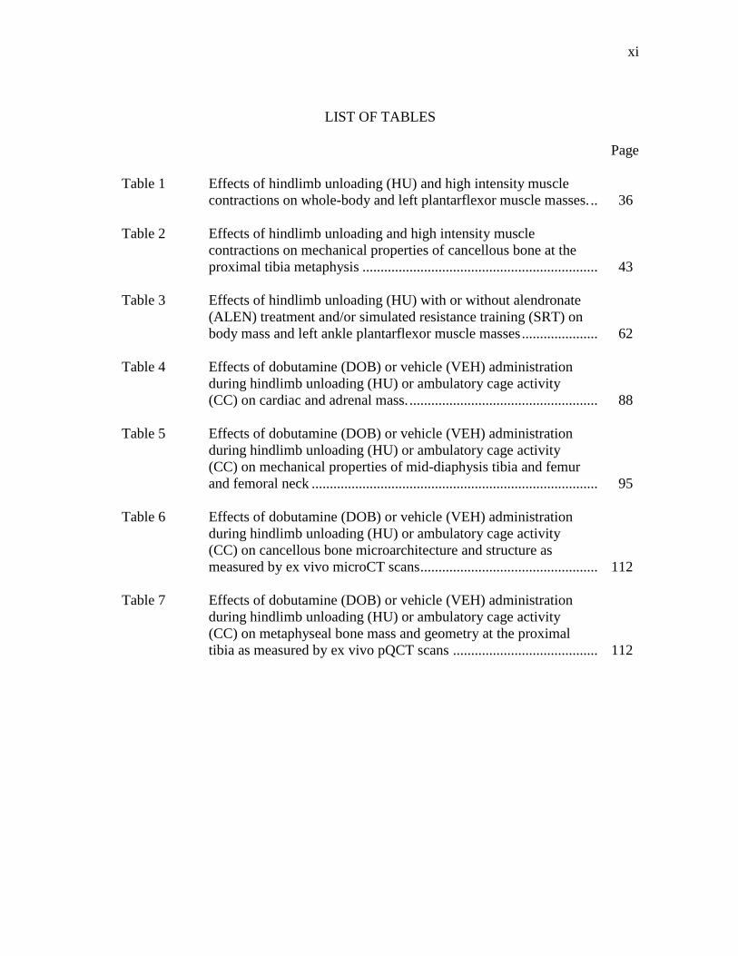
LIST OF TABLES
Page
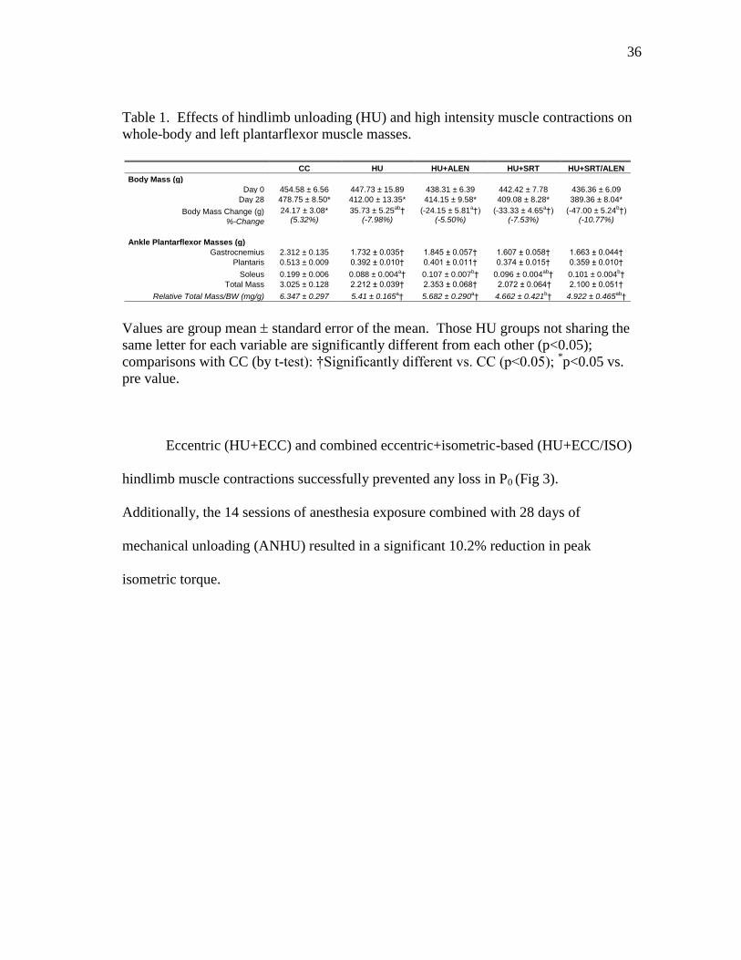
Table 1 Effects of hindlimb unloading (HU) and high intensity muscle
contractions on whole-body and left plantarflexor muscle masses. .. 36
Table 2 Effects of hindlimb unloading and high intensity muscle
contractions on mechanical properties of cancellous bone at the
proximal tibia metaphysis ................................................................. 43
Table 3 Effects of hindlimb unloading (HU) with or without alendronate
(ALEN) treatment and/or simulated resistance training (SRT) on
body mass and left ankle plantarflexor muscle masses ..................... 62
Table 4 Effects of dobutamine (DOB) or vehicle (VEH) administration
during hindlimb unloading (HU) or ambulatory cage activity
(CC) on cardiac and adrenal mass. .................................................... 88
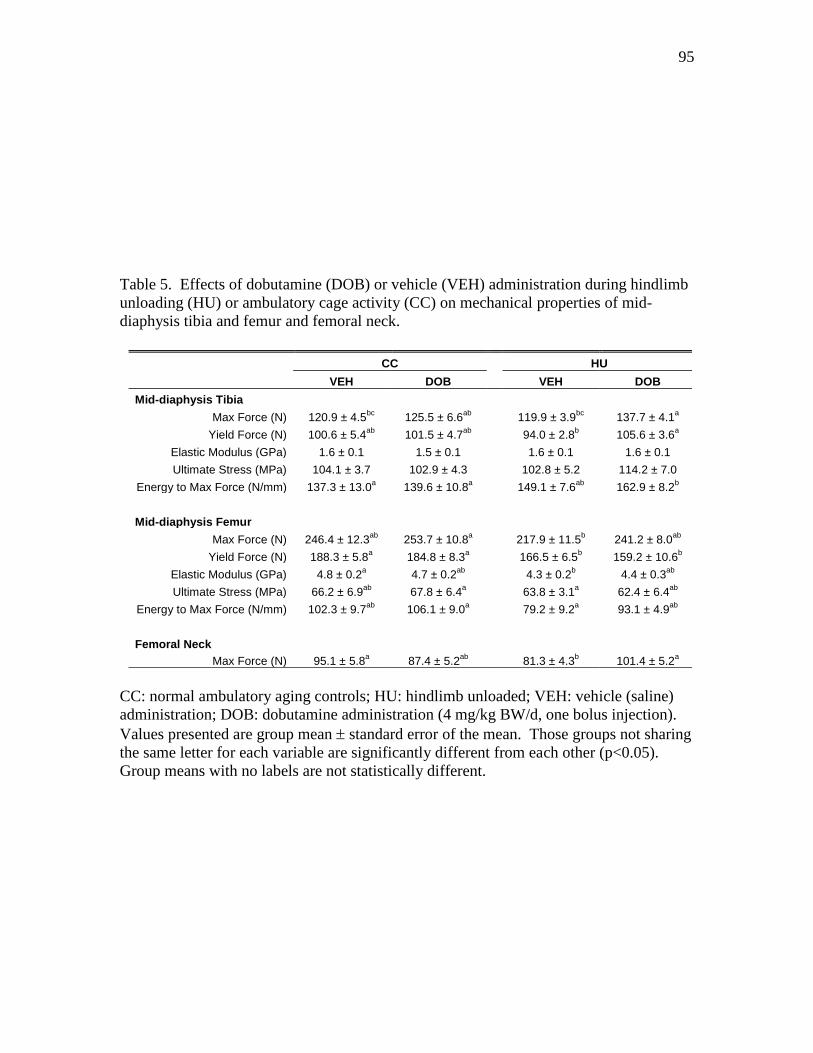
Table 5 Effects of dobutamine (DOB) or vehicle (VEH) administration
during hindlimb unloading (HU) or ambulatory cage activity
(CC) on mechanical properties of mid-diaphysis tibia and femur
and femoral neck ............................................................................... 95
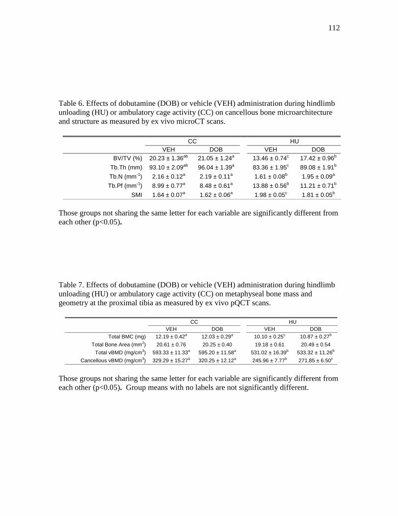
Table 6 Effects of dobutamine (DOB) or vehicle (VEH) administration
during hindlimb unloading (HU) or ambulatory cage activity
(CC) on cancellous bone microarchitecture and structure as
measured by ex vivo microCT scans ................................................. 112
Table 7 Effects of dobutamine (DOB) or vehicle (VEH) administration
during hindlimb unloading (HU) or ambulatory cage activity
(CC) on metaphyseal bone mass and geometry at the proximal
tibia as measured by ex vivo pQCT scans ........................................ 112
Page 12
xii
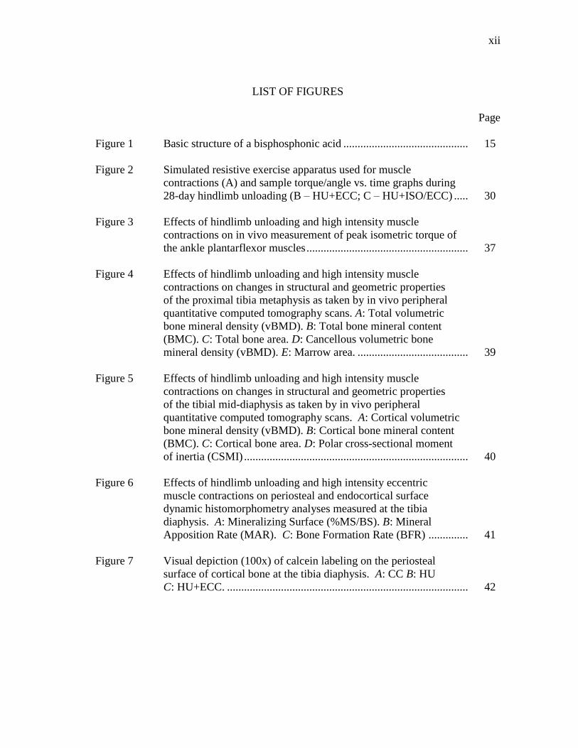
LIST OF FIGURES
Page
Figure 1 Basic structure of a bisphosphonic acid ............................................ 15
Figure 2 Simulated resistive exercise apparatus used for muscle
contractions (A) and sample torque/angle vs. time graphs during
28-day hindlimb unloading (B – HU+ECC; C – HU+ISO/ECC) ..... 30
Figure 3 Effects of hindlimb unloading and high intensity muscle
contractions on in vivo measurement of peak isometric torque of
the ankle plantarflexor muscles ......................................................... 37
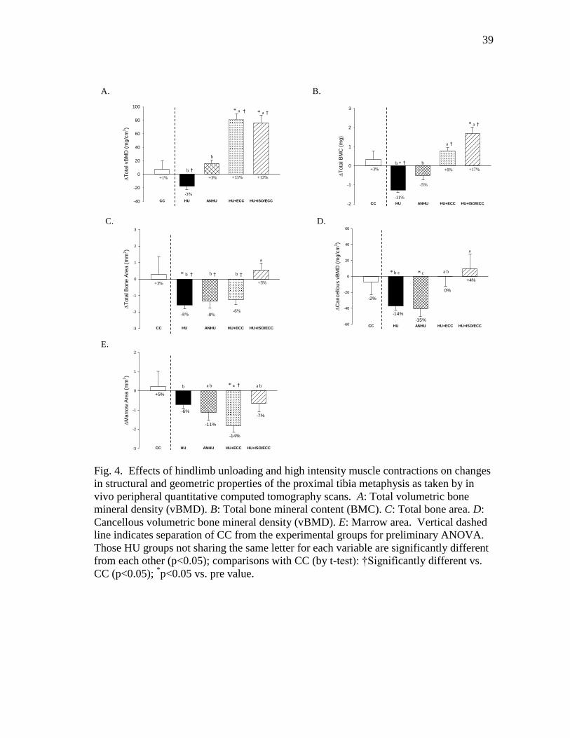
Figure 4 Effects of hindlimb unloading and high intensity muscle
contractions on changes in structural and geometric properties
of the proximal tibia metaphysis as taken by in vivo peripheral
quantitative computed tomography scans. A: Total volumetric
bone mineral density (vBMD). B: Total bone mineral content
(BMC). C: Total bone area. D: Cancellous volumetric bone
mineral density (vBMD). E: Marrow area. ....................................... 39
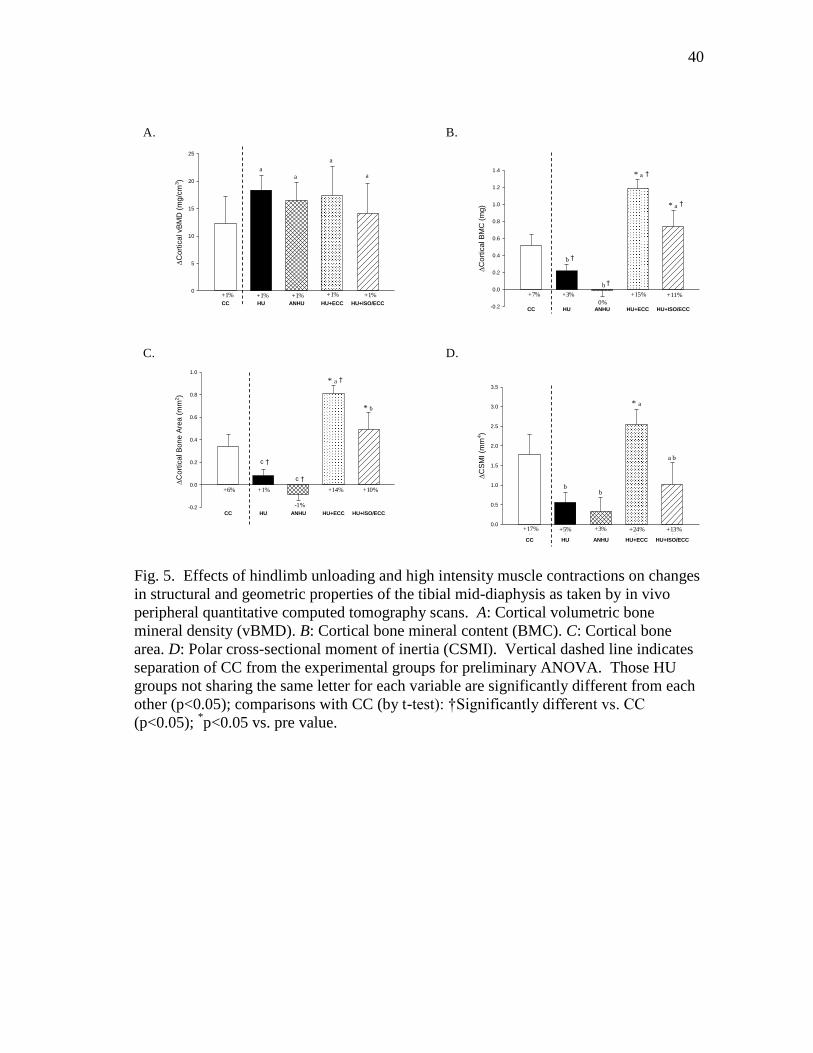
Figure 5 Effects of hindlimb unloading and high intensity muscle
contractions on changes in structural and geometric properties
of the tibial mid-diaphysis as taken by in vivo peripheral
quantitative computed tomography scans. A: Cortical volumetric
bone mineral density (vBMD). B: Cortical bone mineral content
(BMC). C: Cortical bone area. D: Polar cross-sectional moment
of inertia (CSMI) ............................................................................... 40
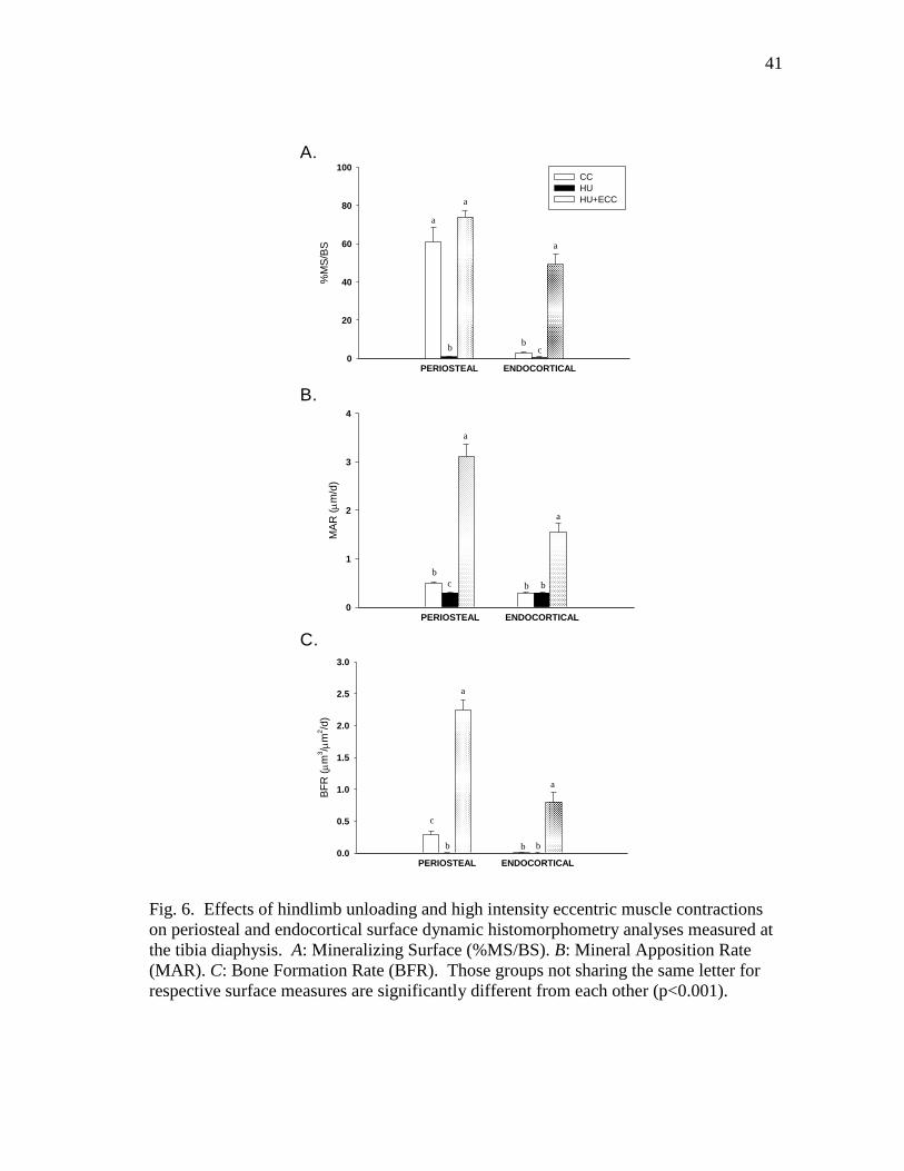
Figure 6 Effects of hindlimb unloading and high intensity eccentric
muscle contractions on periosteal and endocortical surface
dynamic histomorphometry analyses measured at the tibia
diaphysis. A: Mineralizing Surface (%MS/BS). B: Mineral
Apposition Rate (MAR). C: Bone Formation Rate (BFR) .............. 41
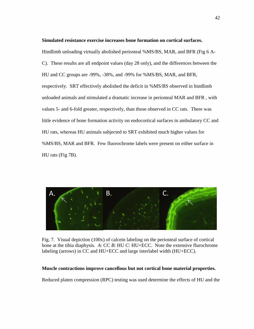
Figure 7 Visual depiction (100x) of calcein labeling on the periosteal
surface of cortical bone at the tibia diaphysis. A: CC B: HU
C: HU+ECC. ..................................................................................... 42
Page 13
xiii
Page
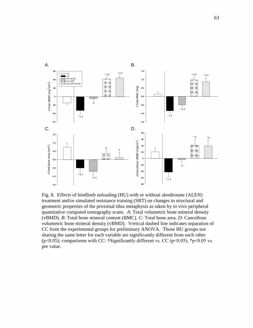
Figure 8 Effects of hindlimb unloading (HU) with or without alendronate
(ALEN) treatment and/or simulated resistance training (SRT) on
changes in structural and geometric properties of the proximal
tibia metaphysis as taken by in vivo peripheral quantitative
computed tomography scans. A: Total volumetric bone mineral
density (vBMD). B: Total bone mineral content (BMC). C: Total
bone area. D: Cancellous volumetric bone mineral density
(vBMD) ............................................................................................. 63
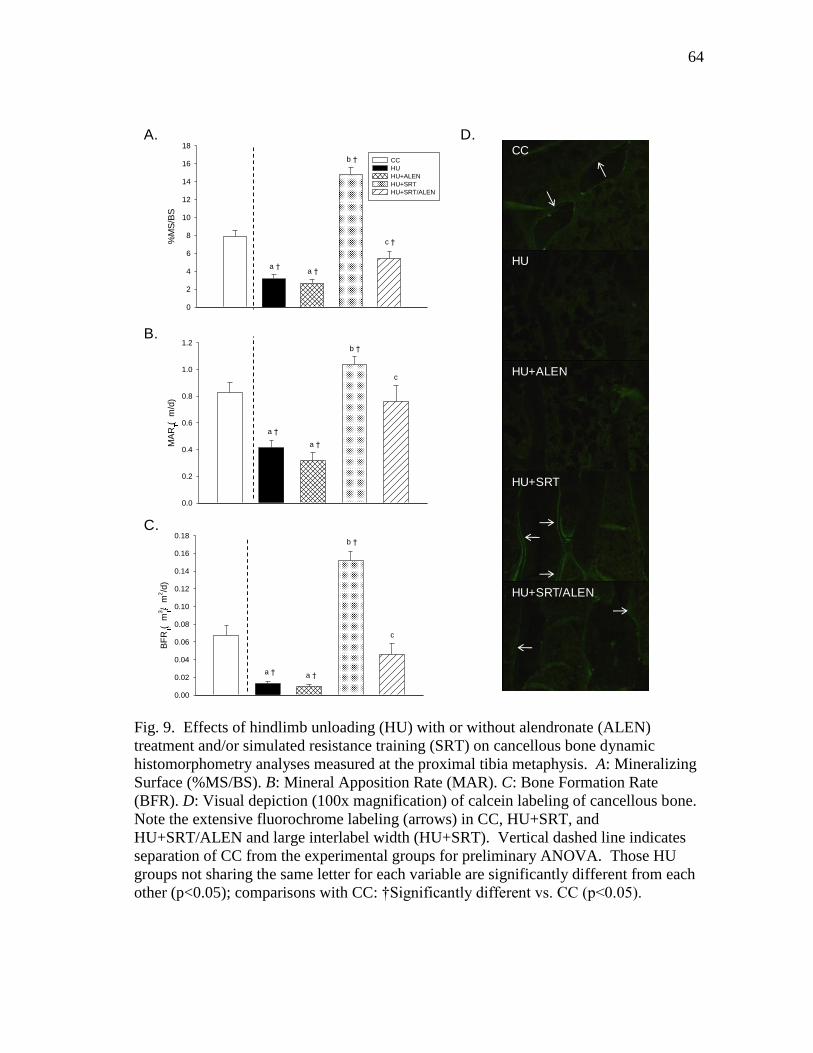
Figure 9 Effects of hindlimb unloading (HU) with or without alendronate
(ALEN) treatment and/or simulated resistance training (SRT) on
cancellous bone dynamic histomorphometry analyses measured
at the proximal tibia metaphysis. A: Mineralizing Surface
(%MS/BS). B: Mineral Apposition Rate (MAR). C: Bone
Formation Rate (BFR). D: Visual depiction (100x magnification)
of calcein labeling of cancellous bone .............................................. 64
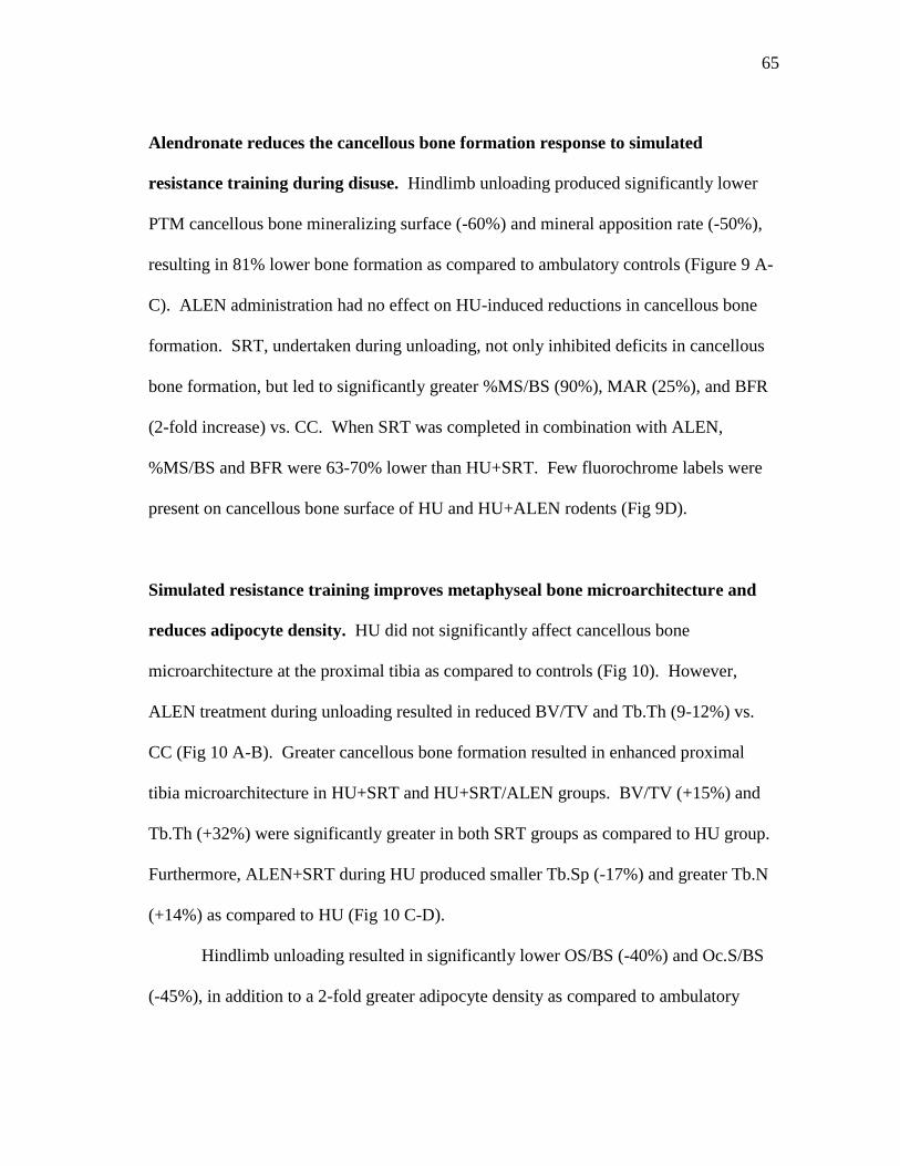
Figure 10 Effects of hindlimb unloading (HU) with or without alendronate
(ALEN) treatment and/or simulated resistance training (SRT) on
cancellous bone microarchitecture. A: Bone Volume (%BV/TV).
B: Trabecular Thickness (Tb.Th.). C: Trabecular Spacing (Tb.Sp.).
D: Trabecular Number (Tb.N.) ......................................................... 66
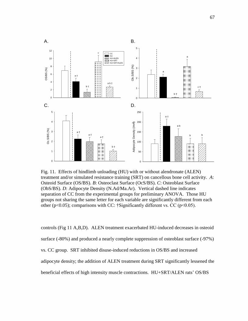
Figure 11 Effects of hindlimb unloading (HU) with or without alendronate
(ALEN) treatment and/or simulated resistance training (SRT) on
cancellous bone cell activity. A: Osteoid Surface (OS/BS).
B: Osteoclast Surface (OcS/BS). C: Osteoblast Surface (ObS/BS).
D: Adipocyte Density (N.Ad/Ma.Ar) ................................................ 67
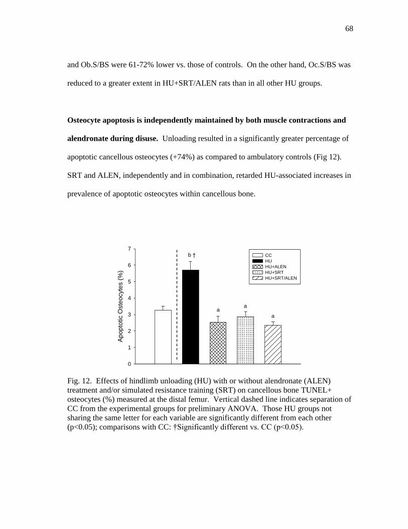
Figure 12 Effects of hindlimb unloading (HU) with or without alendronate
(ALEN) treatment and/or simulated resistance training (SRT) on
cancellous bone TUNEL+ osteocytes (%) measured at the distal
femur. ................................................................................................ 68
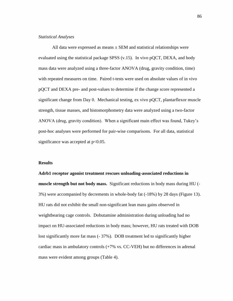
Figure 13 Effects of dobutamine (DOB) or vehicle (VEH) administration
during hindlimb unloading (HU) or ambulatory cage activity (CC)
on changes in body and tissue masses as measured by in vivo
DEXA scans on days -1 and 27 ......................................................... 87
Page 14
xiv
Page
Figure 14 Effects of dobutamine (DOB) or vehicle (VEH) administration
during hindlimb unloading (HU) or ambulatory cage activity (CC)
on in vivo measurement of peak isometric torque of the ankle
plantarflexor muscles ........................................................................ 89
Figure 15 Effects of dobutamine (DOB) or vehicle (VEH) administration
during hindlimb unloading (HU) or ambulatory cage activity (CC)
on changes in structural and geometric properties of the proximal
tibia metaphysis as taken by in vivo peripheral quantitative
computed tomography scans. A: Total bone mineral content
(BMC). B: Total bone area. C: Total volumetric bone mineral
density (vBMD) ................................................................................. 90
Figure 16 Effects of dobutamine (DOB) or vehicle (VEH) administration
during hindlimb unloading (HU) or ambulatory cage activity (CC)
on structural and geometric properties of the femoral neck as taken
by ex vivo peripheral quantitative computed tomography scans.
A: Total bone mineral content (BMC). B: Total volumetric bone
mineral density (vBMD). C: Total bone area .................................... 91
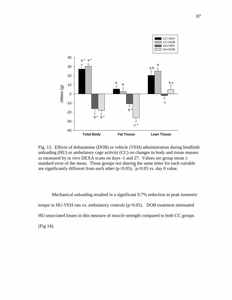
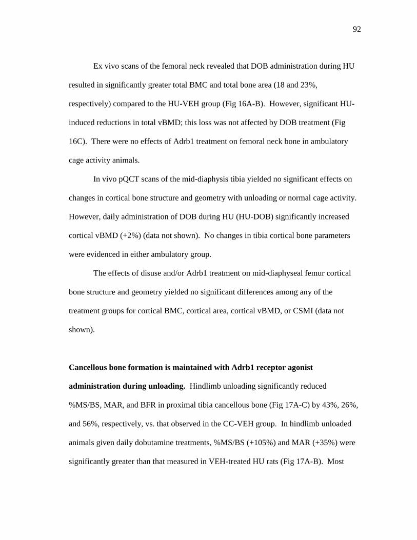
Figure 17 Effects of dobutamine (DOB) or vehicle (VEH) administration
during hindlimb unloading (HU) or ambulatory cage activity (CC)
on cancellous bone dynamic histomorphometry analyses measured
at the proximal tibia metaphysis. A: Mineralizing Surface
(%MS/BS). B: Mineral Apposition Rate (MAR). C: Bone
Formation Rate (BFR) ....................................................................... 93
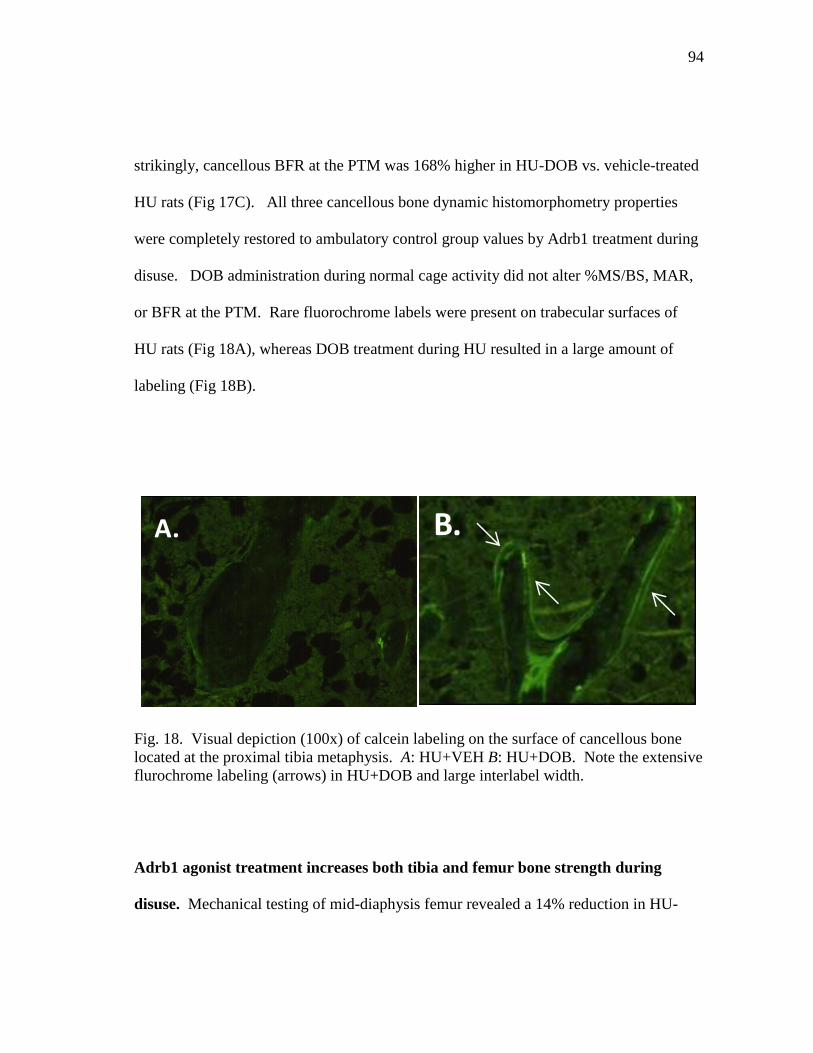
Figure 18 Visual depiction (100x) of calcein labeling on the surface of
cancellous bone located at the proximal tibia metaphysis.
A: HU+VEH B: HU+DOB ................................................................ 94
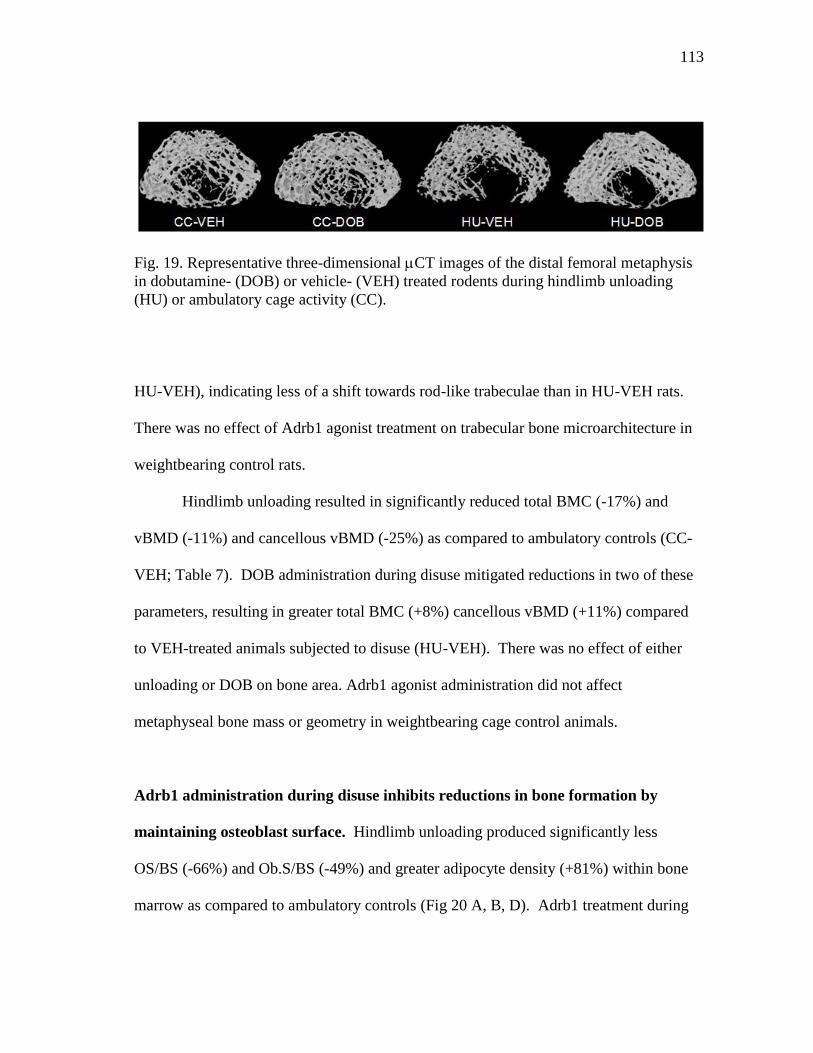
Figure 19 Representative three-dimensional CT images of the distal
femoral metaphysis in dobutamine- (DOB) or vehicle- (VEH)
treated rodents during hindlimb unloading (HU) or ambulatory
cage activity (CC) .............................................................................. 113
Figure 20 Effects of dobutamine (DOB) or vehicle (VEH) administration
during hindlimb unloading (HU) or ambulatory cage activity
(CC) on cancellous bone measures of histomorphometry.
A: Osteoid Surface (OS/BS). B: Osteoblast Surface (ObS/BS).
Page 15
xv
Page
C: Osteoclast Surface (OcS/BS). D: Adipocyte Density
(N.Ad/Ma.Ar) .................................................................................... 114
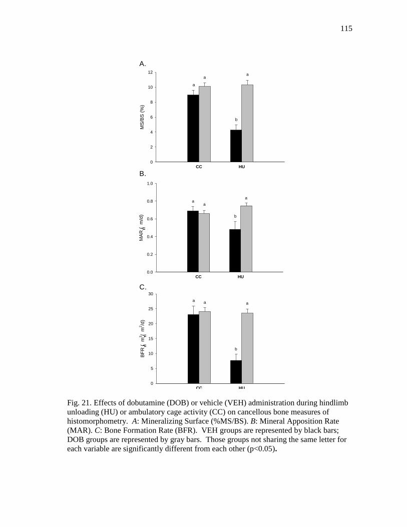
Figure 21 Effects of dobutamine (DOB) or vehicle (VEH) administration
during hindlimb unloading (HU) or ambulatory cage activity
(CC) on cancellous bone measures of histomorphometry.
A: Mineralizing Surface (%MS/BS). B: Mineral Apposition Rate
(MAR). C: Bone Formation Rate (BFR) ........................................... 115
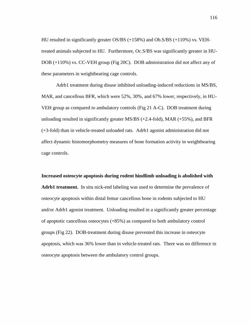
Figure 22 Effects of dobutamine (DOB) or vehicle (VEH) administration
during hindlimb unloading (HU) or ambulatory cage activity
(CC) on cancellous bone TUNEL+ osteocytes (%) measured at
the distal femur .................................................................................. 117
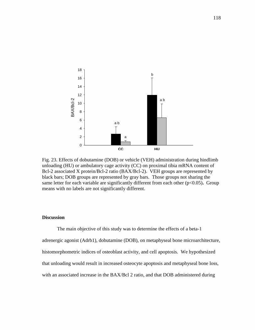
Figure 23 Effects of dobutamine (DOB) or vehicle (VEH) administration
during hindlimb unloading (HU) or ambulatory cage activity (CC)
on proximal tibia mRNA content of Bcl-2 associated X
protein/Bcl-2 ratio (BAX/Bcl-2) ....................................................... 118
Page 16
1
CHAPTER I
INTRODUCTION
During normal weightbearing, bone formation (osteoblasts) and bone resorption
(osteoclasts) remain equal, and the skeleton is in a state of balance. During periods of
decreased skeletal loading, bone formation and resorption become uncoupled, with
increased resorption and decreased formation leading to a net loss of bone mass. This
reduction in skeletal mass, if continued over a prolonged period of time, leads to
osteopenia or osteoporosis and significantly increases the risk of skeletal fracture.
Outside of the realm of microgravity, periods of disuse or reduced mechanical
loading are encountered by a large number of persons and are, therefore, of great
importance for continued investigations. Individuals exposed to prolonged bed rest,
limb immobilization, or spinal cord injury give clinical relevance to the accelerated bone
loss associated with spaceflight. However, understanding the mechanisms underlying
the processes of skeletal tissue loss during disuse and aging have yet to be determined,
and many similarities between bone loss in these two models do exist. Therefore,
research aimed at determining the mechanisms involved in disuse-associated reductions
in skeletal loss will provide essential insight into age-related decrements in bone mass
and osteoporosis.
____________
This dissertation follows the style of Journal of Bone and Mineral Research.
Page 17
2
Bisphosphonates are well established as the leading drugs for the treatment of
osteoporosis and other skeletal diseases characterized by increased bone resorption.
Alendronate (ALEN), a nitrogen-containing bisphosphonate, has been approved by
NASA for in-flight experiments. Alendronate inhibits bone resorption and ultimately
contributes to osteoclast apoptosis, thereby maintaining skeletal mass and strength.
Additionally, ALEN is a proven agent in minimizing bone loss due to estrogen
deficiency and disuse in rats.
Resistance exercise is an anabolic agent proven to stimulate bone growth, even
during periods of bed rest and disuse. The high strains imparted on bone, typically
encountered during high intensity resistance exercise, activate mechanical and chemical
signaling within bone and ultimately lead to “modeling activity.” As of yet, no animal
studies have successfully inhibited reductions in cancellous bone mass and formation
using resistance exercise protocols. Furthermore, no studies have addressed the effects
that combining bisphosphonate treatment with a resistance exercise protocol, completed
during disuse, has on disuse-sensitive cancellous bone.
The sympathetic nervous system (SNS) has been shown to mediate bone
metabolism. However, the exact role that beta-1 adrenergic (Adrb1) receptors have in
this process has not been elucidated. Although stimulation of the SNS has been
documented to increase bone resorption, resulting in reduced cancellous bone mass and
microarchitecture, this has been primarily attributed to stimulation of Adrb2 receptors.
The role that Adrb1 receptor stimulation during reduced mechanical loading has on
Page 18
3
disuse-sensitive cancellous bone is important to further understanding the underlying
mechanisms responsible for bone loss.
In the first study included in this dissertation (Chapter III), we investigated the
effects of two separate simulated resistance training (SRT) protocols, engaged during
rodent hindlimb unloading, on bone and muscle mass and strength. Results from this
investigation demonstrated that high-intensity muscle contractions, independent of
weightbearing forces, can effectively mitigate losses in muscle strength and provide a
potent stimulus to bone during prolonged disuse. Based on results from the first study,
we hypothesized that by adding an anti-resorptive agent, alendronate (ALEN;
bisphosphonate), to an anabolic stimulus, SRT, disuse-associated reductions in
cancellous bone formation and microarchitecture would be completely inhibited. In the
second study (Chapter IV), we investigated the individual and combined effects of a
reduced volume and intensity of SRT and ALEN treatment during HU on cancellous
bone parameters. Results from this investigation further demonstrate the anabolic effect
of a low volume of high intensity muscle contractions during disuse and suggest that
both bone resorption and formation are suppressed when SRT is combined with
bisphosphonate treatment. Furthermore, this study describes the potential inhibitory
effects of ALEN treatment on cancellous bone’s response to SRT during rodent
hindlimb unloading.
In the third study (Chapter V), we evaluated the effects of a beta-1 adrenergic
(Adrb1) agonist, dobutamine (DOB), on unweighted tibiae and femoral bone content,
density, area, and strength during 28 days unloading. DOB administration during HU
Page 19
4
effectively attenuated significant declines in total volumetric bone mineral density
(vBMD) at the proximal tibia by mitigating associated decrements in bone formation.
The significant positive effects of DOB on unweighted bone are not observed in animals
experiencing normal gravitational loading and provide evidence for the importance of
Adrb1 signaling in maintaining bone mass. Based on results from the third
investigation, we wanted to determine if the positive effects of DOB on unweighted
cancellous bone resulted in enhanced metaphyseal microarchitecture and were directly
related to reductions in osteocyte apoptosis. In the fourth study (Chapter VI) we tested
the effects of an Adrb1 agent on osteocyte apoptosis and expression of a pro- and anti-
apoptotic protein (Bax, Bcl-2) in unloaded bone. Results from this investigation
illustrated the effectiveness of DOB treatment during HU to mitigate reductions in
cancellous bone microarchitecture and increases in osteocyte apoptosis, perhaps by
inhibiting increased Bax/Bcl-2 mRNA content.
Results of these investigations highlight the potential ability of high-intensity
muscle contractions to completely abolish disuse-associated reductions in cancellous
bone mass, formation, and deleterious changes in microarchitecture, and further
emphasize the role that both resistance exercise and bisphosphonate treatment have to
inhibit osteocyte apoptosis. In addition, these studies demonstrate the dynamic role that
beta-1 adrenergic agonist signaling has on maintaining cancellous bone during periods of
reduced weightbearing or unloading. Hence, these findings have important implications
for humans experiencing extended periods of reduced weightbearing activity.
Page 20
5
CHAPTER II
REVIEW OF LITERATURE
Bone Remodeling and Modeling in the Adult Skeleton
Skeletal remodeling, occurring throughout the lifetime of a mammal, is defined
as the collaborative and sequential efforts of a BMU (bone multicellular unit). BMU’s
operate on periosteal, endosteal, and trabecular surfaces, as well as within cortical bone
by replacing old with new bone. Osteonal BMU’s originate on the periosteal or
endosteal surfaces of bone, or along the walls of Haversian canals, and are activated by
chemical, mechanical, or electrical signals. The process of bone remodeling is based
upon the coupled action of bone-resorbing cells (osteoclasts) and bone-forming cells
(osteoblasts). Osteoblasts, as well as chondrocytes, adipocytes, fibroblasts, and
myoblasts, differentiate from mesenchymal stem cells (1). Osteoclasts, however, are
derived from the hematopoietic mononuclear lineage (2). The primary functions of the
remodeling cycle include: (1) the preventative maintenance of mechanical strength
through replacement of fatigued bone by new, mechanically sound bone, and (2)
maintenance of mineral homeostasis by providing access to stores of calcium and
phosphorous.
There are 4 basic phases of the remodeling cycle, which include (1) activation,
(2) resorption, (3) reversal, and (4) formation. Activation involves the recruitment of
multinucleated osteoclast precursors from cells in the monocyte-macrophage lineage.
Most remodeling is likely random, but can be targeted towards specific sites that need to
Page 21
6
be repaired. Pre-osteoclasts, from hematopoietic stem cells, affix themselves to the bone
matrix through binding between integrin receptors and organic matrix, creating a sealing
zone. Osteoclasts are then able to create a bone-resorbing compartment between itself
and the bone matrix. Activation of osteoclasts is regulated by local cytokines like
RANKL, IL-1 and IL-6 (3,4).
Resorption, the second phase of remodeling, involves the catabolic actions of
osteoclasts. Osteoclasts are multinucleated, macrophage-like cells that attach to the
bone surface, form a peripheral seal, and break down bone within a sealed area by means
of enzymes or chemicals (1). The osteoclast’s membrane transfers protons to the
resorbing compartment (attached to bone) lowering its pH to 4. This compartment
becomes acidified by the secretion of lysosomal enzymes like TRAPC and cathespin K,
which dissolves and digests the mineral and organic phases of the matrix. This
resorption of bone creates a saucer-shaped cavity, called Howship’s lacunae.
The process of bone resorption ends with osteoclast apoptosis and the reversal
phase begins. During reversal, signals are sent out to “summon” osteoblasts into the
newly formed resorption cavities to replace removed bone. Without efficient coupling
mechanisms, remodeling would result in net bone loss. There are two theories of
coupling signals: (1) osteoclasts release growth factors from bone matrix during
resorption, which attract osteoblast precursors and stimulate osteoblast proliferation and
differentiation; or (2) strain-regulated theory: strain levels are lower ahead of osteoclasts
and higher behind them (in BMU as it moves through bone). Osteoblasts are activated
in response to high strain, and osteoclasts are activated in response to reduced strain.
Page 22
7
Following reversal, formation of new bone occurs. Formation is a two step
process, whereby osteoblasts synthesize organic matrix and then regulate its
mineralization. Osteoblasts mineralize organic matrix by deposition of Ca2+
and
phosphate ions. After completion of their bone formation function, osteoblasts have
three cell fates: (1) die by apoptosis, (2) become entombed in the mineralizing matrix as
osteocytes, or (3) remain on the surface as bone lining cells. Osteocytes maintain
contact with each other and cells on bone surface via gap junctions between cytoplasmic
processes that extend through canaliculae. Osteocytes become part of network which
can sense changes in mechanical properties surrounding bone and transmit this
information to cells on bone surface (i.e. osteoblasts) to initiate or regulate bone
remodeling when necessary.
Modeling differs from remodeling, as this process is not defined by the
coordinated actions of osteoblasts and osteoclasts. Instead, modeling involves the
shaping or reshaping of bones by independent actions of these cells. During modeling,
bone formation is not coupled to resorption, and vice versa. Modeling occurs during
growth or, in an adult skeleton, in response to increased mechanical loading.
Additionally, modeling occurs less frequently than remodeling in adults, particularly in
cancellous bone.
Page 23
8
Disuse Bone Loss
Spaceflight Effects on Bone (Human)
Mechanical loading is an integral component to maintaining bone mass during
periods of disuse (i.e. bedrest or casting) or reduced weightbearing activity. In-flight
measures of bone mass and geometry are not possible to obtain, therefore serum and
urine biomarkers of bone formation and resorption have become useful pieces of
evidence to our understanding how bone responds to microgravity. Early studies from
Skylab missions (1973-1974) sought to determine transient changes in bone metabolic
biomarkers during 28, 59, and 84 days of space flight. Data from these missions
demonstrated nearly 2-fold increases in-flight in urinary excretion of bone resorption
markers type 1 cross-linked N-telopeptide of (NTX), deoxypyridinoline (DPD), and
pyridinoline (PYD), and hydroxyproline vs. pre flight values (5,6). Later investigations
on MIR cosmonauts extended results from these Skylab missions. Extensive
microgravity exposure (up to 180 days) significantly reduced bone alkaline phosphatase
(BAP; bone formation biomarker) and increased DPD and type 1 cross-linked C-
telopeptide (CTX; bone resorption biomarkers) up to 80% (7-10). A study by Smith and
colleagues (10) demonstrated no change in BAP or osteocalcin during 180 days of space
flight. However, these authors noted a significant loss of Ca++
absorption (~250mg/d)
during microgravity. Assessment of changes in biomarkers of bone metabolism pre vs.
post-flight, demonstrated that, although bone formation markers were unchanged during
microgravity exposure, osteocalcin and BAP were significantly increased post-flight
(11).
Page 24
9
Although bone biomarkers provide readily accessible data in-flight data (from
urine or serum), their amounts vary considerably from day to day and from subject to
subject (12). For this reason, more reliable ground-based measures of bone mass and
geometry were necessary to document skeletal changes attributable to space flight. Long
duration exposure to microgravity leads to an accelerated loss of bone mass (~1-
2%/month) and results in osteopenia (13,14). Vico and colleagues (14) completed
peripheral quantitative computed tomography (pQCT) scans on tibiae and radii of
cosmonauts before and after long-duration MIR missions and demonstrated that
prolonged microgravity exposure (6+ months) results in greater reductions in cancellous
than cortical bone mineral density (BMD), and that these reductions in tibia cancellous
BMD are not fully restored by 6 months of recovery. Furthermore, microgravity
exposure affects bone mass in weightbearing (i.e. lower body) bones only. Recent data
gathered from crew members on the International Space Station (ISS) illustrates the
significant losses of bone mineral density (BMD) and geometry of the femoral neck
(15). Dual-energy x-ray absorptiometry (DXA) and QCT scans were taken on 14 ISS
astronauts before and after 4-6 month missions. Data from this investigation
demonstrated that the greatest reductions in areal BMD (aBMD) and trabecular
volumetric BMD (vBMD) occurred at the hip at a rate of 1.4-1.5% and 2.2-2.7%/month,
respectively (15). A subsequent study by Lang and colleagues (16) illustrated a
significant increase in estimated fracture risk during 4.5-6 month ISS missions that
remains even 1 year after returning to Earth. Pre- and post-flight peripheral pQCT scans
were administered to astronauts serving a single 4-6.5 month mission aboard ISS, and
Page 25
10
finite element models (FEM) of these scans were completed to assess estimated
proximal femoral strength during stance or a fall onto the posterolateral aspect of the
greater trochanter. For those crew members experiencing the greatest bone loss,
reductions in modeled proximal femur strength after a microgravity exposure
approached the estimated lifetime loss in stance strength for Caucasian women (17).
Furthermore, lack of recovery of BMD in ISS and MIR crew members has been
documented 6 months post-flight (14), with indications that it may not be fully restored
for 3 years (18).
Taken together, these data suggest that microgravity results in reduced bone
mass, which is attributable to increased bone resorption and decreased (or unchanged)
bone formation and negative calcium balance (in-flight). Furthermore, these reductions
in bone mass put astronauts at increasingly greater risk of fracturing their hip upon
return to Earth’s 1-gravity atmosphere. Most importantly, losses in bone typical of
exposure to microgravity are not fully restored by an equal amount of recovery time (i.e.
6 months).
Simulating Bone Loss with Spaceflight in Rodents: Hindlimb Unloading/Tail Suspension
The rodent hindlimb unloading (HU) model was developed by Emily Morey-
Holton in the 1970’s to most effectively mimic the effects of spaceflight on multiple
systems of the body (19). This model was the first to successfully model microgravity-
induced alterations in cardiovascular, muscular, and skeletal tissues, all the while not
overly stressing the animal. The HU model has become the gold-standard Earth-based
Page 26
11
model to simulate microgravity effects, as it allows the animal ambulation using only the
forelimbs, unloads the hindlimbs without paralysis, and produces cephalic fluid shifts
(20). Furthermore, ground-based studies using the HU model provide a more efficient
avenue to continue to study the effects of microgravity on the body, in comparison to
actual shuttle experiments, which are costly and extremely competitive.
Similar to the effects of microgravity in humans, rodent hindlimb unloading
significantly reduces cancellous bone within the metaphyses of the unweighted femur
and tibia (14,21). The effects of HU in adult male rats are compartment-specific, as 28
days of unloading primarily impacts cancellous and not cortical bone sites (21). HU
results in significant reductions in disuse-sensitive cancellous bone mass, architecture
and material properties, due to early increases in bone resorption followed by prolonged
depressions in bone formation rate (BFR) (21-24). These reductions in cancellous bone
mass are accounted for by decreased trabecular thickness (25-27) and number (27) and
increased trabecular spacing (24).
Resistance Exercise Effects on Skeletal Tissue
It has been well established that bone responds to increased and reduced
mechanical loading in opposing manners. Resistance exercise (RE) training incurs an
integrated physiological response typical of intense exercise (e.g., increased sympathetic
nervous system outflow, blood flow, and IGF-1 production), but which has not been
demonstrated with other models of mechanical loading (28-31). Squats, a lower-body
Page 27
12
form of RE, produces significant lower leg muscle hypertrophy and increased skeletal
muscle protein synthesis (32-34).
Resistance exercise that includes eccentric contractions provides an anabolic
stimulus for both skeletal muscle and bone in human and rodent models (35-38), and has
generally proven more effective in promoting increased bone and muscle mass than
endurance exercise protocols such as running. Furthermore, eccentric contractions
(during muscle lengthening) generate larger muscle forces than do concentric
contractions (muscle shortening), providing greater increases in BMD (36).
Resistance Exercise Effects on Bone (Humans)
In humans, bone responds to increased mechanical stress, evidenced during high-
impact and resistance exercise (RE), and has demonstrated the ability to increase lumbar
spine and femoral neck (FN) bone mass and strength (39-41). Resistance exercise
provides the necessary load to increase bone mass in healthy individuals and prevents
skeletal losses in estrogen deficient women, thereby decreasing the risk of fracture
during a fall (30,42-45). Furthermore, high-intensity RE (39,46) elicits a greater
anabolic response on bone than lower-impact activities like running or walking (47),
making it an attractive countermeasure to inhibit bone loss encountered in persons
experiencing reduced activity.
Page 28
13
Modeling Resistance Exercise Effects on Bone in Small Animal Models
Animal studies demonstrate similar results, as high-intensity RE and jump
training provide greater anabolic stimulus to bone than repetitive, low-load, high-
frequency exercise (i.e. running) (48,49). Drop training, whereby an animal is dropped
from a set height and lands with high amount of force on its hindlimbs, results in greater
proximal tibia total and cancellous volumetric bone mineral density (vBMD), (as a
consequence of greater trabecular thickness (TbTh) compared to ambulatory controls)
(50,51). Jump exercise, experienced when an animal jumps vertically in response to a
visual stimulus, in rats and mice leads to greater bone volume (BV/TV) and osteoblast
activity (MAR) at cancellous-rich bone sites (48,49) as well as increased cortical bone
mass and strength (50,52). Furthermore, jumping with weighted backpacks (i.e. jump
RE) has demonstrated the ability to increase trabecular thickness (TbTh), trabecular
number (TbN), and bone volume in the proximal tibia (38).
Resistance Exercise Effects on Bone During Extended Bed Rest (Human)
Only a few long-term bed rest investigations have successfully mitigated bone
loss with exercise paradigms. Combined supine flywheel resistive and treadmill
exercise during 90-day bed rest in young men attenuates reductions in trochanter and hip
BMD (53). Vigorous resistance training during 17-week bed rest increased lumbar spine
and diminished reductions in total hip BMD, as well as attenuated losses of leg muscle
masses and strength compared with bed rest controls (54). These initial studies provide
promising data suggesting that losses in skeletal BMD, at sites where greatest bone loss
Page 29
14
occurs during disuse, can be attenuated if a high-intensity resistance exercise training
program is begun immediately upon initiation of unloading.
Applying Resistance Exercise During Rodent Hindlimb Unloading
Previous investigations have sought to define the osteogenic effect of mechanical
loading during periods of disuse. Rubin et al (55) ,employing low-magnitude, high-
frequency loads, found that 10min/d of loading was effective at maintaining metaphyseal
bone volume (BV/TV) and did attenuate losses in bone formation at the proximal tibia.
More recent studies have utilized electrical stimulation as a countermeasure to disuse-
induced bone loss. Transcutaneous stimulation of the thigh, producing peak strains of
~200, did not effectively prevent disuse osteopenia in hindlimb unloaded rodents, but
did reduce tibial bone loss (56). A novel rodent resistance exercise device using
flywheel technology was used by Fluckey et al (57) to demonstrate the effects of
maximal voluntary squats, performed during suspension, on changes in metaphyseal
bone mass. The flywheel exercise protocol maintained distal femur BMD and inhibited
unloading-induced losses in bone mass at this site. Lam and Qin (58) recently
demonstrated the beneficial effects of high frequency electrical stimulation of the leg
musculature on bone during disuse. Although this treatment did effectively mitigate
losses of cancellous bone volume in the unloaded distal femur, there was no
demonstrable effect on bone formation. These studies provide initial evidence outlining
the role of resistance exercise in mitigating reductions in metaphyseal bone mass and
microarchitecture during periods of disuse or unloading.
Page 30
15
Bisphosphonate Effects on Bone and Disue-Induced Bone Loss
Pharmacology of Bisphosphonates
Bisphosphonates are a class of anti-resorptive drugs which are strongly attracted
to bone and inhibit osteoclast-mediated bone resorption. This characteristic makes
bisphosphonates especially suitable for the treatment of many skeletal diseases all
characteristic of severe bone loss. There are two main classifications of
bisphosphonates: non-nitrogenous and nitrogenous. All bisphosphonates have a
common phosphate-carbon-phosphate (P-C-P) moiety as part of their basic structure
(59). Attached to the central P-C-P backbone are two side chains (R1 and R2), as shown
in Figure 1. Each bisphosphonate differs in its side chains, which are primarily
responsible for the binding affinity to mineral (R1) and biochemical activity on
osteoclast enzymatic activity (R2).
Fig. 1. Basic structure of a bisphosphonic acid.
R1 R2
P-OH
OH O
HO-P
O OH
C
Page 31
16
Bisphosphonate Mechanisms of Action
The major effect of bisphonates on the skeleton is the inhibition of the osteoclast-
mediated bone resorption. The phosphate ends of the bisphosphonate bind to calcium
hydroxyapetite on the bone surface and are incorporated into bone during remodeling.
Uptake of bisphosphonates by osteoclasts during remodeling leads to osteoclast
apoptosis and the inhibition of key enzymes in the mevalonate pathway (also known as
the HMG-CoA reductase pathway), preventing the generation of lipids necessary for the
prenylation of small GTPase proteins (60). This restricts osteoclastic ability to bind to
and resorb bone, as the ruffled border of the osteoclast is unable to be maintained,
resulting in significantly reduced bone loss. Bisphosphoantes work to slow overall bone
turnover. Bone remodeling units exit for longer time periods, which results in a greater
degree of mineralization of skeletal tissue.
In addition to inhibiting osteoclast-mediated bone resorption, recent studies
suggest that osteocytes may be important target cells for bisphosphonates in bone. Many
bisphosphonates appear to protect osteocytes (and osteoblasts) from apoptosis (61). The
ability of bisphosphonates to inhibit apoptosis in osteocytes contrasts with their ability to
induce apoptosis in osteoclasts. The inhibition of osteocyte apoptosis by
bisphosphonates appears to be mediated by activation of extracellular signal-regulated
kinases (ERKs) (62). This dual role of bisphosphonates and potential interaction with
osteocytes in vivo needs further investigation before the exact mechanisms of
bisphosphonates’ actions can be fully understood.
Page 32
17
Alendronate Effects on Skeletal Tissue During Prolonged Human Bed Rest
Few bed rest investigations on the effects of alendronate (ALEN) have attempted
to elucidate the mechanics of this bisphosphonate’s action on bone. Ruml and
colleagues (63) administered 20 mg/day of ALEN to healthy males during 3 weeks of
bed rest and observed a reduction in urinary calcium excretion as compared to controls.
The only other bed rest study investigating the anti-resorptive capabilities of alendronate
was by Le Blanc and colleagues (64). A dose of 10 mg/d of ALEN was administered
daily to eight male subjects during17 weeks of bed rest. Daily ALEN treatment
successfully prevented reductions in lumbar spine and femoral neck BMD and
attenuated increases in urinary markers of bone resorption [cross-linked N-teleopeptide
of type I collagen (NTX), pyridinium (Pyd), an deoxypyridinium (D-Pyd)]. However,
ALEN during bed rest resulted in significant reductions in serum markers of bone
formation (alkaline phosphatase, bone-specific alkaline phosphatase, and osteocalcin),
which remained unchanged in bed rest subjects not administered ALEN.
Alendronate Effects on Skeletal Tissue During Rodent Hindlimb Unloading
The effectiveness of alendronate to inhibit disuse-induced bone loss has been
demonstrated in numerous rodent hindlimb unloading investigations. Alendronate (100
g/kg/d), administered prior to HU, prevented reductions in total and proximal tibia
bone mass and proximal tibia calcium content. ALEN treatment decreased relative
osteoclast surface and mitigated reductions in bone formation (26). Additionally,
ALEN, administered during 14-day HU, abolished losses in tibia and femur BMD, but
Page 33
18
was unable to rescue disuse-induced reductions in bone strength (65). Apseloff et al.
(66) administered 30 g/kg alendronate during 28 days of unloading and revealed
significant reductions in osteoid perimeter, cancellous bone formation, and bone
resorption with bisphosphonate treatment during HU (as compared to controls). The
resulting increase in bone mass with alendronate treatment resulted from greater
inhibition of resorption than from stimulation of formation.
Beta-Adrenergic Signaling in Skeletal Tissue
Sympathetic Nervous System and Beta-Adrenergic Receptors
Beta-adrenergic receptor agonists, activated by the sympathetic nervous system
(SNS), may affect bone metabolism through separate avenues and have opposing effects
on bone mass. Three subunits of β-adrenergic receptors (Adrb1, 2, 3) are present in
tissues within the body and variant effects. Adrb2 receptors, present in the lungs, cause
bronchile dilation, and on osteoblasts and osteoclasts, stimulate apoptosis and result in
diminished bone mass (67,68). Adrb1 receptors are present on the heart, increasing
cardiac contractility when activated, and are also present on osteoblasts and osteoclasts
(69,70). Activation of β-3 adrenergic receptors, the primary adrenoreceptor on
adipocytes, results in enhanced lipolysis (71,72). Adrb1 and Adrb2 receptors are both
present on osteoblasts, but β-2 adrenergic receptors are the predominant subtype
(67,68,73-75).
Page 34
19
Beta-1 Adrenergic Agonist Effects on Bone
Dobutamine (DOB) is a non-specific, Adrb receptor agonist with dominant β-1
adrenergic receptor activity and a small amount of Adrb2 activity (76). The ability of
DOB to primarily activate Adrb1 receptors make it an attractive synthetic catecholamine
to study in coordination with rodent disuse. To our knowledge, only one study has
attempted to assess the effects of chronic Adrb1 receptor stimulation on skeletal tissue.
DOB administered during HU significantly blunted reductions in femoral midshaft
cortical bone area and cross-sectional moment of inertia (CSMI), as well as mitigated the
HU-associated decrease in femoral mid-diaphyseal cortical bone mineral apposition rate
(77). This preliminary investigation, while lacking mechanisms, provides initial data
outlining the potential positive effects of increased Adrb1 receptor stimulation during
disuse.
Beta-2 Adrenergic Agonist Effects on Bone
Few studies have attempted to define the role that β-2 receptor activation has on
skeletal tissue, beyond a few investigations completed in female rats. Adrb2 receptor
agonist administration leads to increased bone resorption, resulting in reduced cancellous
bone mass and microarchitecture. Bonnet and colleagues (78) treated skeletally
immature, female rats daily (4mg/kg/d) with salbutamol or clenbuterol for 6 weeks. This
study demonstrated the significantly deleterious effects of Adrb2 receptor agonists on
metaphyseal bone, and clenbuterol-treated rats had lower distal femur trabecular number
(-40%), connectivity (-3-fold), and bone volume (-43%) as compared to controls.
Page 35
20
Furthermore, although bone formation was not measured, based on data from urinary
markers the authors concluded the deleterious effects of Adrb2 agonists were related to
increased resorption and not decreased formation. A subsequent study by Bonnet and
colleagues (79) further emphasized the significant reductions in metaphyseal bone in a
subsequent investigation. The authors demonstrated that chronic treatment of
salbutamol or clenbuterol in growing female rats significantly decreased proximal tibia
and lumbar spine bone volume, trabecular thickness, trabecular number, and
biomechanical properties. Furthermore, Bonnet and colleagues (80) verified that
salbutamol further increases the deleterious effects of estrogen deficiency
(ovariectomized) on cancellous bone loss in adult rodent. Additionally, stimulation of
Adrb2 receptors on osteoblasts increases osteoclast differentiation and activity, resulting
in increased bone loss (81).
Adrenergic Receptor Knock-Out Mouse Models and Effects on Skeletal Tissue
A very limited number of in vivo investigations have attempted to systematically
define the precise roles of β-1 and β-2 receptors and their relation to bone mass. Adrb2
receptor knock-out (KO) and Adrb1 receptor KO mice demonstrate a high and low bone
mass phenotype, respectively, vs. wild type controls (67,82). Furthermore, reduced bone
volume in β-less receptor mice (β-1,2,3 KO) is attributed to a marked reduction in
cancellous BFR (83). Estrogen deficiency (OVX) further increased the significant
reductions in metaphyseal bone volume and trabecular number in mice lacking β-
adrenergic receptors as compared to controls. Finally, Adrb1 receptor deficient mice
Page 36
21
demonstrated an inability to respond to mechanical loading, whereas Adrb2 receptor and
wild-type littermates were found to respond normally (82).
Taken together, these data suggest that the higher bone mass phenotype in Adrb2
receptor KO mice may be caused by enhanced β-1 adrenergic receptor activity
stimulating bone formation in the absence of the inhibitory effects of β-2 adrenergic
receptors on osteoblasts. Therefore, defining the role that Adrb1 receptor stimulation
during reduced mechanical loading has on disuse-sensitive cancellous bone is important
to fully understanding underlying mechanisms responsible for bone loss with disuse.
Page 37
22
CHAPTER III
SIMULATED RESISTANCE TRAINING DURING HINDLIMB UNLOADING
ABOLISHES DISUSE BONE LOSS AND MAINTAINS MUSCLE STRENGTH*
Introduction
President George W. Bush announced his Exploration Vision for the National
Aeronautics and Space Agency (NASA) in 2004, proposing lengthy stays at a Lunar
outpost by 2020 and eventual expeditions to Mars thereafter (84). Mission success may
ultimately depend on improvements in our ability to maintain astronaut health and
functional capabilities during such lengthy trips. To date, current exercise
countermeasures (cycle ergometer, treadmill, and resistance exercise device) (85) used
by crew on the International Space Station (ISS) have not effectively mitigated losses of
bone mineral density (BMD) or geometry (15), resulting in a significant increase in
estimated fracture risk that remains even 1 year after returning to Earth (16). For those
crew members experiencing the greatest bone loss, reductions in modeled proximal
femur strength after a 6-month ISS mission approach the estimated lifetime loss in
stance strength for Caucasian women (16,17). Furthermore, lack of recovery of BMD in
ISS and MIR crew members has been documented 6 months post-flight (14), with
indications that it may not be fully restored for 3 years (18). In addition to skeletal
losses, reductions in ISS crew member skeletal muscle volume of the lower leg are
____________
*Reprinted with permission from Swift JM, Nilsson MI, Hogan HA, Sumner LR,
Bloomfield SA 2010 Simulated resistance training during hindlimb unloading abolishes
disuse bone loss and maintains muscle strength. J Bone Miner Res 25(3): 564-74.
Copyright 2010 by John Wiley and Sons, Inc.
Page 38
23
coupled with significant decrements (nearly 30%) in plantarflexor muscle strength (85).
These changes in humans exposed to microgravity parallel alterations in bone
and muscle mass observed during prolonged bedrest (53,54,86,87) and during the more
severe losses incurred after spinal cord injury (SCI). Electrical stimulation has been
utilized in patients with SCI to enable active contractions of the paralyzed lower limbs to
perform aerobic (cycling) and resistive training. If this training is started months to
years after the injury, acting on bone and muscle that is severely compromised by disuse,
only marginal improvements in muscle strength and BMD in the paralyzed limbs are
achieved (88-90). Far more successful is a strategy that starts early after the onset of
paralysis; stimulated contractions of the plantarflexor muscles begun within 6 weeks of a
SCI significantly mitigated the loss of muscle torque and contractile speed of the
paralyzed lower leg, as well as the loss of cancellous bone in the distal tibia (91).
The rodent hindlimb unloading (HU) model is a well-established ground-based
model for investigating disuse effects on bone and muscle (20). Hindlimb unloading
results in significant reductions in disuse-sensitive cancellous bone mass, architecture
and material properties, due to early increases in bone resorption followed by prolonged
depressions in bone formation rate (BFR) (21-24). Additionally, skeletal muscle atrophy
and reduced functional properties (i.e. strength) have been demonstrated as early as 4
days after HU begins and may remain depressed for some time upon reambulation
(57,92,93). To date, no method has successfully prevented deficits in both muscle and
bone during long duration unloading.
Page 39
24
Resistance exercise that includes eccentric contractions provides an anabolic
stimulus for both skeletal muscle and bone in both humans and rodent models (35-38),
and has generally proven more effective in promoting increased bone and muscle mass
than endurance exercise protocols such as running. Eccentric contractions (during
muscle lengthening) generate larger muscle forces than do concentric contractions
(muscle shortening), providing greater increases in BMD (36). Until the launching of
improved training equipment late in 2008, ISS astronauts were unable to train at the high
intensities (>70% maximum) demonstrated as osteogenic in land-based studies (94).
The purpose of the present investigation was to determine the effectiveness of
high intensity simulated resistance training (SRT), achieved without weightbearing, in
maintaining hindlimb muscle and bone (tibia) mass and strength during hindlimb
unloading in skeletally mature rats. We sought to maximize the ability of high intensity
exercise, begun early in the unloading period, to mitigate loss of both muscle and bone
during unloading and so tested two separate contraction paradigms, an eccentric-based
(HU+ECC) and a protocol combining both isometric and eccentric contractions
(HU+ISO/ECC). We hypothesized that SRT would effectively attenuate disuse-induced
bone and muscle loss and that doubling muscle contraction time with the addition of an
isometric component would significantly enhance the effectiveness.
Materials and Methods
Animals and Experimental Design
Fifty male Sprague-Dawley rats were obtained from Harlan (Houston, TX) at 6
months of age and allowed to acclimate to their surroundings for 14 days prior to
Page 40
25
initiation of the study. All animals were housed in a temperature-controlled (23 ± 2ºC)
room with a 12-hour light-dark cycle in an American Association for Accreditation of
Laboratory Animal Care-accredited animal care facility and were provided standard
rodent chow (Harlan Teklad 8604) and water ad-libitum. Animal care and all
experimental procedures described in this investigation were conducted in accordance
with the Texas A&M University Laboratory Animal Care Committee rules.
Five experimental groups were studied: (1) cage control (CC, n=12), (2)
hindlimb unloaded (HU, n=12), (3) HU animals exposed to similar duration of isoflurane
anesthesia (Minrad Inc., Bethlehem, PA) as trained HU rats (ANHU, n=6), (4) HU
subjected to eccentric muscle contraction training (HU+ECC, n=10), and (5) HU rats
subjected to a combined isometric and eccentric hindlimb simulated resistive exercise
(HU+ISO/ECC, n=10). Due to the labor-intensive nature of these experiments, it was
necessary to perform experiments in two successive cohorts of animals. Rats in cohort I
(CC, HU, and HU+ECC) and II (ANHU and HU+ISO/ECC) were randomly assigned to
their respective groups by total vBMD at the proximal tibia metaphysis one day prior to
study initiation. HU+ECC and HU+ISO/ECC animals underwent 14 sessions of
simulated resistive exercise conducted every other day during the 28 day protocol, with
each session consisting of 4 sets of 5 contractions. The HU and ANHU groups were
similarly unloaded for 28 days, whereas the CC animals were allowed normal
ambulatory cage activity; all animals were singly housed.
Page 41
26
Hindlimb Unloading
Hindlimb unloading was achieved by tail suspension as previously described
(77). Briefly, while the rat was under anesthesia, the tail was cleaned and dried
thoroughly. A thin layer of adhesive (Amazing Goop, Eclectic Products, LA) was
applied to the proximal half of the tail along the medial and lateral sides. A standard
porous tape (Kendall, Mansfield, MA) harness was pressed firmly to the glue and
allowed to dry (~30 min). A paper clip was used to attach the animal’s tail harness to a
swivel apparatus on the wire spanning the top of an 18” x 18” x 18” cage. The height of
the animal’s hindquarters was adjusted to prevent any contact of the hindlimbs with the
cage floor, resulting in approximately a 30° head-down tilt. The forelimbs of the animal
maintained contact with the cage bottom, allowing the rat full access to the entire cage.
Calcein injections (25 mg/kg body mass) were given subcutaneously 9 and 2
days prior to sacrifice to label mineralizing bone for histomorphometric analysis. HU
animals were anesthetized before removal from tail suspension at the end of the study to
prevent any weight bearing by the hindlimbs. At necropsy, left soleus, plantaris, and
gastrocnemius muscles were excised and wet weights were recorded. Additionally,
proximal left tibiae were removed, cleaned of soft tissue, and stored at -80°C in PBS
soaked gauze for mechanical testing, whereas the distal portions of the left tibia were
stored in 70% ethanol at 4°C for cortical histomorphometry of the TFJ (tibiofibular
junction) region.
Page 42
27
Muscle Contraction and Training Paradigms
Left plantarflexor muscles from animals in the HU+ECC and HU+ISO/ECC
groups were trained every other day during 28-day HU using a custom-made rodent
isokinetic dynamometer, as previously described (Fig 2A) (95). Animals were
anesethetized with isoflurane gas (~2.5%) mixed with oxygen while remaining
suspended, to prevent any weight bearing of the hindlimbs. Once unconscious, the
lateral side of the rat’s left leg was shaved and aseptically prepared. Each rat was then
placed in right lateral recumbency on a platform, the left foot was secured onto the foot
pedal, and the left knee was clamped so that the lower leg was perpendicular to the foot
and the femur and tibia were at right angles to each other. This was referred to as the
resting, 0° position. For isometric contractions, the foot pedal was held fixed in this
position. For all contractions, the footplate was rotated in synchronization with muscle
stimulation by a Cambridge Technology servomotor system (Model 6900) interfaced
with a 80486 66-MHz PC using custom software written in TestPoint (v.4.0; Capital
Equipment Corp., Billerica, MA). In either case, the torque generated around the
footplate pivot (at the rat’s ankle joint) was measured through the servomotor.
Plantarflexor muscle stimulation was performed with fine wire electrodes consisting of
insulated chromium nickel wire (Stablohm 800B, H-ML Size 003, California Fine Wire
Co.), inserted intramuscularly straddling the sciatic nerve in the proximal thigh region.
The stimulation wires were then attached to the output poles of a Grass Instruments
stimulus isolation unit (Model SIU5; Astro-Med, Inc; W. Warwick, RI) interfaced with a
Page 43
28
stimulator (S88; Astro-Med, Inc; W. Warwick, RI) which delivered current to the sciatic
nerve and induced muscle contraction.
Eccentric-Based Simulated Resistive Exercise Training
An eccentric-based SRT protocol was utilized for training (HU+ECC). At the
beginning of each session, the daily peak isometric torque (P0) was determined. The
foot was held fixed at 0° and successive stimulations were conducted at different
voltages, with the frequency kept constant at 175 Hz (~4-6 contractions, 200ms duration,
45s rest between contractions). The voltage and magnitude of P0 were recorded. This
voltage was maintained for the eccentric contractions, and the stimulation frequency was
varied until the torque matched P0 (i.e., 100% peak isometric torque). This normally
took 3-5 contractions, each at 200ms, with 45s of rest between contractions). A rest
period of 2 minutes was given between the isometric and eccentric contraction
optimization protocols. Upon completion of eccentric frequency optimization, the
training program was initiated. Four sets of 5 repetitions (12 and 120 seconds of rest
inserted between contractions and sets, respectively) were completed during each
training session, which lasted approximately 25 minutes. Each eccentric contraction
stimulus was 1000 ms long. During each plantarflexor muscle lengthening contraction,
the footplate was rotated in dorsiflexion through a 40° arc (centered at the 0° position)
(Fig 2B). After each contraction, the foot and pedal returned to the initial 0° resting
position. Plantarflexor muscles fatigued by an average of 17.8% (maximum torque
generated during the last set vs. the first set) during a typical training session.
Page 44
29
Isometric Plus Eccentric Simulated Resistive Exercise Training
The ability of a longer duration contraction with isometric and eccentric
components (both at 100% P0) to maintain musculoskeletal health during unloading was
also tested (HU+ISO/ECC). Voltage optimization of the peak isometric torque and
frequency optimization of the eccentric torque were performed at the beginning of each
session, as described in the previous paragraph. The total number sets, repetitions, and
training sessions were identical to those of the HU+ECC group. In this case, however,
the training protocol consisted of a 1000ms isometric contraction (175 Hz; 0°),
immediately followed by a 1000ms eccentric contraction the same as those used for the
eccentric contractions in experiment 1 (Fig 2C). By the end of a typical training session,
plantarflexor muscles fatigued by an average of 34.2% and 39.8% for isometric and
eccentric contractions, respectively. These reductions in torque are similar to previous
investigations utilizing 2-second muscle stimulation contractions (95).
Muscle Function Testing
Functional assessments of the left leg plantarflexor muscles were completed on
days 0 and 28 on animals in the ANHU, HU+ECC, and HU+ISO/ECC groups using the
same isokinetic dynamometer (93) used for the SRT protocols. The anesthesia used,
preparation of the animal, and electrodes’ placements were identical to those used during
training. Briefly, stimulation voltage was optimized using 0.1-ms pulses at 300 Hz to
yield the peak isometric torque during a trial of 4-6 tetanic contractions. Peak torque
produced about the ankle was recorded using custom software written in TestPoint.
Page 45
30
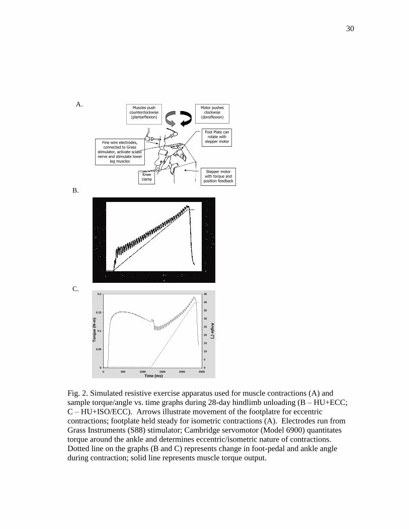
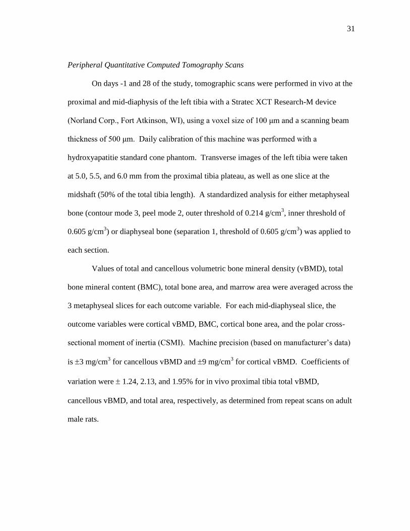
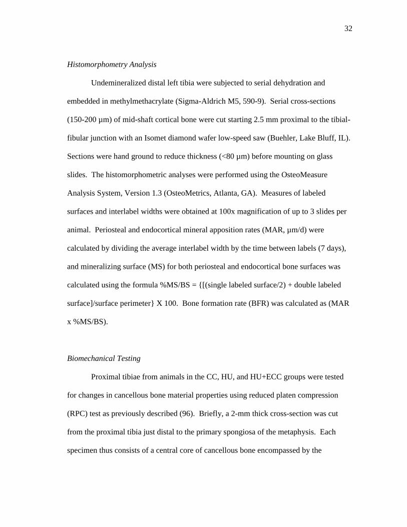
Fig. 2. Simulated resistive exercise apparatus used for muscle contractions (A) and
sample torque/angle vs. time graphs during 28-day hindlimb unloading (B – HU+ECC;
C – HU+ISO/ECC). Arrows illustrate movement of the footplatre for eccentric
contractions; footplate held steady for isometric contractions (A). Electrodes run from
Grass Instruments (S88) stimulator; Cambridge servomotor (Model 6900) quantitates
torque around the ankle and determines eccentric/isometric nature of contractions.
Dotted line on the graphs (B and C) represents change in foot-pedal and ankle angle
during contraction; solid line represents muscle torque output.
0
0.05
0.1
0.15
0.2
0 500 1000 1500 2000 2500
Time (ms)
To
rqu
e (
N-m
)
0
5
10
15
20
25
30
35
40
45
An
gle
(°)
Foot Plate can rotate with
stepper motor Fine wire electrodes, connected to Grass
stimulator, activate sciatic nerve and stimulate lower
leg muscles
Stepper motor with torque and
position feedback
Muscles push counterclockwise (plantarflexion)
Motor pushes clockwise
(dorsiflexion)
Knee clamp
A.
B.
C.
Page 46
31
Peripheral Quantitative Computed Tomography Scans
On days -1 and 28 of the study, tomographic scans were performed in vivo at the
proximal and mid-diaphysis of the left tibia with a Stratec XCT Research-M device
(Norland Corp., Fort Atkinson, WI), using a voxel size of 100 μm and a scanning beam
thickness of 500 μm. Daily calibration of this machine was performed with a
hydroxyapatitie standard cone phantom. Transverse images of the left tibia were taken
at 5.0, 5.5, and 6.0 mm from the proximal tibia plateau, as well as one slice at the
midshaft (50% of the total tibia length). A standardized analysis for either metaphyseal
bone (contour mode 3, peel mode 2, outer threshold of 0.214 g/cm3, inner threshold of
0.605 g/cm3) or diaphyseal bone (separation 1, threshold of 0.605 g/cm
3) was applied to
each section.
Values of total and cancellous volumetric bone mineral density (vBMD), total
bone mineral content (BMC), total bone area, and marrow area were averaged across the
3 metaphyseal slices for each outcome variable. For each mid-diaphyseal slice, the
outcome variables were cortical vBMD, BMC, cortical bone area, and the polar cross-
sectional moment of inertia (CSMI). Machine precision (based on manufacturer’s data)
is 3 mg/cm3 for cancellous vBMD and 9 mg/cm
3 for cortical vBMD. Coefficients of
variation were 1.24, 2.13, and 1.95% for in vivo proximal tibia total vBMD,
cancellous vBMD, and total area, respectively, as determined from repeat scans on adult
male rats.
Page 47
32
Histomorphometry Analysis
Undemineralized distal left tibia were subjected to serial dehydration and
embedded in methylmethacrylate (Sigma-Aldrich M5, 590-9). Serial cross-sections
(150-200 µm) of mid-shaft cortical bone were cut starting 2.5 mm proximal to the tibial-
fibular junction with an Isomet diamond wafer low-speed saw (Buehler, Lake Bluff, IL).
Sections were hand ground to reduce thickness (<80 µm) before mounting on glass
slides. The histomorphometric analyses were performed using the OsteoMeasure
Analysis System, Version 1.3 (OsteoMetrics, Atlanta, GA). Measures of labeled
surfaces and interlabel widths were obtained at 100x magnification of up to 3 slides per
animal. Periosteal and endocortical mineral apposition rates (MAR, µm/d) were
calculated by dividing the average interlabel width by the time between labels (7 days),
and mineralizing surface (MS) for both periosteal and endocortical bone surfaces was
calculated using the formula %MS/BS = {[(single labeled surface/2) + double labeled
surface]/surface perimeter} X 100. Bone formation rate (BFR) was calculated as (MAR
x %MS/BS).
Biomechanical Testing
Proximal tibiae from animals in the CC, HU, and HU+ECC groups were tested
for changes in cancellous bone material properties using reduced platen compression
(RPC) test as previously described (96). Briefly, a 2-mm thick cross-section was cut
from the proximal tibia just distal to the primary spongiosa of the metaphysis. Each
specimen thus consists of a central core of cancellous bone encompassed by the
Page 48
33
surrounding cortical shell. Contact radiographs were made of each specimen to
determine the appropriate size for the loading platens such that the platens contact only
the cancellous bone and not the cortical shell. Quasi-static loading was applied at 2.54
mm/min to compress the specimen until failure occurred using an Instron 1125 load
frame (Norwood, MA). Load and displacement were recorded digitally in real time at
10Hz. Load-displacement data were analyzed to determine the stiffness (slope of linear
loading portion) and the ultimate load (maximum force during test). Cancellous bone
material properties (elastic modulus, EM; and ultimate stress, US) were estimated
assuming uni-axial compression of the cancellous bone material only, that is, assuming
an "effective" specimen with a height equal to the specimen thickness and a cross-
sectional area equivalent to that of the platen surface area. The equations used are: EM
= (stiffness x specimen thickness) / platen surface area, and US = ultimate load/platen
surface area.
Whole left tibiae were tested in three-point bending to assess cortical bone
structural and material properties at the mid-diaphyseal pQCT sampling site (50% of
total bone length). Thawed tibia from animals in the ANHU and HU+ISO/ECC groups
were placed lateral side down on custom-built metal pin supports having a span of
18mm. The loading pin was centered above the lower supports and contacted the medial
surface at the midpoint of the specimen (mid-diaphysis). Quasi-static loading was
applied at 2.54 mm/min through the loading pin until fracture occurred. Load and
displacement were recorded at 10 Hz. Load-displacement curves were analyzed to
determine the structural variables of ultimate force (UF) and stiffness (S), the latter of
Page 49
34
which was defined to be the slope of the elastic linear portion of the loading curve. The
yield point was defined to be the intersection of the load-displacement curve with a line
having a slope 90% of the stiffness. The corresponding load value was the yield force
(YF). Material properties were estimated by normalizing structural properties for the
bone geometry using the bending cross-sectional moment of inertia (bCSMI; from
pQCT), bone diameter (D; measured by calipers), and the support span distance
(L=18mm). The cross-sectional moment of inertia for bending, bCSMI, was estimated
as half of the polar CSMI. Standard beam theory was applied to estimate the material
properties of elastic modulus (EM) and ultimate stress (US) as follows: EM = (S x L3) /
(48 x bCSMI x 1000); US = [UF x L x (D/2)] / (4 x bCSMI).
Statistical Analyses
All data were expressed as means ± SEM and evaluated using the statistical
package SPSS (v.15; Chicago, Ill). In vivo pQCT and body mass delta scores and
muscle mass at sacrifice were first analyzed using a two-factor ANOVA to compare
group differences between HU, ANHU, HU+ECC, and HU+ISO/ECC. A Tukey’s post-
hoc test was used for pair-wise comparisons. Subsequently, a one-factor ANOVA was
used to compare individual group change scores vs. that of the comparator cage control
(CC) group (Tukey’s post-hoc test for pairwise comparisons), and paired t-tests to
determine (using absolute date values pre-post within group) whether that change score
represented a significant change from Day 0. Comparisons of mechanical properties of
cancellous bone, cortical bone histomorphometric variables, and delta scores for muscle
Page 50
35
peak torque values were tested using a one-factor ANOVA, with a Tukey’s post-hoc test
for pairwise comparisons. Paired t-tests were used on absolute values of muscle peak
torque pre- and post-values to determine if the change score represented a significant
change from Day 0. Cortical bone mechanical properties in HU+ISO/ECC and ANHU
animals were compared with unpaired t-tests. For all data, statistical significance was
accepted at p<0.05.
Results
Two animals from the HU+ECC group and one from the HU+ISO/ECC group
died from complications due to isoflurane aministration. No other animals suffered any
deleterious effects of either HU or anesthesia during the study that led to their removal.
Simulated resistance training attenuates muscle mass and strength losses during
disuse. HU alone resulted in no change in body mass, whereas all 3 HU groups
receiving anesthesia did exhibit some loss in body mass (-5.5 to -7.8%) by 28 days
(Table 1). Simulated resistance training paradigms did not attenuate HU-induced losses
in body mass. Unloading resulted in significantly lower left plantarflexor muscle masses
as compared to CC rats, and this reduction was partially attenuated by both exercise
paradigms (Table 1). Additionally, when normalized to body mass, total plantarflexor
muscle mass of the trained left leg in both SRT groups was significantly higher than both
HU and ANHU groups (p<0.05).
Page 51
36
Table 1. Effects of hindlimb unloading (HU) and high intensity muscle contractions on
whole-body and left plantarflexor muscle masses.
Values are group mean standard error of the mean. Those HU groups not sharing the
same letter for each variable are significantly different from each other (p<0.05);
comparisons with CC (by t-test): †Significantly different vs. CC (p<0.05); *p<0.05 vs.
pre value.
Eccentric (HU+ECC) and combined eccentric+isometric-based (HU+ECC/ISO)
hindlimb muscle contractions successfully prevented any loss in P0 (Fig 3).
Additionally, the 14 sessions of anesthesia exposure combined with 28 days of
mechanical unloading (ANHU) resulted in a significant 10.2% reduction in peak
isometric torque.
CC HU HU+ALEN HU+SRT HU+SRT/ALEN
Body Mass (g)
Day 0 454.58 ± 6.56 447.73 ± 15.89 438.31 ± 6.39 442.42 ± 7.78 436.36 ± 6.09
Day 28 478.75 ± 8.50* 412.00 ± 13.35* 414.15 ± 9.58* 409.08 ± 8.28* 389.36 ± 8.04*
Body Mass Change (g) 24.17 ± 3.08* (5.32%)
35.73 ± 5.25ab† (-7.98%)
(-24.15 ± 5.81a†) (-5.50%)
(-33.33 ± 4.65a†) (-7.53%)
(-47.00 ± 5.24b†) (-10.77%) %-Change
Ankle Plantarflexor Masses (g) Gastrocnemius 2.312 ± 0.135 1.732 ± 0.035† 1.845 ± 0.057† 1.607 ± 0.058† 1.663 ± 0.044†
Plantaris 0.513 ± 0.009 0.392 ± 0.010† 0.401 ± 0.011† 0.374 ± 0.015† 0.359 ± 0.010†
Soleus 0.199 ± 0.006 0.088 ± 0.004a† 0.107 ± 0.007b† 0.096 ± 0.004ab† 0.101 ± 0.004b†
Total Mass 3.025 ± 0.128 2.212 ± 0.039† 2.353 ± 0.068† 2.072 ± 0.064† 2.100 ± 0.051†
Relative Total Mass/BW (mg/g) 6.347 ± 0.297 5.41 ± 0.165a† 5.682 ± 0.290a† 4.662 ± 0.421b† 4.922 ± 0.465ab†
Page 52
37
Fig. 3. Effects of hindlimb unloading and high intensity muscle contractions on in vivo
measurement of peak isometric torque of the ankle plantarflexor muscles. Values are
change in group mean standard error of the mean. Those groups not sharing the same
letter for each variable are significantly different from each other (p<0.05).
*Significantly different for Post vs. Pre value (p<0.05).
Simulated resistance training not only mitigates loss of bone mass but stimulates
gain during disuse. Hindlimb unloading resulted in reduced total BMC (-11%) and
total bone area (-8%) at the PTM (Fig 4B-C); most of this bone loss appears to occur in
the cancellous compartment, as cancellous vBMD declined by 14% (Fig 4D). SRT
during HU not only prevented the loss of total metaphyseal vBMD (Fig 4A) but resulted
in significantly increased total vBMD (+12-13% post vs. pre values; both SRT groups)
and total BMC (+17% post vs. pre; ISO/ECC group) at this disuse-sensitive site. In
ANHU HU+ECC HU+ISO/ECC
P
eak Isom
etr
ic T
orq
ue (
N*m
)
-0.04
-0.03
-0.02
-0.01
0.00
0.01
0.02
a b
a
b *
-10%
-2%
+1%
Page 53
38
addition, both SRT paradigms led to a complete maintenance of cancellous bone during
unloading. HU+ECC rats experienced significantly greater reductions in marrow area as
compared to HU rats (Fig 4E), possibly resulting from endocortical formation.
Similar to the effects at the PTM, both SRT protocols increased bone mass at the
midshaft tibia after 28 days (Fig 5). Although no significant changes were noted in
cortical vBMD with either hindlimb unloading or SRT (Fig 5A), cortical BMC increased
by 15 and 11% in HU+ECC and HU+ISO/ECC rats, respectively, surpassing the 7%
gain observed in CC rats (Fig 5B). For structural and geometric properties, unloading
resulted in a suppression of growth-related increases (Fig 5C-D; HU and ANHU
groups), but the beneficial effect of both SRT protocols was again strongly evident.
Bone area increased by 14 and 10% in the HU+ECC and HU+ISO/ECC groups,
respectively (Fig 5C), and the change in polar CSMI was significantly greater in the
HU+ECC group as compared to non-exercised unloaded animals (Fig 5D).
Page 54
39
Fig. 4. Effects of hindlimb unloading and high intensity muscle contractions on changes
in structural and geometric properties of the proximal tibia metaphysis as taken by in
vivo peripheral quantitative computed tomography scans. A: Total volumetric bone
mineral density (vBMD). B: Total bone mineral content (BMC). C: Total bone area. D:
Cancellous volumetric bone mineral density (vBMD). E: Marrow area. Vertical dashed
line indicates separation of CC from the experimental groups for preliminary ANOVA.
Those HU groups not sharing the same letter for each variable are significantly different
from each other (p<0.05); comparisons with CC (by t-test): †Significantly different vs.
CC (p<0.05); *p<0.05 vs. pre value.
CC HU ANHU HU+ECC HU+ISO/ECC
T
ota
l vB
MD
(m
g/c
m3)
-40
-20
0
20
40
60
80
100
b
* a * a
b
+1%
-3%
+3% +13% +13%
† †
†
CC HU ANHU HU+ECC HU+ISO/ECC
T
ota
l B
MC
(m
g)
-2
-1
0
1
2
3
* c
* a
b b *
a
+3%
-11%
-5%
+6% +17%
†
†
†
A. B.
CC HU ANHU HU+ECC HU+ISO/ECC
T
ota
l B
on
e A
rea
(m
m2)
-3
-2
-1
0
1
2
3
a
b b * b
+3%
-8% -8% -6%
+3%
† † †
CC HU ANHU HU+ECC HU+ISO/ECC
C
ancello
us v
BM
D (
mg/c
m3)
-60
-40
-20
0
20
40
60
a
a b * b c * c
-2%
-14%
-15%
0%
+4%
CC HU ANHU HU+ECC HU+ISO/ECC
M
arr
ow
Are
a (
mm
2)
-3
-2
-1
0
1
2
a b a b b * a
+5%
-6%
-11%
-14%
-7%
†
C. D.
E.
Page 55
40
Fig. 5. Effects of hindlimb unloading and high intensity muscle contractions on changes
in structural and geometric properties of the tibial mid-diaphysis as taken by in vivo
peripheral quantitative computed tomography scans. A: Cortical volumetric bone
mineral density (vBMD). B: Cortical bone mineral content (BMC). C: Cortical bone
area. D: Polar cross-sectional moment of inertia (CSMI). Vertical dashed line indicates
separation of CC from the experimental groups for preliminary ANOVA. Those HU
groups not sharing the same letter for each variable are significantly different from each
other (p<0.05); comparisons with CC (by t-test): †Significantly different vs. CC
(p<0.05); *p<0.05 vs. pre value.
CC HU ANHU HU+ECC HU+ISO/ECC
C
ort
ical B
MC
(m
g)
-0.2
0.0
0.2
0.4
0.6
0.8
1.0
1.2
1.4
* a
* a
b
b
+7% +3%
0%
+15% +11%
†
†
†
†
CC HU ANHU HU+ECC HU+ISO/ECC
C
ort
ical B
one
Are
a (
mm
2)
-0.2
0.0
0.2
0.4
0.6
0.8
1.0
* a
* b
+6% +1%
-1%
+14% +10%
†
c †
c †
CC HU ANHU HU+ECC HU+ISO/ECC
C
ort
ical vB
MD
(m
g/c
m3)
0
5
10
15
20
25
a a
a
a
+1% +1% +1% +1% +1%
CC HU ANHU HU+ECC HU+ISO/ECC
C
SM
I (m
m4)
0.0
0.5
1.0
1.5
2.0
2.5
3.0
3.5
a b
* a
b b
+17% +5% +3% +24% +13%
C. D.
A. B.
Page 56
41
Fig. 6. Effects of hindlimb unloading and high intensity eccentric muscle contractions
on periosteal and endocortical surface dynamic histomorphometry analyses measured at
the tibia diaphysis. A: Mineralizing Surface (%MS/BS). B: Mineral Apposition Rate
(MAR). C: Bone Formation Rate (BFR). Those groups not sharing the same letter for
respective surface measures are significantly different from each other (p<0.001).
PERIOSTEAL ENDOCORTICAL
%M
S/B
S
0
20
40
60
80
100CC
HU
HU+ECC a
a
a
c b
b
PERIOSTEAL ENDOCORTICAL
MA
R (
m/d
)
0
1
2
3
4
a
a
b b
b
c
PERIOSTEAL ENDOCORTICAL
BF
R (
m3/
m2/d
)
0.0
0.5
1.0
1.5
2.0
2.5
3.0
a
a
c
b b b
A.
B.
C.
Page 57
42
Simulated resistance exercise increases bone formation on cortical surfaces.
Hindlimb unloading virtually abolished periosteal %MS/BS, MAR, and BFR (Fig 6 A-
C). These results are all endpoint values (day 28 only), and the differences between the
HU and CC groups are -99%, -38%, and -99% for %MS/BS, MAR, and BFR,
respectively. SRT effectively abolished the deficit in %MS/BS observed in hindlimb
unloaded animals and stimulated a dramatic increase in periosteal MAR and BFR , with
values 5- and 6-fold greater, respectively, than those observed in CC rats. There was
little evidence of bone formation activity on endocortical surfaces in ambulatory CC and
HU rats, whereas HU animals subjected to SRT exhibited much higher values for
%MS/BS, MAR and BFR. Few fluorochrome labels were present on either surface in
HU rats (Fig 7B).
Fig. 7. Visual depiction (100x) of calcein labeling on the periosteal surface of cortical
bone at the tibia diaphysis. A: CC B: HU C: HU+ECC. Note the extensive flurochrome
labeling (arrows) in CC and HU+ECC and large interlabel width (HU+ECC).
Muscle contractions improve cancellous but not cortical bone material properties.
Reduced platen compression (RPC) testing was used determine the effects of HU and the
A.
B. C.
Page 58
43
eccentric-based SRT protocol on material properties of cancellous bone. Eccentric-
based exercise during HU resulted in significantly higher estimated values for both the
elastic modulus and ultimate stress values (Table 2). The elastic modulus of the
HU+ECC animals was 3.1 and 4.6-fold higher than values in CC and HU rats,
respectively. The ultimate stress of the HU+ECC animals was 2.6 times higher than in
CC rats and 4.6 times higher than in HU animals. Although HU rats exhibited lower
elastic modulus and ultimate stress values (by 33% and 4%, respectively), as compared
to ambulatory aging controls, these differences were not statistically significant.
The effects of eccentric-based muscle contractions on changes in mechanical
properties of left tibia cortical bone with unloading yielded no significant effects of a
combined isometric and eccentric muscle stimulation protocol during 28-day hindlimb
unloading (data not shown). Extrinsic (maximal force, yield force, and stiffness) and
intrinsic (elastic modulus, ultimate stress, and yield stress) mechanical properties of
cortical bone were not significantly different in ANHU compared to HU+ISO/ECC
groups.
Table 2. Effects of hindlimb unloading and high intensity muscle contractions on
mechanical properties of cancellous bone at the proximal tibia metaphysis.
Values are group mean standard error of the mean. Those HU groups not sharing the
same letter for each variable are significantly different from each other (p<0.05); group
comparisons made with ANOVA.
CC HU HU+ECC
Elastic Modulus (MPa) 21.0 ± 7.7b
14.1 ± 3.4b
65.5 ± 19.2a
Ultimate Stress (MPa) 0.9 ± 0.2b
0.5 ± 0.1b
2.3 ± 0.5a
Page 59
44
Discussion
Our main objective was to determine if high intensity simulated resistance
training during hindlimb unloading would effectively mitigate disuse associated losses in
musculoskeletal mass and strength. We hypothesized that the in vivo exercise
paradigms tested would lessen any HU-associated losses in both tibial bone and
plantarflexor muscles and that doubling the contraction time would further increase this
response.
Most significantly, our data demonstrate that, if an isometric component is added
to eccentric muscle contractions, bone loss is not only attenuated but an absolute
increase in total and cancellous vBMD at the disuse-sensitive proximal tibia metaphysis
results. Additionally, we demonstrate large gains in mid-shaft cortical bone area and
BMC with SRT, both of which were suppressed in HU animals (Fig 5B-C). The
increased bone mass at the tibial mid-diaphysis resulted from strikingly higher cortical
bone formation on the periosteal surface and activation of endocortical formation in
eccentrically trained animals that was virtually absent in ambulatory controls and
hindlimb unloaded rats. The HU+ECC and HU+ECC/ISO simulated resistive exercise
paradigms successfully preserved in vivo muscle isometric strength (post vs. pre),
whereas unloaded animals experienced a 10% reduction in strength. The SRT protocols
also mitigated unloading-associated losses in muscle mass. Thus, our data suggest that a
low volume of high intensity muscle contractions, with isometric and eccentric
components, can effectively maintain musculoskeletal mass and strength during 28 days
of hindlimb unloading.
Page 60
45
Lower leg disuse in rodents leads to significant hindlimb muscle atrophy in as
little as 4 days (97,98). Previous studies have shown that maximal voluntary isometric
exercise during HU effectively maintains soleus mass and strength (99), but muscle
stimulation is unable to diminish reductions in medial gastrocnemius mass or protein
content (100). Additionally, more physiological contractions (combined isometric +
concentric + eccentric) during unloading were able to maintain muscle mass and
myofibril protein content (95). None of these previous studies tested changes in
unloaded muscle beyond 14 days of unloading, and few examine outcomes for a
functional muscle group (as opposed to the disuse-sensitive soleus muscle). Our
investigation sought to prevent unloading-induced changes in plantarflexor muscle mass
and strength over a much longer duration and provides the first report of longitudinal in
vivo measures of muscle strength (in rodents). Both resistive exercise protocols used in
the present study mitigated losses in gastrocnemius and soleus mass, as well as total and
relative plantarflexor masses (Table 1). Importantly, isometric strength of these muscles
was maintained at pre-HU levels, whereas hindlimb unloading without intervention
significantly reduced muscle strength by more than 10% (Fig 3).
Previous investigations have sought to define the osteogenic effect of mechanical
loading during periods of disuse. Rubin et al. (55),employing low-magnitude, high-
frequency loads, found that 10min/d of loading was effective at maintaining metaphyseal
bone volume (BV/TV) and did attenuate losses in bone formation at the proximal tibia.
More recent studies have utilized electrical stimulation as a countermeasure to disuse-
induced bone loss. Transcutaneous stimulation of the thigh, producing peak strains of
Page 61
46
~200, did not effectively prevent disuse osteopenia in hindlimb unloaded rodents, but
did reduce tibial bone loss (56). Lam and Qin (58) recently demonstrated the beneficial
effects of high frequency electrical stimulation of the leg musculature on bone during
disuse. Although this treatment did effectively mitigate losses of cancellous bone
volume in the unloaded distal femur, there was no demonstrable effect on bone
formation. Their model stimulates muscle contraction in an unrestrained leg, generating
relatively small muscle forces and presumably low strain levels on the associated bone.
By contrast, our simulated resistive exercise protocols provide a more physiological
loading of muscle with quantifiable torque production at the restrained ankle. The
loading stimulus can be manipulated to any desired intensity, even exceeding 100% peak
isometric torque during eccentric contractions, and can generate concentric, eccentric or
isometric contractions.
Significant increases in periosteal bone formation rate in the HU+ECC group
(Fig 6C) contributed to absolute gains in cortical bone mass as compared to controls,
more than reversing the losses observed in HU animals (Fig 5). Periosteal bone
formation at mid-shaft tibia was approximately 7-fold higher in HU+ECC rats than in
CC animals, which was largely attributable to a 5-fold higher mineral apposition rate.
Both of our resistive exercise paradigms effectively mitigated bone loss in
unloaded hindlimbs and actually led to gains in cortical bone mass (Fig 5), while
maintaining disuse-sensitive cancellous bone at the proximal tibia (Fig 4D). Combined
isometric and eccentric contractions resulted in a 5% increase in cancellous vBMD over
28 days, whereas untreated HU animals exhibited 14-15% reductions. Those animals
Page 62
47
treated with only eccentric-based contractions effectively maintained cancellous vBMD
during the period of disuse. Importantly, this cancellous bone compartment exhibited
dramatic increases in material properties, with ultimate stress and elastic modulus values
approximately 4-fold higher in HU+ECC vs. unloaded animals (Table 2). Discrepancies
in effects on cancellous vBMD versus changes in mechanical properties are not
uncommon because bone density, volume, and mass can sometimes be imperfect
measures of bone condition, particularly for cancellous bone. Other prominent factors
contributing to mechanical performance include the cancellous architecture and the
quality of the solid bone tissue, with the latter encompassing mineral and organic matrix
compositional and ultrastructural features. Changes in these bone quality-related
variables sometimes gives rise to a disassociation between density or volume and bone
strength. An example of this effect was reported by Allen et al. (101) describing
changes in mechanical properties of cancellous bone with raloxifene treatment. The
treated and control groups had essentially the same BV/TV, yet material properties were
substantially higher in the treated group (ultimate stress 130% higher, elastic modulus
89% higher). Accordingly, our results suggest that the exercise protocol may very well
have induced positive adaptations in the organic matrix of the cancellous bone and/or
enhancements in cancellous bone microarchitecture. To our knowledge, these are the
first exercise paradigms utilized during disuse that effectively produce absolute gains in
cancellous bone mass and/or in cancellous bone material properties.
The simulated resistance training protocols tested were successful in achieving,
at minimum, maintenance of cortical and cancellous bone mass and attenuation of
Page 63
48
muscle mass and strength losses associated with hindlimb unloading. However, the
degree to which each regimen achieved these was not equal. Both exercise regimens
significantly increased cortical BMC and bone area, yet only in the HU+ECC animals
did the gain in area result in a significant increase in polar cross-sectional moment of
inertia. When an isometric component was added to the eccentric contraction, greater
benefits to disuse-sensitive cancellous bone occurred. Although both SRT groups
experienced equivalent increases in total vBMD at the proximal tibia, only the
HU+ISO/ECC protocol significantly increased total BMC and mitigated reductions in
bone area. In sum, these data suggest a bone compartment-specific response to the
different contraction paradigms. Training using eccentric-only contractions had the
greater effect on the mid-diaphysis cortical bone shell, whereas a combined
eccentric+isometric training paradigm resulted in greater effects in cancellous bone, as
measured by in vivo pQCT scans. Additionally, both exercise protocols were successful
in preventing hindlimb unloading-associated losses in muscle strength and mitigating
loss of muscle mass.
The effectiveness of our simulated resistance training protocols in maintaining
plantarflexor muscle strength and increasing bone mass during disuse may offer some
important insight regarding the efficacy of exercise interventions for human subjects
exposed to prolonged bed rest. Only a few long-term bed rest investigations have
successfully mitigated bone loss with exercise paradigms. Combined supine flywheel
resistive and treadmill exercise during 90-day bed rest in young men attenuates
reductions in trochanter and hip BMD (53). Treadmill exercise combined with lower-
Page 64
49
body negative pressure effectively maintains total hip and femoral shaft BMD (87) and
lumbar spine muscle strength (86). Vigorous resistance training during 17-week bed rest
increased lumbar spine and diminished reductions in total hip BMD, as well as
attenuated losses of leg muscle masses and strength compared with bed rest controls
(54). These data mirror the findings of our rodent protocol, with the exception that our
exercised animals experienced absolute gains in bone mass and not merely mitigation of
losses. Our present study, using invasive measures feasible only in an animal model,
illustrates the efficacy of a high intensity resistive exercise intervention in maintaining
musculoskeletal mass and strength during long duration disuse. Our results suggest that,
should newer exercise equipment now being installed in the ISS more effectively
provide for higher intensity resistance training, the currently intractable problem of bone
loss during prolonged missions may be partially solved.
There were several limitations to the current investigation. This study utilized
two separate cohorts of animals, which were not equally and randomly assigned to all
representative groups. To most fairly represent the data across these two cohorts, we
normalized all longitudinal data (in vivo pQCT and muscle strength) by expressing them
as change values. Additionally, lacking detailed histomorphometric measures at the
proximal tibia metaphysis, we cannot verify tissue-level mechanisms (increased
formation and/or decreased resorption) for the gains in cancellous vBMD and in material
properties in this bone compartment observed with our exercise interventions.
In summary, our data demonstrate that high intensity simulated resistance
training during unloading leads to absolute increases in disuse-sensitive cancellous bone
Page 65
50
mass and material properties, while maintaining muscle strength. The exercise
paradigms used in this investigation during disuse also stimulated increases in midshaft
BMC and bone area, which gains were found to be attributed to dramatic increases in
periosteal bone formation. These data provide the first direct evidence that high-
intensity, eccentric-based resistive exercise, begun early during the period of unloading,
can prevent the loss of bone mass and muscle strength routinely observed during a
period of disuse or exposure to microgravity.
Page 66
51
CHAPTER IV
CANCELLOUS BONE RESPONSE TO SIMULATED RESISTANCE TRAINING IS
BLUNTED BY CONCOMITANT ALENDRONATE TREATMENT DURING DISUSE
Introduction
Significant bone loss remains a persistent problem for humans exposed to
microgravity, with little evidence of consistent recovery upon return to Earth. Recently,
Lang and colleagues (15) have demonstrated significant reductions in bone mineral
density (BMD) and geometry in astronauts aboard the International Space Station (4-6
month missions), which losses result in increased estimated fracture risk up to 1 year
after returning to Earth (16,17). Additionally, lack of recovery of BMD in ISS and MIR
crew members has been documented 6 months post-flight (14), with indications that it
may not be fully restored for 3 years (18). The ability of current resistance exercise
countermeasures installed on ISS (iRED, interim resistance exercise device; and aRED
advanced resistance exercise device) to mitigate reductions in lower leg bone mass and
strength has yet to be validated (85). Furthermore, if these exercise devices are unable to
provide sufficient mechanical loading and a crew member fracture did occur in
microgravity, it would be debilitating and may compromise mission objectives,
particularly if crews are to be working on the lunar or Martian surface as currently
planned in the National Aeronautics and Space Administration’s (NASA) “Vision for
Space Exploration” (102).
Page 67
52
The rodent hindlimb unloading (HU) model is a well-established ground-based
model for investigating disuse effects on bone and muscle (20). Hindlimb unloading
results in significant reductions in disuse-sensitive cancellous bone mass, architecture and
material properties, due to early increases in bone resorption followed by prolonged
depressions in bone formation rate (BFR) (21,103-105). These reductions in metaphyseal
bone mass are associated with increased osteocyte and osteoblast apoptosis. Recent data
demonstrate a striking 66% increase in apoptotic osteocytes at the proximal tibia with 14
days of HU and increased cancellous and cortical osteocyte apoptosis in unweighted
tibiae by day 3 of HU (106,107). Reducing osteocyte apoptosis within the metaphyseal
region of hindlimb bone during unloading may be crucial to preserving cancellous bone
mass and reducing bone resorption.
Previously, our lab has demonstrated the significant positive effects of high
intensity muscle contractions, produced during rodent simulated resistance training
(SRT), on unweighted tibia bone. SRT, completed every other day, results in absolute
increases in disuse-sensitive cancellous bone mass and material properties, while
maintaining muscle strength (105). Furthermore, significant gains in mid-diaphyseal
tibia cortical bone mineral density were associated with a 5-fold greater periosteal bone
formation rate (BFR) as compared to control animals. However, we have not yet
identified the cellular mechanisms by which our SRT protocol inhibits unloading-induced
reductions in cancellous bone mineral density (BMD).
The inability of current exercise hardware to prevent microgravity-induced losses
in skeletal tissue may be inconsequential if the use of pharmacological agents in-flight
Page 68
53
proves to be a more effective and less time-intensive countermeasure. Alendronate
(ALEN), an anti-resorptive agent, has been approved by NASA for in-flight experiments
(108). Alendronate is a nitrogen-containing bisphosphonate that inhibits bone resorption
by adsorbing to bone mineral; it interferes with osteoclast activity by inhibiting enzymes
of the mevalonate pathway, and ultimately contributes to osteoclast apoptosis (109).
Additionally, ALEN is currently in use for treatment of various disorders characterized
by increased osteoclast-mediated bone resorption, and is a proven agent in minimizing
bone loss due to estrogen deficiency and disuse in rats (65,66,110,111). Combining anti-
resorptive therapy with the muscle-anabolic effects of resistance exercise might prove to
be an effective approach for protecting both bone and skeletal muscle during bed rest and
spaceflight.
The aim of this current investigation was to determine whether combining the
anabolic effects of SRT with the anti-resorptive effects of ALEN during 28 days of HU
positively impacts cancellous bone. Furthermore, we sought to define the effects of our
SRT protocol and ALEN on the prevalence of cancellous osteocyte apoptosis. We
hypothesized that administering ALEN in rats also subjected to SRT during HU will
better prevent or ameliorate deleterious changes in cancellous bone than will ALEN or
SRT administration alone and that this effect may be due to both treatments’ ability to
mitigate disuse associated increases in prevalence of apoptotic osteocytes.
Page 69
54
Materials and Methods
Animals and Experimental Design
Sixty male Sprague-Dawley rats were obtained from Harlan (Houston, TX) at 6
months of age and allowed to acclimate to their surroundings for 14 days prior to
initiation of the study. All animals were singly housed in a temperature-controlled (23 ±
2ºC) room with a 12-hour light-dark cycle in an American Association for Accreditation
of Laboratory Animal Care-accredited animal care facility and were provided standard
rodent chow (Harlan Teklad 8604) and water ad-libitum. Animal care and all
experimental procedures described in this investigation were conducted in accordance
with the Texas A&M University Laboratory Animal Care Committee rules.
Five experimental groups were studied: (1) cage control (CC, n=12), (2) hindlimb
unloaded (HU, n=12), (3) HU animals administered 0.01 mg/kg alendronate via
subcutaneous injection 3 times/week (HU+ALEN, n=12), (4) HU subjected to simulated
resistance training one time/3 days (HU+SRT, n=12), and (5) HU rats subjected to both
ALEN and SRT (HU+SRT/ALEN, n=12). HU+SRT and HU+ALEN/SRT animals
underwent 9 sessions of simulated resistive exercise conducted once every three days
during the 28 day protocol. The HU group was similarly unloaded for 28 days and
exposed to the same duration (25 min) of isoflurane anesthesia (Minrad Inc., Bethlehem,
PA) as trained HU rats, whereas the CC animals were allowed normal ambulatory cage
activity.
Calcein injections (25 mg/kg body mass) were given subcutaneously 9 and 2 days
prior to sacrifice to label mineralizing bone for histomorphometric analysis. HU animals
Page 70
55
were anesthetized before removal from tail suspension at the end of the study to prevent
any weight bearing by the hindlimbs. At necropsy, left soleus, plantaris, and
gastrocnemius muscles were excised and wet weights were recorded. Additionally,
proximal left tibiae were removed, cleaned of soft tissue, and stored in 70% ethanol at
4°C for cancellous histomorphometry of the proximal tibia metaphysis. Distal left
femora were decalcified and stored at 4°C for paraffin embedding.
Hindlimb Unloading
Hindlimb unloading was achieved by tail suspension as previously described
(105). Briefly, while the rat was under anesthesia, the tail was cleaned and dried
thoroughly. A thin layer of adhesive (Amazing Goop, Eclectic Products, LA) was
applied to the proximal half of the tail along the medial and lateral sides. A standard
porous tape (Kendall, Mansfield, MA) harness was pressed firmly to the glue and
allowed to dry (~30 min). A paper clip was used to attach the animal’s tail harness to a
swivel apparatus on the wire spanning the top of an 18” x 18” x 18” cage. The height of
the animal’s hindquarters was adjusted to prevent any contact of the hindlimbs with the
cage floor, resulting in approximately a 30° head-down tilt. The forelimbs of the animal
maintained contact with the cage bottom, allowing the rat full access to the entire cage.
Simulated Resistance Training (SRT) Paradigm
Simulated resistance training was completed as previously mentioned (105).
Briefly, left plantarflexor muscles from animals in the HU+SRT group were trained once
Page 71
56
every 3 days during 28-day HU using a custom-made rodent isokinetic dynamometer.
Animals were anesthetized with isoflurane gas (~2.5%) mixed with oxygen while
remaining suspended, to prevent any weight bearing of the hindlimbs. Once
unconscious, the lateral side of the rat’s left leg was shaved and aseptically prepared.
Each rat was then placed in right lateral recumbency on a platform, the left foot was
secured onto the foot pedal, and the left knee was clamped so that the lower leg was
perpendicular to the foot and the femur and tibia were at right angles to each other. This
was referred to as the resting, 0° position. For isometric contractions, the foot pedal was
held fixed in this position. For all contractions, the footplate was rotated in
synchronization with muscle stimulation by a Cambridge Technology servomotor system
(Model 6900) interfaced with a 80486 66-MHz PC using custom software written in
TestPoint (v.4.0; Capital Equipment Corp., Billerica, MA). In either case, the torque
generated around the footplate pivot (at the rat’s ankle joint) was measured through the
servomotor. Plantarflexor muscle stimulation was performed with fine wire electrodes
consisting of insulated chromium nickel wire (Stablohm 800B, H-ML Size 003,
California Fine Wire Co.), inserted intramuscularly straddling the sciatic nerve in the
proximal thigh region. The stimulation wires were then attached to the output poles of a
Grass Instruments stimulus isolation unit (Model SIU5; Astro-Med, Inc; W. Warwick,
RI) interfaced with a stimulator (S88; Astro-Med, Inc; W. Warwick, RI) which delivered
current to the sciatic nerve and induced muscle contraction.
Voltage optimization of the peak isometric torque and frequency optimization of
the eccentric torque were performed at the beginning of each session, as described
Page 72
57
previously (105). The eccentric phase of the muscle contraction was titrated to equal
~75% of each animal’s daily peak isometric torque. The HU+SRT and HU+SRT/ALEN
animals completed a combined isometric+eccentric simulated resistance training (SRT)
exercise paradigm, consisting of 4 sets of 5 repetitions, once every 3 days during HU
(n=9 total exercise sessions). The training paradigm consisted of a 1000ms isometric
contraction (175 Hz; 0°), immediately followed by a 1000ms eccentric contraction (75%
of the peak isometric contraction).
Bisphosphonate Treatment
Animals in the HU+ALEN, and HU+SRT/ALEN groups were administered 10
g/kg alendronate (Merck and Co.; Rathway, New Jersey) via subcutaneous injection
3x/week for the duration of the 28-day study. The ALEN dose of 10 g/kg was the
lowest dose found to effectively mitigate reductions in cancellous vBMD at the proximal
tibia during 28-day HU in a small pilot study (unpublished data) and is similar to the 15
g/kg ALEN (2x/week) shown to maintain femur and lumbar spine bone mass and
strength in ovariectomized (OVX) rodents (112). Furthermore, this dose of ALEN is
lower than the dose used in previously published clinical studies in OVX rats
demonstrating pronounced increases in bone mass and strength (113). Rats in the CC,
HU, and HU+SRT groups were administered an equal volume of vehicle (phosphate-
buffered saline).
Page 73
58
Peripheral Quantitative Computed Tomography Scans
On days -1 and 28 of the study, tomographic scans were performed in vivo at the
proximal metaphysis of the left tibia with a Stratec XCT Research-M device (Norland
Corp., Fort Atkinson, WI), using a voxel size of 100 μm and a scanning beam thickness
of 500 μm. Daily calibration of this machine was performed with a hydroxyapatite
standard cone phantom. Transverse images of the left tibia were taken at 5.0, 5.5, and 6.0
mm from the proximal tibia plateau. A standardized analysis for metaphyseal bone
(contour mode 3, peel mode 2, outer threshold of 0.214 g/cm3, inner threshold of 0.605
g/cm3) was applied to each section.
Values of total volumetric bone mineral density (vBMD), total bone mineral
content (BMC), total bone area, and cancellous vBMD were averaged across the 3 slices
at the proximal tibia to yield a mean value. Machine precision (based on manufacturer’s
data) is 3 mg/cm3 for cancellous vBMD. Coefficients of variation were 0.6, 1.6, 1.9,
and 2.13% for in vivo proximal tibia total vBMD, total BMC, total area, and cancellous
vBMD respectively, as determined from 3 repeat scans on each of 6 adult male rats.
Histomorphometry Analysis
Undemineralized proximal left tibia were subjected to serial dehydration and
embedded in methylmethacrylate (Aldrich M5, 590-9). Serial frontal sections were cut 8
μm thick and left unstained for fluorochrome label measurements. Additionally, 4 μm
thick sections treated with von Kossa staining were used for measurement of cancellous
bone volume normalized to tissue volume (%BV/TV), and osteoid (Os/BS), osteoblast
Page 74
59
(ObS/BS), and osteoclast (OcS/BS) surfaces as a percent of total cancellous surface.
Adipocyte density was calculated as number of adipocytes (Ad.N) divided by the marrow
area (Ma.Ar) of the region of measurement. The histomorphometric analyses were
performed by using the OsteoMeasure Analysis System, Version 1.3 (OsteoMetrics,
Atlanta, GA). A defined region of interest was established ~1 mm from the growth plate
and within the endocortical edges encompassing 8-9 mm2
at x40 magnification. Total
bone surface (BS), single labeled surface (SLS), double-labeled surface (DLS), interlabel
distances, bone volume, and osteoid/osteoclast/osteoblast surfaces were measured at x200
magnification. Mineral apposition rate (MAR, μm/day) was calculated by dividing the
average interlabel width by the time between labels (7 days), and mineralizing surface
(MS) for cancellous bone surfaces (BS) was calculated by using the formula %MS/BS =
{[(SLS/2) + DLS]/surface perimeter} X 100. Bone formation rate (BFR) was calculated
as (MAR x MS/BS). All nomenclature for cancellous histomorphometry follows
standard usage (114).
Osteocyte Apoptosis
Distal left femora were fixed in 4% phosphate-buffered formalin for 48 hours at
4°C and then decalcified in 10% EDTA and 4% phosphate-buffered formalin for 14 days.
Following decalcification, the distal left femora were embedded in paraffin and serial
frontal sections were cut 10 μm thick and mounted on slides. Apoptosis of osteocytes
was detected by in situ terminal deoxynucleotidyl transferase dUTP nick end labeling
(TUNEL) using the DNA fragmentation TdT enzyme and fluorescein-dUTP label (Roche
Page 75
60
Diagnostics Corp., Indianapolis, IN) in distal femoral sections counterstained with
hematoxylin QS (Vector Laboratories; Burlingame, CA). A defined region of interest
was established ~1 mm from the growth plate and within the endocortical edges
encompassing 8-9 mm2
at x40 magnification. Quantification of osteocytes residing in
trabeculae within the region of interest was performed by using the OsteoMeasure
Analysis System, Version 1.3 (OsteoMetrics, Atlanta, GA). The total number of
osteocytes (N.Ot) within the region was first counted (under normal light), followed by
identification of TUNEL+ osteocytes using ultraviolet light at 200x magnification. The
percentage of apoptotic osteocytes was calculated as (N.Ot /TUNEL+ Ot) x 100.
Statistical Analyses
All data were expressed as means ± SEM and evaluated using the statistical
package SPSS (v.15; Chicago, Ill). In vivo pQCT, body mass delta scores, muscle mass
at sacrifice, histomorphometry, and apoptosis assays were first analyzed using a two-
factor ANOVA (exercise and alendronate) to compare group differences between HU
groups (HU, HU+ALEN, HU+SRT, and HU+SRT/ALEN). A Tukey’s post-hoc test was
used for pair-wise comparisons. Subsequently, a one-factor ANOVA was used to
compare individual group change scores vs. that of the comparator cage control (CC)
group (Tukey’s post-hoc test for pairwise comparisons), and paired t-tests to determine
(using absolute date values pre-post within group) whether that change score represented
a significant change from Day 0 (pQCT and body mass data only). For all data, statistical
significance was accepted at p<0.05.
Page 76
61
Results
Simulated resistance training does not prevent disuse-associated reductions in total
body or ankle plantarflexor muscle masses. Hindlimb unloading significantly reduced
total body mass (-8%), which was not attenuated by ALEN (-5.5%), SRT (-7.5%), or the
combination of both ALEN and SRT (-11%) during 28-day HU (Table 3). Unloading
resulted in lower gastrocnemius, plantaris, and soleus muscle masses as compared to
ambulatory controls (24-56%). Reduced left ankle plantarflexor muscle masses were not
affected by SRT alone, although ALEN and SRT/ALEN mitigated the decrease in soleus
mass. Additionally, when normalized to body mass, SRT did not prevent reduced total
plantarflexor muscle mass of the trained left leg (Table 3).
High intensity muscle contractions, performed during unloading, inhibit reductions
in cancellous bone mass. Hindlimb unloading significantly reduced PTM total vBMD (-
5%), total BMC (-8%), and total bone area (-3%). ALEN prevented HU-associated
losses in total vBMD, but not the reductions in total BMC and bone area (Fig 8A-C).
SRT and SRT/ALEN prevented HU-associated reductions in these parameters, resulting
in significant increases in PTM total vBMD (7%) and total BMC (8-10%). Cancellous
vBMD, reduced 8% by HU-alone, was maintained with ALEN treatment (Fig 8D).
Simulated resistance training not only inhibited disuse-associated reductions in
cancellous vBMD, but resulted in absolute gains.
Page 77
62
Table 3. Effects of hindlimb unloading (HU) with or without alendronate (ALEN)
treatment and/or simulated resistance training (SRT) on body mass and left ankle
plantarflexor muscle masses.
Values are group mean standard error of the mean. Those HU groups not sharing the
same letter for each variable are significantly different from each other (p<0.05);
comparisons with CC: †Significantly different vs. CC (p<0.05); *p<0.05 vs. pre value.
Group means with no labels are not significantly different.
CC HU HU+ALEN HU+SRT HU+SRT/ALEN
Body Mass (g)
Day 0 454.58 ± 6.56 447.73 ± 15.89 438.31 ± 6.39 442.42 ± 7.78 436.36 ± 6.09
Day 28 478.75 ± 8.50* 412.00 ± 13.35* 414.15 ± 9.58* 409.08 ± 8.28* 389.36 ± 8.04*
Body Mass Change (g) 24.17 ± 3.08* (5.32%)
35.73 ± 5.25ab† (-7.98%)
(-24.15 ± 5.81a†) (-5.50%)
(-33.33 ± 4.65a†) (-7.53%)
(-47.00 ± 5.24b†) (-10.77%) %-Change
Ankle Plantarflexor Masses (g) Gastrocnemius 2.312 ± 0.135 1.732 ± 0.035† 1.845 ± 0.057† 1.607 ± 0.058† 1.663 ± 0.044†
Plantaris 0.513 ± 0.009 0.392 ± 0.010† 0.401 ± 0.011† 0.374 ± 0.015† 0.359 ± 0.010†
Soleus 0.199 ± 0.006 0.088 ± 0.004a† 0.107 ± 0.007b† 0.096 ± 0.004ab† 0.101 ± 0.004b†
Total Mass 3.025 ± 0.128 2.212 ± 0.039† 2.353 ± 0.068† 2.072 ± 0.064† 2.100 ± 0.051†
Relative Total Mass/BW (mg/g) 6.347 ± 0.297 5.41 ± 0.165a† 5.682 ± 0.290a† 4.662 ± 0.421b† 4.922 ± 0.465ab†
Page 78
63
Fig. 8. Effects of hindlimb unloading (HU) with or without alendronate (ALEN)
treatment and/or simulated resistance training (SRT) on changes in structural and
geometric properties of the proximal tibia metaphysis as taken by in vivo peripheral
quantitative computed tomography scans. A: Total volumetric bone mineral density
(vBMD). B: Total bone mineral content (BMC). C: Total bone area. D: Cancellous
volumetric bone mineral density (vBMD). Vertical dashed line indicates separation of
CC from the experimental groups for preliminary ANOVA. Those HU groups not
sharing the same letter for each variable are significantly different from each other
(p<0.05); comparisons with CC: †Significantly different vs. CC (p<0.05); *p<0.05 vs.
pre value.
T
ota
l vB
MD
(m
g3/c
m2)
-60
-40
-20
0
20
40
60
CC
HU
HU+ALEN
HU+SRT
HU+SRT/ALEN
* a † * a †
b
* c †
T
ota
l vB
MD
(m
g3/c
m2)
-60
-40
-20
0
20
40
60
CC
HU
HU+ALEN
HU+SRT
HU+SRT/ALEN
* a † * a †
b
* c †
T
ota
l B
MC
(m
g)
-1.5
-1.0
-0.5
0.0
0.5
1.0
1.5
* b †
* c †
* a † * a †
T
ota
l B
on
e A
rea
(m
m2)
-1.5
-1.0
-0.5
0.0
0.5
1.0
1.5
* b †
* b †
a a
T
ota
l B
on
e A
rea
(m
m2)
-1.5
-1.0
-0.5
0.0
0.5
1.0
1.5
* b †
* b †
a a
Can
ce
llous v
BM
D (
mg
/cm
3)
-40
-30
-20
-10
0
10
20
30
40
* c †
b
* a * a
Can
ce
llous v
BM
D (
mg
/cm
3)
-40
-30
-20
-10
0
10
20
30
40
* c †
b
* a * a
A. B.
D.C.
Page 79
64
Fig. 9. Effects of hindlimb unloading (HU) with or without alendronate (ALEN)
treatment and/or simulated resistance training (SRT) on cancellous bone dynamic
histomorphometry analyses measured at the proximal tibia metaphysis. A: Mineralizing
Surface (%MS/BS). B: Mineral Apposition Rate (MAR). C: Bone Formation Rate
(BFR). D: Visual depiction (100x magnification) of calcein labeling of cancellous bone.
Note the extensive fluorochrome labeling (arrows) in CC, HU+SRT, and
HU+SRT/ALEN and large interlabel width (HU+SRT). Vertical dashed line indicates
separation of CC from the experimental groups for preliminary ANOVA. Those HU
groups not sharing the same letter for each variable are significantly different from each
other (p<0.05); comparisons with CC: †Significantly different vs. CC (p<0.05).
%M
S/B
S
0
2
4
6
8
10
12
14
16
18
CC
HU
HU+ALEN
HU+SRT
HU+SRT/ALEN
a † a †
b †
c †
BF
R (
m3/
m2/d
)
0.00
0.02
0.04
0.06
0.08
0.10
0.12
0.14
0.16
0.18
c
a † a †
b †
MA
R (
m/d
)
0.0
0.2
0.4
0.6
0.8
1.0
1.2
a †
a †
b †
c
A.
C.
B.
D.CC
HU
HU+ALEN
HU+SRT
HU+SRT/ALEN
Page 80
65
Alendronate reduces the cancellous bone formation response to simulated
resistance training during disuse. Hindlimb unloading produced significantly lower
PTM cancellous bone mineralizing surface (-60%) and mineral apposition rate (-50%),
resulting in 81% lower bone formation as compared to ambulatory controls (Figure 9 A-
C). ALEN administration had no effect on HU-induced reductions in cancellous bone
formation. SRT, undertaken during unloading, not only inhibited deficits in cancellous
bone formation, but led to significantly greater %MS/BS (90%), MAR (25%), and BFR
(2-fold increase) vs. CC. When SRT was completed in combination with ALEN,
%MS/BS and BFR were 63-70% lower than HU+SRT. Few fluorochrome labels were
present on cancellous bone surface of HU and HU+ALEN rodents (Fig 9D).
Simulated resistance training improves metaphyseal bone microarchitecture and
reduces adipocyte density. HU did not significantly affect cancellous bone
microarchitecture at the proximal tibia as compared to controls (Fig 10). However,
ALEN treatment during unloading resulted in reduced BV/TV and Tb.Th (9-12%) vs.
CC (Fig 10 A-B). Greater cancellous bone formation resulted in enhanced proximal
tibia microarchitecture in HU+SRT and HU+SRT/ALEN groups. BV/TV (+15%) and
Tb.Th (+32%) were significantly greater in both SRT groups as compared to HU group.
Furthermore, ALEN+SRT during HU produced smaller Tb.Sp (-17%) and greater Tb.N
(+14%) as compared to HU (Fig 10 C-D).
Hindlimb unloading resulted in significantly lower OS/BS (-40%) and Oc.S/BS
(-45%), in addition to a 2-fold greater adipocyte density as compared to ambulatory
Page 81
66
Fig. 10. Effects of hindlimb unloading (HU) with or without alendronate (ALEN)
treatment and/or simulated resistance training (SRT) on cancellous bone
microarchitecture. A: Bone Volume (%BV/TV). B: Trabecular Thickness (Tb.Th.). C:
Trabecular Spacing (Tb.Sp.). D: Trabecular Number (Tb.N.). Vertical dashed line
indicates separation of CC from the experimental groups for preliminary ANOVA.
Those HU groups not sharing the same letter for each variable are significantly different
from each other (p<0.05); comparisons with CC: †Significantly different vs. CC
(p<0.05).
A. B.
D.C.
BV
/TV
(%
)
0
5
10
15
20
25
30 CC
HU
HU+ALEN
HU+SRT
HU+SRT/ALEN
a a †
b b
Tb
.Th
. (
m)
0
10
20
30
40
50
60
a
b †
c c
Tb
.Sp
. (
m)
0
50
100
150
200
250
300
a
a b a b
b †
Tb
.N.
(/m
m)
0
1
2
3
4
5
a
b a b a b
Page 82
67
Fig. 11. Effects of hindlimb unloading (HU) with or without alendronate (ALEN)
treatment and/or simulated resistance training (SRT) on cancellous bone cell activity. A:
Osteoid Surface (OS/BS). B: Osteoclast Surface (OcS/BS). C: Osteoblast Surface
(ObS/BS). D: Adipocyte Density (N.Ad/Ma.Ar). Vertical dashed line indicates
separation of CC from the experimental groups for preliminary ANOVA. Those HU
groups not sharing the same letter for each variable are significantly different from each
other (p<0.05); comparisons with CC: †Significantly different vs. CC (p<0.05).
controls (Fig 11 A,B,D). ALEN treatment exacerbated HU-induced decreases in osteoid
surface (-80%) and produced a nearly complete suppression of osteoblast surface (-97%)
vs. CC group. SRT inhibited disuse-induced reductions in OS/BS and increased
adipocyte density; the addition of ALEN treatment during SRT significantly lessened the
beneficial effects of high intensity muscle contractions. HU+SRT/ALEN rats’ OS/BS
A. B.
D.C.
OS
/BS
(%
)
0
2
4
6
8
10
12
CC
HU
HU+ALEN
HU+SRT
HU+SRT/ALEN
a †
b †
a b †
c
Ob
.S/B
S (
%)
0
1
2
3
4
5
a
a
b †
c †
Oc.S
/BS
(%
)
0
1
2
3
4
5
a † a †
a †
b †
Ad
ipo
cyte
De
nsity (
/mm2
)
0
50
100
150
200
250
a †
b b
a b
Page 83
68
and Ob.S/BS were 61-72% lower vs. those of controls. On the other hand, Oc.S/BS was
reduced to a greater extent in HU+SRT/ALEN rats than in all other HU groups.
Osteocyte apoptosis is independently maintained by both muscle contractions and
alendronate during disuse. Unloading resulted in a significantly greater percentage of
apoptotic cancellous osteocytes (+74%) as compared to ambulatory controls (Fig 12).
SRT and ALEN, independently and in combination, retarded HU-associated increases in
prevalence of apoptotic osteocytes within cancellous bone.
Fig. 12. Effects of hindlimb unloading (HU) with or without alendronate (ALEN)
treatment and/or simulated resistance training (SRT) on cancellous bone TUNEL+
osteocytes (%) measured at the distal femur. Vertical dashed line indicates separation of
CC from the experimental groups for preliminary ANOVA. Those HU groups not
sharing the same letter for each variable are significantly different from each other
(p<0.05); comparisons with CC: †Significantly different vs. CC (p<0.05).
Oste
ocyte
Density (
/mm
2)
0
200
400
600
800
1000
1200
1400
1600
†
Apopto
tic O
ste
ocyte
s (
%)
0
1
2
3
4
5
6
7CC
HU
HU+ALEN
HU+SRT
HU+SRT/ALEN
a a
a
b †
Page 84
69
Discussion
The main purpose of our study was to determine if a reduced volume of high
intensity muscle contractions simulating resistance training (RT), coupled with the anti-
resorptive effects of alendronate (ALEN), would benefit unloaded cancellous bone. We
hypothesized that, by combining simulated RT (SRT) and alendronate, the metaphyseal
bone response would be greater than either of them alone and would result in decreased
prevalence of apoptotic osteocytes.
Contrary to our hypothesis, adding alendronate to simulated resistance training
during disuse did not have a greater positive impact on metaphyseal bone as compared to
either of these interventions alone. SRT with ALEN during HU resulted in similar
changes in proximal tibia bone mass and microarchitecture. Both the HU+SRT and
HU+SRT/ALEN groups experienced significant increases in proximal tibia cancellous
vBMD (Fig 8D), associated with greater bone volume, trabecular thickness and
trabecular number as compared to untreated HU rats (Fig 9). Additionally, both exercise
trained groups exhibited a reduced prevalence of cancellous osteocyte apoptosis (Fig
12). However, when simulated RT was added to alendronate treatment during disuse,
we observed a 70% suppression in cancellous bone formation as measured during the
final week of disuse (Fig 9C). Furthermore, ALEN treatment inhibited the dramatic
response of osteoid and osteoblast cells to SRT (Fig 11 A-B), with values similar to the
HU-only group. Since the combined treatment did not provide any advantage in terms
of cancellous bone mass gains beyond that observed in the HU+SRT animals, our data
suggest that absolute gains in metaphyseal bone mass in the HU+SRT/ALEN group
Page 85
70
occurred because of the anabolic effect on bone with the loading provided by these high-
force muscle contractions and not because of anti-resorptive effects of alendronate.
Although this is the first study, to our knowledge, to investigate the effects of
alendronate treatment in conjunction with exercise (engaged without weightbearing)
during hindlimb unloading, there are a limited number of investigations on this topic
testing their efficacy during estrogen deficiency using ovariectomized (OVX) rodents.
Fuchs et al. (112) administered 15 g/kg ALEN (given 2x/week) to OVX rats in
combination with moderate treadmill running and found that the effects of the combined
therapy are superior in maintaining bone mass and strength as compared to either
individual treatment. Similar to this study, Tamaki et al. (115) documented significant
interactions between exercise and bisphosphonate treatment on BMD of the proximal
femur and trabecular bone of the proximal tibia. These studies, in contrast to our
investigation, employed moderate intensity treadmill running which has been shown to
be less effective than resistance exercise at increasing bone mass in rodents (49). The
individual and combined effects of 12 months of jump exercise training and daily
alendronate administration (5 mg/d) on bone mass in post-menopausal women
demonstrates no additive effects of alendronate and exercise on bone mass or
mechanical properties, although each treatment was found to independently impact bone
(116).
Combined exercise and alendronate treatment has been investigated in few
human studies assessing bone loss typically evidenced during long-duration spaceflight.
The inability of combined anabolic and anti-resorptive therapy to inhibit reductions in
Page 86
71
disuse-sensitive metaphyseal bone mass has also been documented during bed rest.
Administration of an early generation bisphosphonate (ethane-1-hydroxy-1-
disphosphonate) combined with treadmill exercise and cycling (modeling exercise
regimens used by cosmonauts) during 360 days of bed rest reduced negative calcium
balance, but did not significantly mitigate losses in femoral neck BMD (117). Neither
flywheel resistance exercise or pamidronate (another nitrogen-containing
bisphosphonate was able to rescue metaphyseal bone loss in the tibia during 90 days of
bed rest (118). However, combined exercise and bisphosphonate treatment significantly
mitigates reductions in tibia diaphyseal bone mass.
The simulated resistance training protocol utilized in the current study produced
similar positive effects on the unweighted tibia consistent with our previous results
(105). However, lacking detailed histomorphometry data, we were unable to confirm
that our SRT protocol had an anabolic effect on cancellous bone. In the current
investigation, we used a reduced intensity of combined isometric and eccentric muscle
contractions (75% vs. 100% peak isometric torque), administered fewer training sessions
(9 vs. 14), and still demonstrated absolute increases in proximal tibia cancellous bone
vBMD (Fig 8D). This increase in cancellous bone mass was associated with
significantly greater bone formation (Fig 9C), osteoid and osteoblast surface (Fig 11A-
B), along with a suppression of the increase in adipocyte density observed in unloaded
bones (Fig 11D). Furthermore, simulated RT resulted in greater trabecular thickness as
compared to both unloaded and ambulatory control animals (Fig 10 B). However, the
reduced volume of our training protocol used in this study was not able to mitigate the
Page 87
72
loss of left ankle plantarflexor muscle mass (Table 1) as did our previous study. We
were unable to detect any effect of alendronate treatment on unweighted skeletal muscle.
Taken together, these data emphasize the dramatic anabolic response of cancellous bone
to a low volume of high intensity muscle contractions engaged during periods of disuse.
However, reducing the training intensity and volume (versus our previous protocol)
resulted in a loss of the mitigating effect on disuse-induced muscle atrophy.
The effectiveness of alendronate to inhibit disuse-induced bone loss has been
demonstrated in numerous rodent hindlimb unloading investigations. Alendronate (0.1
mg/kg), administered prior to HU, prevented reductions in unweighted bone mass by
decreasing relative osteoclast surface and mitigating reductions in bone formation (26).
Furthermore, alendronate treatment during 14-day HU abolished losses in tibia and
femur BMD, but was unable to rescue disuse-induced reductions in bone strength (65).
Apseloff et al. (66) administered 0.3 mg/kg alendronate during 28 days of unloading and
revealed significant reductions in osteoid perimeter, cancellous bone formation, and
bone resorption during hindlimb unloading. The resulting increase in bone mass with
alendronate treatment resulted from greater inhibition of resorption. Bed rest
investigations on human subjects testing the effects of alendronate have elucidated
similar mechanisms of this bisphosphonate’s action on bone during periods of disuse.
Ruml and colleagues (63) administered 20 mg/day of alendronate during 3 weeks of bed
rest and observed a reduction in urinary calcium excretion as compared to untreated
controls. During a longer duration bed rest investigation (17 weeks), 10 mg/day of
Page 88
73
alendronate successfully prevented reductions in lumbar spine and femoral neck BMD
and attenuated increases in urinary markers of bone resorption (64).
Data from these previous investigations parallel findings in our current study.
We found that 10 g/kg alendronate (3x/week), given during disuse, maintained both
total and cancellous vBMD at the proximal tibia, despite lower bone formation and
osteoid and osteoblast surface. We were unable to detect significant reductions in
osteoclast surface in alendronate-treated rodents subjected to unloading. We did
measure serum TRACP 5b, a systemic marker of osteoclast activity. In postmenopausal
females, serum TRACP 5b correlates significantly with changes in BMD and has been
accepted as a useful marker for monitoring alendronate treatment (119). Serum TRACP
5b was significantly reduced in both HU+ALEN and HU+SRT/ALEN groups as
compared to unloaded animals (data not shown). These data suggest that, although
osteoclast surface measured in our proximal tibia region of interest was not suppressed
with alendronate treatment during disuse, osteoclast activity across the entire skeleton
may have been reduced.
Similar to previously published results, we found that 28 days of hindlimb
unloading significantly increased cancellous osteocyte apoptosis (106,120). However, to
our knowledge, no prior investigation has defined the role of mechanical loading,
engaged during a period of imposed disuse, on osteocyte apoptosis within cancellous
bone. Basso et al. (107) previously demonstrated that 2 weeks of resumption of normal
weightbearing after an equal duration of hindlimb unloading returns cancellous
osteocyte apoptosis to control levels. Additionally, ulnar loading has been shown to
Page 89
74
significantly reduce cortical osteocyte apoptosis (121). Surprisingly, our data
demonstrate that high intensity muscle contractions completely prevent disuse-induced
increases in osteocyte apoptosis (Fig 12A). Furthermore, simulated RT prevented the
reductions in cancellous bone osteocyte density observed in untreated hindlimb unloaded
rats (Fig 12B). Alendronate treatment resulted in a similar protective effect. Although
previously published studies have confirmed that alendronate inhibits osteocyte
apoptosis (61,122), our data are the first to demonstrate the anti-apoptotic effects of
alendronate during disuse.
There were a few limitations to this current investigation. We were unable to
detect significant changes in cancellous bone microarchitecture by 2-D
histomorphometry. Employing a more sensitive technique of measuring these variables
may result in more definitive answers to the combined and individual effects of
simulated RT and alendronate during unloading. Additionally, our study design did not
include weightbearing control animals (CC) receiving either simulated RT or
alendronate; therefore we are unable to determine the important clinical question as to
whether or not our regimen of alendronate treatment combined with RT has similar
negative effects on cancellous bone formation in weightbearing rodents.
In conclusion, data from this study suggests that bisphosphonate treatment, when
combined with high-intensity muscle contractions during disuse, significantly reduces
the anabolic response of cancellous bone to simulated resistance training. This
beneficial effect of mechanical loading and alendronate treatment (acting independently
and in combination) may be affected in part by inhibiting disuse-associated increases in
Page 90
75
cancellous osteocyte apoptosis. The suggested inhibitory effects of bisphosphonate
treatment on the cancellous bone formation response to high intensity resistance exercise
has important implications for the efficacy of exercise countermeasures utilized during
periods of disuse in any population using these pharmaceutical agents.
Page 91
76
CHAPTER V
ADMINISTRATION OF A BETA-1 ADRENERGIC AGONIST ATTENUATES
METAPHYSEAL BONE LOSS DURING UNLOADING BY MAINTAINING
FORMATION
Introduction
Mechanical loading is essential to maintain bone mass during periods of disuse
(i.e. bedrest or casting) or reduced weightbearing activity. Long duration exposure to
microgravity leads to an accelerated loss of bone mass (~1-2%/month) and results in
osteopenia (13,14). For those astronauts experiencing the greatest bone loss, reductions
in modeled proximal femur strength after 6 months of microgravity exposure approach
the estimated lifetime loss in stance strength for Caucasian women (16,17). Ground-
based models demonstrate similar deleterious effects when mechanical loading of
weightbearing bones ceases. Prolonged bed rest reduces femoral neck, lumbar spine,
and lower body bone mineral density (BMD), resulting in decreased bone volume
fraction (BV/TV) and trabecular thickness (Tb.Th) (54,123). In addition, spinal cord
injury (SCI) patients experience severe reductions in bone, predominantly in cancellous
bone compartments (124-126).
The rodent hindlimb unloading (HU) model is a well-established ground-based
model for investigating disuse effects on bone and muscle (20). Hindlimb unloading
results in significant reductions in disuse-sensitive cancellous bone mass, architecture
Page 92
77
and material properties due to early increases in bone resorption followed by prolonged
depression of bone formation rate (BFR) (21-24,104,105).
Beta-adrenergic receptor agonists, activated by the sympathetic nervous system
(SNS), may affect bone metabolism through separate avenues and elicit opposing effects
on bone mass. Three subunits of β-adrenergic receptors (Adrb1, 2, 3) are present in
tissues within the body. Adrb2 receptors present in the lungs mediate bronchiolar
dilation; those on osteoblasts and osteoclasts stimulate apoptosis and result in diminished
bone mass (67,68). Adrb1 receptors on the heart, increase cardiac contractility when
activated, and are also present on osteoblasts and osteoclasts (69,70). Activation of β-3
adrenergic receptors, the primary adrenoreceptor on adipocytes, upregulates lipolysis
(71,72).
Adrb1 and Adrb2 receptors are both present on osteoblasts, but β-2 adrenergic
receptors are the predominant subtype (67,68,73-75). Adrb2 receptor agonist
administration leads to increased bone resorption, resulting in reduced cancellous bone
mass and negative changes to microarchitecture (78,79). Stimulation of Adrb2 receptors
on osteoblasts increases osteoclast differentiation and activity (81). Salbutamol, an
Adrb2 receptor agonist, is unable to inhibit the deleterious effects of ovariectomy-
induced cancellous bone loss (80). In addition, Adrb2 receptor knock-out (KO) and
Adrb1 receptor KO mice demonstrate a high and low bone mass phenotype,
respectively, whereas Adrb1/2 receptor double-KO mice exhibit a marked reduction in
cancellous BFR vs. wild types (67,82,127). Adrb1 receptor deficient mice do not
respond to mechanical loading, whereas Adrb2 receptor deficient and wild-type
Page 93
78
littermates were found to respond normally (82). Taken together, these data suggest that
the high bone mass phenotype in Adrb2 receptor KO mice may be caused by enhanced
β-1 adrenergic receptor activity stimulating bone formation in the absence of the
inhibitory effects of β-2 adrenergic receptors on osteoblasts. Therefore, the role that
Adrb1 receptor stimulation during reduced mechanical loading has on disuse-sensitive
cancellous bone is important to further understanding the underlying mechanisms
responsible for bone loss.
Dobutamine (DOB) is a non-specific, Adrb receptor agonist with dominant β-1
adrenergic receptor activity and a small amount of Adrb2 activity (76). Its ability to
primarily activate Adrb1 and makes it an attractive synthetic catecholamine to study in
coordination with HU. Our previous data demonstrated that DOB administered during
HU significantly blunts disuse-induced reductions in femoral midshaft bone area and
cross-sectional moment of inertia (CSMI) by mitigating the decline in periosteal bone
mineral apposition rate (MAR) (77).
Our aim with these experiments was to elucidate the mechanisms by which an
Adrb1 agonist (DOB) mitigates losses bone (tibia) mass and strength during HU in
skeletally mature rats. Secondary to this, we sought to characterize Adrb1 agonist
effects on unloaded skeletal muscle strength and lean tissue mass. We hypothesized that
DOB treatment during HU would diminish HU-associated reductions in bone mass and
strength, and that the effects of DOB treatment would be greater during normal
gravitational loading.
Page 94
79
Materials and Methods
Animals and Experimental Design
Forty-eight male Sprague-Dawley rats were obtained from Harlan (Houston, TX)
at 6 months of age and allowed to acclimate to their surroundings for 14 days prior to
initiation of the study. All animals were acclimated to their surroundings for 14 days,
and were then singly housed in a temperature-controlled (23 ± 2ºC) room with a 12-hour
light-dark cycle in an American Association for Accreditation of Laboratory Animal
Care-accredited animal care facility, where standard rodent chow (Harlan Teklad 8604)
and water were provided ad-libitum. Animal care and all experimental procedures
described in this investigation were approved by the Texas A&M University Institutional
Animal Care and Use Committee.
One day prior to initiation of experimental treatments, all animals underwent
peripheral quantitative computed tomography (pQCT) scans and were rank ordered by
total volumetric bone mineral density (vBMD) of the proximal tibia metaphysis (PTM)
and randomly assigned to one of two activity groups: normal ambulatory cage activity
(CC; n=24) or hindlimb unloading (HU; n=24). Within each activity group, rats were
randomly assigned to receive daily intraperitoneal injections of dobutamine (DOB; 4
mg/kg body mass/d; n=12) or an equal volume of saline (VEH; n=12), administered
within the first 2 hours of the animals’ dark cycle. The one bolus dose of DOB was
found to be effective at mitigating reductions in cancellous vBMD at the PTM during
28-day HU in a small pilot study (unpublished data). Dobutamine hydrochloride
Page 95
80
solution (Sigma-Aldrich Corp., St. Louis, MO) was made daily with sterile saline and
stored at 4ºC until use.
Calcein injections (25 mg/kg body mass) were given subcutaneously on 9 and 2
days prior to sacrifice to label mineralizing bone for histomorphometric analyses. HU
animals were anesthetized before removal from tail suspension at the end of the study to
prevent any weight bearing by the hindlimbs. At necropsy, both right and left soleus,
plantaris, and gastrocnemius muscles were excised and wet weights were recorded.
Right tibia and femur were removed, cleaned of soft tissue, and stored at -80°C in PBS
soaked gauze for ex vivo pQCT scans and/or mechanical testing, whereas left tibia were
stored in 70% ethanol at 4°C for histology. Additionally, adrenal glands were dissected
free and wet weights obtained as a marker of stress; heart wet weight was also recorded.
Hindlimb Unloading
Hindlimb unloading was achieved by tail suspension as previously described
(21,103,105). Briefly, while the rat was under anesthesia, the tail was cleaned and dried
thoroughly. A thin layer of adhesive (Amazing Goop, Eclectic Products, LA) was
applied to the proximal half of the tail along the medial and lateral sides. A standard
porous tape (Kendall, Mansfield, MA) harness was pressed firmly to the glue and
allowed to dry (~30 min). A paper clip was used to attach the animal’s tail harness to a
swivel apparatus on the wire spanning the top of an 18” x 18” x 18” cage. The height of
the animal’s hindquarters was adjusted to prevent any contact of the hindlimbs with the
Page 96
81
cage floor, resulting in approximately a 30° head-down tilt. The forelimbs of the animal
maintained contact with the cage bottom, allowing the rat full access to the entire cage.
Muscle Function Testing
Peak isometric torque of the left leg plantarflexor muscles was determined on
day 28 on all animals using an isokinetic dynamometer as previously described (128).
Briefly, animals were anesethetized with isoflurane gas (~2.5%) mixed with oxygen
while remaining suspended to prevent any weight bearing of the hindlimbs. Each rat
was then placed in right lateral recumbency on a platform, the left foot was secured onto
the foot pedal, and the left knee was clamped so that the lower leg was perpendicular to
the foot and the femur and tibia were at right angles to each other. Plantarflexor muscle
stimulation was performed with percutaneous electrodes inserted straddling the sciatic
nerve in the proximal thigh region. The stimulation wires were then attached to the
output poles of a Grass Instruments stimulus isolation unit (Model SIU5; Astro-Med,
Inc; W. Warwick, RI) interfaced with a stimulator (S88; Astro-Med, Inc; W. Warwick,
RI), which delivered current to the sciatic nerve and induced muscle contraction.
Peripheral Quantitative Computed Tomography (pQCT)
On days -1 and 28 of the study, tomographic scans were performed in vivo at the
proximal and mid-diaphysis of the left tibia with a Stratec XCT Research-M device
(Norland Corp., Fort Atkinson, WI), using a voxel size of 100 μm and a scanning beam
thickness of 500 μm. Daily calibration of this machine was performed with a
Page 97
82
hydroxyapatite standard cone phantom. Transverse images of the left tibia were taken at
5.0, 5.5, and 6.0 mm from the proximal tibia plateau, as well as one slice at the midshaft
(50% of the total tibia length). A standardized analysis for either metaphyseal bone
(contour mode 3, peel mode 2, outer threshold of 0.214 g/cm3, inner threshold of 0.605
g/cm3) or diaphyseal bone (separation 1, threshold of 0.605 g/cm
3) was applied to each
slice.
For ex vivo scans, thawed femur and humeri were placed in a 1 mol/L PBS-filled
vial to maintain hydration during the scan, after which time they were returned to the -
80°C freezer. Femoral neck scanning was performed with the bone shaft, wrapped in
PBS-soaked gauze, positioned on a platform with only the neck exposed in order to scan
slices (2 images, 0.5 mm apart) perpendicular to the femoral neck’s long axis.
Additionally, one scan slice was taken at the femur mid-diaphysis. Femoral neck slices
were analyzed using contour mode 3, peel mode 5, and attenuation threshold 0.214
g/cm3. Scan speed was set at 2.5 mm/sec with a voxel resolution of 0.07 x 0.07 x 0.50
mm.
Values of total volumetric bone mineral density (vBMD), total bone mineral
content (BMC) and total bone area were averaged across slices at each metaphyseal bone
site to yield a mean value. Additionally, mid-diaphyseal cross-sectional moment of
inertia (CSMI) was obtained with respect to the neutral bending axis during three-point
bending for later calculation of material properties. Machine precision (based on
manufacturer’s data) is and 9 mg/cm3 for cortical BMD. Coefficients of variation
were 0.6, 1.6, and 1.9% for in vivo proximal tibia total vBMD, total BMC, and total
Page 98
83
area, respectively, as determined from repeat scans on each of (n=6) adult male rats. In
vivo coefficients of variation for cortical vBMD (0.7%), cortical BMC (1.2%), cortical
bone area (1.5%), and CSMI (3.0%) were determined in a similar manner as described
above.
Dual –Energy X-Ray Absorptiometry (DEXA)
One day before both initiation and cessation of the investigation, all animals were
anesthetized with ketamine/medetomidine cocktail (ketamine 50 mg/kg, medetomidine
0.5 mg/kg) and dual-energy x-ray absorptiometry (DEXA; GE Lunar Prodigy using
small animal software) scans were completed to assess changes in whole body
composition (lean mass, fat mass). In vivo total body coefficients of variation for lean
mass and fat mass are 1.07 and 2.99%, respectively, as determined from repeat scans on
each of adult male rats (n=6).
Histomorphometry Analyses
Undemineralized proximal left tibia were subjected to serial dehydration and
embedded in methylmethacrylate (Aldrich M5, 590-9). Serial frontal sections were cut 8
μm thick and left unstained for fluorochrome label measurements. Additionally, 4 μm
thick sections for von Kossa staining for measurement of cancellous bone volume
normalized to tissue volume and quantification of osteoid, osteoblast, and osteoclast
surfaces as a percent of total cancellous surface. The histomorphometric analyses were
performed by using the OsteoMeasure Analysis System, Version 1.3 (OsteoMetrics,
Page 99
84
Atlanta, GA). A defined region of interest was established ~1 mm from the growth plate
and within the endocortical edges encompassing 8-9 mm2
at x40 magnification. Total
bone surface (BS), single labeled surface (SLS), double-labeled surface (DLS), interlabel
distances, bone volume, and osteoid/osteoclast/osteoblast surfaces were measured at
x200 magnification. Mineral apposition rate (MAR, μm/day) was calculated by dividing
the average inter-label width by the time between labels (7 days); mineralizing surface
(MS) for cancellous bone surfaces (BS) was calculated using the formula %MS/BS =
{[(SLS/2) + DLS]/surface perimeter} X 100. Bone formation rate (BFR) was calculated
as (MAR x MS/BS).
Biomechanical Testing
Femur and tibia mid-diaphyseal cortical bone mechanical properties were
determined using three-point bending to failure and femoral neck maximal force
measured on an Instron 1125 machine as previously described (129). Sites of mechanical
testing were matched to pQCT sampling sites (e.g., the femoral mid-diaphyses were
analyzed at 50% of total bone length). Prior to testing, the length of each specimen was
measured and the half-length was marked with Absolute Digamatic calipers (Mitutoyo
Corp., Japan). In addition, the anterioposterior (AP) and mediolateral (ML) surface
diameters at each testing site were similarly measured at the previously marked mid-
point. On the day of testing, bones were thawed at room temperature and placed either
anterior (femur) or lateral (tibia) side down on metal pin supports located ±9 mm (tibia)
and ±7.5 mm (femur) from the predetermined mid-diaphysis testing site. All specimens
Page 100
85
were sprayed with PBS immediately preceding testing to maintain hydration. Femoral
necks were tested by placing the distal portion of the proximal half of the femur
perpendicularly into a metal fixture and loading the femoral head in vertical direction,
parallel to the long axis of the femur (129). Loading of the femoral head in this fashion
creates a combination of bending, shear, and compression. Using a 1,000 lb load cell
calibrated to 100 lb maximum load, all tests used quasi-static loading at a rate of 2.54
mm/min applied to the posterior surface of the femora, the medial surface of the tibia or
the femoral head until fracture. Displacements of the servo-controlled Instron were
monitored by a linear variable differential transformer (LVDT) interfaced with a
personal computer (Gardener Systems software).
Raw data, collected at 10 Hz as load vs. displacement curves, were analyzed with
Table-Curve 2.0 (Jandel Scientific; San Rafael, CA). Structural variables were obtained
directly from load/displacement curves. The maximum load attained was defined as
maximum force (MF) and the slope of the elastic portion of the curve defined as stiffness
(S). Yield force (90%) was ascribed as a 10% deviation from the slope of the elastic
portion of the load/displacement curve. Material properties of tibia and femur were
calculated as previously described (swift jbmr 2010). Briefly, structural properties were
normalized to bone geometry at the site of testing using cross-sectional moment of
inertia (CSMI; from pQCT), bone diameter (D) measured by calipers, and a support span
distance of either 18mm (tibia) or 15mm (femur). Formulas for elastic modulus (EM)
and ultimate stress (US) were as follows: EM = (S x support span3) / (48 x CSMI x
1,000); US = [MF x support span x (D / 2)] / (4 x CSMI).
Page 101
86
Statistical Analyses
All data were expressed as means ± SEM and statistical relationships were
evaluated using the statistical package SPSS (v.15). In vivo pQCT, DEXA, and body
mass data were analyzed using a three-factor ANOVA (drug, gravity condition, time)
with repeated measures on time. Paired t-tests were used on absolute values of in vivo
pQCT and DEXA pre- and post-values to determine if the change score represented a
significant change from Day 0. Mechanical testing, ex vivo pQCT, plantarflexor muscle
strength, tissue masses, and histomorphometry data were analyzed using a two-factor
ANOVA (drug, gravity condition). When a significant main effect was found, Tukey’s
post-hoc analyses were performed for pair-wise comparisons. For all data, statistical
significance was accepted at p<0.05.
Results
Adrb1 receptor agonist treatment rescues unloading-associated reductions in
muscle strength but not body mass. Significant reductions in body mass during HU (-
3%) were accompanied by decrements in whole-body fat (-18%) by 28 days (Figure 13).
HU rats did not exhibit the small non-significant lean mass gains observed in
weightbearing cage controls. Dobutamine administration during unloading had no
impact on HU-associated reductions in body mass; however, HU rats treated with DOB
lost significantly more fat mass (- 37%). DOB treatment led to significantly higher
cardiac mass in ambulatory controls (+7% vs. CC-VEH) but no differences in adrenal
mass were evident among groups (Table 4).
Page 102
87
Fig. 13. Effects of dobutamine (DOB) or vehicle (VEH) administration during hindlimb
unloading (HU) or ambulatory cage activity (CC) on changes in body and tissue masses
as measured by in vivo DEXA scans on days -1 and 27. Values are group mean
standard error of the mean. Those groups not sharing the same letter for each variable
are significantly different from each other (p<0.05); *p<0.05 vs. day 0 value.
Mechanical unloading resulted in a significant 9.7% reduction in peak isometric
torque in HU-VEH rats vs. ambulatory controls (p<0.05). DOB treatment attenuated
HU-associated losses in this measure of muscle strength compared to both CC groups
(Fig 14).
Total Body Fat Tissue Lean Tissue
M
ass (
g)
-40
-30
-20
-10
0
10
20
30
40
CC+VEH
CC+DOB
HU+VEH
HU+DOB
a * a *
b *
a a
b *
c *
a a b
b c
c
b *
Page 103
88
Table 4. Effects of dobutamine (DOB) or vehicle (VEH) administration during hindlimb
unloading (HU) or ambulatory cage activity (CC) on cardiac and adrenal mass.
CC: normal ambulatory aging controls; HU: hindlimb unloaded; VEH: vehicle (saline)
administration; DOB: dobutamine administration (4 mg/kg BW/d, one bolus injection).
Values presented are group mean standard error of the mean. Those groups not
sharing the same letter for each variable are significantly different from each other
(p<0.05). Group means with no labels are not statistically different.
Loss in bone mass and changes in bone geometry with disuse is attenuated with
Adrb1 treatment. In vivo pQCT scans on days -1 and 28 revealed significant
reductions in total BMC (-17%), total bone area (-8%), and total vBMD (-9%) at the
PTM attributable to unloading (HU-VEH group) (Fig 15A-C). Dobutamine
administration during HU (HU-DOB) was unable to mitigate associated reductions in
both total BMC (-16%) or total bone area (-10%), but did significantly attenuate the loss
in total vBMD (vs. HU-VEH). Additionally, beta-adrenergic agonist treatment
significantly increased total bone area (+5%) at the PTM (Fig 15B) in weightbearing
controls.
CC HU
VEH DOB VEH DOB
Tissue Mass (g)
Cardiac 1.350 ± 0.038a 1.442 ± 0.031
b
1.294 ± 0.038
a 1.375 ± 0.039
ab
Adrenal 0.030 ± 0.003 0.030 ± 0.001 0.041 ± 0.010 0.061 ± 0.027
Page 104
89
Fig. 14. Effects of dobutamine (DOB) or vehicle (VEH) administration during hindlimb
unloading (HU) or ambulatory cage activity (CC) on in vivo measurement of peak
isometric torque of the ankle plantarflexor muscles. VEH groups are represented by
black bars; DOB groups are represented by gray bars. Values are group mean standard
error of the mean. Those groups not sharing the same letter for each variable are
significantly different from each other (p<0.05).
CC HU
Peak Isom
etr
ic T
orq
ue (
N*m
)
0.000
0.200
0.225
0.250
0.275
0.300
0.325
0.350
a
a
ab
b
Page 105
90
Fig. 15. Effects of dobutamine (DOB) or vehicle (VEH) administration during hindlimb
unloading (HU) or ambulatory cage activity (CC) on changes in structural and geometric
properties of the proximal tibia metaphysis as taken by in vivo peripheral quantitative
computed tomography scans. A: Total bone mineral content (BMC). B: Total bone area.
C: Total volumetric bone mineral density (vBMD). VEH groups are represented by
black bars; DOB groups are represented by gray bars. Those groups not sharing the
same letter for each variable are significantly different from each other (p<0.05);
*p<0.05 vs. day 0 value.
A.
B.
C.
CC HU
T
ota
l B
MC
(m
g)
-3.0
-2.5
-2.0
-1.5
-1.0
-0.5
0.0
0.5
1.0
a
a
b * b *
CC HU
T
ota
l B
one A
rea (
mm
2)
-4
-3
-2
-1
0
1
2a *
b
c *
c *
CC HU
T
ota
l vB
MD
(m
g/c
m3)
-70
-60
-50
-40
-30
-20
-10
0
a
a *
b *
a *
Page 106
91
Fig. 16. Effects of dobutamine (DOB) or vehicle (VEH) administration during hindlimb
unloading (HU) or ambulatory cage activity (CC) on structural and geometric properties
of the femoral neck as taken by ex vivo peripheral quantitative computed tomography
scans. A: Total bone mineral content (BMC). B: Total volumetric bone mineral density
(vBMD). C: Total bone area. VEH groups are represented by black bars; DOB groups
are represented by gray bars. Those groups not sharing the same letter for each variable
are significantly different from each other (p<0.05).
A.
B.
C.
CC HU
Tota
l B
MC
(m
g)
0
1
2
3
4
5
6
a
b
a b a b
CC HU
Tota
l B
one A
rea (
mm
2)
0
1
2
3
4
5
6
a a a
b
CC HU
To
tal vB
MD
(m
g/c
m3)
0
700
800
900
1000
1100
1200a
a
b b
Page 107
92
Ex vivo scans of the femoral neck revealed that DOB administration during HU
resulted in significantly greater total BMC and total bone area (18 and 23%,
respectively) compared to the HU-VEH group (Fig 16A-B). However, significant HU-
induced reductions in total vBMD; this loss was not affected by DOB treatment (Fig
16C). There were no effects of Adrb1 treatment on femoral neck bone in ambulatory
cage activity animals.
In vivo pQCT scans of the mid-diaphysis tibia yielded no significant effects on
changes in cortical bone structure and geometry with unloading or normal cage activity.
However, daily administration of DOB during HU (HU-DOB) significantly increased
cortical vBMD (+2%) (data not shown). No changes in tibia cortical bone parameters
were evidenced in either ambulatory group.
The effects of disuse and/or Adrb1 treatment on mid-diaphyseal femur cortical
bone structure and geometry yielded no significant differences among any of the
treatment groups for cortical BMC, cortical area, cortical vBMD, or CSMI (data not
shown).
Cancellous bone formation is maintained with Adrb1 receptor agonist
administration during unloading. Hindlimb unloading significantly reduced
%MS/BS, MAR, and BFR in proximal tibia cancellous bone (Fig 17A-C) by 43%, 26%,
and 56%, respectively, vs. that observed in the CC-VEH group. In hindlimb unloaded
animals given daily dobutamine treatments, %MS/BS (+105%) and MAR (+35%) were
significantly greater than that measured in VEH-treated HU rats (Fig 17A-B). Most
Page 108
93
Fig. 17. Effects of dobutamine (DOB) or vehicle (VEH) administration during hindlimb
unloading (HU) or ambulatory cage activity (CC) on cancellous bone dynamic
histomorphometry analyses measured at the proximal tibia metaphysis. A: Mineralizing
Surface (%MS/BS). B: Mineral Apposition Rate (MAR). C: Bone Formation Rate
(BFR). VEH groups are represented by black bars; DOB groups are represented by gray
bars. Those groups not sharing the same letter for each variable are significantly
different from each other (p<0.001).
A.
B.
C.
CC HU
%M
S/B
S
0
2
4
6
8
10
12
14
16
18 a
b
a b
c
CC HU
MA
R (
um
/d)
0.0
0.2
0.4
0.6
0.8
1.0
1.2
1.4
1.6
1.8
a a a
b
CC HU
BF
R (
m3/
m2/d
)
0.00
0.05
0.10
0.15
0.20
0.25
0.30
a
a a
b
Page 109
94
strikingly, cancellous BFR at the PTM was 168% higher in HU-DOB vs. vehicle-treated
HU rats (Fig 17C). All three cancellous bone dynamic histomorphometry properties
were completely restored to ambulatory control group values by Adrb1 treatment during
disuse. DOB administration during normal cage activity did not alter %MS/BS, MAR,
or BFR at the PTM. Rare fluorochrome labels were present on trabecular surfaces of
HU rats (Fig 18A), whereas DOB treatment during HU resulted in a large amount of
labeling (Fig 18B).
Fig. 18. Visual depiction (100x) of calcein labeling on the surface of cancellous bone
located at the proximal tibia metaphysis. A: HU+VEH B: HU+DOB. Note the extensive
flurochrome labeling (arrows) in HU+DOB and large interlabel width.
Adrb1 agonist treatment increases both tibia and femur bone strength during
disuse. Mechanical testing of mid-diaphysis femur revealed a 14% reduction in HU-
A. B.
Page 110
95
Table 5. Effects of dobutamine (DOB) or vehicle (VEH) administration during hindlimb
unloading (HU) or ambulatory cage activity (CC) on mechanical properties of mid-
diaphysis tibia and femur and femoral neck.
CC: normal ambulatory aging controls; HU: hindlimb unloaded; VEH: vehicle (saline)
administration; DOB: dobutamine administration (4 mg/kg BW/d, one bolus injection).
Values presented are group mean standard error of the mean. Those groups not sharing
the same letter for each variable are significantly different from each other (p<0.05).
Group means with no labels are not statistically different.
CC HU
VEH DOB VEH DOB
Mid-diaphysis Tibia Max Force (N) 120.9 ± 4.5
bc 125.5 ± 6.6
ab
119.9 ± 3.9
bc 137.7 ± 4.1
a
Yield Force (N) 100.6 ± 5.4ab
101.5 ± 4.7ab
94.0 ± 2.8b 105.6 ± 3.6
a
Elastic Modulus (GPa) 1.6 ± 0.1 1.5 ± 0.1
1.6 ± 0.1 1.6 ± 0.1
Ultimate Stress (MPa) 104.1 ± 3.7 102.9 ± 4.3
102.8 ± 5.2 114.2 ± 7.0
Energy to Max Force (N/mm) 137.3 ± 13.0a 139.6 ± 10.8
a
149.1 ± 7.6
ab 162.9 ± 8.2
b
Mid-diaphysis Femur Max Force (N) 246.4 ± 12.3
ab 253.7 ± 10.8
a
217.9 ± 11.5
b 241.2 ± 8.0
ab
Yield Force (N) 188.3 ± 5.8a 184.8 ± 8.3
a
166.5 ± 6.5
b 159.2 ± 10.6
b
Elastic Modulus (GPa) 4.8 ± 0.2a 4.7 ± 0.2
ab
4.3 ± 0.2
b 4.4 ± 0.3
ab
Ultimate Stress (MPa) 66.2 ± 6.9ab
67.8 ± 6.4a
63.8 ± 3.1
a 62.4 ± 6.4
ab
Energy to Max Force (N/mm) 102.3 ± 9.7ab
106.1 ± 9.0a
79.2 ± 9.2
a 93.1 ± 4.9
ab
Femoral Neck Max Force (N) 95.1 ± 5.8
a 87.4 ± 5.2
ab 81.3 ± 4.3
b 101.4 ± 5.2
a
Page 111
96
VEH maximum force vs. CC-DOB (Table 5). Additionally, hindlimb unloading
significantly reduced femur elastic modulus by 10% vs. ambulatory cage controls (CC-
VEH). DOB treatment during disuse inhibited reductions in mid-diaphysis femur
maximum force and elastic modulus. Dobutamine treatment during hindlimb unloading
significantly enhanced tibial mid-diaphysis maximum and yield forces vs. HU-VEH
(+10 and 12%, respectively).
Hindlimb unloading significantly reduced femoral neck maximum force (15%)
vs. CC-VEH. This reduction in strength was abolished by DOB treatment during HU,
resulting in a 25% higher max force vs. the mean value in HU-VEH animals (Table 5).
Discussion
The main objective of this study was to determine the effects of a beta-1
adrenergic agonist (Adrb1) on disuse-induced bone loss. We hypothesized that daily
treatment of dobutamine (DOB), an Adrb1 agonist, during hindlimb unloading (HU)
would diminish disuse-associated reductions in unweighted bone mass, and strength, and
that these effects would be enhanced during normal gravitational loading.
This experiment is the first to demonstrate that DOB administration during disuse
significantly blunts the catabolic effect of HU on bone mass at a mixed bone site
(proximal tibia). Most significantly, daily DOB treatment during HU not only
attenuated but abolished reductions in cancellous bone formation at the proximal tibia
metaphysis (PTM) observed in vehicle-treated HU rats; both , mineral apposition rate
and % mineralizing surface were enhanced as compared to HU-VEH (Fig 17A-C).
Page 112
97
Additionally, we observe similar effects of this beta-agonist on disuse bone loss at
another mixed bone site (femoral neck) in the unloaded hindlimb (Table 5; Fig 16A).
The maintenance of bone formation at the PTM in the HU-DOB group mitigated
reductions in vBMD as compared to vehicle (VEH) treatment (Fig 15C). DOB
treatment during HU also resulted in significant fat, but not lean mass losses, resulting in
significant reductions in total body mass (Fig 13). Furthermore, dobutamine
administration during unloading attenuated reductions in lower leg muscle strength (Fig
14). Thus, our data demonstrate that daily Adrb1 agonist administration during disuse
can effectively mitigate reductions in metaphyseal bone mass by maintaining cancellous
bone formation.
Previous studies testing the efficacy of β-2 adrenoreceptor agonists have found
no benefit in preventing bone loss due to estrogen deficiency or to disuse. Salbutamol, a
selective Adrb2 agonist, is unable to inhibit ovariectomy-induced reductions in femur
BMD and trabecular bone microarchitecture, resulting in decreased trabecular number
and thickness (80). Zeman et. al. (130) demonstrated the ineffectiveness of another
Adrb2 agonist, clenbuterol, in attenuating losses in tibia or femur bone mass during
hindlimb unloading or in ovariectomized rats. However, clenbuterol was able to
mitigate reductions in lower leg bone mass after surgical denervation. These data
suggest that activation of Adrb2 receptors during reduced mechanical loading or
estrogen deficiency does not result in any beneficial effects on cancellous bone.
We speculate that the positive effects of a β-1 adrenergic receptor agonist
treatment on metaphyseal bone during disuse may be a function of reduced prevalence of
Page 113
98
osteoblast and osteocyte apoptosis. Reductions in metaphyseal bone mass during
hindlimb unloading are associated with significant and immediate increases in
cancellous and cortical osteocyte apoptosis in unweighted proximal tibiae by day 3 of
HU, which effect persisted for 18 days (106,107,120). Isoproterenol, an Adrb agonist
(equally stimulating Adrb1 and Adrb2 receptors) has anti-apoptotic effects on cultured
osteoblasts (131). Adrb1 stimulation may attenuate increases in mechanosensing
osteocytes and osteoblasts, providing an explanation for the lack of reduction in
cancellous bone formation in unloaded animals administered dobutamine. Evaluation of
changes in osteocyte and osteoblast apoptosis, as well as bone marrow stromal cell
differentiation within metaphyseal region of unweighted bone would be desirable.
We hypothesized that DOB treatment would mitigate reductions in bone mass
during HU and that any effects on bone geometry or mass would be further enhanced
with normal weightbearing activity in control animals. Although we did demonstrate
that Adrb1 receptor agonist treatment attenuated reductions in disuse-sensitive
metaphyseal bone, our data do not support the latter hypothesis. Dobutamine given
during normal gravitational loading did not stimulate any additional gains in cortical or
cancellous bone mass or strength as compared to the CC-VEH group. Cancellous bone
mineralizing surface was greater in animals treated with DOB, but without any
concordant effect on mineral apposition rate, there was no increase in bone formation
rate (Fig 17 A,C).
Data from this investigation demonstrates no deleterious effects of beta-1
adrenergic agonist administration on bone during normal ambulatory activity. There
Page 114
99
was a significant decrease in CC-DOB total vBMD at the proximal tibia, but this
reduction was not significantly different from the CC-VEH group (Fig 15C).
Furthermore, the mild reduction in CC-DOB total vBMD was attributable to a greater
increase in proximal tibia total bone area as compared to total BMC (Fig 15A-B).
Numerous other investigations have revealed significant reductions in bone mass and
microarchitecture with β-2 adrenergic agonist administration. Previous studies have
observed diminished tibia and femur metaphyseal BMD, trabecular bone
microarchitecture, and diaphyseal bone strength in animals chronically administered
clenbuterol or salbutamol (78,79,132). These detrimental effects are attributable to an
increase in bone resorption, and not reduced formation (133). Contrary to these results,
our data demonstrate that Adrb1 agonist administration does not result in deleterious
effects on cortical or cancellous bone during normal gravitational loading, consistent
with our previous findings (77).
Data from this investigation indicate that, during a period of reduced mechanical
loading, dobutamine’s effects are more pronounced in primarily cancellous bone
compartments than at cortical bone sites. Hindlimb unloaded animals given DOB
exhibited mitigated reductions in proximal tibia vBMD (Fig 15C) and higher cancellous
bone formation than in vehicle-treated unloaded animals (Fig 17C). Most significantly,
femoral neck BMC and bone area (Fig 16), and maximal force during mechanical testing
were greater in dobutamine-treated HU rats vs. vehicle-treated unloaded animals,
contributing to significantly higher femoral neck strength in the former group (Table 5).
The only benefits observed to purely cortical bone sites were a slight increase in tibia
Page 115
100
cortical vBMD (data not shown) and mid-diaphysis bone strength (Table 5); otherwise,
Adrb1 agonist treatment during disuse did not result in any further enhancement of
cortical bone properties.
There were several limitations in the current study. This investigation utilized a
non-specific Adrb agonist (dobutamine), which may have some effects on beta 2
receptor-mediated functions. Although dobutamine functions primarily as an Adrb1
agonist, future studies involving a specific Adrb1 agonist are necessary for definitively
characterizing these effects. Beta-2 adrenergic (Adrb2) stimulation increases bone
resorption and results in significantly reduced bone mass, whereas Adrb2-deficient mice
display a high bone mass phenotype with increased cancellous BV/TV as compared to
wild-type littermates (67,69). Dobutamine’s ability to stimulate Adrb2’s on osteoblasts
may have contributed to the reduced metaphyseal vBMD during unloading as compared
to ambulatory controls (Fig 15C). An Adrb1 agonist, with no stimulation of Adrb2s,
may demonstrate greater attenuation in losses of cancellous bone during disuse.
Secondly, in this study, we did not assess the effect DOB administration on trabecular
bone microarchitecture. Assessment of metaphyseal bone structure and bone cell
surfaces are necessary to fully elucidate the effects of Adrb1 receptor agonist stimulation
on metaphyseal bone during disuse.
The ability of DOB to attenuate reductions in metaphyseal bone mass during 28-
day HU may be related to some mitigation of the reduced blood flow to bone during
unloading. Perfusion pressure and increased vascular resistance in unweighted tibia and
femur bones are observed within 10 minutes of assuming the head-down posture of
Page 116
101
hindlimb unloading, with maintained adverse effects with 28 days of continued
suspension (134). Distal femur and proximal tibia blood flow are reduced by ~40% after
a 28-day HU protocol (135). Dobutamine has previously been shown to increase
hindlimb blood flow during normal ambulation (136). Maintained bone blood flow to
unweighted tibiae with DOB treatment may be an alternate explanation for the
attenuated reductions in metaphyseal bone mass.
In summary, β-1 adrenergic receptor agonist administration during disuse
attenuates reductions in metaphyseal bone mass by effectively maintaining cancellous
bone formation. Dobutamine treatment significantly increased mid-diaphyseal tibia
cortical vBMD in and during 28-day HU, resulting in increased bone strength as
compared to VEH-treated unloaded rats. Importantly, contrary to documented effects of
β-2 agonist treatment, we found no deleterious effects of dobutamine on cortical or
cancellous bone with normal ambulation. These data demonstrate a potential role for β-
1 adrenergic signaling in the bone response to mechanical unloading. Further
investigations are necessary to define the exact mechanisms responsible for the positive
effects of dobutamine or more specific Adrb1 agonists on disuse-induced bone loss.
Page 117
102
CHAPTER VI
BETA-ADRENGERIC AGONIST ADMINISTRATION MITIGATES NEGATIVE
CHANGES IN CANCELLOUS BONE MICROARCHITECTURE AND INIHIBITS
OSTEOCYTE APOPTOSIS DURING DISUSE
Introduction
Osteoporosis is a debilitating skeletal disorder reportedly affecting nearly 44
million in the United States alone (137). Fragility fractures common in those with
advanced osteoporosis can result in a reduced quality of life (138-141). Recently, it has
been estimated that the cost associated with treating new osteoporotic fractures in the US
will total $16.9 billion (142). Furthermore, a significant number of osteoporotic patients
are bedridden, resulting in greater risk for debilitating secondary physiological effects
and even death (143).
Similar to the effects of prolonged bed rest in humans, rodent hindlimb unloading
(HU) significantly reduces cancellous bone mass and leads to deleterious changes in
microarchitecture due to early increases in bone resorption followed by prolonged
depressions in bone formation rate (BFR) (21-24). Unloading-associated reductions in
metaphyseal bone mass are associated with increased osteocyte and osteoblast apoptosis.
Dramatic increases are observed in the number of apoptotic osteocytes in cancellous
bone as early as 3 days after initiation of HU (106,107). Additionally, HU increases the
Bax/Bcl-2 (pro- and anti-apoptotic proteins) ratio by 2-fold in metaphyseal bone which
may be responsible for the increased prevalence of osteoblast and osteocyte apoptosis
Page 118
103
(120). Isoproterenol, an Adrb agonist (equally stimulating Adrb1 and Adrb2 receptors),
has been found to have anti-apoptotic effects on cultured osteoblasts (131). Taken
together, these data suggest that reducing osteocyte apoptosis during the early stages of
unloading may be an effective strategy to preserve cancellous bone mass and maintain
osteoblast function.
Although stimulation of the SNS has been documented to increase bone
resorption, resulting in reduced cancellous bone mass and microarchitecture, this has
been primarily attributed to stimulation of beta-2 adrenergic (Adrb2) receptors (78-
80,144). However, the exact role that beta-1 adrenergic (Adrb1) receptors have in this
process has not been elucidated. Dobutamine (DOB), primarily an Adrb1 receptor
agonist, significantly blunts HU-induced reductions in cortical bone area and cross-
sectional moment of inertia (CSMI), as well as mitigating the decreases in femoral mid-
diaphyseal cortical bone MAR (77). Furthermore, we have previously demonstrated the
ability of DOB to inhibit reductions of cancellous bone formation and mitigate losses in
bone mass at the proximal tibia and femoral neck (145,146). Demonstrating a direct
relationship between alterations in osteoblast activity, osteocyte apoptosis, and
cancellous microarchitecture resulting from Adrb1 agonist administration during
hindlimb unloading may provide useful insights into underlying mechanisms involved in
disuse-induced bone loss.
Hence, the purpose of the current project was to characterize the independent and
combined effects of DOB and hindlimb unloading on cancellous bone microarchitecture,
osteoblast activity, and osteocyte apoptosis. Furthermore, we sought to define the
Page 119
104
effects of DOB on the expression of pro- and anti-apoptotic genes within metaphyseal
bone during normal weightbearing activity and during disuse. We hypothesized that
unloading would increase the BAX/Bcl-2 ratio and osteocyte apoptosis in animals
experiencing metaphyseal bone loss and that DOB administered during HU would
mitigate deleterious changes in cancellous bone microarchitecture and increase osteocyte
cell survival.
Materials and Methods
Animals and Experimental Design
Thirty-six male Sprague-Dawley rats were obtained from Harlan (Houston, TX)
at 6 months of age and allowed to acclimate to their surroundings for 14 days prior to
initiation of the study. All animals were housed in a temperature-controlled (23 ± 2ºC)
room with a 12-hour light-dark cycle in an American Association for Accreditation of
Laboratory Animal Care-accredited animal care facility and were provided standard
rodent chow (Harlan Teklad 8604) and water ad-libitum. Animal care and all
experimental procedures described in this investigation were conducted in accordance
with the Texas A&M University Laboratory Animal Care Committee rules.
All animals were randomly assigned to one of two activity groups according to
body mass on day -1: normal ambulatory cage activity (CC; n=18) or hindlimb
unloading (HU; n=18). Each activity group was further divided by random assignment
to either one daily, bolus intraperitoneal (IP) injection of 4 mg/kg body mass/day
dobutamine solution (DOB; n=9) or an equal volume of inactive saline solution (VEH;
Page 120
105
n=9), which was administered within the first 2 hours of the animals’ dark cycle.
Dobutamine hydrochloride solution (Sigma-Aldrich Corp.) was made daily and stored,
along with saline solution, at 4ºC until usage.
Calcein injections (25 mg/kg body mass) were given subcutaneously on 9 and 2
days prior to sacrifice to label mineralizing bone for histomorphometric analyses. HU
animals were anesthetized before removal from tail suspension at the end of the study to
prevent any weight bearing by the hindlimbs. At necropsy, both right and left soleus,
plantaris, and gastrocnemius muscles were excised and wet weights were recorded.
Right femur were removed, cleaned of soft tissue, and stored at 4°C in 70% ethanol for
ex vivo CT and pQCT scans and subsequent histomorphormetry, whereas distal right
femur were stored in paraformaldehyde for paraffin embedding. Right tibia were
extracted, cleaned of soft tissue, and immediately frozen in liquid nitrogen and stored at
-80°C for mRNA analysis.
Hindlimb Unloading
Hindlimb unloading was achieved by tail suspension as previously described
(21,105). Briefly, while the rat was under anesthesia, the tail was cleaned and dried
thoroughly. A thin layer of adhesive (Amazing Goop, Eclectic Products, LA) was
applied to the proximal half of the tail along the medial and lateral sides. A standard
porous tape (Kendall, Mansfield, MA) harness was pressed firmly to the glue and
allowed to dry (~30 min). A paper clip was used to attach the animal’s tail harness to a
swivel apparatus on the wire spanning the top of an 18” x 18” x 18” cage. The height of
Page 121
106
the animal’s hindquarters was adjusted to prevent any contact of the hindlimbs with the
cage floor, resulting in approximately a 30° head-down tilt. The forelimbs of the animal
maintained contact with the cage bottom, allowing the rat full access to the entire cage.
Ex vivo Micro Computed Tomography (CT)
Microarchitecture of cancellous bone located in the distal left femur was
determined using a Skyscan 1172 (Kontich, Belgium) high-resolution desk-top micro-
computed tomography system. Bones were wrapped in parafilm to prevent drying
during the scanning. Scans were obtained using an x-ray source set at 60kV and 167 A
over an angular range of 180 degrees (rotational steps of 0.40 degrees) with a 6 m pixel
size. Projection images were reconstructed using standard Skyscan software. The
trabecular bone compartment was segmented from the cortical shell for 50 slices in a
region ~0.5 mm below the most distal portion of the growth plate for each animal.
Outcomes variables include bone volume/tissue volume (BV/TV, %), trabecular number
(Tb.N, m-1
), and trabecular thickness (Tb.Th, m). Values of topological parameters
were calculated to describe the 3D orientation of the trabecular bone. Trabecular pattern
factor (Tb.Pf) representing the amount of concave (plate-shaped bone) and convex
(rodlike bone) structures was calculated; the higher the Tb.Pf, the more trabecular bone
is organized in the form of rodlike structures. The structure modeling index (SMI) was
measured for the prevalence of platelike or rodlike structures, where 0 represents
“plates” and 3 represents “rods.”
Page 122
107
Ex vivo Peripheral Quantitative Computed Tomography (pQCT)
Scans were performed ex vivo at the distal metaphysis of the left femur with a
Stratec XCT Research-M device (Norland Corp., Fort Atkinson, WI), using a voxel size
of 70 μm and a scanning beam thickness of 500 μm. Daily calibration of this machine
was performed with a hydroxyapatitie standard cone phantom. Transverse images of the
right femur were taken at 4.5, 5.0, 5.5, and 6.0 mm from the distal femur plateau. A
standardized analysis for metaphyseal bone (contour mode 3, peel mode 2, outer
threshold of 0.214 g/cm3, inner threshold of 0.605 g/cm
3) was applied to each section.
Femora were placed in a 70% ethanol filled vial during the course of the scan. Values of
total and cancellous volumetric bone mineral density (vBMD), total bone mineral
content (BMC) and total bone area were averaged across slices at each bone site to yield
a mean value.
Histomorphometry Analysis
Undemineralized proximal left femur were subjected to serial dehydration and
embedded in methylmethacrylate (Sigma Aldrich, Inc.; Milwaukee, WI, USA). Serial
frontal sections were cut 8 μm thick and left unstained for fluorochrome label
measurements. Additionally, 4 μm thick sections treated with von Kossa staining were
used for measurement of cancellous bone volume normalized to tissue volume
(%BV/TV), and osteoid (OS/BS), osteoblast (Ob.S/BS), and osteoclast (Oc.S/BS)
surfaces as a percent of total cancellous surface. Adipocyte density was calculated as
number of adipocytes (Ad.N) divided by the marrow area (Ma.Ar) of the region of
Page 123
108
measurement. The histomorphometric analyses were performed by using the
OsteoMeasure Analysis System, Version 1.3 (OsteoMetrics, Atlanta, GA). A defined
region of interest was established ~1 mm from the growth plate and within the
endocortical edges encompassing 8-9 mm2
at x40 magnification. Total bone surface
(BS), single labeled surface (SLS), double-labeled surface (DLS), interlabel distances,
bone volume, and osteoid/osteoclast/osteoblast surfaces were measured at x200
magnification. Mineral apposition rate (MAR, μm/day) was calculated by dividing the
average interlabel width by the time between labels (7 days), and mineralizing surface
(MS) for cancellous bone surfaces (BS) was calculated by using the formula %MS/BS =
{[(SLS/2) + DLS]/surface perimeter} X 100. Bone formation rate (BFR) was calculated
as (MAR x MS/BS). All nomenclature for cancellous histomorphometry follows
standard usage (114).
Osteocyte Apoptosis
Distal left femora were fixed in 4% phosphate-buffered formalin for 48 hours at
4°C and then decalcified in 10% EDTA and 4% phosphate-buffered formalin for 14
days. Following decalcification, the distal left femora were embedded in paraffin and
serial frontal sections were cut 10 μm thick and mounted on slides. Apoptosis of
osteocytes were detected by in situ terminal deoxynucleotidyl transferase dUTP nick end
labeling (TUNEL) using the DNA fragmentation TdT enzyme and fluorescein-dUTP
label (Roche Diagnostics Corp., Indianapolis, IN) in distal femoral sections
counterstained with hematoxylin QS (Vector Laboratories; Burlingame, CA).
Page 124
109
Quantification of osteocytes within individual trabeculae was performed by using the
OsteoMeasure Analysis System, Version 1.3 (OsteoMetrics, Atlanta, GA). A defined
region of interest was established ~1 mm from the growth plate and within the
endocortical edges encompassing 8-9 mm2
at x40 magnification. Positive staining
consisted in sections treated with DNase I (Roche Diagnostics Corp., Indianapolis, IN).
Negative controls consisted of sections which were not incubated with the substrate.
Apoptotic osteocytes were characterized by TUNEL-stained nucleus with condensed
chromatin. The total number of osteocytes (N.Ot) within the region was first counted
(under normal light), followed by identification of TUNEL+ osteocytes using ultraviolet
light at 200x magnification. The number of apoptotic osteocytes was quantified in the
metaphysis in the same area as for other histomorphometry variables, and was evaluated
as percentage of total osteocytes.
RNA Extraction and Quantitative Real-Time PCR (RT-PCR)
Right tibiae metaphyses (proximal 25% of bone) were pulverized using a SPEX
freezer mill (Model 6750; Metchum, NJ, USA) cooled with liquid nitrogen. The bone
powder was suspended and isolated using RNA STAT-60 reagent (Tel-Test, Inc;
Friendswood, TX, USA) and samples were then centrifuged at 12,000 g for 10 min at
4°C to precipitate RNA. Total RNA was isolated, dried, and samples were centrifuged
at 7,500 g for 5 min. Samples were dried again, and then dissolved in
diethylpyrocarbonate (DEPC)-treated water before their concentration was measured
using a Nano-Drop spectrophotometer (ND-1000; Wilmington, DE, USA) to insure that
Page 125
110
the A260/A280 (sample purity) ratio was > 1.6. One g of total RNA from each sample
was reverse transcribed using the Taqman RT Reagents gene expression assay kit
(Applied Biosystems, Inc.; Branchburg, NJ, USA) with oligo (dT) primers. The
resulting cDNA portions of the Bax, Bcl-2, and 18S sequences were amplified using
Taqman RT Reagents kit. RT-PCR analysis was performed using the program SDS
2.2.2 on using a 7900 HT Fast Real-time PCR System (Applied Biosystems, Inc.; Foster
City, CA, USA). For each reaction, 1-4 l of the total RT product was amplified using
the SYBR green master mix (Applied Biosystems, Inc.; Branchburg, NJ, USA) in a total
of 20 l reaction volume according to the manufacturer’s standard cycling conditions.
The –fold change in expression is calculated using a ΔΔCt comparative threshold cycle
method. The BAX/Bcl-2 mRNA content ratio was subsequently calculated for each
sample.
A negative control, consisting of sterile water, was used in place of RNA and
ensured no contamination of reaction materials. 18S was used as a housekeeping gene
control. Relative expression levels of Bax and Bcl-2, relative to 18S, were calculated.
Primers specific for Bax and Bcl-2 were designed and purchased from Elim
Biopharmaceuticals, Inc. (Hayward, CA, USA), and all were previously used in
published investigations (147,148). The sequences are as follows: 5’-ATC ATG AAG
ACA GGG GCC TT-3’ (Bax, forward), 5’-TCT GGA AGA AGA TGG GCT GA-3’
(Bax, reverse), 5’-CAG CAT GCG ACC TCT GTT TG-3’ (Bcl-2, forward), 5’-TCT
GCT GAC CTC ACT TGT GG-3’ (Bcl-2, reverse), 5’-CCT GTA ATT GGA ATG AGT
Page 126
111
CCA CTT T-3’ (18S, forward), and 5’-ATA CGC TAT TGG AGC TGG AAT TAC C-
3’ (18S, reverse).
Statistical Analyses
All data were expressed as means ± SEM, and their statistical relationships were
evaluated using the statistical package SPSS (v.15). Ex vivo CT and pQCT,
histomorphometry, mRNA content, and osteocyte apoptosis data were analyzed using a
two-factor ANOVA (drug and gravity). When a significant main effect was found,
Tukey’s post-hoc analyses were performed for pairwise comparisons. For all data,
statistical significance was accepted at p<0.05.
Results
Reductions in metaphyseal bone microarchitecture and mass during unloading are
mitigated with Adrb1 treatment. To assess changes in cancellous bone
microarchitecture, ex vivo CT scans were performed on the distal femur. Unloading
resulted in significantly lower BV/TV (-33%), Tb.Th (-11%), and Tb.N (-25%)
compared to ambulatory controls (CC-VEH) (Table 6, Fig 19). Additionally, both Tb.Pf
(+54%) and SMI (+21%) were significantly greater than in the CC-VEH group,
signifying more rod-like trabeculae. Adrb1 treatment during disuse attenuated these
changes in cancellous bone microarchitecture. BV/TV (+29%), Tb.Th (+7%), and Tb.N
(+21%) were significantly greater in DOB-treated vs. VEH-treated animals subjected to
disuse. In addition, SMI was 8% lower in animals administered DOB during HU (vs.
Page 127
112
Table 6. Effects of dobutamine (DOB) or vehicle (VEH) administration during hindlimb
unloading (HU) or ambulatory cage activity (CC) on cancellous bone microarchitecture
and structure as measured by ex vivo microCT scans.
Those groups not sharing the same letter for each variable are significantly different from
each other (p<0.05).
Table 7. Effects of dobutamine (DOB) or vehicle (VEH) administration during hindlimb
unloading (HU) or ambulatory cage activity (CC) on metaphyseal bone mass and
geometry at the proximal tibia as measured by ex vivo pQCT scans.
Those groups not sharing the same letter for each variable are significantly different from
each other (p<0.05). Group means with no labels are not significantly different.
CC HU
VEH DOB
VEH DOB
BV/TV (%) 20.23 ± 1.36ab 21.05 ± 1.24a
13.46 ± 0.74c 17.42 ± 0.96b
Tb.Th (mm) 93.10 ± 2.09ab 96.04 ± 1.39a
83.36 ± 1.95c 89.08 ± 1.91b
Tb.N (mm-1) 2.16 ± 0.12a 2.19 ± 0.11a
1.61 ± 0.08b 1.95 ± 0.09a
Tb.Pf (mm-1) 8.99 ± 0.77a 8.48 ± 0.61a
13.88 ± 0.56b 11.21 ± 0.71b
SMI 1.64 ± 0.07a 1.62 ± 0.06a 1.98 ± 0.05c 1.81 ± 0.05b
CC HU
VEH DOB
VEH DOB
Total BMC (mg) 12.19 ± 0.42a 12.03 ± 0.29a
10.10 ± 0.25c 10.87 ± 0.27b
Total Bone Area (mm2) 20.61 ± 0.76 20.25 ± 0.40
19.18 ± 0.61 20.49 ± 0.54
Total vBMD (mg/cm3) 593.33 ± 11.33a 595.20 ± 11.58a
531.02 ± 16.39b 533.32 ± 11.26b
Cancellous vBMD (mg/cm3) 329.29 ± 15.27a 320.25 ± 12.12a 245.96 ± 7.77b 271.85 ± 6.50c
Page 128
113
Fig. 19. Representative three-dimensional CT images of the distal femoral metaphysis
in dobutamine- (DOB) or vehicle- (VEH) treated rodents during hindlimb unloading
(HU) or ambulatory cage activity (CC).
HU-VEH), indicating less of a shift towards rod-like trabeculae than in HU-VEH rats.
There was no effect of Adrb1 agonist treatment on trabecular bone microarchitecture in
weightbearing control rats.
Hindlimb unloading resulted in significantly reduced total BMC (-17%) and
vBMD (-11%) and cancellous vBMD (-25%) as compared to ambulatory controls (CC-
VEH; Table 7). DOB administration during disuse mitigated reductions in two of these
parameters, resulting in greater total BMC (+8%) cancellous vBMD (+11%) compared
to VEH-treated animals subjected to disuse (HU-VEH). There was no effect of either
unloading or DOB on bone area. Adrb1 agonist administration did not affect
metaphyseal bone mass or geometry in weightbearing cage control animals.
Adrb1 administration during disuse inhibits reductions in bone formation by
maintaining osteoblast surface. Hindlimb unloading produced significantly less
OS/BS (-66%) and Ob.S/BS (-49%) and greater adipocyte density (+81%) within bone
marrow as compared to ambulatory controls (Fig 20 A, B, D). Adrb1 treatment during
Page 129
114
Fig. 20. Effects of dobutamine (DOB) or vehicle (VEH) administration during hindlimb
unloading (HU) or ambulatory cage activity (CC) on cancellous bone measures of
histomorphometry. A: Osteoid Surface (OS/BS). B: Osteoblast Surface (ObS/BS). C:
Osteoclast Surface (OcS/BS). D: Adipocyte Density (N.Ad/Ma.Ar). VEH groups are
represented by black bars; DOB groups are represented by gray bars. Those groups not
sharing the same letter for each variable are significantly different from each other
(p<0.05).
A. B.
CC HU
OS
/BS
(%
)
0
1
2
3
4
5
6
7 a a
a b
b
CC HU
ObS
/BS
(%
)
0.0
0.5
1.0
1.5
2.0
2.5
a
a
a b
b
CC HU
OcS
/BS
(%
)
0.0
0.5
1.0
1.5
2.0
2.5
3.0
a
a b
a b b
CC HU
Adip
ocyte
Density (
/mm
2)
0
20
40
60
80
100
120
140
a a
a b
b
C. D.
Page 130
115
Fig. 21. Effects of dobutamine (DOB) or vehicle (VEH) administration during hindlimb
unloading (HU) or ambulatory cage activity (CC) on cancellous bone measures of
histomorphometry. A: Mineralizing Surface (%MS/BS). B: Mineral Apposition Rate
(MAR). C: Bone Formation Rate (BFR). VEH groups are represented by black bars;
DOB groups are represented by gray bars. Those groups not sharing the same letter for
each variable are significantly different from each other (p<0.05).
A.
C.
CC HU
MS
/BS
(%
)
0
2
4
6
8
10
12
a
a a
b
B.
CC HU
MA
R (
m/d
)
0.0
0.2
0.4
0.6
0.8
1.0
a a
a
b
CC HU
BF
R (
m3/
m2/d
)
0
5
10
15
20
25
30
a a a
b
Page 131
116
HU resulted in significantly greater OS/BS (+158%) and Ob.S/BS (+110%) vs. VEH-
treated animals subjected to HU. Furthermore, Oc.S/BS was significantly greater in HU-
DOB (+110%) vs. CC-VEH group (Fig 20C). DOB administration did not affect any of
these parameters in weightbearing cage controls.
Adrb1 treatment during disuse inhibited unloading-induced reductions in MS/BS,
MAR, and cancellous BFR, which were 52%, 30%, and 67% lower, respectively, in HU-
VEH group as compared to ambulatory controls (Fig 21 A-C). DOB treatment during
unloading resulted in significantly greater MS/BS (+2.4-fold), MAR (+55%), and BFR
(+3-fold) than in vehicle-treated unloaded rats. Adrb1 agonist administration did not
affect dynamic histomorphometry measures of bone formation activity in weightbearing
cage controls.
Increased osteocyte apoptosis during rodent hindlimb unloading is abolished with
Adrb1 treatment. In situ nick-end labeling was used to determine the prevalence of
osteocyte apoptosis within distal femur cancellous bone in rodents subjected to HU
and/or Adrb1 agonist treatment. Unloading resulted in a significantly greater percentage
of apoptotic cancellous osteocytes (+85%) as compared to both ambulatory control
groups (Fig 22). DOB-treatment during disuse prevented this increase in osteocyte
apoptosis, which was 36% lower than in vehicle-treated rats. There was no difference in
osteocyte apoptosis between the ambulatory control groups.
Page 132
117
Fig. 22. Effects of dobutamine (DOB) or vehicle (VEH) administration during hindlimb
unloading (HU) or ambulatory cage activity (CC) on cancellous bone TUNEL+
osteocytes (%) measured at the distal femur. VEH groups are represented by black bars;
DOB groups are represented by gray bars. Those groups not sharing the same letter for
each variable are significantly different from each other (p<0.05).
Adrb1 treatment during disuse prevents pro-apoptotic state of metaphyseal bone.
To elucidate the molecular bases underlying the effects of DOB treatment on unloading-
induced gains in osteocyte apoptosis, mRNA content in the proximal tibia was analyzed.
Unloading significantly enhanced the Bax/Bcl-2 mRNA content ratio in bone as
compared to the CC-DOB group (Fig 23C). However, DOB-treated animals subjected
to unloading did not significantly increase the Bax/Bcl-2 mRNA content ratio in
metaphyseal bone. Adrb1 treatment in weightbearing cage control animals did not affect
Bax/Bcl-2 levels.
CC HU
Oste
ocyte
Ap
op
tosis
(%
)
0
2
4
6
8
10
a
a
a
b
Page 133
118
Fig. 23. Effects of dobutamine (DOB) or vehicle (VEH) administration during hindlimb
unloading (HU) or ambulatory cage activity (CC) on proximal tibia mRNA content of
Bcl-2 associated X protein/Bcl-2 ratio (BAX/Bcl-2). VEH groups are represented by
black bars; DOB groups are represented by gray bars. Those groups not sharing the
same letter for each variable are significantly different from each other (p<0.05). Group
means with no labels are not significantly different.
Discussion
The main objective of this study was to determine the effects of a beta-1
adrenergic agonist (Adrb1), dobutamine (DOB), on metaphyseal bone microarchitecture,
histomorphometric indices of osteoblast activity, and cell apoptosis. We hypothesized
that unloading would result in increased osteocyte apoptosis and metaphyseal bone loss,
with an associated increase in the BAX/Bcl 2 ratio, and that DOB administered during
CC HU
BA
X/B
cl-2
0
2
4
6
8
10
12
14
16
18
a
b
a b
a b
Page 134
119
HU would mitigate deleterious changes in cancellous bone microarchitecture and
increase osteocyte cell survival.
Most strikingly, our data confirm that dobutamine treatment during unloading
inhibited reductions in cancellous bone formation, resulting in attenuated losses in
cancellous bone mineral density (vBMD) and bone volume (BV/TV). Dobutamine
treatment prevented increased cancellous osteocyte apoptosis and may have mitigated
the increase in Bax/Bcl-2 levels evidenced during disuse. We found no effect of DOB
treatment on any of these outcomes when it was administered to rats experiencing
normal ambulatory cage activity. Taken together, these data demonstrate the dynamic
role that beta-1 adrenergic agonist signaling has on maintaining cancellous bone during
periods of reduced weightbearing or unloading.
Data from this experiment demonstrate an overall increase in forming and
resorbing surfaces with dobutamine treatment during unloading. DOB administration
resulted in significantly greater osteoblast (+110%) and osteoid (2.6-fold) surface within
cancellous bone of the distal femur as compared to vehicle-treated hindlimb unloaded
rodents (Fig 20 A-B). Most strikingly, these increases in osteoblast surface were
accompanied by 2.4 to 3-fold greater mineralizing surface and bone formation rate (Fig
3 A, C) and 55% greater mineral apposition rate (Fig 21B) vs. HU-VEH group.
Additionally, dobutamine treatment during unloading resulted in significantly greater
osteoclast surface (+110%) compared to vehicle treated ambulatory controls (Fig 20C).
Only one investigation each has assessed the effects of Adrb2 receptor agonist
treatment on cancellous bone cell activity using ovariectomized (OVX) rodents or mice
Page 135
120
subjected to unloading. Salbutamol, a β-2 adrenoreceptor agonist, administered to OVX
rodents did not significantly alter osteoblast or osteoclast activity as compared to
untreated estrogen deficient rats (80). However, salbutamol-treated OVX animals
significantly increased both active bone resorbing and forming surfaces as compared to
sham-controls. Mice administered isoproterenol (equally stimulating β-1 and β-2
adrenergic receptors) during unloading exhibit significant reductions in cancellous
BV/TV, attributable to significantly lowered bone formation (149). Numerous
investigations have demonstrated that Adrb2 receptor agonist administration in
weightbearing rats reduces net bone formation by inhibiting osteoblast and increasing
osteoclast differentiation. Takeda et al. (75) showed that isoproterenol, a non-specific β-
adrenergic agonist, decreased cancellous BV/TV by significantly reducing osteoblast
number and bone formation rate (-24-42% vs. controls). Furthermore, salbutamol
treatment induced significant reductions in both tibia and femur cancellous bone volume
as a consequence of significantly increased osteoclast surface (144). Deletion of all
adrenergic receptors in skeletally mature mice results in even greater reductions in distal
femur bone formation (-63% vs wild-type littermates) (83). Taken together, these data
suggest opposing roles of Adrb1 and Adrb2 receptors on osteoblasts, and that, when
activated, Adrb2 receptors inhibit osteoblast activity, whereas signaling via Adrb1
receptors promotes bone formation.
Administration of β-2 adrenergic agonists results in significant and deleterious
modifications to cancellous bone microarchitecture. Both salbutamol and clenbuterol
treatment significantly reduces metaphyseal bone volume by reducing Tb.Th and
Page 136
121
increasing Tb.Sp, resulting in increased rodlike trabecular structures (78,79,144).
Trabecular microarchitecture was similarly affected by concurrent Adrb2 administration
in OVX rats (80). Data from our current investigation provide evidence for an opposing
effect of Adrb1 treatment in the context of disuse. Dobutamine administration
significantly mitigated unloading associated reductions in BV/TV by attenuating reduced
Tb.Th and Tb.N (Table 6). Furthermore, hindlimb unloading resulted in a prevalence of
more rod-like trabecular structures within the distal femur, which change was attenuated
with dobutamine treatment. In contrast to the aforementioned deleterious effects of β-2
adrenergic agonists, Adrb1 administration to rats allowed normal cage activity did not
produce any significant alterations in cancellous bone microarchitecture.
Reductions in metaphyseal bone mass and structure during hindlimb unloading
have been characterized by immediate increases in osteocyte and osteoblast apoptosis.
Recent data have demonstrated a significant increase in cancellous osteocyte apoptosis
in unweighted proximal tibiae by day 3 of HU, persisting for up to 18 days after disuse
(106,107,120). An in vitro study using isoproterenol, an Adrb agonist (equally
stimulating Adrb1 and Adrb2 receptors), demonstrated beta-adrenergic stimulation to
have anti-apoptotic effects on cultured osteoblasts (131). Data from our investigation
provides further evidence that Adrb1-signaling protects osteocytes from apoptosis, as
dobutamine treatment during unloading inhibited disuse-induced increases in osteocyte
apoptosis in cancellous bone of the distal femur (Fig 22A). Furthermore, our current
investigation implies the ability of Adrb1 agonist treatment to potentially mitigate
associated increases in Bax/Bcl-2 mRNA content of metaphyseal bone (Fig 23C),
Page 137
122
although these data are not definitive. In unloaded animals treated with vehicle, the
trend towards increased Bax/Bcl-2 mRNA content of disuse-sensitive cancellous bone
was attributable to a significant increase in Bax and no effect on Bcl-2 mRNA. While
this finding is in agreement with data of Dufour et al. (120), the earlier study found that
the HU-induced increased Bax/Bcl-2 ratio was due to decreased Bcl-2 mRNA content.
Data from our current study provide preliminary evidence for the role of adrenergic
signaling in inhibiting apoptosis of mechano-sensing cancellous osteocytes, possibly by
mitigating increases in Bax mRNA levels during disuse.
Previously we demonstrated that dobutamine treatment during unloading
significantly mitigated longitudinal reductions in proximal tibia total vBMD by
maintaining bone formation (145,146). Furthermore, data from these studies showed
that effects of Adrb1 treatment occurred mainly in cancellous (and mixed bone sites) and
not cortical bone compartments of unweighted tibia and femur. However, lacking
details about alterations in bone cell activity and cancellous bone microarchitecture, we
were unable to adequately describe the effects of Adrb1 administration during normal
and reduced gravitational loading. Data from this current study are in agreement with
our previous studies, demonstrating that dobutamine treatment maintains cancellous
bone formation during disuse. Furthermore, similar to our previous study, we
demonstrate that Adrb1 receptor agonist administration mitigates losses in cancellous
vBMD at the distal femur.
Hindlimb unloading increases adipocyte number, while simultaneously inhibiting
osteoblast differentiation, resulting in increased adipocyte number and volume and
Page 138
123
reducing bone mass and strength at the proximal tibia (150-152). Furthermore, beta-
adrenergic stimulation has been shown to increase bone marrow adipocyte lipolysis
(153). Interestingly, we demonstrated that dobutamine treatment mitigated unloading
associated increases in marrow area adipocyte density (Fig 20D). While we did not
measure adipocyte differentiation, data from our current study provide preliminary
evidence that Adrb1 agonist administration during disuse may reduce adipocyte
proliferation and result in increased osteoblast formation and activity (Fig 20, 21).
Knock-out mouse models have demonstrated the potentially unique effects of β-1
and β-2 adrenergic receptors on osteoclasts and osteoblasts, ultimately affecting
cancellous bone mass and microarchitecture. Adrb2 and Adrb1 receptor knock-out (KO)
mice have demonstrated a high and low bone mass phenotype, respectively, whereas
Adrb1/2 receptor double-KO mice exhibit a marked reduction in cancellous BFR vs.
wild types (67,82,127). Furthermore, complete deletion of all 3 Adrb receptors results in
a significant reduction in bone formation, although trabecular microarchitecture and
bone volume remain unchanged (83). Bonnet and colleagues (82) demonstrated that
Adrb1 receptor-deficient mice do not respond to mechanical loading, whereas Adrb2
receptor KO mice and wild-type littermates were found to respond normally. Taken
together, these data suggest that the higher bone mass phenotype in Adrb2 receptor KO
mice may be caused by enhanced β-1 adrenergic receptor activity, stimulating bone
formation in the absence of the inhibitory effects of β-2 adrenergic receptors on
osteoblasts. Therefore, delineating the effects of Adrb1 receptor stimulation on disuse-
Page 139
124
sensitive cancellous bone during reduced mechanical loading is important to defining the
underlying mechanisms responsible for bone loss.
There were a few limitations of the current study. To fully elucidate the
individual and, potentially, combined effects of disuse and Adrb1 receptor treatment on
adrenergic receptors, immunohistochemistry analysis of osteoblast and osteoclast
expression of Adrb1 and Adrb2 within weightbearing cancellous bone would be useful
in elucidating mechanisms responsible for the effects demonstrated in this study.
Additionally, although we did quantify osteocyte apoptosis within cancellous bone of the
distal femur, we were unable to describe the effects of Adrb1 treatment during unloading
on osteoblast or osteoclast apoptosis.
In conclusion, β-1 adrenergic receptor agonist treatment during unloading
significantly impacted metaphyseal bone and inhibited associated increases in apoptosis.
DOB administration during disuse abolished reductions in cancellous bone formation
and resulted in significantly greater bone volume, trabecular thickness, and trabecular
number as compared to vehicle-treated animals. Furthermore, Adrb1 treatment inhibited
increased prevalence of apoptotic osteocytes and may have maintained Bax/Bcl-2 levels
within cancellous bone during disuse. Similar to our previous findings, we demonstrate
no deleterious effects of Adrb1 agonist treatment on cancellous bone during normal
ambulation. These data further define the role that β-adrenergic receptors have in
mediating bone’s response to periods of disuse or unloading. However, further studies
are necessary to determine the precise mechanisms involved with adrenergic signaling in
bone during disuse.
Page 140
125
CHAPTER VII
CONCLUSIONS
The ultimate goals of the first two studies were to determine the role that high
intensity muscle contractions and bisphosphonate treatment, initiated during an extended
period of mechanical unloading, have on maintaining disuse-sensitive cancellous bone.
Eccentric- and combined isometric+eccentric-based simulated resistance training (SRT)
protocols were used alone or in combination with alendronate (ALEN) treatment and
compared to unloaded controls. The second two studies were completed to determine
the role that beta-1 adrenergic (Adrb1) agonist administration has on metaphyseal bone
during disuse. Dobutamine (DOB), an Adrb1 receptor agonist, was administered during
28-day hindlimb unloading (HU) or during normal ambulation. Measurements of bone
mass and geometry (via pQCT), metaphyseal bone microarchitecture and structure
(CT), dynamic and static histomorphometry, bone biomechanical properties (reduced
platen compression and 3-point bending tests), osteocyte apoptosis, and mRNA content
of specific genes (real-time quantitative polymerase chain reaction) were used to assess
the efficacy of these different treatments.
Data from these investigations document that (1) high-intensity, eccentric-based
resistive exercise, begun early during the period of unloading, can prevent the loss of
bone mass and muscle strength routinely observed during a period of disuse or exposure
to microgravity; (2) ALEN treatment, when combined with high-intensity muscle
contractions during disuse, significantly reduces the anabolic response of cancellous
Page 141
126
bone to SRT; (3) mechanical loading (due to muscle contractions) and ALEN treatment
(acting independently and in combination) inhibit disuse-associated increases in
cancellous osteocyte apoptosis; (4) Adrb1 receptor agonist administration during HU
attenuates reductions in metaphyseal bone mass by effectively maintaining cancellous
bone formation; (5) DOB administration during rodent HU inhibits disuse-associated
increases in osteocyte apoptosis by preventing increases in the ratio of Bax/Bcl-2 mRNA
content within metaphyseal bone; (6) the mitigation of reductions in metaphyseal bone
microarchitecture and cancellous bone mineral density in DOB-treated animals, as
compared to unloaded controls, is primarily due to greater increases in bone formation
than increased resorption; (7) beta-1 adrenergic receptor agonist administration during
normal ambulation is not deleterious to bone.
The first two studies provide preliminary data on the impact that resistance
exercise has on maintaining bone mass and strength during periods of disuse or reduced
weightbearing activity. Recent data has demonstrated the effectiveness of mechanical
loading to significantly diminish sclerostin (SOST) expression within osteocytes using
immunohistochemistry (IHC) techniques (154). SOST, a key inhibitory regulating
protein in bone, as well as canonical Wnt/β-catenin signaling within osteoblasts may be
crucial mediators of unloading-related reductions in bone mass and formation associated
with disuse. Future, mechanistically-driven investigations are necessary to outline the
effects of resistive exercise during unloading on potential signaling pathways so that
effective interventions targeting these proteins can be established and applied to other
models attempting to address the mechanisms responsible for osteoporosis.
Page 142
127
The second two studies provide preliminary data on the effects of Adrb1 receptor
agonist administration during HU and demonstrate the basic cellular mechanisms
responsible for the positive effects of DOB treatment. Further research defining the
exact role of disuse on Adrb2 and Adrb1 receptors within osteoblasts and osteoclasts is
necessary to help explain data from these current investigations and can be accomplished
with in situ immunohistochemistry. Additionally, Adrb1receptor knock-out mice studies
would help explain the differentiating results on cancellous bone demonstrated in DOB
treatment during disuse and normal ambulation. Data from these investigations can help
further elucidate those mechanisms responsible for bone loss and potentially assist in
determining the exact role of beta-adrenergic signaling within bone cells.
Page 143
128
REFERENCES
1. Duque G, Troen BR 2008 Understanding the mechanisms of senile osteoporosis:
new facts for a major geriatric syndrome. J Am Geriatr Soc 56(5):935-41.
2. Sipos W, Pietschmann P, Rauner M, Kerschan-Schindl K, Patsch J 2009
Pathophysiology of osteoporosis. Wien Med Wochenschr 159(9-10):230-4.
3. Duplomb L, Dagouassat M, Jourdon P, Heymann D 2007 Concise review:
embryonic stem cells: a new tool to study osteoblast and osteoclast
differentiation. Stem Cells 25(3):544-52.
4. Silver IA, Murrills RJ, Etherington DJ 1988 Microelectrode studies on the acid
microenvironment beneath adherent macrophages and osteoclasts. Exp Cell Res
175(2):266-76.
5. Leach CS, Rambaut PC, Di Ferrante N 1979 Amino aciduria in weightlessness.
Acta Astronaut 6(10):1323-33.
6. Smith SM, Nillen JL, Leblanc A, Lipton A, Demers LM, Lane HW, Leach CS
1998 Collagen cross-link excretion during space flight and bed rest. J Clin
Endocrinol Metab 83(10):3584-91.
7. Caillot-Augusseau A, Lafage-Proust MH, Soler C, Pernod J, Dubois F,
Alexandre C 1998 Bone formation and resorption biological markers in
cosmonauts during and after a 180-day space flight (Euromir 95). Clin Chem
44(3):578-85.
8. Caillot-Augusseau A, Vico L, Heer M, Voroviev D, Souberbielle JC, Zitterman
A, Alexandre C, Lafage-Proust MH 2000 Space flight is associated with rapid
decreases of undercarboxylated osteocalcin and increases of markers of bone
resorption without changes in their circadian variation: observations in two
cosmonauts. Clin Chem 46(8 Pt 1):1136-43.
9. Collet P, Uebelhart D, Vico L, Moro L, Hartmann D, Roth M, Alexandre C 1997
Effects of 1- and 6-month spaceflight on bone mass and biochemistry in two
humans. Bone 20(6):547-51.
10. Smith SM, Wastney ME, O'Brien KO, Morukov BV, Larina IM, Abrams SA,
Davis-Street JE, Oganov V, Shackelford LC 2005 Bone markers, calcium
metabolism, and calcium kinetics during extended-duration space flight on the
mir space station. J Bone Miner Res 20(2):208-18.
Page 144
129
11. Smith SM, Wastney ME, Morukov BV, Larina IM, Nyquist LE, Abrams SA,
Taran EN, Shih CY, Nillen JL, Davis-Street JE, Rice BL, Lane HW 1999
Calcium metabolism before, during, and after a 3-mo spaceflight: kinetic and
biochemical changes. Am J Physiol 277(1 Pt 2):R1-10.
12. Smith SM, Heer M 2002 Calcium and bone metabolism during space flight.
Nutrition 18(10):849-52.
13. Holick MF 1998 Perspective on the impact of weightlessness on calcium and
bone metabolism. Bone 22(5 Suppl):105S-111S.
14. Vico L, Collet P, Guignandon A, Lafage-Proust MH, Thomas T, Rehaillia M,
Alexandre C 2000 Effects of long-term microgravity exposure on cancellous and
cortical weight-bearing bones of cosmonauts. Lancet 355(9215):1607-11.
15. Lang T, LeBlanc A, Evans H, Lu Y, Genant H, Yu A 2004 Cortical and
trabecular bone mineral loss from the spine and hip in long-duration spaceflight.
J Bone Miner Res 19(6):1006-12.
16. Lang TF, Leblanc AD, Evans HJ, Lu Y 2006 Adaptation of the proximal femur
to skeletal reloading after long-duration spaceflight. J Bone Miner Res
21(8):1224-30.
17. Keyak JH, Koyama AK, LeBlanc A, Lu Y, Lang TF 2009 Reduction in proximal
femoral strength due to long-duration spaceflight. Bone 44(3):449-53.
18. Sibonga JD, Evans HJ, Sung HG, Spector ER, Lang TF, Oganov VS, Bakulin
AV, Shackelford LC, LeBlanc AD 2007 Recovery of spaceflight-induced bone
loss: bone mineral density after long-duration missions as fitted with an
exponential function. Bone 41(6):973-8.
19. Morey ER, Sabelman EE, Turner RT, Baylink DJ 1979 A new rat model
simulating some aspects of space flight. Physiologist 22(6):S23-4.
20. Morey-Holton E, Globus RK, Kaplansky A, Durnova G 2005 The hindlimb
unloading rat model: literature overview, technique update and comparison with
space flight data. Adv Space Biol Med 10:7-40.
21. Bloomfield SA, Allen MR, Hogan HA, Delp MD 2002 Site- and compartment-
specific changes in bone with hindlimb unloading in mature adult rats. Bone
31(1):149-57.
Page 145
130
22. Allen MR, Bloomfield SA 2003 Hindlimb unloading has a greater effect on
cortical compared with cancellous bone in mature female rats. J Appl Physiol
94(2):642-50.
23. Baek K, Barlow AA, Allen MR, Bloomfield SA 2008 Food restriction and
simulated microgravity: effects on bone and serum leptin. J Appl Physiol
104(4):1086-93.
24. Turner RT, Lotinun S, Hefferan TE, Morey-Holton E 2006 Disuse in adult male
rats attenuates the bone anabolic response to a therapeutic dose of parathyroid
hormone. J Appl Physiol 101(3):881-6.
25. Barou O, Palle S, Vico L, Alexandre C, Lafage-Proust MH 1998 Hindlimb
unloading in rat decreases preosteoblast proliferation assessed in vivo with BrdU
incorporation. Am J Physiol 274(1 Pt 1):E108-14.
26. Bikle DD, Morey-Holton ER, Doty SB, Currier PA, Tanner SJ, Halloran BP
1994 Alendronate increases skeletal mass of growing rats during unloading by
inhibiting resorption of calcified cartilage. J Bone Miner Res 9(11):1777-87.
27. Machwate M, Zerath E, Holy X, Hott M, Godet D, Lomri A, Marie PJ 1995
Systemic administration of transforming growth factor-beta 2 prevents the
impaired bone formation and osteopenia induced by unloading in rats. J Clin
Invest 96(3):1245-53.
28. Casey DP, Joyner MJ 2009 Skeletal muscle blood flow responses to
hypoperfusion at rest and during rhythmic exercise in humans. J Appl Physiol
107(2):429-37.
29. Parkhouse WS, Coupland DC, Li C, Vanderhoek KJ 2000 IGF-1 bioavailability
is increased by resistance training in older women with low bone mineral density.
Mech Ageing Dev 113(2):75-83.
30. Ryan AS, Treuth MS, Rubin MA, Miller JP, Nicklas BJ, Landis DM, Pratley RE,
Libanati CR, Gundberg CM, Hurley BF 1994 Effects of strength training on bone
mineral density: hormonal and bone turnover relationships. J Appl Physiol
77(4):1678-84.
31. Shepherd JT 1987 Circulatory response to exercise in health. Circulation 76(6 Pt
2):VI3-10.
32. Farrell PA, Fedele MJ, Hernandez J, Fluckey JD, Miller JL, 3rd, Lang CH, Vary
TC, Kimball SR, Jefferson LS 1999 Hypertrophy of skeletal muscle in diabetic
rats in response to chronic resistance exercise. J Appl Physiol 87(3):1075-82.
Page 146
131
33. Fluckey JD, Kraemer WJ, Farrell PA 1995 Pancreatic islet insulin secretion is
increased after resistance exercise in rats. J Appl Physiol 79(4):1100-5.
34. Gasier HG, Wiggs MP, Buentello JA, Previs SF, Riechman SE, Fluckey JD 2008
The assessment of in vivo protein synthesis following chronic resistance exercise
using 2H2O. FASEB J 22:LB91.
35. Fluckey JD, Knox M, Smith L, Dupont-Versteegden EE, Gaddy D, Tesch PA,
Peterson CA 2006 Insulin-facilitated increase of muscle protein synthesis after
resistance exercise involves a MAP kinase pathway. Am J Physiol Endocrinol
Metab 290(6):E1205-11.
36. Hawkins SA, Schroeder ET, Wiswell RA, Jaque SV, Marcell TJ, Costa K 1999
Eccentric muscle action increases site-specific osteogenic response. Med Sci
Sports Exerc 31(9):1287-92.
37. Nickols-Richardson SM, Miller LE, Wootten DF, Ramp WK, Herbert WG 2007
Concentric and eccentric isokinetic resistance training similarly increases
muscular strength, fat-free soft tissue mass, and specific bone mineral
measurements in young women. Osteoporos Int 18(6):789-96.
38. Westerlind KC, Fluckey JD, Gordon SE, Kraemer WJ, Farrell PA, Turner RT
1998 Effect of resistance exercise training on cortical and cancellous bone in
mature male rats. J Appl Physiol 84(2):459-64.
39. Fuchs RK, Bauer JJ, Snow CM 2001 Jumping improves hip and lumbar spine
bone mass in prepubescent children: a randomized controlled trial. J Bone Miner
Res 16(1):148-56.
40. Kelley GA, Kelley KS, Tran ZV 2001 Resistance training and bone mineral
density in women: a meta-analysis of controlled trials. Am J Phys Med Rehabil
80(1):65-77.
41. Martyn-St James M, Carroll S 2006 High-intensity resistance training and
postmenopausal bone loss: a meta-analysis. Osteoporos Int 17(8):1225-40.
42. Kerr D, Ackland T, Maslen B, Morton A, Prince R 2001 Resistance training over
2 years increases bone mass in calcium-replete postmenopausal women. J Bone
Miner Res 16(1):175-81.
43. Nichols DL, Sanborn CF, Love AM 2001 Resistance training and bone mineral
density in adolescent females. J Pediatr 139(4):494-500.
Page 147
132
44. Singh JA, Schmitz KH, Petit MA 2009 Effect of resistance exercise on bone
mineral density in premenopausal women. Joint Bone Spine 76(3):273-80.
45. Woitge HW, Friedmann B, Suttner S, Farahmand I, Muller M, Schmidt-Gayk H,
Baertsch P, Ziegler R, Seibel MJ 1998 Changes in bone turnover induced by
aerobic and anaerobic exercise in young males. J Bone Miner Res 13(12):1797-
804.
46. Taaffe DR, Robinson TL, Snow CM, Marcus R 1997 High-impact exercise
promotes bone gain in well-trained female athletes. J Bone Miner Res 12(2):255-
60.
47. Kohrt WM, Bloomfield SA, Little KD, Nelson ME, Yingling VR 2004 American
College of Sports Medicine Position Stand: physical activity and bone health.
Med Sci Sports Exerc 36(11):1985-96.
48. Notomi T, Lee SJ, Okimoto N, Okazaki Y, Takamoto T, Nakamura T, Suzuki M
2000 Effects of resistance exercise training on mass, strength, and turnover of
bone in growing rats. Eur J Appl Physiol 82(4):268-74.
49. Notomi T, Okazaki Y, Okimoto N, Saitoh S, Nakamura T, Suzuki M 2000 A
comparison of resistance and aerobic training for mass, strength and turnover of
bone in growing rats. Eur J Appl Physiol 83(6):469-74.
50. Umemura Y, Baylink DJ, Wergedal JE, Mohan S, Srivastava AK 2002 A time
course of bone response to jump exercise in C57BL/6J mice. J Bone Miner
Metab 20(4):209-15.
51. Welch JM, Turner CH, Devareddy L, Arjmandi BH, Weaver CM 2008 High
impact exercise is more beneficial than dietary calcium for building bone
strength in the growing rat skeleton. Bone 42(4):660-8.
52. Umemura Y, Ishiko T, Yamauchi T, Kurono M, Mashiko S 1997 Five jumps per
day increase bone mass and breaking force in rats. J Bone Miner Res 12(9):1480-
5.
53. Smith SM, Zwart SR, Heer M, Lee SM, Baecker N, Meuche S, Macias BR,
Shackelford LC, Schneider S, Hargens AR 2008 WISE-2005: supine treadmill
exercise within lower body negative pressure and flywheel resistive exercise as a
countermeasure to bed rest-induced bone loss in women during 60-day simulated
microgravity. Bone 42(3):572-81.
Page 148
133
54. Shackelford LC, LeBlanc AD, Driscoll TB, Evans HJ, Rianon NJ, Smith SM,
Spector E, Feeback DL, Lai D 2004 Resistance exercise as a countermeasure to
disuse-induced bone loss. J Appl Physiol 97(1):119-29.
55. Rubin C, Xu G, Judex S 2001 The anabolic activity of bone tissue, suppressed by
disuse, is normalized by brief exposure to extremely low-magnitude mechanical
stimuli. Faseb J 15(12):2225-9.
56. Midura RJ, Dillman CJ, Grabiner MD 2005 Low amplitude, high frequency
strains imposed by electrically stimulated skeletal muscle retards the
development of osteopenia in the tibiae of hindlimb suspended rats. Med Eng
Phys 27(4):285-93.
57. Fluckey JD, Dupont-Versteegden EE, Montague DC, Knox M, Tesch P, Peterson
CA, Gaddy-Kurten D 2002 A rat resistance exercise regimen attenuates losses of
musculoskeletal mass during hindlimb suspension. Acta Physiol Scand
176(4):293-300.
58. Lam H, Qin YX 2008 The effects of frequency-dependent dynamic muscle
stimulation on inhibition of trabecular bone loss in a disuse model. Bone
43(6):1093-1100.
59. Rodan GA, Fleisch HA 1996 Bisphosphonates: mechanisms of action. J Clin
Invest 97(12):2692-6.
60. van Beek ER, Cohen LH, Leroy IM, Ebetino FH, Lowik CW, Papapoulos SE
2003 Differentiating the mechanisms of antiresorptive action of nitrogen
containing bisphosphonates. Bone 33(5):805-11.
61. Plotkin LI, Weinstein RS, Parfitt AM, Roberson PK, Manolagas SC, Bellido T
1999 Prevention of osteocyte and osteoblast apoptosis by bisphosphonates and
calcitonin. J Clin Invest 104(10):1363-74.
62. Plotkin LI, Manolagas SC, Bellido T 2006 Dissociation of the pro-apoptotic
effects of bisphosphonates on osteoclasts from their anti-apoptotic effects on
osteoblasts/osteocytes with novel analogs. Bone 39(3):443-52.
63. Ruml LA, Dubois SK, Roberts ML, Pak CY 1995 Prevention of hypercalciuria
and stone-forming propensity during prolonged bedrest by alendronate. J Bone
Miner Res 10(4):655-62.
64. LeBlanc AD, Driscol TB, Shackelford LC, Evans HJ, Rianon NJ, Smith SM,
Feeback DL, Lai D 2002 Alendronate as an effective countermeasure to disuse
induced bone loss. J Musculoskelet Neuronal Interact 2(4):335-43.
Page 149
134
65. Apseloff G, Girten B, Walker M, Shepard DR, Krecic ME, Stern LS, Gerber N
1993 Aminohydroxybutane bisphosphonate and clenbuterol prevent bone
changes and retard muscle atrophy respectively in tail-suspended rats. J
Pharmacol Exp Ther 264(3):1071-8.
66. Apseloff G, Girten B, Weisbrode SE, Walker M, Stern LS, Krecic ME, Gerber N
1993 Effects of aminohydroxybutane bisphosphonate on bone growth when
administered after hind-limb bone loss in tail-suspended rats. J Pharmacol Exp
Ther 267(1):515-21.
67. Elefteriou F, Ahn JD, Takeda S, Starbuck M, Yang X, Liu X, Kondo H, Richards
WG, Bannon TW, Noda M, Clement K, Vaisse C, Karsenty G 2005 Leptin
regulation of bone resorption by the sympathetic nervous system and CART.
Nature 434(7032):514-20.
68. Moore RE, Smith CK, 2nd, Bailey CS, Voelkel EF, Tashjian AH, Jr. 1993
Characterization of beta-adrenergic receptors on rat and human osteoblast-like
cells and demonstration that beta-receptor agonists can stimulate bone resorption
in organ culture. Bone Miner 23(3):301-15.
69. Bonnet N, Pierroz DD, Ferrari SL 2008 Adrenergic control of bone remodeling
and its implications for the treatment of osteoporosis. J Musculoskelet Neuronal
Interact 8(2):94-104.
70. Zheng M, Han QD, Xiao RP 2004 Distinct beta-adrenergic receptor subtype
signaling in the heart and their pathophysiological relevance. Sheng Li Xue Bao
56(1):1-15.
71. Bojanic D, Nahorski SR 1983 Identification and subclassification of rat
adipocyte beta-adrenoceptors using (+/-)-[125I]cyanopindolol. Eur J Pharmacol
93(3-4):235-43.
72. Kurabayashi T, Tomita M, Matsushita H, Honda A, Takakuwa K, Tanaka K
2001 Effects of a beta 3 adrenergic receptor agonist on bone and bone marrow
adipocytes in the tibia and lumbar spine of the ovariectomized rat. Calcif Tissue
Int 68(4):248-54.
73. Bonnet N, Benhamou CL, Malaval L, Goncalves C, Vico L, Eder V, Pichon C,
Courteix D 2008 Low dose beta-blocker prevents ovariectomy-induced bone loss
in rats without affecting heart functions. J Cell Physiol 217(3):819-27.
74. Elefteriou F 2005 Neuronal signaling and the regulation of bone remodeling. Cell
Mol Life Sci 62(19-20):2339-49.
Page 150
135
75. Takeda S, Elefteriou F, Levasseur R, Liu X, Zhao L, Parker KL, Armstrong D,
Ducy P, Karsenty G 2002 Leptin regulates bone formation via the sympathetic
nervous system. Cell 111(3):305-17.
76. Williams RS, Bishop T 1981 Selectivity of dobutamine for adrenergic receptor
subtypes: in vitro analysis by radioligand binding. J Clin Invest 67(6):1703-11.
77. Bloomfield SA, Girten BE, Weisbrode SE 1997 Effects of vigorous exercise
training and beta-agonist administration on bone response to hindlimb
suspension. J Appl Physiol 83(1):172-8.
78. Bonnet N, Benhamou CL, Brunet-Imbault B, Arlettaz A, Horcajada MN, Richard
O, Vico L, Collomp K, Courteix D 2005 Severe bone alterations under beta2
agonist treatments: bone mass, microarchitecture and strength analyses in female
rats. Bone 37(5):622-33.
79. Bonnet N, Brunet-Imbault B, Arlettaz A, Horcajada MN, Collomp K, Benhamou
CL, Courteix D 2005 Alteration of trabecular bone under chronic beta2 agonists
treatment. Med Sci Sports Exerc 37(9):1493-501.
80. Bonnet N, Laroche N, Beaupied H, Vico L, Dolleans E, Benhamou CL, Courteix
D 2007 Doping dose of salbutamol and exercise training: impact on the skeleton
of ovariectomized rats. J Appl Physiol 103(2):524-33.
81. Togari A, Arai M 2008 Pharmacological topics of bone metabolism: the
physiological function of the sympathetic nervous system in modulating bone
resorption. J Pharmacol Sci 106(4):542-6.
82. Bonnet N, Pierroz DD, Ferrari SL 2008 Low bone mass and decreased
biomechanical response in beta 1 adrenergic receptor ko but not in beta 2
adrenergic receptor ko mice. J Bone Miner Res 23(Supplement 1):S41.
83. Bouxsein ML, Devlin MJ, Glatt V, Dhillon H, Pierroz DD, Ferrari SL 2009 Mice
lacking beta-adrenergic receptors have increased bone mass but are not protected
from deleterious skeletal effects of ovariectomy. Endocrinology 150(1):144-52.
84. Mendell WW 2005 Meditations on the new space vision: the Moon as a stepping
stone to Mars. Acta Astronaut 57(2-8):676-83.
85. Trappe S, Costill D, Gallagher P, Creer A, Peters JR, Evans H, Riley DA, Fitts
RH 2009 Exercise in space: human skeletal muscle after 6 months aboard the
International Space Station. J Appl Physiol 106(4):1159-68.
Page 151
136
86. Macias BR, Cao P, Watenpaugh DE, Hargens AR 2007 LBNP treadmill exercise
maintains spine function and muscle strength in identical twins during 28-day
simulated microgravity. J Appl Physiol 102(6):2274-8.
87. Zwart SR, Hargens AR, Lee SM, Macias BR, Watenpaugh DE, Tse K, Smith SM
2007 Lower body negative pressure treadmill exercise as a countermeasure for
bed rest-induced bone loss in female identical twins. Bone 40(2):529-37.
88. Belanger M, Stein RB, Wheeler GD, Gordon T, Leduc B 2000 Electrical
stimulation: can it increase muscle strength and reverse osteopenia in spinal cord
injured individuals? Arch Phys Med Rehabil 81(8):1090-8.
89. Bloomfield SA, Mysiw WJ, Jackson RD 1996 Bone mass and endocrine
adaptations to training in spinal cord injured individuals. Bone 19(1):61-8.
90. Mohr T, Podenphant J, Biering-Sorensen F, Galbo H, Thamsborg G, Kjaer M
1997 Increased bone mineral density after prolonged electrically induced cycle
training of paralyzed limbs in spinal cord injured man. Calcif Tissue Int
61(1):22-5.
91. Shields RK, Dudley-Javoroski S 2006 Musculoskeletal plasticity after acute
spinal cord injury: effects of long-term neuromuscular electrical stimulation
training. J Neurophysiol 95(4):2380-90.
92. Fitts RH, Riley DR, Widrick JJ 2000 Physiology of a microgravity environment
invited review: microgravity and skeletal muscle. J Appl Physiol 89(2):823-39.
93. Warren GL, Stallone JL, Allen MR, Bloomfield SA 2004 Functional recovery of
the plantarflexor muscle group after hindlimb unloading in the rat. Eur J Appl
Physiol 93(1-2):130-8.
94. Schneider SM, Amonette WE, Blazine K, Bentley J, Lee SM, Loehr JA, Moore
AD, Jr., Rapley M, Mulder ER, Smith SM 2003 Training with the International
Space Station interim resistive exercise device. Med Sci Sports Exerc
35(11):1935-45.
95. Adams GR, Haddad F, Bodell PW, Tran PD, Baldwin KM 2007 Combined
isometric, concentric, and eccentric resistance exercise prevents unloading-
induced muscle atrophy in rats. J Appl Physiol 103(5):1644-54.
96. Hogan HA, Ruhmann SP, Sampson HW 2000 The mechanical properties of
cancellous bone in the proximal tibia of ovariectomized rats. J Bone Miner Res
15(2):284-92.
Page 152
137
97. Fluckey JD, Dupont-Versteegden EE, Knox M, Gaddy D, Tesch PA, Peterson
CA 2004 Insulin facilitation of muscle protein synthesis following resistance
exercise in hindlimb-suspended rats is independent of a rapamycin-sensitive
pathway. Am J Physiol Endocrinol Metab 287(6):E1070-5.
98. Thomason DB, Herrick RE, Surdyka D, Baldwin KM 1987 Time course of
soleus muscle myosin expression during hindlimb suspension and recovery. J
Appl Physiol 63(1):130-7.
99. Hurst JE, Fitts RH 2003 Hindlimb unloading-induced muscle atrophy and loss of
function: protective effect of isometric exercise. J Appl Physiol 95(4):1405-17.
100. Haddad F, Adams GR, Bodell PW, Baldwin KM 2006 Isometric resistance
exercise fails to counteract skeletal muscle atrophy processes during the initial
stages of unloading. J Appl Physiol 100(2):433-41.
101. Allen MR, Hogan HA, Hobbs WA, Koivuniemi AS, Koivuniemi MC, Burr DB
2007 Raloxifene enhances material-level mechanical properties of femoral
cortical and trabecular bone. Endocrinology 148(8):3908-13.
102. Lawler A 2004 Space exploration. Scientists add up gains, losses in Bush's new
vision for NASA. Science 303(5657):444-5.
103. Baek K, Bloomfield SA 2009 Beta-adrenergic blockade and leptin replacement
effectively mitigate disuse bone loss. J Bone Miner Res 24(5):792-9.
104. Dehority W, Halloran BP, Bikle DD, Curren T, Kostenuik PJ, Wronski TJ, Shen
Y, Rabkin B, Bouraoui A, Morey-Holton E 1999 Bone and hormonal changes
induced by skeletal unloading in the mature male rat. Am J Physiol 276(1 Pt
1):E62-9.
105. Swift JM, Nilsson MI, Hogan HA, Sumner LR, Bloomfield SA 2010 Simulated
resistance training during hindlimb unloading abolishes disuse bone loss and
maintains muscle strength. J Bone Miner Res 25(3):564-574.
106. Aguirre JI, Plotkin LI, Stewart SA, Weinstein RS, Parfitt AM, Manolagas SC,
Bellido T 2006 Osteocyte apoptosis is induced by weightlessness in mice and
precedes osteoclast recruitment and bone loss. J Bone Miner Res 21(4):605-15.
107. Basso N, Heersche JN 2006 Effects of hind limb unloading and reloading on
nitric oxide synthase expression and apoptosis of osteocytes and chondrocytes.
Bone 39(4):807-14.
Page 153
138
108. LeBlanc A, Schneider V, Shackelford L, West S, Oganov V, Bakulin A, Voronin
L 2000 Bone mineral and lean tissue loss after long duration space flight. J
Musculoskelet Neuronal Interact 1(2):157-60.
109. Russell RG, Rogers MJ 1999 Bisphosphonates: from the laboratory to the clinic
and back again. Bone 25(1):97-106.
110. Sato M, Grasser W, Endo N, Akins R, Simmons H, Thompson DD, Golub E,
Rodan GA 1991 Bisphosphonate action. Alendronate localization in rat bone and
effects on osteoclast ultrastructure. J Clin Invest 88(6):2095-105.
111. Schenk R, Eggli P, Fleisch H, Rosini S 1986 Quantitative morphometric
evaluation of the inhibitory activity of new aminobisphosphonates on bone
resorption in the rat. Calcif Tissue Int 38(6):342-9.
112. Fuchs RK, Shea M, Durski SL, Winters-Stone KM, Widrick J, Snow CM 2007
Individual and combined effects of exercise and alendronate on bone mass and
strength in ovariectomized rats. Bone 41(2):290-6.
113. Rodan GA, Seedor JG, Balena R 1993 Preclinical pharmacology of alendronate.
Osteoporos Int 3 Suppl 3:S7-12.
114. Parfitt AM, Drezner MK, Glorieux FH, Kanis JA, Malluche H, Meunier PJ, Ott
SM, Recker RR 1987 Bone histomorphometry: standardization of nomenclature,
symbols, and units. Report of the ASBMR Histomorphometry Nomenclature
Committee. J Bone Miner Res 2(6):595-610.
115. Tamaki H, Akamine T, Goshi N, Kurata H, Sakou T 1998 Effects of exercise
training and etidronate treatment on bone mineral density and trabecular bone in
ovariectomized rats. Bone 23(2):147-53.
116. Uusi-Rasi K, Kannus P, Cheng S, Sievanen H, Pasanen M, Heinonen A,
Nenonen A, Halleen J, Fuerst T, Genant H, Vuori I 2003 Effect of alendronate
and exercise on bone and physical performance of postmenopausal women: a
randomized controlled trial. Bone 33(1):132-43.
117. Grigoriev AI, Morukov BV, Oganov VS, Rakhmanov AS, Buravkova LB 1992
Effect of exercise and bisphosphonate on mineral balance and bone density
during 360 day antiorthostatic hypokinesia. J Bone Miner Res 7 Suppl 2:S449-
55.
118. Rittweger J, Frost HM, Schiessl H, Ohshima H, Alkner B, Tesch P, Felsenberg D
2005 Muscle atrophy and bone loss after 90 days' bed rest and the effects of
Page 154
139
flywheel resistive exercise and pamidronate: results from the LTBR study. Bone
36(6):1019-29.
119. Nenonen A, Cheng S, Ivaska KK, Alatalo SL, Lehtimaki T, Schmidt-Gayk H,
Uusi-Rasi K, Heinonen A, Kannus P, Sievanen H, Vuori I, Vaananen HK,
Halleen JM 2005 Serum TRACP 5b is a useful marker for monitoring
alendronate treatment: comparison with other markers of bone turnover. J Bone
Miner Res 20(8):1804-1812.
120. Dufour C, Holy X, Marie PJ 2007 Skeletal unloading induces osteoblast
apoptosis and targets alpha5beta1-PI3K-Bcl-2 signaling in rat bone. Exp Cell Res
313(2):394-403.
121. Noble BS, Peet N, Stevens HY, Brabbs A, Mosley JR, Reilly GC, Reeve J,
Skerry TM, Lanyon LE 2003 Mechanical loading: biphasic osteocyte survival
and targeting of osteoclasts for bone destruction in rat cortical bone. Am J
Physiol Cell Physiol 284(4):C934-43.
122. Plotkin LI, Aguirre JI, Kousteni S, Manolagas SC, Bellido T 2005
Bisphosphonates and estrogens inhibit osteocyte apoptosis via distinct molecular
mechanisms downstream of extracellular signal-regulated kinase activation. J
Biol Chem 280(8):7317-25.
123. Thomsen JS, Morukov BV, Vico L, Alexandre C, Saparin PI, Gowin W 2005
Cancellous bone structure of iliac crest biopsies following 370 days of head-
down bed rest. Aviat Space Environ Med 76(10):915-22.
124. de Bruin ED, Dietz V, Dambacher MA, Stussi E 2000 Longitudinal changes in
bone in men with spinal cord injury. Clin Rehabil 14(2):145-52.
125. Frey-Rindova P, de Bruin ED, Stussi E, Dambacher MA, Dietz V 2000 Bone
mineral density in upper and lower extremities during 12 months after spinal
cord injury measured by peripheral quantitative computed tomography. Spinal
Cord 38(1):26-32.
126. Giangregorio L, McCartney N 2006 Bone loss and muscle atrophy in spinal cord
injury: epidemiology, fracture prediction, and rehabilitation strategies. J Spinal
Cord Med 29(5):489-500.
127. Pierroz DD, Baldock P, Bouxsein ML, Ferrari SL 2006 Low cortical bone mass
in mice lacking beta 1 and beta 2 adrenergic receptors is associated with low
bone formation and and circulating IGF-1. J Bone Miner Res 21(Supplement
1):S277.
Page 155
140
128. Allen MR, Hogan HA, Bloomfield SA 2006 Differential bone and muscle
recovery following hindlimb unloading in skeletally mature male rats. J
Musculoskelet Neuronal Interact 6(3):217-25.
129. Prisby RD, Swift JM, Bloomfield SA, Hogan HA, Delp MD 2008 Altered bone
mass, geometry and mechanical properties during the development and
progression of type 2 diabetes in the Zucker diabetic fatty rat. J Endocrinol
199(3):379-88.
130. Zeman RJ, Hirschman A, Hirschman ML, Guo G, Etlinger JD 1991 Clenbuterol,
a beta 2-receptor agonist, reduces net bone loss in denervated hindlimbs. Am J
Physiol 261(2 Pt 1):E285-9.
131. Chen X, Song IH, Dennis JE, Greenfield EM 2007 Endogenous PKI gamma
limits the duration of the anti-apoptotic effects of PTH and beta-adrenergic
agonists in osteoblasts. J Bone Miner Res 22(5):656-64.
132. Kitaura T, Tsunekawa N, Kraemer WJ 2002 Inhibited longitudinal growth of
bones in young male rats by clenbuterol. Med Sci Sports Exerc 34(2):267-73.
133. Cavalie H, Lac G, Lebecque P, Chanteranne B, Davicco MJ, Barlet JP 2002
Influence of clenbuterol on bone metabolism in exercised or sedentary rats. J
Appl Physiol 93(6):2034-7.
134. Colleran PN, Wilkerson MK, Bloomfield SA, Suva LJ, Turner RT, Delp MD
2000 Alterations in skeletal perfusion with simulated microgravity: a possible
mechanism for bone remodeling. J Appl Physiol 89(3):1046-54.
135. Roer RD, Dillaman RM 1994 Decreased femoral arterial flow during simulated
microgravity in the rat. J Appl Physiol 76(5):2125-9.
136. Robie NW, Nutter DO, Moody C, McNay JL 1974 In vivo analysis of adrenergic
receptor activity of dobutamine. Circ Res 34(5):663-71.
137. National Osteoporosis Foundation 2002 America's bone health: the state of
osteoporosis and low bone mass in our nation. National Osteoporosis
Foundation, Washington, DC.
138. Adachi JD, Loannidis G, Berger C, Joseph L, Papaioannou A, Pickard L,
Papadimitropoulos EA, Hopman W, Poliquin S, Prior JC, Hanley DA, Olszynski
WP, Anastassiades T, Brown JP, Murray T, Jackson SA, Tenenhouse A 2001
The influence of osteoporotic fractures on health-related quality of life in
community-dwelling men and women across Canada. Osteoporos Int
12(11):903-8.
Page 156
141
139. Cooper C 1997 The crippling consequences of fractures and their impact on
quality of life. Am J Med 103(2A):12S-17S; discussion 17S-19S.
140. Ensrud KE, Thompson DE, Cauley JA, Nevitt MC, Kado DM, Hochberg MC,
Santora AC, 2nd, Black DM 2000 Prevalent vertebral deformities predict
mortality and hospitalization in older women with low bone mass. Fracture
Intervention Trial Research Group. J Am Geriatr Soc 48(3):241-9.
141. Oleksik A, Lips P, Dawson A, Minshall ME, Shen W, Cooper C, Kanis J 2000
Health-related quality of life in postmenopausal women with low BMD with or
without prevalent vertebral fractures. J Bone Miner Res 15(7):1384-92.
142. Burge R, Dawson-Hughes B, Solomon DH, Wong JB, King A, Tosteson A 2007
Incidence and economic burden of osteoporosis-related fractures in the United
States, 2005-2025. J Bone Miner Res 22(3):465-75.
143. Ehrlich PJ, Lanyon LE 2002 Mechanical strain and bone cell function: a review.
Osteoporos Int 13(9):688-700.
144. Bonnet N, Benhamou CL, Beaupied H, Laroche N, Vico L, Dolleans E, Courteix
D 2007 Doping dose of salbutamol and exercise: deleterious effect on cancellous
and cortical bones in adult rats. J Appl Physiol 102(4):1502-9.
145. Swift JM, Ali JT, Stallone JL, Hogan HA, Bloomfield SA 2007 Beta-adrenergic
receptor agonist administration during hindlimb unloading attenuates losses in
bone geometry, structure, and mechanical properties. J Bone Miner Res
22(Supplement 1):S134.
146. Swift JM, Swift SN, Bloomfield SA 2008 Beta-adrenergic receptor agonist
administration during hindlimb unloading effectively mitigates reductions in
cancellous bone formation. J Bone Miner Res 23(Supplement 1):S231.
147. Ishijima M, Ezura Y, Tsuji K, Rittling SR, Kurosawa H, Denhardt DT, Emi M,
Nifuji A, Noda M 2006 Osteopontin is associated with nuclear factor kappaB
gene expression during tail-suspension-induced bone loss. Exp Cell Res
312(16):3075-83.
148. Xian CJ, Cool JC, Pyragius T, Foster BK 2006 Damage and recovery of the bone
growth mechanism in young rats following 5-fluorouracil acute chemotherapy. J
Cell Biochem 99(6):1688-704.
149. Kondo H, Nifuji A, Takeda S, Ezura Y, Rittling SR, Denhardt DT, Nakashima K,
Karsenty G, Noda M 2005 Unloading induces osteoblastic cell suppression and
Page 157
142
osteoclastic cell activation to lead to bone loss via sympathetic nervous system. J
Biol Chem 280(34):30192-200.
150. Ahdjoudj S, Lasmoles F, Holy X, Zerath E, Marie PJ 2002 Transforming growth
factor beta2 inhibits adipocyte differentiation induced by skeletal unloading in rat
bone marrow stroma. J Bone Miner Res 17(4):668-77.
151. Hamrick MW, Shi X, Zhang W, Pennington C, Thakore H, Haque M, Kang B,
Isales CM, Fulzele S, Wenger KH 2007 Loss of myostatin (GDF8) function
increases osteogenic differentiation of bone marrow-derived mesenchymal stem
cells but the osteogenic effect is ablated with unloading. Bone 40(6):1544-53.
152. Hino K, Nifuji A, Morinobu M, Tsuji K, Ezura Y, Nakashima K, Yamamoto H,
Noda M 2006 Unloading-induced bone loss was suppressed in gold-thioglucose
treated mice. J Cell Biochem 99(3):845-52.
153. Laharrague P, Larrouy D, Fontanilles AM, Truel N, Campfield A, Tenenbaum R,
Galitzky J, Corberand JX, Penicaud L, Casteilla L 1998 High expression of leptin
by human bone marrow adipocytes in primary culture. FASEB J 12(9):747-52.
154. Robling AG, Niziolek PJ, Baldridge LA, Condon KW, Allen MR, Alam I,
Mantila SM, Gluhak-Heinrich J, Bellido TM, Harris SE, Turner CH 2008
Mechanical stimulation of bone in vivo reduces osteocyte expression of
Sost/sclerostin. J Biol Chem 283(9):5866-75.
Page 158
143
APPENDIX A
TERMINAL DEOXYNUCLEOTIDYL TRANSFERASE dUTP NICK END
LABELING (TUNEL) ASSAY FOR PARAFFIN-EMBEDDED BONE SECTIONS
Melt paraffin on slides
You can complete up to 18 slides (with duplicate bone slices on each slide, of course)
per day; each fluorescein in-situ cell death kit (Roche Diagnostics, Inc.; Indianapolis,
IN) allows for only 50 samples (25 slides). This procedure will take you around 3-4
hours. Place slides in glass holders (empty bottom) and put in a 56-58°C oven for 10-15
minutes, or just until the paraffin melts. After paraffin melts, immediately turn oven
down to 31°C.
1. Deparaffinize slides
Before the paraffin hardens dehydrate the slides in the following graded series of xylenes
and alcohol (thus, the slide holders must physically fit in the ~ 300 ml jars that hold
these liquids):
1. Xylene 5 min
2. Xylene 5 min
3. 100% ethanol 3 min
4. 100% ethanol 3 min
5. 95% ethanol 3 min
6. 70% ethanol 3 min
Don’t reuse these solutions!
2. Rehydrate sections with 0.3% H2O2 (594 ml DI water, 6 ml 0.3% H2O2) in DI water
for 30 minutes. Use the same glass slide holders as before and quench in 300 ml jars.
3. Make “wells” with Liquid Blocker pen (Accurate Chemical and Scientific Corp,
Westbury, NY) around each bone slice. It is very important that these wells will keep
the fluids and don’t leak. Take your time when you are doing this step, but make sure
that the bone samples are well-hydrated.
3. Incubate tissue section for 15-30 minutes at 21-37°C with Proteinase K recombinant
(PCR Grade; Roche Diagnostics, Indianapolis, IN) in oven. Proteinase K has been
aloquated out in 1 ml vials in a concentration of 20 mg/ml. To make up Proteinase K
Page 159
144
solution for TUNEL assay, thaw out aliquot and remove 2 vials of 10 l; dilute 10 l in
1 ml of 10mM Tris-HCl (2 vials total).
After incubation is complete place a steel pan filled with dd H2O and 3 drops of Clear
Bath Algicide (NWR International; Polyscience, Niles, IL) in bottom of oven
(humidifies oven).
4. Rinse slides 2x with freshly made PBS (make with Distilled H2O).
5. Remove TUNEL reaction mixtures from ice.
a) Remove 100 l Label Solution (vial 2) for two negative controls.
b) Add total volume (50 l) of Enzyme Solution (vial 1) to remaining 450 l
Label Solution in vial 2 to obtain 500 l of TUNEL reaction mixture.
c) Mix well to equilibrate components.
d) Remember to complete 1 positive control (DNase I recombinant) for each
TUNEL kit. To make DNase I recombinant (Roche Diagnostics, Inc.;
Indianapolis, IN): 1) combine 0.788g Tris-HCl (157.6g/M; Sigma, St. Louis,
MO) with 100mL Di H2O2, 2) add 1 mg/ml BSA (Bovine Serum Albumin; in this
case, add exactly 0.1g BSA because we are using 150 ml solution), 3) combine
6.7 l of DNase I (concentration is 10 U/l; need concentration of 1500 U/ml;
have 10,000 units in 1 ml; therefore, use 6.7 l DNase I) with 150 l of Tris-
HCl/BSA solution.
6. Rinse slides 2x with PBS (place slides in PBS-filled jar for 2 minutes); subsequently
dry area around samples with cotton-tipped swab.
7. Add 50 l TUNEL reaction mixture on samples (NOTE: for negative control, add 50
l Label Solution each instead of TUNEL reaction mixture; for positive control, add 50
l DNase I solution, then add 50 l TUNEL reaction mixture).
8. Incubate slides in a humidified atmosphere for 60 min at 37°C.
9. Rinse slides 3x with fresh PBS.
10. Counterstain with 1 ml drop of hematoxylin (Hematoxylin QS; Vector Laboratories;
Burlingame, CA) for 10-15 seconds of exposure (less time if stain is too bright).
11. Rinse slides for 3-5 seconds under faucet with tap water and return to slide holder.
Page 160
145
12. Immediately coverslip one slide at a time under dark conditions until all slides are
complete, and place slides in a box. Do not allow for extended UV light exposure by
slides (i.e. sunlight)! This light will fade fluorescent labeling of cells & inhibit accurate
measurement of TUNEL+ cells.
13. Measure positive & negative controls first under UV light (SWB) at x200 or x400
magnification to ensure quality of solutions.
Page 161
146
VITA
Joshua Michael Swift
Texas A&M University
Department of Health and Kinesiology; MS4243
College Station, TX 77843-4243
[email protected]
EDUCATION
2002 B.S. The Pennsylvania State University Kinesiology
2010 Ph.D.* Texas A&M University Kinesiology * certificate in Space Life Sciences, awarded by Texas A&M University
HONORS AND AWARDS
2010 Distinguished Graduate Student Award for Excellence in Doctoral
Research
2010 Research Presentation Award, Annual NASA HRP Workshop
2009 Oral Presentation Award, American Society of Bone and Mineral
Research 31st Annual Meeting
2009 Texas Chapter of the American College of Sports Medicine, Manuscript
Award, 1st Place
2008-2010 US Navy Health Services Collegiate Scholarship; Commissioned Navy
Lieutenant upon Graduation as Research Physiologist
2007 Plenary Poster Presentation Award, American Society of Bone and
Mineral Research 29th
Annual Meeting
2006-2008 National Space Biomedical Research Institute Pre-Doctoral Training
Fellowship
2006 Texas Chapter of the American College of Sports Medicine, Research
Award in the Master’s Category, 1st Place
PUBLICATIONS
1. Prisby, R.D., J.M. Swift, S.A. Bloomfield, H.A. Hogan, M.D. Delp. 2008 Altered
bone mass, geometry, and mechanical properties during the development and
progression of type 2 diabetes in the Zucker diabetic fatty rat. Journal of
Endocrinology 199(3): 379-388.
2. Swift, J.M., H.A. Hogan, M.I. Nilsson, L.R. Sumner, S.A. Bloomfield. 2010
Simulated resistance training during hindlimb unloading abolishes disuse bone loss
and maintains muscle strength. Journal of Bone and Mineral Research (In Press)
3. Swift, J.M., H.G. Gasier, M.P. Wiggs, S.N. Swift, H.A. Hogan, J.D. Fluckey, and
S.A. Bloomfield. Reduced load increases the cancellous bone response to rodent
voluntary jump exercise.(Submitted to Bone)
4. Swift, J.M., S.D. Bouse, H.A. Hogan, S.A. Bloomfield. Cancellous bone response to
simulated resistance training is blunted by concomitant alendronate treatment during
disuse. (In preparation)