88
| Date post: | 16-Apr-2017 |
| Category: |
Technology |
| Upload: | maria-donohue |
| View: | 427 times |
| Download: | 0 times |


Mitosis
Why do cells need to divide?


Recap… Cell theory…
Cells are the basic structural unit of life Cells are the functional units of life Cells come from pre-existing cells

Overview Why do cells need to divide?
Repair, growth, development Types of reproduction
Sexual Genetically different 2 parents Takes time to develop, better chance of survival
asexual Genetically identical One parent Many offspring very quickly

DNA Blueprint of life, nucleic acid Chromatin
Granular genetic material, spread out in nucleus of non-dividing cells
Chromosomes Condensed genetic material, in dividing cells
Sister chromatids Identical copies of Chromosomes joined by
a centromere (“centro-” middle)

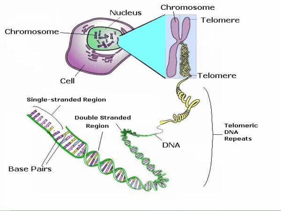

Humans 46 chromosomes
46 sister chromatids One from your mom, one from your dad

Cell Cycle: Life of a Cell
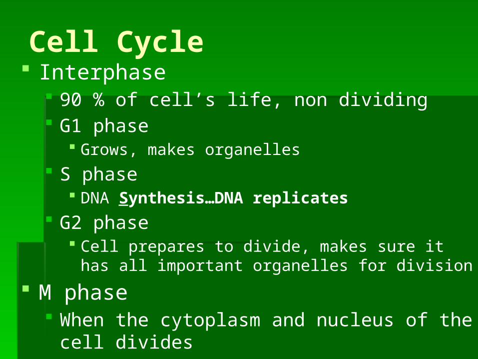
Cell Cycle Interphase
90 % of cell’s life, non dividing G1 phase
Grows, makes organelles S phase
DNA Synthesis…DNA replicates G2 phase
Cell prepares to divide, makes sure it has all important organelles for division
M phase When the cytoplasm and nucleus of the cell
divides

Cell Cycle There are check points in G1, S, and G2
Make sure cell is ready to move onto the next phase (has all necessary organelles, copied DNA, etc.)
Once the cell has past the G1 checkpoint, it will complete the cell cycle
Some cells stay in the G1 phase all their life (muscle cell, brain cells)
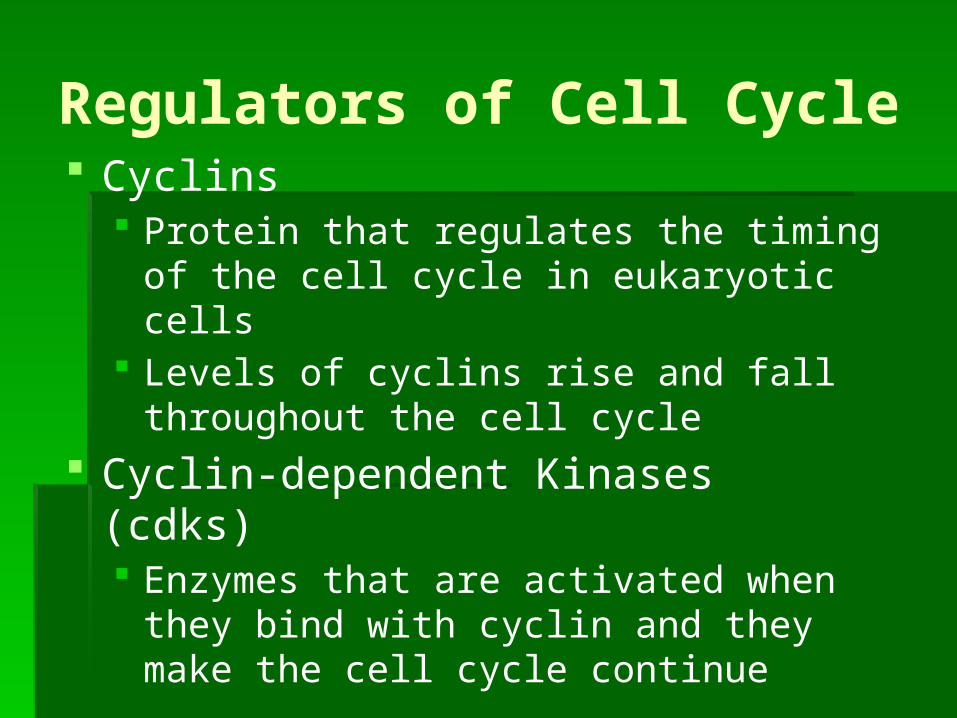
Regulators of Cell Cycle Cyclins
Protein that regulates the timing of the cell cycle in eukaryotic cells
Levels of cyclins rise and fall throughout the cell cycle
Cyclin-dependent Kinases (cdks) Enzymes that are activated when they bind
with cyclin and they make the cell cycle continue


Regulators Internal
Factors within the cell that control cell cycle Cyclin and CDKs
Allow cell cycle to proceed only when certain processes have occurred
Replication of chromosomes Chromosome Attachment to spindle before anaphase
External Factors Outside the cell
Growth factorsmolecules that bind to cell surface that signal cell to divide
Similar cells have molecules that have opposite effect so that when it becomes to crowded, cells stop dividing


M-phase Consists of mitosis and
cytokinesis Mitosis
Process by which the nucleus of a cell divides
One parent cell makes two identical daughter cells
This is how organisms repair tissue and grow and develop
Cytokinesis-division of the cytoplasm

Depending on cell type… Mitosis can take a few minutes or a few
days Muscle cells (non-dividing) Nerve cells (non-dividing) Skin cells (divide all the time) Digestive Tract cells (divide all the time)

Life Span of Some Human Cells
Cell type Life span Cell divisionLining of esophagus 2-3 days Can divide
Lining of small intestine 1-2 days Can divide
Lining of the large intestine
6 days Can divide
Red blood cells Less than 120 days Cannot divide
White blood cells 10 hours to decades Many do not divide
Smooth muscle Long-lived Can divide
Cardiac (heart) muscle Long-lived Cannot divide
Skeletal muscle Long-lived Cannot divide
Neurons (nerve) cells Long-lived Most do not divide

Prophase 50-60% of time Chromosomes become visible Centrioles develop in cytoplasm near nuclear
envelope Centrioles separate and migrate to opposite ends
of nuc. Env. Centrosome
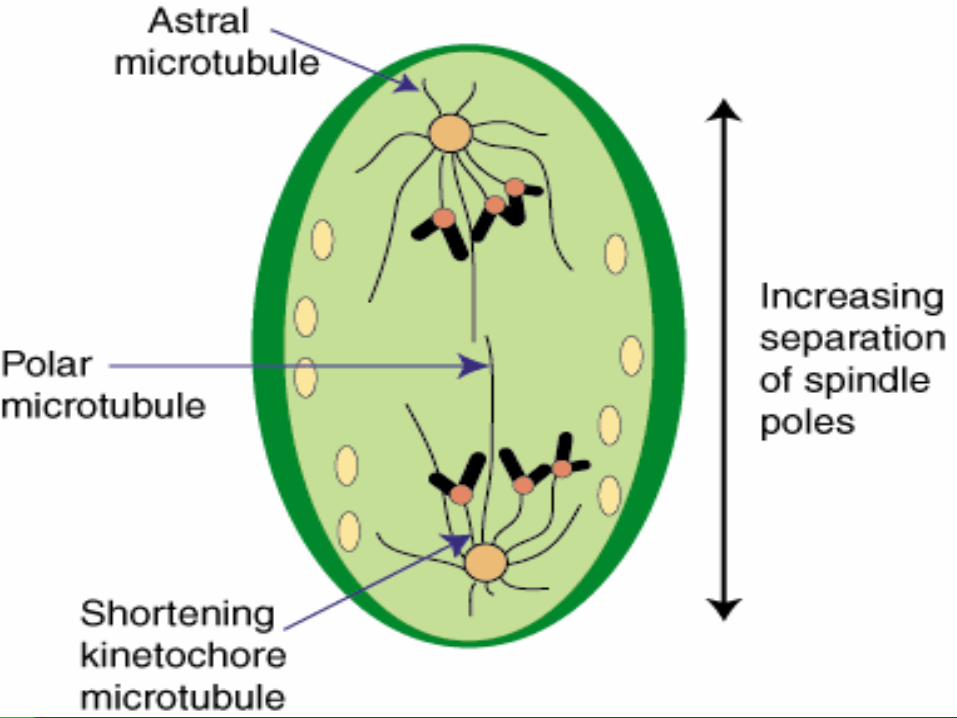
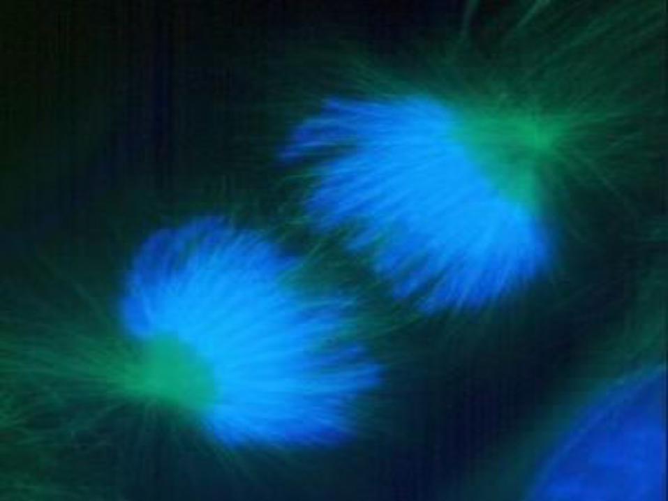
Region where Centrioles are found Organize the “spindle”
Fan like microtubule structure that helps separate chromosomes
Plants do NOT have Centrioles
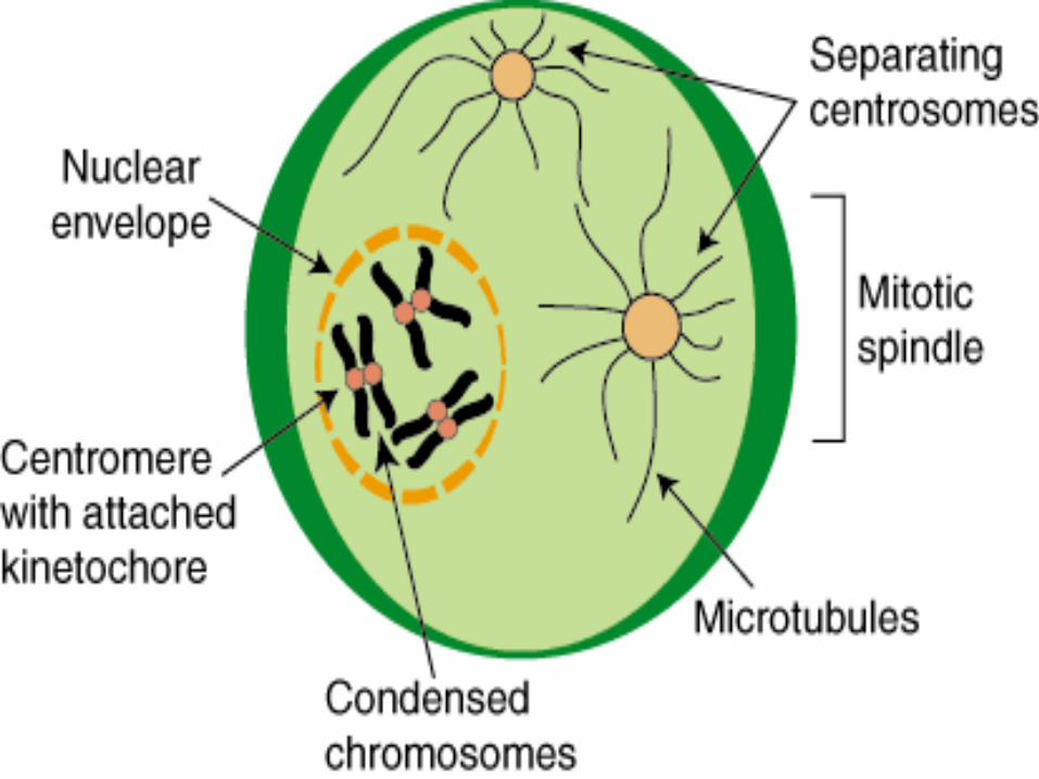

End of prophase Chromosomes coil together tightly Nucleolus disappears Nuclear envelope breaks down
Metaphase Few minutes Chromosomes line up in middle (M in
metaphase MIDDLE) Microtubules connect centromere of each
chromosome to the 2 poles of spindle


Anaphase Centromeres joining sister chromatids
separate and become individual chromosomes
They are dragged by fibers to opposite poles
Ends when chromosomes stop moving


Telophase Opposite of prophase Condensed chromosomes disperse into
tangle of material Nuclear envelope reforms Spindle breaks apart Nucleolus becomes visible At the end 2 identical nuclei in one cell


Cytokinesis Happens at the same time as Telophase Division of cytoplasm Animal Cells
Cell membrane drawn inward until it pinches off and forms 2 id daughter cells
Plant Cells Cell plate forms between nuclei Cell Plate develops into separate membrane Cell wall appears


Regulators of Cell Cycle Cyclins
Protein that regulates the timing of the cell cycle in eukaryotic cells
Levels of cyclins rise and fall throughout the cell cycle
Cyclin-dependent Kinases (cdks) Enzymes that are activated when they bind
with cyclin and they make the cell cycle continue

Regulators Internal
Factors within the cell that control cell cycle Cyclin and CDKs
Allow cell cycle to proceed only when certain processes have occurred
Replication of chromosomes Chromosome Attachment to spindle before anaphase
External Factors Outside the cell
Growth factorsmolecules that bind to cell surface that signal cell to divide
Similar cells have molecules that have opposite effect so that when it becomes to crowded, cells stop dividing

MEIOSIS

Gregor Mendel
1822 Austrian monk University of
Vienna In charge of
the Garden

What Gregor Mendel Knew…
Each organism must inherit a single copy of every gene from each of its “parents”
Each of the organisms gametes must contain just one set genes When gametes are formed, there must be a
process that separates the 2 sets of genes so each gamete gets one set

Karyotype A photograph of a organism’s
chromosomes, arranged according to size
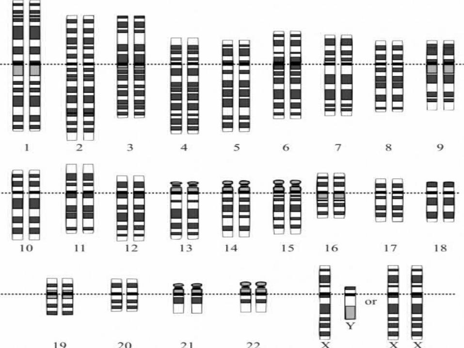


Chromosome Number Homologous chromosomes
Chromosome that has a corresponding chromosome from the opposite-sex parent
Fruit fly has 8 chromosomes 4 from mom 4 from dad

Diploid Di= two sets Cell that contains both sets of homologus
chromosomes Cell contains
2 complete sets of chromosome 2 complete sets of genes
Number of chrms in diploid cell represented by 2N
For Drosophilia (fruit fly) 2N=8 Mendel said:
Each adult cell contains two copies of each gene
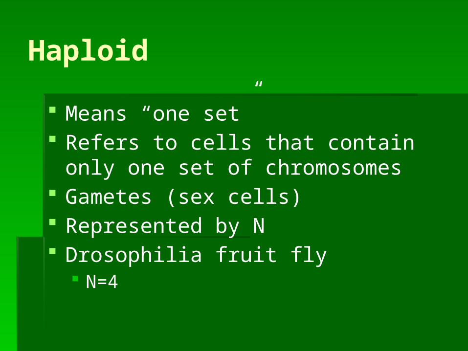
Haploid Means “one set” Refers to cells that contain only one set
of chromosomes Gametes (sex cells) Represented by N Drosophilia fruit fly
N=4

How are haploid (N) gametes made from diploid (2N) cells?

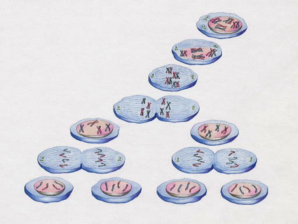
Meiosis Process of reduction division in which the
number of chromosomes per cell is cut in half through the separation of homologous chromosomes in a diploid cell

Meiosis 2 distinct stages Meiosis I
A diploid cell enters here Meiosis II
At the end of this, the diploid cell that entered meiosis has become 4 haploid cells


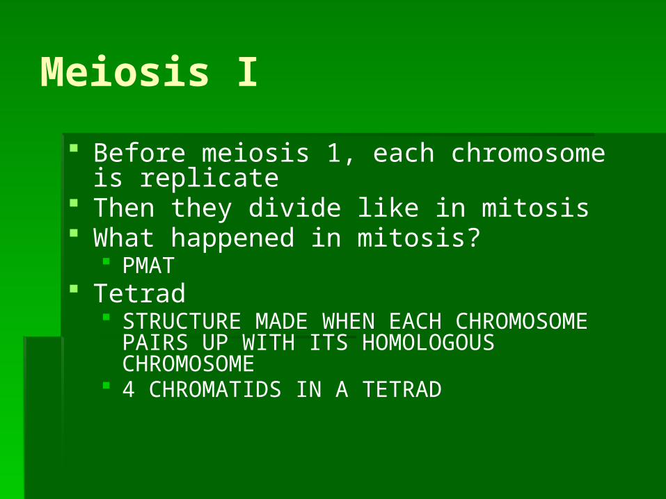
Meiosis I Before meiosis 1, each chromosome is
replicate Then they divide like in mitosis What happened in mitosis?
PMAT Tetrad
STRUCTURE MADE WHEN EACH CHROMOSOME PAIRS UP WITH ITS HOMOLOGOUS CHROMOSOME
4 CHROMATIDS IN A TETRAD

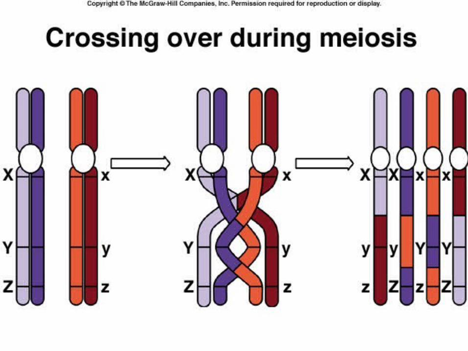
Prophase 1 Each chromosome pairs with its
homologous chromosome making a tetrad
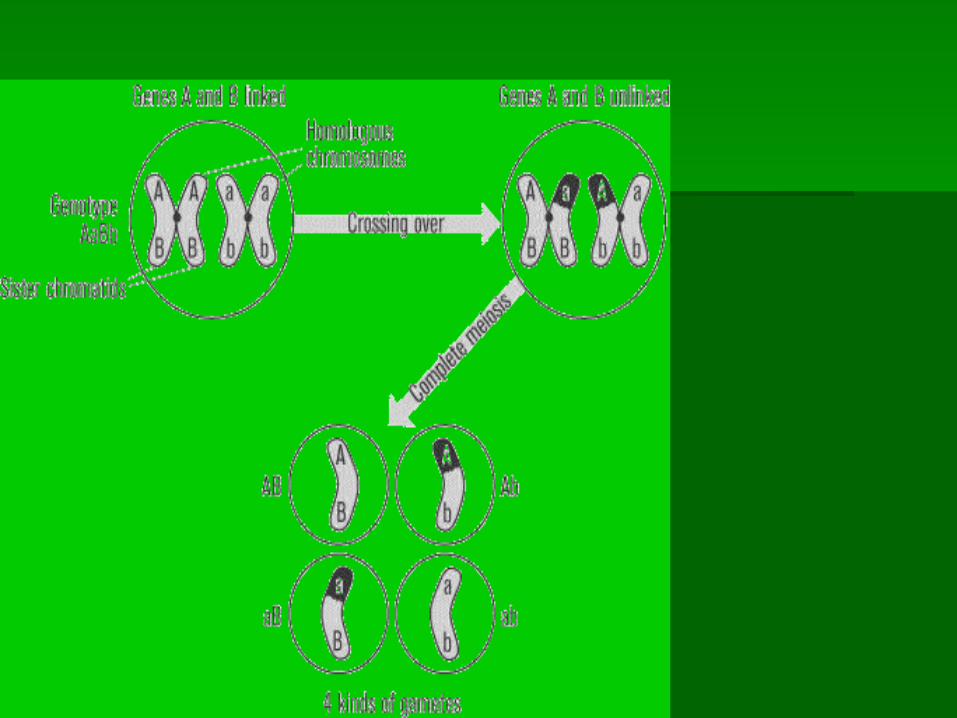
As they pair up in tetrads, chromosomes exchange portions of their chromatids in the process …. CROSSING OVER

Crossing Over

Metaphase1 Spindle fibers attach to chromosomes
Anaphase 1• The spindles pull homologous
chromosomes apart to opposite poles/ends
Telophase 1• Nuclear membranes form and cell
separates into two new cells


Now what do we have? 2 new daughter cells Are they identical to the parents?
No The parent has 4 chromosomes Each daughter cell has 4 chromosomes but they
are different because of crossing-over Each daughter cell has a set of chromosomes
and alleles different from each other and different from the parent diploid cell

Meiosis II Unlike Mitosis, Neither cell goes through
a round of chromosome replication Each cell’s chromosome has 2
chromatids

Prophase II Meiosis I resulted in 2 “seemingly” diploid
cells Remember they are genetically different b/c
of crossing over in prophase I We still need to cut this number in half to
reach our goal of 4 haploid cells

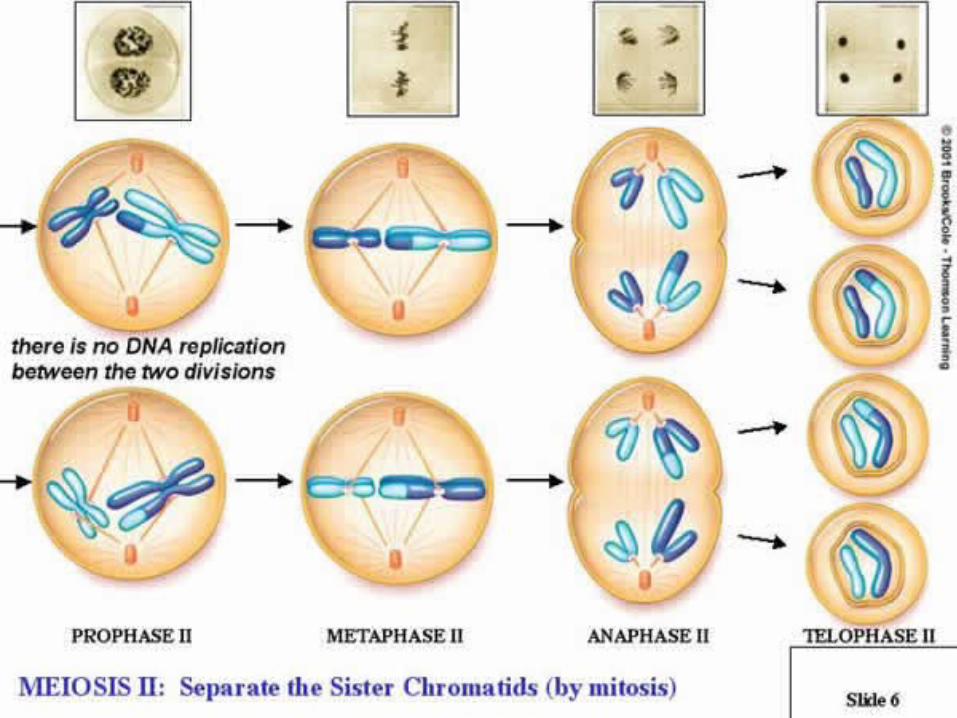
Metaphase 2 Chromosomes line up in middleAnaphase 2
• Sister chromatids separate and move to opposite poles
Telophase 2• Meiosis II results in 4 haploid (N)
daughter cells• 4 daughter cells contain haploid number
of chromosomes, just 2 each
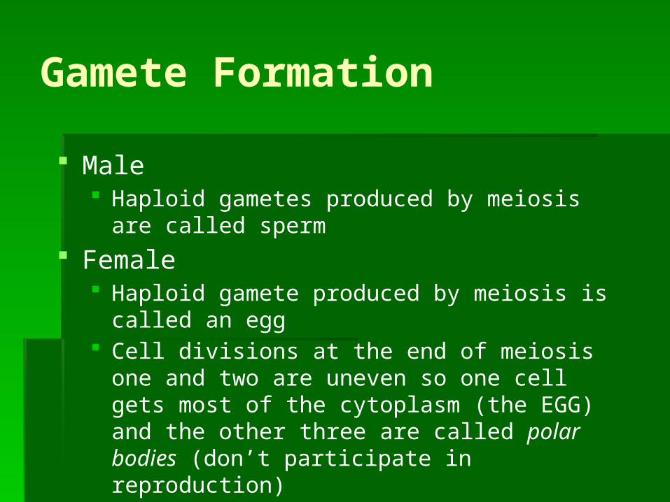
Gamete Formation
Male Haploid gametes produced by meiosis are called
sperm Female
Haploid gamete produced by meiosis is called an egg
Cell divisions at the end of meiosis one and two are uneven so one cell gets most of the cytoplasm (the EGG) and the other three are called polar bodies (don’t participate in reproduction)


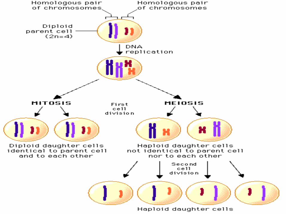


Mitosis vs. Meiosis Mitosis
Results in the production of two genetically identical DIPLOID cells
Daughter cells have sets of chromosomes identical to each other and to parent cell
MITOSIS allows body to grow and replace other cells
Asexual reproduction Meiosis
Results in four genetically different HAPLOID cells MEIOSIS is how sexually reproducing organisms
make gametes


Genes


Microscope Lab Analysis Mitosis/Meiosis
Microscope Lab Lab notebooks Title “Cell
Division Microscope Lab”
MUST sketch each stage and label the power
Label slide name Stage of mitosis or meiosis Power of the objective
used to observe cell Need to observe each
stage of mitosis and meiosis


