MMP expression alteration and MMP-1production control by syringic acid via AP-1mechanismJi Young Ryu1 and Eun Ju Na2*
Abstract
Background: Syringic acid is a phenolic compound that can be produced through selective hydrolysis of eudesmicacid containing 20% sulfuric acid. The acid is obtained by breaking down components, such as anthocyanin andlignin acid, present in the oils of acai berries and other fruits. Recently, the anti-inflammatory, selective toxicity,anticancer, and antioxidant effects of syringic acid have been studied, but few studies on the effects of syringicacid on human keratinocytes (HaCaT) cells have been published. The present study investigated the antioxidanteffects of syringic acid, as a potential cosmetic ingredient, on matrix metalloproteinase (MMP) expressionalteration and MMP production control through the activator protein-1 (AP-1) mechanism in HaCaT cellsexposed to ultraviolet B radiation.
Methods: A reactive oxygen species (ROS)-scavenging assay using a luciferase reporter that utilizes the AP-1 response element, an enzyme-linked immunosorbent assay (ELISA), and quantitative reverse transcriptionpolymerase chain reaction (qRT-PCR) were used. To confirm if ROS in HaCaT cells damaged by ultraviolet Bare eliminated by syringic acid, 2′-7′-dichlorofluorescein diacetate was used to measure the ROS quantity.qRT-PCR analysis was used to measure the expressions of SOD1 mRNA, GPx1 mRNA, and catalase (CAT) mRNA,which are related to oxidation inhibition. To measure the anti-aging effects of syringic acid, qRT-PCR was used tomeasure the expression levels of MMP mRNA, c-Jun, and c-Fos.
Results: ROS were eliminated by syringic acid, and cell aging due to ultraviolet B was suppressed. Results of qRT-PCR analysis confirmed that syringic acid suppressed oxidation in HaCaT cells damaged by ultraviolet B. Further,syringic acid was found to suppress the expression of MMP mRNA, c-Jun, and c-Fos in a concentration-dependentmanner. ELISA showed that MMP-1 production decreased in a concentration-dependent manner. The luciferasereporter analysis revealed a concentration-dependent decrease in the transcriptional activity of AP-1 promotercaused by syringic acid.
Conclusions: Syringic acid was shown to be involved in altering MMP expression and controlling MMP-1 productionthrough the AP-1 mechanism. Thus, the antioxidant and anti-aging effects of syringic acid increased the survival rate ofHaCaT cells damaged by ultraviolet B, suggesting that it can be used as a natural phytochemical in cosmetic products.
Keywords: Syringic acid, HaCaT , Ultraviolet B, ROS, Antioxidant, MMPs, Procollagen type I
* Correspondence: [email protected] of Biological Engineering, Konkuk University, 120Neungdong-ro, Gwangjin-gu, Seoul 05029, Republic of KoreaFull list of author information is available at the end of the article
BackgroundExternal stimuli, including the sun’s rays as well as directand continuous exposure of keratinocytes to the externalenvironment, lead to oxidative stress and skin aging.When the skin is in a continuous oxidative state withslow recovery, it becomes rough and dull, which causesskin aging characterized by loss of elasticity and wrinkles(Agarwal et al. 1988; Wong et al. 2007). Therefore, im-proving the body’s antioxidant system is important forprotecting keratinocytes and delaying cellular aging(Applegate et al. 1995).Skin aging can be divided into two classes: intrinsic
aging caused by natural genetics and extrinsic agingcaused by exposure to the external environment(Naylor et al. 2011). Short-wavelength ultraviolet light(UVB) induces the production of matrix metallopro-teinases (MMPs), which harm the skin (Pygmalion etal. 2010). The primary function of MMPs is to breakdown proteins and enzymes in the extra cellularmatrix (ECM) (Egeblad and Werb 2002). Thus, MMPshave a destructive effect on the ECM and cause a de-crease in fibrous collagen (Scharffetter-Kochanek etal. 2000). UCB passes through the skin keratin, causesDNA damage, and interacts with photosensitizers andchromatophores to induce oxidative stress (Ma et al.2001). Consequently, UVB promotes the productionof activator protein-1 (AP-1) and vitalization of nu-clear factor kappa-light-chain-enhancer of activated Bcells and induces the production of reactive oxygenspecies (ROS) on cell surface receptors, such asmitogen-activated kinases (MAPK) (Xu and Fisher2005; Jiang et al. 2006). As such, UV is a major causeof skin aging. To delay aging, continuous researchand development have been conducted in a variety offields to normalize the signal systems within skin cellsand prevent harm caused by UV and external stimuli.Syringic acid, a type of phenolic compound, can be
obtained through the selective hydrolysis of eudesmicacid containing 20% sulfuric acid (Bogert and Ehrlich1919). It is contained in large amounts in oils of acaiberries and other fruits (Pacheco-Palencia et al. 2008).Syringic acid is isolated from medicinal plants and bio-synthesized through the shikimic acid pathway(Andrade et al. 2001; De Heredia et al. 2001; Dawidaret al. 2000). Studies on the selective effects of syringicacid (Shim et al. 1995; Goldberg et al. 1999; Ferguson2001), such as strong anticancer and anti-inflammatoryeffects (Lü et al. 1998; Sun et al. 2002; Rekha et al.2014), antioxidant effects (Srinivasn et al. 2014), andDPPH radical-scavenging activity, have been conducted.Compared with traditional chemical agents, natural in-gredients extracted from plants are known to be lesstoxic to normal cells but are selectively toxic to cancercells. Accordingly, there has been a growing interest in
the mechanisms and extracted components of naturalactive compounds (Shim et al. 1995).The present study investigated the antioxidant ef-
fects of syringic acid on MMP expression alterationand MMP production control through the AP-1mechanism in human keratinocytes exposed to UVBradiation and confirmed the protective effects ofsyringic acid against cell damage and its potential asa cosmetic component.
MethodsCell culture and sample treatmentCell cultureA human keratinocyte (HaCaT) cell line was obtainedfrom American Type Culture Collection (USA) and cul-tured Dulbecco’s modified Eagle’s medium (Hyclone,USA) containing HaCaT cells in 10% fetal bovine serum(Hyclone) and 1% penicillin/streptomycin (penicillin100 IU/mL, streptomycin 100 μg/mL; Invitrogen, USA)and incubated at 37 °C in 5% CO2.
Syringic acid treatmentSyringic acid was purchased from Sigma-Aldrich (USA)in refined powder form and was dissolved in dimethylsulfoxide (Sigma-Aldrich) for the experiment. Afterculturing HaCaT cells (1 × 10⁶ cells/well) in culturedishes for 24 h, syringic acid was added to the medium,and the culture was incubated for 6 h. The cells werethen irradiated with a UVB lamp (UVP, USA). UVBwavelengths were measured using a USB 2000 fiberopticspectrometer system (Ocean Optics, USA). To investi-gate the effects of UVB on HaCaT cells, the medium wasremoved from the culture plate and washed withphosphate-buffered saline (PBS; pH 7.4). To prevent thecells from drying, 1-mL PBS was added to the washedHaCaT cells, which were then irradiated with UVB withthe lid open. After UBS irradiation, PBS was removed,fresh medium was added, and the cells were furthercultured for 24 h.
Measurement of cell viabilityThe cell viability was measured using the principles ofthe water-soluble tetrazolium salts (WST)-1 assay, whichmeasures the absorbance of formazan, a chromogenicmaterial obtained by the reaction of mitochondrialdehydrogenase and soluble tetrazolium salts. The cellswere inoculated into 96-well plates at a concentration of3 × 103 cells/well in 100-μL amounts and incubated for24 h. The cells were then treated with syringic acid atconcentrations of 1, 2, 5, and 10 μM and incubated foranother 24 h after exposure to UVB. Subsequently, 10-μL aliquots of EZ-Cytox cell viability assay kit reagent(ItsBio, Korea) was added to the cell culture plates. After1-h incubation, a microplate reader (Bio-Rad, USA) was
Ryu and Na Biomedical Dermatology (2018) 2:15 Page 2 of 10
used to measure the absorbance at 490 nm to determinethe cell viability; this process was repeated thrice toderive the mean and standard deviation of cell viability.
RNA extraction and cDNA productionAfter extracting RNA for a quantitative analysis of thechanges in gene expression pattern in HaCaT cells dueto syringic acid, cDNA was synthesized, and the expres-sion level of the desired gene was determined throughquantitative real-time polymerase chain reaction (qRT-PCR). After dissolving the incubated cells in TRIzolreagent (Invitrogen, USA), 0.2-mL chloroform (Biopure,Canada) was added, and the cells were kept at roomtemperature before centrifuging at 12000 rpm at 4 °Cfor 20 min. Subsequently, the supernatant fluid, includ-ing mRNA from the infranatant liquid with protein, wasseparated, and 0.5-mL isopropanol was added to it. Thecells were kept at room temperature for 10 min and thencentrifuged at 12000 rpm at 4 °C. After precipitating theRNA, it was washed with 75% ethanol, ethanol wasremoved, and the RNA was dried in room temperature.The dried mRNA was dissolved in diethylpyrocarbonate(DEPC; Biopure) water for use in the experiment, andonly the extracted RNA that exceeded the purity level of260/280 nm (1.8 ratio), as determined using nanodrop(Maestrogen, USA), was used in the experiment. Afterobtaining 10 μL of 1-μg RNA, 0.5-ng oligo dT18 withDEPC water was added in a PCR tube and kept at 70 °Cfor 10 min. After inducing RNA denaturation, RNA wasincubated with M-MLV reverse transcriptase (Enzy-nomics, Korea) at 37 °C for 1 h to synthesize cDNA.
Measurement of gene expressionqRT-PCR is used to measure the amount of amplifica-tion products by measuring the real-time fluorescence offluorescent material, such as double-strand DNA SYBRgreen, bound by PCR products. The threshold at whichfluorescence can be detected was set to the thresholdcycle (ct), and after measuring the number of cycles thatwere needed to reach the ct, the difference in expressionlevels was determined. If the expression level is high, ct
is reached quickly. As the number of cycles decreases,expression decreases. Therefore, the difference in onecycle results in twice the expression.qRT-PCR was performed using Linegene K (BioER,
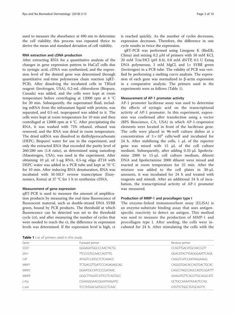
China) and mixing 0.2 μM of primers with 50 mM KCl,20 mM Tris/HCl (pH 8.4), 0.8 mM dNTP, 0.5 U ExtaqDNA polymerase, 3 mM MgCl, and 1× SYBR green(Invitrogen) in a PCR tube. The validity of PCR was veri-fied by performing a melting curve analysis. The expres-sion of each gene was normalized to β-actin expressionin a comparative analysis. The primers used in theexperiments were as follows (Table 1).
Measurement of AP-1 promoter activityAP-1 promoter luciferase assay was used to determinethe effects of syringic acid on the transcriptionalactivity of AP-1 promoter. In this experiment, expres-sion was confirmed after transfection using a vector(BPS Bioscience, CA, USA) in which AP-1-responsiveelements were located in front of the luciferase gene.The cells were placed in 96-well culture dishes at aconcentration of 3 × 10⁴ cells/well and incubated for24 h. After stabilizing the cells, 1 μL of the reportergene was mixed with 15 μL of the cell culturemedium. Subsequently, after adding 0.35-μL lipofecta-mine 2000 to 15-μL cell culture medium, diluentDNA and lipofectamine 2000 diluent were mixed andreacted at room temperature for 25 min. After themixture was added to the cell plates in 30-μLamounts, it was incubated for 24 h and treated withreagents and stimuli. After an additional 24 h of incu-bation, the transcriptional activity of AP-1 promoterwas measured.
Production of MMP-1 and procollagen type IThe enzyme-linked immunosorbent assay (ELISA) isan enzyme-substrate binding assay that uses antigen-specific reactivity to detect an antigen. This methodwas used to measure the production of MMP-1 andprocollagen type I. After seeding, the cells were in-cubated for 24 h. After stimulating the cells with the
Ryu and Na Biomedical Dermatology (2018) 2:15 Page 3 of 10
sample, the cells were cultured for another 24 h, andthe cell culture medium was then separated. ELISAof MMP-1 and procollagen were performed usingthe MMP-1 ELISA kit (QIA55; Merck & Co., Inc.,USA) and the procollagen type I C-peptide enzymeimmunoassay kit (MK 101; Takara, Japan). A 100-μLaliquot of cell culture medium was added to thesurface of each plastic cell culture dish well fixatedwith MMP-1 monoclonal antibody and incubated atroom temperature for 2 h. The cells were washedfive times with 1× washing buffer, 100-μL horserad-ish peroxide-conjugated anti-MMP-1 antibody wasadded, and the cells were incubated at roomtemperature for 1 h. Subsequently, 100-μL of3,3′,5,5′-tetramethylbenzidine (TMB) substrate wasadded and incubated in a dark room at roomtemperature for 30 min. The absorbance wasmeasured at 450 nm.Subsequently, 100 μL of the culture medium was dis-
pensed into the anti-procollagen type I C-peptide (PIP)monoclonal antibody-coated plate and kept at roomtemperature for 2 h. The medium was washed fivetimes with 1× washing buffer and procollagen type-IC-peptide (anti-PIP) monoclonal antibody, which waslabeled with peroxidase, and hydrogen peroxide usedto catalyze the dehydrogenation of substrates wasadded and reacted for 3 h. After adding 100-μL TMBsubstrate and incubating at room temperature for15 min, the absorbance was measured at 450 nm.
Statistical analysisAll experiments were independently repeated ≥threetimes under the same conditions to obtain theexperimental results. The results of each experimentwere analyzed using the non-paired Student’s t test.The p value was calculated, and any value < 0.05 wasconsidered to indicate statistical significance.
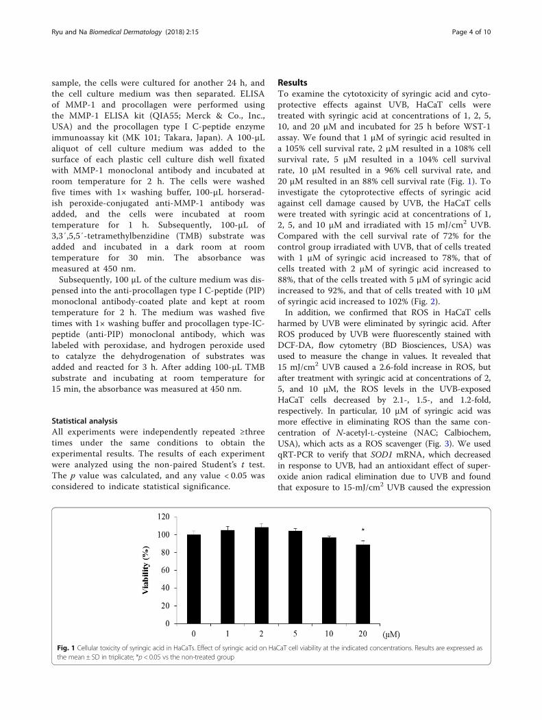
ResultsTo examine the cytotoxicity of syringic acid and cyto-protective effects against UVB, HaCaT cells weretreated with syringic acid at concentrations of 1, 2, 5,10, and 20 μM and incubated for 25 h before WST-1assay. We found that 1 μM of syringic acid resulted ina 105% cell survival rate, 2 μM resulted in a 108% cellsurvival rate, 5 μM resulted in a 104% cell survivalrate, 10 μM resulted in a 96% cell survival rate, and20 μM resulted in an 88% cell survival rate (Fig. 1). Toinvestigate the cytoprotective effects of syringic acidagainst cell damage caused by UVB, the HaCaT cellswere treated with syringic acid at concentrations of 1,2, 5, and 10 μM and irradiated with 15 mJ/cm2 UVB.Compared with the cell survival rate of 72% for thecontrol group irradiated with UVB, that of cells treatedwith 1 μM of syringic acid increased to 78%, that ofcells treated with 2 μM of syringic acid increased to88%, that of the cells treated with 5 μM of syringic acidincreased to 92%, and that of cells treated with 10 μMof syringic acid increased to 102% (Fig. 2).In addition, we confirmed that ROS in HaCaT cells
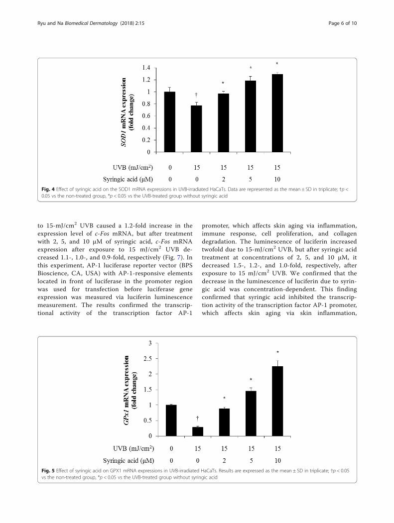
harmed by UVB were eliminated by syringic acid. AfterROS produced by UVB were fluorescently stained withDCF-DA, flow cytometry (BD Biosciences, USA) wasused to measure the change in values. It revealed that15 mJ/cm2 UVB caused a 2.6-fold increase in ROS, butafter treatment with syringic acid at concentrations of 2,5, and 10 μM, the ROS levels in the UVB-exposedHaCaT cells decreased by 2.1-, 1.5-, and 1.2-fold,respectively. In particular, 10 μM of syringic acid wasmore effective in eliminating ROS than the same con-centration of N-acetyl-L-cysteine (NAC; Calbiochem,USA), which acts as a ROS scavenger (Fig. 3). We usedqRT-PCR to verify that SOD1 mRNA, which decreasedin response to UVB, had an antioxidant effect of super-oxide anion radical elimination due to UVB and foundthat exposure to 15-mJ/cm2 UVB caused the expression
Fig. 1 Cellular toxicity of syringic acid in HaCaTs. Effect of syringic acid on HaCaT cell viability at the indicated concentrations. Results are expressed asthe mean ± SD in triplicate; *p < 0.05 vs the non-treated group
Ryu and Na Biomedical Dermatology (2018) 2:15 Page 4 of 10
level of SOD1 to decrease 0.7-fold, but after treatmentwith 2, 5, and 10 μM of syringic acid, the SOD1 mRNAexpression after exposure to 15 mJ/cm2 UVB increased0.9-, 1.1-, and 1.2-fold, respectively (Fig. 4). We also usedqRT-PCR to verify that GPX1 mRNA, which decreasedin response to UVB, had an antioxidant effect of super-oxide anion radical elimination caused by UVB andfound that 15-mJ/cm2 UVB caused the expression levelof GPX1 to decrease 0.2-fold, but after treatment with 2,5, and 10 μM of syringic acid, the SOD1 mRNA expres-sion after exposure to 15 mJ/cm2 UVB increased 0.8-,1.4-, and 2.2-fold, respectively (Fig. 5). In this experi-ment, qRT-PCR was used to determine whether catalase(CAT) mRNA, which decreased in response to UVB,could be recovered to some extent by syringic acid and
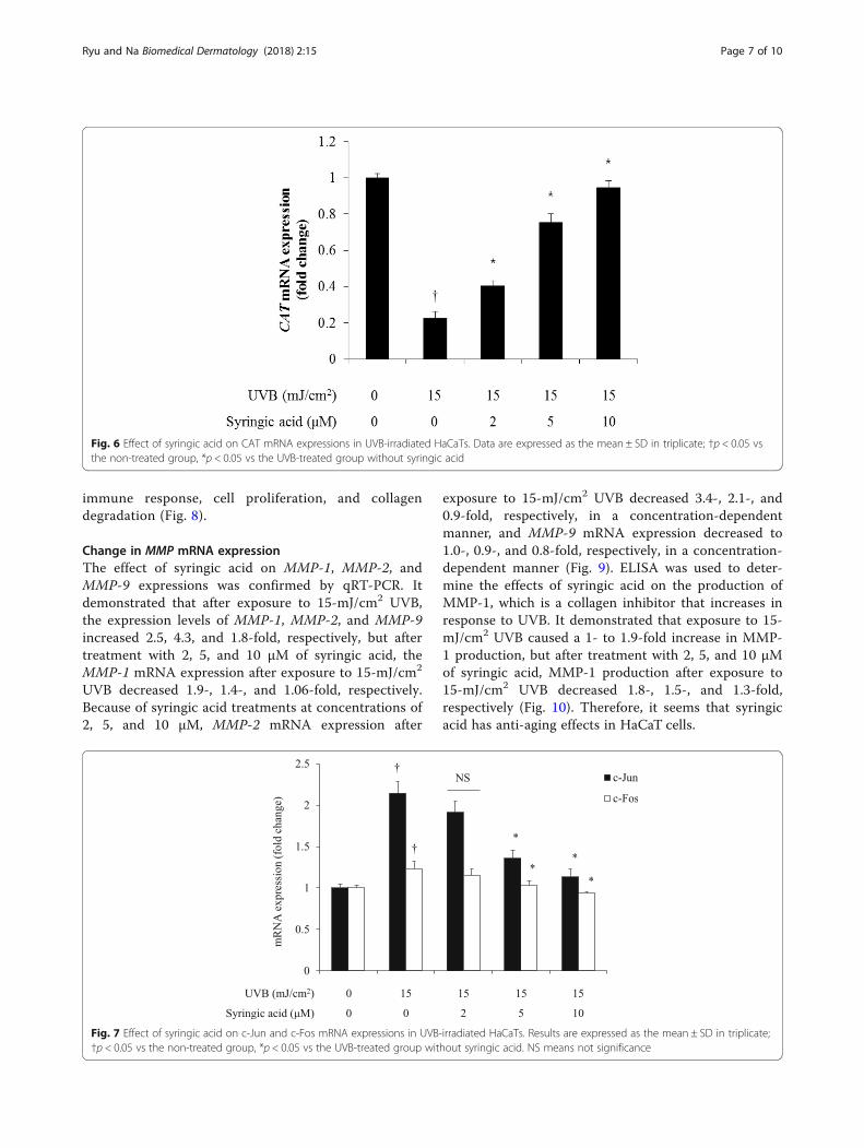
found that 15-mJ/cm2 UVB caused the expression levelof CAT mRNA to decrease 0.2-fold, but after treatmentwith 2, 5, and 10 μM of syringic acid, CAT mRNAexpression after exposure to 15 mJ/cm2 UVB increased0.4-, 0.7-, and 0.9-fold, respectively (Fig. 6).To examine the changes in MMP expression and
MMP-1 production caused by syringic acid throughthe AP-1 mechanism, qRT-PCR was used to determinethe degree of reduction in c-Jun and c-Fos mRNA ex-pression due to syringic acid after their increase in re-sponse to UVB. Our results revealed that 15-mJ/cm2
UVB caused a 2.1-fold increase in the expression levelof c-Jun mRNA, but after treatment with 2, 5, and10 μM of syringic acid, c-Jun mRNA expression de-creased 1.9-, 1.3-, and 1.1-fold, respectively. Exposure
Fig. 2 Effect of syringic acid on the viability of UVB-irradiated HaCaTs. Data are expressed as the mean ± SD in triplicate; †p < 0.05 vs the non-treatedgroup, *p < 0.05 vs the UVB-treated group without syringic acid
Fig. 3 ROS scavenging effect of syringic acid in UVB-irradiated HaCaTs. Results are expressed as the mean ± SD in triplicate; †p < 0.05 vs the non-treatedgroup, *p < 0.05 vs the UVB-treated group without syringic acid
Ryu and Na Biomedical Dermatology (2018) 2:15 Page 5 of 10
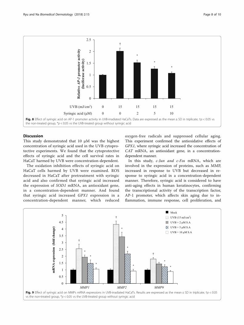
to 15-mJ/cm2 UVB caused a 1.2-fold increase in theexpression level of c-Fos mRNA, but after treatmentwith 2, 5, and 10 μM of syringic acid, c-Fos mRNAexpression after exposure to 15 mJ/cm2 UVB de-creased 1.1-, 1.0-, and 0.9-fold, respectively (Fig. 7). Inthis experiment, AP-1 luciferase reporter vector (BPSBioscience, CA, USA) with AP-1-responsive elementslocated in front of luciferase in the promoter regionwas used for transfection before luciferase geneexpression was measured via luciferin luminescencemeasurement. The results confirmed the transcrip-tional activity of the transcription factor AP-1
promoter, which affects skin aging via inflammation,immune response, cell proliferation, and collagendegradation. The luminescence of luciferin increasedtwofold due to 15-mJ/cm2 UVB, but after syringic acidtreatment at concentrations of 2, 5, and 10 μM, itdecreased 1.5-, 1.2-, and 1.0-fold, respectively, afterexposure to 15 mJ/cm2 UVB. We confirmed that thedecrease in the luminescence of luciferin due to syrin-gic acid was concentration-dependent. This findingconfirmed that syringic acid inhibited the transcrip-tion activity of the transcription factor AP-1 promoter,which affects skin aging via skin inflammation,
Fig. 4 Effect of syringic acid on the SOD1 mRNA expressions in UVB-irradiated HaCaTs. Data are represented as the mean ± SD in triplicate; †p <0.05 vs the non-treated group, *p < 0.05 vs the UVB-treated group without syringic acid
Fig. 5 Effect of syringic acid on GPX1 mRNA expressions in UVB-irradiated HaCaTs. Results are expressed as the mean ± SD in triplicate; †p < 0.05vs the non-treated group, *p < 0.05 vs the UVB-treated group without syringic acid
Ryu and Na Biomedical Dermatology (2018) 2:15 Page 6 of 10
immune response, cell proliferation, and collagendegradation (Fig. 8).
Change in MMP mRNA expressionThe effect of syringic acid on MMP-1, MMP-2, andMMP-9 expressions was confirmed by qRT-PCR. Itdemonstrated that after exposure to 15-mJ/cm2 UVB,the expression levels of MMP-1, MMP-2, and MMP-9increased 2.5, 4.3, and 1.8-fold, respectively, but aftertreatment with 2, 5, and 10 μM of syringic acid, theMMP-1 mRNA expression after exposure to 15-mJ/cm2
UVB decreased 1.9-, 1.4-, and 1.06-fold, respectively.Because of syringic acid treatments at concentrations of2, 5, and 10 μM, MMP-2 mRNA expression after
exposure to 15-mJ/cm2 UVB decreased 3.4-, 2.1-, and0.9-fold, respectively, in a concentration-dependentmanner, and MMP-9 mRNA expression decreased to1.0-, 0.9-, and 0.8-fold, respectively, in a concentration-dependent manner (Fig. 9). ELISA was used to deter-mine the effects of syringic acid on the production ofMMP-1, which is a collagen inhibitor that increases inresponse to UVB. It demonstrated that exposure to 15-mJ/cm2 UVB caused a 1- to 1.9-fold increase in MMP-1 production, but after treatment with 2, 5, and 10 μMof syringic acid, MMP-1 production after exposure to15-mJ/cm2 UVB decreased 1.8-, 1.5-, and 1.3-fold,respectively (Fig. 10). Therefore, it seems that syringicacid has anti-aging effects in HaCaT cells.
Fig. 6 Effect of syringic acid on CAT mRNA expressions in UVB-irradiated HaCaTs. Data are expressed as the mean ± SD in triplicate; †p < 0.05 vsthe non-treated group, *p < 0.05 vs the UVB-treated group without syringic acid
Fig. 7 Effect of syringic acid on c-Jun and c-Fos mRNA expressions in UVB-irradiated HaCaTs. Results are expressed as the mean ± SD in triplicate;†p < 0.05 vs the non-treated group, *p < 0.05 vs the UVB-treated group without syringic acid. NS means not significance
Ryu and Na Biomedical Dermatology (2018) 2:15 Page 7 of 10
DiscussionThis study demonstrated that 10 μM was the highestconcentration of syringic acid used in the UVB cytopro-tective experiments. We found that the cytoprotectiveeffects of syringic acid and the cell survival rates inHaCaT harmed by UVB were concentration-dependent.The oxidation inhibition effects of syringic acid on
HaCaT cells harmed by UVB were examined. ROSdecreased in HaCaT after pretreatment with syringicacid and also confirmed that syringic acid increasedthe expression of SOD1 mRNA, an antioxidant gene,in a concentration-dependent manner. And foundthat syringic acid increased GPX1 expression in aconcentration-dependent manner, which reduced
oxygen-free radicals and suppressed cellular aging.This experiment confirmed the antioxidative effects ofGPX1, where syringic acid increased the concentration ofCAT mRNA, an antioxidant gene, in a concentration-dependent manner.In this study, c-Jun and c-Fos mRNA, which are
involved in the expression of proteins, such as MMP,increased in response to UVB but decreased in re-sponse to syringic acid in a concentration-dependentmanner. Therefore, syringic acid is considered to haveanti-aging effects in human keratinocytes, confirmingthe transcriptional activity of the transcription factor,AP-1 promoter, which affects skin aging due to in-flammation, immune response, cell proliferation, and
Fig. 8 Effect of syringic acid on AP-1 promoter activity in UVB-irradiated HaCaTs. Data are expressed as the mean ± SD in triplicate; †p < 0.05 vsthe non-treated group, *p < 0.05 vs the UVB-treated group without syringic acid
Fig. 9 Effect of syringic acid on MMPs mRNA expressions in UVB-irradiated HaCaTs. Results are expressed as the mean ± SD in triplicate; †p < 0.05vs the non-treated group, *p < 0.05 vs the UVB-treated group without syringic acid
Ryu and Na Biomedical Dermatology (2018) 2:15 Page 8 of 10
collagen degradation. Therefore, syringic acid ap-peared to be an effective anti-aging agent in humankeratinocytes.Proteinases, such as MMP-1, play a crucial role in skin
aging by abnormally suppressing collagen in dermaltissues (Chen et al. 2008). In the present study, we con-firmed that syringic acid decreased the production ofcollagen-inhibiting enzyme MMP-1 in a concentration-dependent manner. Thus, syringic acid has an anti-agingeffect in the human keratinocyte cell line HaCaT.Therefore syringic acid was shown to be involved in
altering MMP expression and controlling MMP-1production through the AP-1 mechanism. Thus, theantioxidant and anti-aging effects of syringic acid in-creased the survival rate of HaCaT cells damaged byultraviolet B, suggesting that it can be used as a naturalphytochemical in cosmetic products.
ConclusionMost studies related to syringic acid have focused on itsantioxidant and anti-inflammatory roles in the fields offood, medicine, pharmacy, and life sciences. No studyhas been conducted on its role in anticancer treatmentsor cosmetics. In particular, no research on the effects ofsyringic acid in human keratinocytes has been published.The present study investigated the effects of syringicacid, which has been shown to protect against cell dam-age via anti-inflammatory, antioxidant, and anticancereffects. It examined antioxidative effects of syringic acidin HaCaT cells exposed to UVB and the effect on MMPexpression alteration and MMP production controlthrough AP-1 regulation. Syringic acid was tested for itscytoprotective effect and its potential as a cosmetic in-gredient. Syringic acid was shown to be involved in
MMP gene expression and MMP production controlthrough AP-1 regulation, thereby protecting human ker-atinocytes from damage and restoring the survival rateof the cells harmed by UVB. Syringic acid was shownto have antioxidant and anti-aging effects; therefore,it can potentially be used as a natural phytochemicalingredient in cosmetic products.
Authors’ contributionsBoth authors contributed to all the research background such as the experiments,data collection, and statistical analysis as well as manuscript draft. Both authorsread and approved the final manuscript.
Ethics approval and consent to participateNot applicable
Consent for publicationNot applicable
Competing interestsThe authors declare that they have no competing interests.
Fig. 10 Effect of syringic acid on MMP-1 production in UVB-irradiated HaCaTs. Data are represented as the mean ± SD in triplicate; †p < 0.05 vsthe non-treated group, *p < 0.05 vs the UVB-treated group without syringic acid
Ryu and Na Biomedical Dermatology (2018) 2:15 Page 9 of 10
Publisher’s NoteSpringer Nature remains neutral with regard to jurisdictional claims in publishedmaps and institutional affiliations.
Author details1Halla University, 28, Halladae-gil, Heungeop-myeon, Wonju-si, Gangwon-do25404, Republic of Korea. 2Department of Biological Engineering, KonkukUniversity, 120 Neungdong-ro, Gwangjin-gu, Seoul 05029, Republic of Korea.
Received: 25 September 2017 Accepted: 5 March 2018
ReferencesAgarwal S, Drysdale BE, Shin HS. Tumor necrosis factor-mediated cytotoxicity
Analysis of phenolic compounds in Spanish Albrariño and PortugueseAlvarinho and Loureiro wines by capillary zone electrophoresis and high-performance liquid chromatography. Electrophoresis. 2001;22(8):1568–72.
Applegate LA, Noël A, Vile G, Frenk E, Tyrrell RM. Two genes contribute todifferent extents to the heme oxygenase enzyme activity measured incultured human skin fibroblasts and keratinocytes. Photochem Photobiol.1995;61(3):285–91.
Bogert M, Ehrlich J. The synthesis of certain pyrogallol ethers, including a newacetophenetide derived from the ethyl ether of syringic acid. J Am ChemSoc. 1919;41(5):798–810.
Chen W, Kang J, Xia J, Li Y, Yang B, Chen B, Sun W, Song X, Xiang W, Wang X,Wang F, Wan Y, Bi Z. p53-related apoptosis resistance and tumor suppressionactivity in UVB-induced premature senescent human skin fibroblasts. Int JMol Med. 2008;21(5):645–53.
Dawidar AM, Ezmiriy ST, Abdel-Mogib M, el-Dessouki Y, Angawi RF. New stilbenecarboxylic acid from Convolvulus hystrix. Pharmazie. 2000;55(11):848–9.
De Heredia JB, Torregrosa J, Dominguez JR, Peres JA. Kinetic model for phenoliccompound oxidation by Fenton’s reagent. Chmosphere. 2001;45(1):85–90.
Egeblad M, Werb Z. New functions for the matrix metalloproteinases in cancerprogression. Nat Rev Cancer. 2002;2(3):161–74.
Ferguson LR. Role of plant polyphenols in genomic stability. Mutat Res. 2001;475(1–2):89–111.
Goldberg DM, Hoffman B, Yang J, Soleas GJ. Phenolic constituents, furans, and totalantioxidant status of distilled spirits. J Agric Food Chem. 1999;47(10):3978–85.
Jiang Q, Zhou C, Healey S, Chu W, Kouttab N, Bi Z, Wan Y. UV radiation down-regulates Dsg-2 via Rac/NADPH oxidase-mediated generation of ROS inhuman lens epithelial cells. Int J Mol Med. 2006;18(2):381–7.
Lü W, Shi J, Zhang S, Du Z. Determination of ferulic acid and peoniflorin in siwudecoction prepared by different methods of yellow rice wine. ZhongguoZhong Yao Za Zhi. 1998;23(9):531–3. 575
Ma W, Wlaschek M, Tantcheva-Poór I, Schneider LA, Naderi L, Razi-Wolf Z,Schüller J, Scharffetter-Kochanek. Chronologicalageing and photoageing ofthe fibroblasts and thedermal connective tissue. Clin Exp Dermatol. 2001;26(7):592–9.
Naylor EC, Watson Rachel EB, Sherratt MJ. Molecular aspects of skin ageing.Maturitas. 2011;69(3):249–56.
Pacheco-Palencia LA, Mertens-Talcott S, Talcott ST. Chemical composition,antioxidant properties, and thermal stability of a phytochemicalenriched oil from acai (Euterpe oleracea Mart). J Agric Food Chem.2008;56(12):4631–6.
Pygmalion MJ, Ruiz L, Popovic E, Gizard J, Portes P, Marat X, Lucet-Levannier K,Muller B, Galey JB. Skin cell protection against UVA by Sideroxyl, a newantioxidant complementary to sunscreens. Free Radic Biol Med. 2010;49(11):1629–37.
Rekha KR, Selvakumar GP, Sivakamasundari RI. Effects of syringic acid on chronicMPTP/probenecid induced motor dysfunction, dopaminergic markersexpression and neuroinflammation in C57BL/6 mice. Biomed Aging Pathol.2014;4(2):95–104.
Scharffetter-Kochanek K, Brenneisen P, Wenk J, Herrmann G, Ma W, Kuhr L,Meewes C, Wlaschek M. Photoaging of the skin from phenotype tomechanisms. Exp Gerontol. 2000;35(3):307–16.
Shim JS, Kang MH, Kim YH, Roh JK, Roberts C, Lee IP. Chemopreventive effect ofgreen tea (Camellia sinensis) among cigarette smokers. Cancer EpidermiolBiomarkers Prev. 1995;4(4):387–911.
Srinivasn S, Muthukumaranb J, Muruganathana U, Venkatesanb RS, JalaludeencAM. Antihyperglycemic effect of syringic acid on attenuating the keyenzymes of carbohydrate metabolism in experimental diabetic rats. BiomedPrev Nutr. 2014;4(4):595–602.
Sun J, Chu YF, Wu X, Liu RH. Antioxidant and antiproliferative activities ofcommom fruit. Agric Food Chem. 2002;50(25):7449–54.
Wong T, McGrath JA, Navsaria H. The role of fibroblasts in tissue engineering andregeneration. Br J Dermatol. 2007;156(6):1149–55.
Xu Y, Fisher GJ. Ultraviolet (UV) light irradiation induced signal transduction inskin photoaging. J Dermatol Sci Suppl. 2005;1(2):S1–8.
• We accept pre-submission inquiries
• Our selector tool helps you to find the most relevant journal
• We provide round the clock customer support
• Convenient online submission
• Thorough peer review
• Inclusion in PubMed and all major indexing services
• Maximum visibility for your research
Submit your manuscript atwww.biomedcentral.com/submit
Submit your next manuscript to BioMed Central and we will help you at every step:
Ryu and Na Biomedical Dermatology (2018) 2:15 Page 10 of 10