Molecular-Level Insights into Photocatalysis from Scanning ProbeMicroscopy Studies on TiO2(110)Michael A. Henderson*,† and Igor Lyubinetsky*,‡
†Physical Sciences Division, Pacific Northwest National Laboratory, P.O. Box 999, MS K8-87 Richland, Washington 99352, UnitedStates‡Environmental Molecular Sciences Laboratory, Pacific Northwest National Laboratory, P.O. Box 999, MS K8-93 Richland,Washington 99352, United States
CONTENTS
1. Introduction A2. SPM Imaging of TiO2(110) in Relation to Photo-
catalysis B3. Photochemistry on TiO2(110) C
3.1. Oxygen C3.1.1. Adsorption of Molecular Oxygen D3.1.2. Photoreactions of Molecular Oxygen E
3.2. Carbon Monoxide H3.2.1. Adsorption of Carbon Monoxide H3.2.2. Photoreactions of Carbon Monoxide I
3.3. Carboxylic Acids J3.3.1. Trimethylacetic Acid K3.3.2. Other Carboxylic Acids N
3.4. Alcohols O3.5. Organic Carbonyls P3.6. Water R3.7. Miscellaneous Molecules R
4. Conclusions and Future Considerations S4.1. Charge Trapping Sites S4.2. Surface Modifiers S4.3. Adsorption State, Proximity, and Coverage
Dependence U4.4. Adsorbate Dynamics U4.5. Beyond the Rutile TiO2(110) Surface V
Author Information VCorresponding Author VNotes VBiographies W
Acknowledgments WGlossary of Acronyms and Abbreviations WReferences W
1. INTRODUCTION
The field of heterogeneous photocatalysis has grownconsiderably in the decades since Fujishima and Honda’sground-breaking publications on photoelectrochemistry onTiO2.
1 Numerous review articles2−18 continue to point toboth the progress made in the use of heterogeneous materials(such as TiO2) to perform photoconversion processes and themany opportunities and challenges in heterogeneous photo-catalysis research, such as solar energy conversion andenvironmental remediation. The past decade has also seen anincrease in the use of molecular-level approaches applied tomodel single crystal surfaces in an effort to obtain new insightsinto photocatalytic phenomena. In particular, scanning probemicroscopy (SPM) techniques have enabled researchers to takea “nanoscale” approach to photocatalysis that includesinterrogation of the reactivities of specific sites and adsorbateson a model photocatalyst surface. The rutile TiO2(110) surfacehas become the prototypical oxide single crystal surface forfundamental studies of many interfacial phenomena.2,4,5,19−26
In particular, TiO2(110) has become an excellent model surfacefor probing photochemical and photocatalytic reactions at themolecular-level.4 A variety of experimental approaches haveemerged as being ideally suited for studying photochemicalreactions on TiO2(110), including desorption-oriented ap-proaches and electronic spectroscopies, but perhaps the mostpromising techniques for evaluating site-specific properties arethose of SPM. In this review, we highlight the growing use ofSPM techniques in providing molecular-level insights intosurface photochemistry on the model photocatalyst surface ofrutile TiO2(110). Our objective is both to illustrate the uniqueknowledge that scanning probe techniques have alreadyprovided the field of photocatalysis and also to motivate anew generation of effort into the use of such approaches toobtain new insights into the molecular-level details ofphotochemical events occurring at interfaces. Discussion willstart with an examination of how scanning probe techniques arebeing used to characterize the TiO2(110) surface in ways thatare relevant to photocatalysis. We will then discuss specificclasses of photochemical reaction on TiO2(110) for which SPMhas proven indispensible in providing unique molecular-levelinsights, and we will conclude with discussion of future areas in
which SPM studies may prove valuable to photocatalysis onTiO2.
2. SPM IMAGING OF TIO2(110) IN RELATION TOPHOTOCATALYSIS
The rutile TiO2(110) surface is likely the most well-studiedsingle crystal oxide surface, particularly with SPM ap-proaches.19,20,24 The popularity of this surface amongresearchers interested in oxide surface phenomenon stems, inpart, from the rich insights obtained about surface physical andelectronic structures using SPM. Figure 1 presents a typicalempty-state scanning tunneling microscopy (STM) image ofthe rutile TiO2(110) surface (right) along with a correspondingball model for the surface (left). The utility of this surface forSPM study is seen in the relative ease with which surface sitescan be imaged with atomic resolution. The two mainundercoordinated surface sites, five-coordinate surface Ti4+
cations (Ti5c) and two-coordinate “bridging” surface O2‑ anions(Ob), appear as bright spots/rows and dark rows, respectively,when tunneling into the surface. SPM techniques also readilydetect surface abnormalities, such as oxygen vacancy sites (VO,see Figure 1), steps, and other point or extended defects, on theTiO2(110) surface. The ability of SPM to provide both physicaland electronic structure at the atomic scale for a variety ofsurface sites on TiO2(110) makes this surface useful as a modelfor many oxide surface chemical and photochemical studies.The electronic structure of a photocatalyst surface dictates
the potential energies at which charge carriers ultimately settle
as they diffuse from the bulk to the surface. The surfaceelectronic structure also influences acceptor and donor levels inadsorbed species, which in turn sets the conditions for electrontransfer events important in photocatalysis. In concept, SPMcan provide a site-by-site assessment of both the filled andempty electronic states of a surface structure. This type ofanalysis can focus on the initial state of the surface (prior tolight exposure) and/or a final state in which carriers becometrapped at the surface, that is assuming they are long-livedenough to be imaged. Several studies and reviews19,24,28−36
have examined the use of STM to image electronic defects inthe TiO2(110) surface and changes resulting from lightexposure. In some cases, features observed after irradiationwere linked to trapping of charge carriers at the surface. STMstudies of the “initial” TiO2(110) electronic structure are moreabundant, especially when imaging empty states. Theseconditions provide ample differentiation between cation andanion surface states, and identification ability for surface defectsand adsorbates. STM and scanning tunneling spectroscopy(STS) can be used to characterize the “initial state” electronicstructure of TiO2(110), and the findings are interpreted interms of how the surface might be involved either in photonabsorption events or in charge trapping (from bulk absorption).For example, Minato et al.29 varied the tunneling direction toprovide unique insights into the degree of charge delocalizationat vacancy sites on TiO2(110). Papageorgiou et al.35 exploredthe electronic structure of surface oxygen vacancy sites andadsorbed species on TiO2(110) by varying the sample bias insuch a way as to image both filled and empty states. Figure 2
Figure 1. (a) Ball model and (b) typical empty-state STM image of the rutile TiO2(110) surface. Modified with permission from ref 27. Copyright2006 American Chemical Society.
Figure 2. Experimental STM images simultaneously recorded for a 44 Å2 area of TiO2(110) at +2 V (left) and −2 V (right) sample bias. The squaresmark the positions of VO (green) and OHb (purple) sites, and features adjacent to several VO sites (black). (An impurity is marked with a whitesquare.) Modified with permission from ref 35. Copyright 2010 National Academy of Sciences, USA.
shows STM images generated from tunneling into the surfacewith a +2 V sample bias (left) and out of the surface with a −2V bias (right). Common reference points in the two images areoxygen vacancies (light green squares on “dark” rows), vacancy-related charges residing on the Ti rows (black squares), andbridging OH groups (purple squares at bright features on darkrows). When the bias conditions are changed so that electronswere tunneled from the TiO2 filled states to the tip, theresulting STM image was markedly charged (right in Figure 2).The bridging OH groups (OHb) and VO sites showed little orno contrast, whereas the features on the Ti rows in the vicinityof VO sites showed strong contrast changes (dark squares).These features imaged to the right in Figure 2, recorded with a−2 V bias on the sample, were suggestive of tunneling fromsurface “gap states” located slightly above the valence band(VB) edge. These surface states, associated with O vacancysites, might act as hole trapping sites or potentially even asvisible light absorption centers. (The relative importance oflight absorption at the surface as opposed to in the bulkbecomes proportionally more significant as the particle size isdecreased.)Similar gap surface states have been detected by STS on
another rutile TiO2 surface. Tao et al.37 used STS tocharacterize the rutile TiO2(011) surface before and after“regrowth” in O2. As has been seen by several authors on therutile TiO2(110) surface,
38−44 Tao and co-workers have shownthat Ti interstitials diffusing in the bulk of rutile can be broughtto the TiO2(011) surface at elevated temperatures and in a highO2 chemical potential, resulting in regrowth of TiO2 on thissurface. A “zigzag” (2 × 1) surface structure is typicallyobtained on TiO2(011) by annealing in UHV (left side ofFigure 3). (Details on the surface structure of the (2 × 1)
reconstruction can be found elsewhere.24) Tao et al. were ableto prepare a new surface phase on TiO2(011) by controlling theregrowth processes. Figure 4 (left) shows an STM image withboth phases (the (2 × 1) and the new phase) in spatiallydistinct regions on the surface. By STS, the (2 × 1)reconstruction has the expected ∼3 eV band gap (blue squaresin Figure 4b and c). In contrast, the regrown regions show asignificant tunneling current at negative sample bias (red circlesin Figure 4b and c), corresponding to new surface states formed
within the TiO2 surface band gap. The surface structure of theregrowth is also different from that of the (2 × 1) surface,consisting of a hexagonal array of sites (right side image inFigure 3), presumably indicating a greater degree of under-coordinated cation sites. Evidence for narrowing of the surfaceband gap was also obtained by Tao et al. from UPS, whichshowed a gap state at 2.1 eV below the Fermi level only for theregrown surface.Although neither Papageorgiou et al.35 or Tao et al.37
conducted photochemical studies, their STS studies illustratehow this approach could be useful in making correlationsbetween surface electronic structure and photocatalyticperformance on a site-by-site basis. In particular, there is thepotential that reconstructed or defected surfaces may becomeactive for sub-band-gap photochemical reactivity, as (forexample) has been proposed by Ariga and co-workers for thefaceted surfaces of rutile TiO2(001).
45
3. PHOTOCHEMISTRY ON TIO2(110)
STM is the ideal technique for connecting “local” surfaceelectronic structural information (the previous section) withassessments of “local” surface structure−reactivity relationships.These kinds of connections are becoming commonplace inmany areas of surface science (e.g., catalysis and electronicmaterials) and are made most useful by reaching a consensusabout what STM is actually imaging. Clearly, the field of SPM isstill maturing in its chemical recognition ability. Connectionsbetween surface structure (atomic and electronic) andadsorbate reactivity are less common in the surface photo-chemistry literature. The remainder of this review willconcentrate on examples in which SPM techniques successfullyprovided unique insights into photochemical reactionsoccurring on the TiO2(110) surface. Molecular systems arehighlighted in which there is a high degree of certainty in SPM’sability to identify the reactant. These include adsorbed oxygen,CO, carboxylates, and a variety of other molecular systems. Insome cases, photochemistry has been observed directly withSPM by a change in the reactant’s image. Direct correlationscan then be made between observations of photoreactivity bySPM and the actual charge transfer event itself (e.g., whichcarrier was involved, the relative rates of adsorbates at differentsites, and the reaction mechanism). In other cases, SPM wasused to characterize a reactant-covered surface prior to photonirradiation, with other techniques (e.g., photon stimulateddesorption (PSD)) used to characterized the photochemistry.In these cases, SPM measurements provided valuable insightsinto the local arrangements of adsorbates and their proximitiesto important surface sites (such as VO sites).
3.1. Oxygen
Oxygen plays multiple important roles in many photocatalyticreactions on TiO2, ranging from organic oxidation to watersplitting.12,142−6,13,15,16 In particular, O2 is perhaps the simplestoxidation agent in photooxidation reactions, acting as anelectron scavenger, facilitating photoreactions by assisting thecharge separation, preventing an electron−hole recombination,and indirectly generating other oxidation species (such as OHradicals and H2O2).
4,46 In addition, since O2 is commonlypresent in TiO2 application environments, surface-boundoxygen species may considerably affect TiO2 catalytic proper-ties.2,47 Both the surface thermal chemistry and photochemistryof the O2 on TiO2 have been intensively investigated in the pastwith a variety of ensemble-averaging techniques.4,19,20,24
Figure 3. STM images of the (2 × 1) rutile TiO2(011) reconstructedsurface (left; 50 × 50 nm2) and of the new TiO2 surface phase formedafter annealing in 1 × 10⁻⁶ Torr O2. The surface unit cells areindicated in each image. (Line profiles along the indicated lines can befound in the original figure.) Modified with permission from ref 37.Reprinted by permission from Macmillan Publishers Ltd., copyright2011.
However, only very recent breakthroughs in STM imaging ofmolecular O2 on TiO2(110) have allowed a direct atomisticcorrelation between O2 photochemistry and specific adsorbedO2-related species on this surface, as will be discussed below.This section will start with a brief overview of SPM findingsregarding the thermal properties of O2 on TiO2(110) sincethese set the stage for the photochemistry.3.1.1. Adsorption of Molecular Oxygen. Knowledge of
the adsorption behavior of dioxygen is critical for acomprehensive understanding of O2 photochemistry on TiO2.The temperature, the reduction state of the TiO2 surface, andthe O2 coverage all affect the adsorption state of oxygen. Inparticular, at sufficiently low temperatures (T < 60 K), O2 onlyphysisorbs on the stoichiometric TiO2(110).
48,49 This isconsistent with density functional theory (DFT) calculationsthat show that charge transfer from reduced TiO2 to theadsorbed species is required for O2 chemisorption.50−55 Atelevated temperatures (above ∼150 K), O2 molecules dissociateat VO sites on TiO2(110), and STM studies have directlydemonstrated that each dissociation event leads to healing of asingle VO site, leaving an oxygen adatom (Oa) at a nearby Ti5csite.56−59 In addition, STM studies have revealed a minor(though potentially important) second O2 dissociation channeloccurring on the Ti5c rows, which leads to the formation of theOa pairs.
52,60,61 Wendt and co-workers have suggested that thecharge required for this minor channel of O2 dissociation isprovided by subsurface Ti interstitials.52 However, Petrik, Du,and co-workers suggested that the delocalized unpairedelectrons associated with the VO sites could be utilized in thischannel as well, with the total surface charge being a limitingfactor in the extent of O2 dissociation (at all sites).54,62
The molecular chemisorption of O2 occurs on the reducedTiO2(110) surface at T < 150 K.49,51,63 Chemisorption of O2 isenabled by an electron transfer from a surface source ofelectrons near the Fermi level, usually associated with defects(e.g., VO and Ti interstitials). This chemisorption event hasbeen noted by a variety of spectroscopic means. For example, anew O2-related loss feature appeared in electron energy lossspectroscopy (EELS) at ∼2.8 eV when O2 was exposed to thevacuum-annealed TiO2(110) at low temperature, with itsappearance correlated with a decrease in the Ti3+ defect state at∼0.8 eV.51,64 DFT calculations generally support the formationof nominally O2
− species,52,54,55,65 although the exact extent ofelectron transfer is not well-understood and other species suchas O2
2− have also been suggested.65−69 On the basis of acorrelation between STM and EELS,64 it would appear that theO2 species active for photodesorption is best characterized as aperoxo species (O2
2−) rather than a superoxo species (O2−).
Calculations suggest that the most stable configuration for asingle chemisorbed O2 molecule on TiO2(110) is adsorption ina VO with the O−O bond nearly parallel to the surface andperpendicular to an Ob row, resulting in a O−O distancecharacteristic of O2
2−.55,57,67,68 DFT results also indicate thatchemisorbed anionic oxygen species bind rather strongly (∼2eV) at VO sites57,68,70 and also at neighboring Ti5c sites(although the latter is energetically less favorable).67 Themaximum amount of chemisorbed O2 has been foundempirically to be about twice the VO concentration.49
The molecular-level details associated with O2 chemisorptionon TiO2(110) would be significantly limited without theinsights provided by SPM. Recently, Scheiber et al.71,72 andWang et al.71,72 were both able to directly image with STM atlow temperatures O2 molecules chemisorbed at VO sites onTiO2(110). Single chemisorbed O2 molecules are evidenced bya virtual disappearance of the VO replaced by an extremely faintfeature, as shown in Figure 5b. The STM tip can easilydissociate these adsorbed O2 species, as illustrated in Figure5c.72 While Wang et al.72 reported that O2 dissociation by theSTM tip results in healing of the original VO and deposition ofa single Oa nearby, Scheiber et al.
71 described the formation oftwo Oa’s at opposite sides of the original VO. Lately, Tan et al.
73
Figure 4. STM and STS data for the reconstructed rutile TiO2(011) surface. (a) The surface prepared so the (2 × 1) reconstruction (shown in blue)and a new TiO2 surface phase (red) coexist. (b) I−V spectra taken in the two surface areas (“blue” and “red”). (c) Numerically differentiated dI/dVcurves for the two surface phases. Reprinted with permission from ref 37 and from Macmillan Publishers Ltd., copyright 2011.
Figure 5. STM images of the same (3.0 × 3.2) nm2 area of TiO2(110)(a) before and (b) after O2 exposure at 50 K, taken at V = 0.8 V, I = 3pA, and showing the O2 chemisorption at VO sites, and then (c)imaged at V = 1.5 V, I = 3 pA. Corresponding ball models of themarked rectangular region illustrate the observed events. Reprintedwith permission from ref 72. Copyright 2010 American ChemicalSociety.
have attributed the results of Scheiber and co-workers to ametastable, fast-switching intermediate state driven by the STMtip, thus confirming the final state invoked by Wang et al.72 Inaddition, Tan et al. have observed that, at the saturation O2coverage (corresponding to ∼2 O2 per VO), the chemisorbedO2 species are likewise barely visible in STM images.73
Interestingly, these authors also found that two or more O2molecules in or near a vacancy tend to be more stable againsttip-induced dissociation than a single O2 at VO. Evidently, thisindicates a different bonding and/or charge configuration of O2at coverages above and below the 1:1 ratio of O2 to VO, as hasbeen anticipated in the literature.49,69
Focusing on the initial stages of O2 adsorption, Wang et al.72
have observed another distinctive channel of O2 chemisorptionoccurring at Ti5c sites (far from VO’s). Figure 6 shows that, in
contrast to adsorption at VO sites, an O2 molecule at a Ti5c siteis readily distinguishable as a single protrusion on the Ti5c row.The O2 species chemisorbed at Ti5c sites are found to beimmobile at 50 K,72 in agreement with DFT calculations thatindicate O2 diffusion along Ti5c troughs is hindered by anactivation barrier of ∼0.6 eV.67 On the other hand, the O2species at Ti5c is even more prone to STM tip-induceddissociation that converts it into two adjacent Oa, as shown inFigure 6c, unless extremely mild tunneling parameters are used.Note that accounting for a possible tip-induced dissociationmay clarify the results of previous STM studies that describedO2 dissociation at temperatures as low as 80 K74 and 120 K,52,75
contrary to other literature that stipulates dissociation occursabove 150 K.49,51,62 Comparing two site-specific O2 chem-isorption channels, Wang et al. have reported a highconcentration difference between them, with much less O2species observed at Ti5c sites than at VO sites.72 These authorsalso suggested that the O2 species chemisorbed at the VO andTi5c sites are most likely precursors for the two O2 dissociationchannels on TiO2(110) observed at elevated temper-atures.49,51,62
3.1.2. Photoreactions of Molecular Oxygen. Discussionof the photochemistry of O2 on TiO2(110) in the literature washistorically restricted to the subject of photodesorp-
tion.2,5,14,46,66,76,77 However, more recent results, highlightedbelow, show that both photodesorption and photodissociationoccur when chemisorbed O2 on TiO2(110) is irradiated withUV light. In this section, we will highlight how SPM methodshave revealed new insights into both of these photochemicalchannels of O2 on TiO2(110).
O2 Photodesorption. The photodesorption of molecularoxygen from rutile TiO2(110) is the most comprehensivelystudied photoreaction on TiO2 (by traditional ensemble-averaging techniques). The most extensive research here hasbeen carried out by the Yates group, with the mainaccomplishments described in their recent reviews.2,5,78 Inparticular, they have developed a hole-mediated desorptionmodel for O2 desorption,5,66,78 where photogenerated holesmove to the surface and react with chemisorbed O2 anions,leading to neutral O2 desorption (reaction 1).On the basis of
their results, this group also suggested the existence of twodistinct states of chemisorbed O2 species that contribute to thePSD signal. This has been recently confirmed by Sporleder etal.79 using time-of-flight (TOF) measurements of the velocitydistribution of photodesorbing O2, which revealed twodesorption channels with different mean translational energies.The identities of these two chemisorbed states were unclearthough. Yates and co-workers have also used O2 PSD as amonitor of the charge carrier recombination kinetics inTiO2(110) that governs the survival probability of holes.46,80
In particular, they showed that the O2 PSD kinetics are first-order in charge carrier concentration when surface traps play astrong role in the electron−hole recombination process.80 Incontrast, the O2 PSD kinetics for charge carriers originatingdeeper in the bulk are second-order, dominated by directelectron−hole recombination due to the presence of fewerdefects in the bulk.46 In addition, the kinetics of O2photodesorption were multiexponential, which was interpretedin terms of a percolating model influenced by a network of VOsites.46
While most of the results reported by the Yates group werefor a saturation coverage of chemisorbed O2 (i.e., Θ(O2) ≈2Θ(VO)), the changes in the O2 photodesorption versus O2coverage have been explored by Petrik and Kimmel.81,82 Theyfound that the PSD yield was a complex function of Θ(O2),increasing linearly with different rates over two differentcoverage ranges separated by Θ(O2) ≈ Θ(VO) (see Figure7). These authors argued that differences in the photo-desorption yield were related to changes in the charge state ofO2 below and above Θ(O2) ≈ Θ(VO). In particular, O2molecules chemisorbed at Ti5c sites could have a larger PSDyield than those O2 molecules bound at VO sites.81,82 However,there is some inconsistency for a single O2 at VO, since O2
2−
reacting with a hole should not lead to photodesorption ofneutral O2 because the species would still be negatively chargedand electrostatically bound to the surface. The situationbecomes more complicated if O2 molecules bound at Ti5csites are more O2
2− in character than O2−.64 Interestingly, for
the results in Figure 7, only part (less than 40%) of thechemisorbed O2 desorbed even after prolonged UV irradiation,while the rest remained on the surface in some form that wasinactive for PSD (see below).
Figure 6. STM images of the same (2.3 × 2.6) nm2 area of TiO2(110)(a) before and (b) after O2 exposure at 50 K, taken at V = 0.3 V, I = 1pA, showing the O2 chemisorption at Ti5c sites, and then (c) imaged atV = 0.6 V, I = 3 pA. Reprinted with permission from ref 72. Copyright2010 American Chemical Society.
The photochemical properties of O2 on TiO2(110) wererecently examined by Lyubinetsky and co-workers with STM.83
These authors demonstrated that the photochemical behaviorof O2 bound at non-VO sites must be taken into account inorder to properly describe O2 photochemistry on TiO2(110).For Θ(O2) < Θ(VO), Figure 8 shows that, after UV irradiation,
the O2 molecule located on the Ti5c row (far from a VO)disappeared without emergence of any new feature, indicatingthe desorption of O2 from the Ti5c site. The O2 photo-desorption event from Ti5c sites (reaction 2, modified from
reaction 1) is consistent with a hole-mediated process. On theother hand, no O2 desorption from VO sites was detected. Onthe basis of these STM results, O2 PSD signals occurring at low
Θ(O2) in Figure 782 originate only from O2 molecules bound at
Ti5c rather than VO sites. While this O2 desorption channel isexpected to be active also for Θ(O2) > Θ(VO), an additionaldesorption channel may kick-in at higher O2 coverages, possiblyoriginating from the second O2 species chemisorbed near Ovacancies. Presumably, these two O2 desorption channelsaccount for the two distinctive peaks in the velocity distributionof O2 PSD observed by Sporleder and co-workers.79
The photochemical reactions between chemisorbed, post-annealed O2 and weakly bound, physisorbed O2 have recentlybeen examined by Petrik and Kimmel at annealing temper-atures up to 600 K using PSD of various O2 isotopologues.
84
The distinction between chemisorbed and physisorbed O2 restsin the availability of charge that can be donated from thesurface to O2 to form O2
− or O22− species. In the absence of
available charge, O2 is held on the surface only through its weakpolarizability. They found that UV irradiation resulted inexchange of O atoms between chemisorbed and physisorbedoxygen. The amount of O2 produced in these exchangereactions was maximized for annealing temperatures of ∼350 K,as shown in Figure 9. Annealing above ∼400 K brought Ti
interstitials from the bulk to the surface which reacted with theadsorbed O2 to form TiOx islands. This was evidenced by adecrease in the 16O2 and
16O18O PSD yields.40,52 Furthermore,the O2 produced in the exchange photoreactions was observedto preferentially desorb at an angle of ∼45° with respect to thesurface normal in a plane perpendicular to the Ob rows, whilethe chemisorbed O2 desorbed normal to the surface.Interestingly, the chemisorbed species retained its photo-chemical activity through repeated cycles of UV irradiation andO2 adsorption. The authors argued that the photoactive
Figure 7. Integrated 18O2 PSD (blue) and TPD (orange) yields versus18O2 coverage from TiO2(110). While no thermal desorption isobserved for Θ(O2) < 0.19 ML (∼2Θ(VO)), PSD is readily observed,and it increases linearly over two different coverage ranges. Reprintedwith permission from ref 82. Copyright 2011 American ChemicalSociety.
Figure 8. STM images of the same (1.8 × 2.2) nm2 area of TiO2(110)(a) before and (b) after O2 exposure (Θ(O2) ∼ 0.08 ML), and (c)after UV irradiation for 15 min (all at 50 K), showing the O2photodesorption from the Ti5c site. Corresponding ball models ofthe marked rectangular region illustrate the observed event. Modifiedwith permission from ref 83. Copyright 2012 American ChemicalSociety.
+ → +− +hO /Ti Ti O2 5c 5c 2(g) (2)
Figure 9. Integrated (a) 16O2,18O2, and (b) 16O18O PSD yields versus
annealing T. Saturation coverages of chemisorbed 18O2 on TiO2(110)were annealed; then, 0.43 ML 16O2 was adsorbed at 30 K, and the O2PSD signals were monitored during UV irradiation. Reprinted withpermission from ref 84. Copyright 2011 American Chemical Society.
exchange complex should contain at least four O atoms,whereas among likely candidates are O2 adsorbed at VO and/ortetraoxygen (O4).
84
Yates and co-workers have also investigated the effect ofcoadsorbates on O2 PSD and electron stimulated desorption(ESD).85,86 In particular, they found that single adsorbed Auatoms decreased the O2 PSD yield, as shown in Figure 10.86
This was qualitatively attributed to the reducing of the holetransport rate from the bulk to the surface, presumably causedby a downward band bending induced by the positively chargedAu atoms in the n-type semiconductor of reduced TiO2.Complementary DFT calculations indicated that ∼0.2 electrontransfers from a single Au atom to TiO2(110), leading to a ∼0.6eV downward band bending. The diminishing effect on the O2PSD yield with increasing Au coverage shown in Figure 10 hasbeen ascribed to the notion that the charge per Au atomdecreased due to the formation of Au cluster at highercoverages.86
O2 Photodissociation. Hole-induced O2 photodesorptionhas been the prevailing consideration in oxygen photochemistrystudies in the literature.5,66,78 Recently, Petrik and Kimmel,81,82
using TPD of O2 af ter UV irradiation of adsorbed O2 as adiagnostic tool, revealed that photolysis of adsorbed O2 resultedin dissociation at the VO sites. These authors proposed anelectron-mediated mechanism, resulting in healing of theoriginal VO and deposition of Oa at an adjacent Ti5c site(similar to the thermally induced O2 dissociation at VO),according to reaction 3. Figure 11 summarizes the experimental
results and shows that, for low O2 coverages (i.e., Θ(O2) ≤Θ(VO)), only a relatively small portion of the O2 photodesorbswhile the remaining O2 either photodissociates or stays on thesurface in a nondissociated (“photoblind”) state.81 (In Figure11, Θsat is defined as the saturation coverage of O2, Θrem is thetotal molecular and dissociative oxygen coverages after UVirradiation, Θocc is the number of adsorption sites occupied bythe oxygen, and Θsecond is the O2 coverage resulting from O2exposure after UV irradiation. Θrem was further broken downinto dissociated, Θdiss, and nondissociated, ΘX, components
(see ref 81).) For the maximum coverage of chemisorbed O2,the fractions of both photodesorbed and nondissociated O2increased considerably. Petrik and Kimmel argued that becausethe charge state of the chemisorbed O2 depends on the totaloxygen coverage, the coverage should influence the photo-desorption process. They also proposed that reactions betweenthe chemisorbed O2 and conduction band electrons (e.g.,through an electron attachment mechanism) were responsiblefor both the dissociated and nondissociated O2. Regarding thelatter O2 species, these authors pointed out that this species waslikely different from that of the originally adsorbed O2. UVirradiation likely converted them to a species that either did notreact with electrons or did not result in dissociation.The first spatially resolved evidence of O2 photodissociation
on TiO2(110) was recently presented by Wang et al.83 on thebasis of STM results. For Θ(O2) < Θ(VO), Figure 12demonstrates that after UV irradiation Oa species appearedon Ti5c sites adjacent to the original VO positions as a result ofO2 photodissociation at VO sites (reaction 3). However, someO2/VO species remained intact after UV irradiation (e.g., one inthe left-bottom in Figure 12), confirming the finding of Petrikand Kimmel81 that some O2/VO species remained undis-sociated. In addition, Wang et al. reported that completeness ofO2 dissociation depends on the coverage, whereas the reactionis quenched only above a critical coverage (Θcrit of ∼0.05 ML),whereas all chemisorbed O2/VO species could be dissociated atlower coverages.83 (Note also that O2 dissociation at Ti5c sitesdoes not occur because only O2 desorption has been observedat those sites.) These authors also remarked that while STMstudies have not yet been extended to saturation O2 coverages,the conclusions obtained here (for Θ(O2) up to Θ(VO)) likelycan be projected to higher O2 coverages. One may expect thatcomparable, site-specific photoreactive channels also operatethere through similar mechanisms.The complex site-specific and coverage-dependent photo-
chemical behaviors of O2 on TiO2(110) are still far from beingwell-understood. It should be noted the photoinducedprocesses of electron-mediated dissociation and hole-mediateddesorption of O2 can be considered as complementaryreductive and oxidative surface reactions, respectively. Inorder for photocatalysis on a TiO2 surface to be effective,both reactions should occur at substantial and balanced rates. Incontrast, as Wang and co-workers argued, these conditions arenot satisfied here due to a considerable concentration differencebetween the chemisorbed O2/VO and O2/Ti5c species. In thiscase, a larger total number of electrons should be consumed inO2 dissociation compared to the number of holes consumed inthe O2 photodesorption process.83 Consequently, an increasingnumber of “excess” holes should be trapped at the surface afterphotodesorption is completed but as photodissociationcontinues, as schematically shown in Figure 13. Since anelectron subsequently reaching the surface would have a higherprobability of recombining with trapped holes, the electron-mediated O2 dissociation rate should become diminished whenthe concentration of excess holes exceeds some critical level (atΘcrit).Comparison of the photoinduced dissociation and desorp-
tion of O2 shows that these two reactions originate from thedistinct adsorption sites of VO and Ti5c, respectively, asdiscussed above. Furthermore, Wang et al.83 found that tworeactions follow rather different kinetics. Figure 14 shows thatthe concentration of chemisorbed O2/Ti5c species promptlydecayed to zero (due to photodesorption) with UV irradiation
Figure 10. Initial PSD yield of 18O2 versus Au coverage on the 18O2/TiO2(110) surface. Modified with permission from ref 86. Copyright2011 American Chemical Society.
time. In contrast, for the dissociation channel the chemisorbedO2/VO number decreased considerably more slowly, reachingzero only for Θ(O2) < Θcrit. In addition, they observed thatneither plot could be described by a simple exponentialdependence, as the inset in Figure 14 shows. However, theinitial rate constant for O2 desorption was observed to be (∼16
times) larger than that of O2 dissociation. This is somewhatsurprising, since it is generally believed that holes diffuse moreslowly than electrons.4,5 These authors have contemplated thatthe considerable disparity in the reaction rates may beattributed to a substantial difference in the dynamics ofelectron and hole transfer to chemisorbed O2/VO and O2/Ti5cspecies, respectively.3.2. Carbon Monoxide
Beyond being one of the most frequently employed moleculesas a probe in surface science, carbon monoxide is a keyparticipant in a variety of important catalytic processes, such ashydrogenation, water-gas shift, and thermal/photochemicaloxidation, that employ TiO2 as a support or catalyst.12,14,46
Hence, knowledge of the mechanistic details of these processeson TiO2 surfaces is of fundamental interest. Extensiveexperimental insights into CO on TiO2(110) have beenobtained with various ensemble-averaged techniques.4,14
However, detailed insights from atomically-resolved techniqueshave been largely missing until now. As with O2 on TiO2(110),we will start with insights into the thermal properties of COand then discuss those for photochemistry, focusing on theutility of SPM approaches.
3.2.1. Adsorption of Carbon Monoxide. In general, theadsorption of CO can be considered as a logical starting pointto provide fundamental information on the surface chemistry of
Figure 11. Amount of adsorbed, photodesorbed, photodissociated, and nondissociated (“photoblind”) O2 before and after UV irradiation for (a)Θ(O2) ≈ Θ(VO) and (b) Θ(O2) ≈ 2Θ(VO) on TiO2(110). Reprinted with permission from ref 81. Copyright 2010 American Chemical Society.
Figure 12. STM images of the same (2.6 × 2.6) nm2 area ofTiO2(110) (a) before and (b) after O2 exposure, and (c) after UVirradiation for 15 min, showing the O2 dissociation at the VO site.Modified with permission from ref 83. Copyright 2012 AmericanChemical Society.
Figure 13. Schematic diagram illustrating the tentative mechanismresponsible for the hindering of the electron-mediated O2 dissociationon TiO2(110) upon increasing of a concentration of excess holes. (O2/VO, O2/Ti5c, and Oa species are depicted in red, green, and black,respectively).
Figure 14. STM-derived concentrations of O2 species chemisorbed atTi5c and VO sites of TiO2(110) as a function of UV irradiation time.Inset shows the initial regions of the plots in a semilog scale (the linesare linear regressions). Reprinted with permission from ref 83.Copyright 2012 American Chemical Society.
CO on TiO2. On the stoichiometric TiO2(110), CO is thoughtto be weakly bound and essentially neutral.48,87 On the otherhand, molecular orbital calculations predicted an electrondonation from the reduced TiO2(110) to adsorbed CO, leadingto a strongly enhanced interaction.87 Yet, the identification ofCO adsorption sites on reduced TiO2(110) has been somewhatcontroversial. In particular, the X-ray photoelectron spectros-copy (XPS) and EELS studies have pointed out VO sites as theadsorption centers for CO on reduced TiO2(110),
88 supportedby the calculation results.89 In contrast, TPD studies havesuggested the regular Ti5c sites act as adsorption sites,90 inagreement with other theoretical works.91,92
A recent STM investigation of CO adsorption on TiO2(110)at 80 K by Zhao et al.93 indicated that CO adsorbed at Ti5c sitesclose to VO sites but not in the VO themselves, as shown inFigure 15. Compilation from multiple STM images provided a
distribution of adsorption sites in which CO rarely adsorbs atthe VO site or its first-nearest neighboring Ti5c site. The mostpreferred adsorption sites were found to be the second- andthird-nearest Ti5c neighbors (with adsorption probabilities of∼40% and ∼20%, respectively). These CO adsorptionbehaviors were correlated with results from DFT calculationsthat showed that the adsorption energy at the most preferredsite (experimentally) was by ∼5.0 kcal/mol more favorable thanthat at the VO site.93 The authors noted that this result mayindicate that the excess charge of VO sites is delocalized overmultiple neighboring Ti5c sites, from which adsorbed COspecies receive the negative charge. Zhao et al. have alsodetected CO diffusion, both along Ti5c troughs and across Obrows.93 These authors argued that while VO is not a stableadsorption site, it could serve as an intermediate state for COdiffusion across Ob rows.An interesting aspect of the interaction of CO with
TiO2(110) is the role of various coadsorbed O-related species(such as Oa) in catalytic CO oxidation. STM studies by Wang
et al.74 have demonstrated that CO molecules prefer to adsorbat 80 K close to the Oa species, potentially forming CO−Oa andCO−Oa−CO surface complexes. These complexes were foundto be stable against the alteration by both STM tip and UVirradiation. Taking into account the high DFT-calculatedactivation energy barrier (∼0.56 eV) for CO oxidation forthe CO−Oa complex, these authors concluded that the Oaspecies was not likely responsible for any catalytic CO oxidationseen on Au-covered TiO2(110) surfaces at low temperatures.74
A combined STM, ESD, and DFT study by Lee et al.94 hasconfirmed earlier conclusions and also showed that attractiveelectrostatic interactions occurred between Oa and CO species.In addition, this study explored the effect of adsorbateinteractions on ESD, which lead to a substantial decline inthe O+ yield upon CO adsorption. On the other hand, inanother investigation employing medium energy ion scattering(MEIS) and XPS techniques, Mitsuhara et al.95,96 suggested achemical reaction between CO and Oa species at 300 K. Themain experimental evidence in this study was an observedreduction in the Oa concentration after exposure of the oxidizedTiO2(110) to ∼105 L of CO, though the necessity of largeexposures can lead to unaccounted artifacts. In particular, oneshould be concerned that a large CO exposure may release anon-negligible amount of reactive species (e.g., H2O) from wallreactions in the UHV chamber. It has been shown thatinteractions of oxidized TiO2(110) with trace amounts of waterinduce high Oa mobility and formation of reactive intermediatesthat consume Oa species.
97−101
3.2.2. Photoreactions of Carbon Monoxide. CO Photo-oxidation. The photooxidation of CO on TiO2(110) is aninteresting model system for study because it does not ideallyfit the typical redox mechanism of a heterogeneous photo-catalytic reaction and because of the potential role of thevarious different forms of oxygen available (see above). Yates’group, using knowledge acquired from studies of O2 photo-desorption, have also examined the CO photooxidation onTiO2(110).
66,102,103 In particular, they have observed that UVirradiation at ∼100 K induced both the photooxidation of COto CO2 and O2 photodesorption. They proposed that twodistinctive forms of chemisorbed O2 were involved in theseprocesses. They also found that the photoreactions occurredonly on the reduced surface and not on the oxidized surface,suggesting that dissociative forms of O2, as well as latticeoxygen, were not involved.Recently, the photooxidation of CO on TiO2(110) has been
re-examined by Petrik and Kimmel.104 Using isotope labelingexperiments, these authors showed that, in addition to CO2 andO2 as photoproducts, CO was also detected, with all threespecies having similar PSD yields (Figure 16). The CO2 PSDsignals for both Θ(O2) = 0.5Θsat, corresponding to a single O2per VO, and Θ(O2) = Θsat, corresponding to one O2 per VO andan additional O2 on adjacent Ti5c, were comparable (Figure 16band a, respectively), suggesting that the O2 species chemisorbedin VO was more reactive for CO photooxidation than the O2adjacent to a vacancy. On the other hand, the O2 PSD isotopedetected was attributed to O2 species not involved in the COphotooxidation reaction. This suggests that O2 moleculesbound at VO sites oxidize CO and that O2 molecules bound atTi5c sites are (mostly) spectators. The charge carrier (electrons,holes, or both) mediating CO photooxidation remains unclear.Petrik and Kimmel propose that O2 photodissociation at a VOsite is activated by electrons,81 and if the same process initiatesCO oxidation, then CO photooxidation would appear to
Figure 15. STM images acquired in the same area (8.2 × 8.2 nm2) of aTiO2(110) surface (a) before and (b) after CO dosing at 80 K. (c, d)Corresponding magnified images with superposed structure. Greensquares denote VO’s and blue dots the CO adsorption sites on a Ti5crow. (Position 1 represents the second-nearest Ti5c neighbor to VO).Reprinted with permission from ref 93. Copyright 2009 AmericanChemical Society.
involve an initial reduction step of O2. Furthermore, Zhang etal.105 recently reported that CO oxidation induced by electronimpact is electron-mediated and a similar electronic excitationmay occur in photon excitation.80
The angular distribution of the photodesorbing CO2 productwas peaked at ∼40° from the surface normal in the [11 0]direction (perpendicular to the Ob rows). On the basis of thisresult, Petrik and Kimmel argued that CO2 is likely producedfrom CO molecules adsorbed in Ti5c sites adjacent to O2molecules residing in VO sites that are being photo-activated.92,106 They propose that a transition state existed inwhich O2 and CO were tilted toward each other asymmetricallyrelative to the original VO site, consistent with theoreticalpredictions.106 Interestingly, CO also exhibited an off-normalPSD signal, which Petrik and Kimmel suggested originatedfrom unsuccessful oxidation attempts but a common transitionstate.104
As a model photoreaction on TiO2, CO oxidation hasrecently been used by Xu et al.107 to probe the differences inthe photocatalytic activities of the rutile and anatase phases ofTiO2. Employing reflection−absorption infrared spectroscopy(RAIRS) for the rutile (110) and anatase (101) surfaces withadsorbed CO at 100 K, they monitored the depletion of theν(CO) of adsorbed CO (at 2180 cm−1) and the appearance ofthe να(OCO) feature (at 2340 cm−1) due to adsorbed CO2upon UV irradiation in a background of O2, as illustrated inFigure 17a for the anatase. On the basis of RAIRS data, thephotoreaction cross section for anatase was an order ofmagnitude larger than that for rutile (Figure 17b). Theseauthors have linked a substantial difference in the photoactivitywith a large disparity in the lifetime of e‑/h+ pairs in the bulkbetween the two polymorphs, which, in turn, was verified fromthe transient photoconductance measurements.107 They arguedthat longer e‑/h+ pair lifetimes in the anatase would assist thetransport of bulk-generated carriers to the surface, facilitatingthe surface photoreactions. An issue of importance in this studywould appear to be a difference in active site and mechanism onthese two surfaces. The anatase TiO2(101) surface does notpossess oxygen vacancy sites,108 so the active site and
photooxidation mechanism on this surface may be verydifferent from that on rutile TiO2(110).
CO Photodesorption. As discussed in the previous section,CO photodesorption was observed as a byproduct of the COphotooxidation. Petrik and Kimmel104 have also detected COphotodesorption from TiO2(110) in the absence of coadsorbedoxygen, as shown in Figure 16c. Because CO photodesorptioncan be disadvantageous for CO oxidation (because it deprivesthe surface of chemisorbed CO), it is important to understandthe charge carrier dynamics and efficiencies of this processrelative to the CO photochemical process. Petrik and Kimmelobserved that the CO PSD signal decayed more slowly and wasmuch smaller in comparison with the signal obtained withcoadsorbed O2 (Figure 16).
104 Hence, the authors argued thatthe CO species alone interact less efficiently with photo-generated charge carriers. Nevertheless, prolonged UVirradiation could result in the eventual depletion ofchemisorbed CO from TiO2(110).Although the CO photodesorption process is believed to be
substrate-mediated, detailed molecular-level insights into themechanism have not yet been achieved. Recent embeddedcluster and quantum dynamic theoretical studies by Mehring etal.109 of CO desorption from a stoichiometric TiO2(110)surface have assumed an internal 5σ → 2π* excitation withinthe CO molecule. (Such direct excitations in adsorbed COwould require a photon energy of ∼6 eV.) The authors tracedthe desorption mechanism back to a specific interplay betweenthe potential energy surfaces of the ground and excited states ofthe adsorbed CO molecule.3.3. Carboxylic Acids
Various photocatalytic applications of titanium dioxide ofteninvolve interfaces with carboxylate-anchored organic molecules,e.g. in dye-sensitized solar cells.110 Carboxylate species are alsooften key intermediates in many catalytic and photocatalyticreactions on TiO2.
4,26,111,112 Dosing TiO2(110) with acarboxylic acid typically results in acid−base surface chemistrywhereby the acidic proton attaches to a surface oxygen anion
Figure 16. 13C16O (black lines), 16O2 (red line), and 13C16O2 (bluelines) PSD signals versus time. (a) For Θ(16O2) = Θsat and Θ(13C16O)≈ 1 ML, the 13C16O and 13C16O2 PSD signals are comparable. (b) ForΘ(16O2) = 0.5Θsat and Θ(13C16O) ≈ 1 ML, the initial 13C16O2 signal iscomparable to that of part a but decays more quickly. (c) The 13COand 13C16O2 PSD signals from 1 ML of 13C16O without predosed 16O2are very small. The curves are displaced for clarity. Reprinted withpermission from ref 104. Copyright 2010 American Chemical Society.
Figure 17. Photooxidation of CO on TiO2 monitored by RAIRS. (a)RAIRS data of CO and CO2 during photoinduced CO oxidationreaction on the anatase TiO2(101) single-crystal surface at 100 K. Thesample was first exposed to CO (10−7 mbar) and then exposed to O2(10−7 mbar) and UV light (3.4 eV, 2 × 10¹⁴ photons/(cm2 s)) fordifferent times. (b) Comparison of the reaction cross section of COphotooxidation on rutile TiO2(110) and anatase TiO2(101). Plotted isthe ln(C0/Ct) as a function of the UV irradiation time. C0 is the initialCO coverage before UV irradiation, and Ct is the CO coverage afterirradiation at time t. Reprinted with permission from ref 107.Copyright 2011 the American Physical Society.
forming a hydroxyl group and the accompanying basiccarboxylate anion strongly attaches to a pair of surface titaniumcations.20,26 This strong binding facilitates an important role ofcarboxylate species as an anchor for linking organic functionalgroups to the TiO2 surface. These characteristics of adsorbedcarboxylic acids have stimulated extensive interest in themolecular-level fundamental studies of carboxylic acids onTiO2. Moreover, while carboxylic acids serve as useful probes insurface chemistry studies, they also represent one of the mostinteresting classes of molecules for photocatalytic studies onTiO2(110).
4
3.3.1. Trimethylacetic Acid. The most frequentlyemployed carboxylic acid used in probing photochemistry onTiO2(110) is trimethylacetic acid (TMAA; also known aspivalic acid). This is largely because the adsorbed trimethylacetate (TMA) group exhibits relatively straightforward photo-chemistry involving a single electron transfer event associatedwith a valence band hole extracting an electron from thecarboxylate’s π system (photo-Kolbe mechanism) and withreasonably fast kinetics. This event, in UHV, results in readilydetected photodesorption products at RT from TiO2(110).
4
Furthermore, TMA photodecomposition at RT does not leaveany carbon-containing surface species, allowing STM to easilydetect molecules photodecomposed. Both the thermal andphotochemical behaviors of TMAA on TiO2(110) have beenintensively explored with a variety of ensemble-averagingspectroscopic methods, and, to a lesser extent, with molecularlyresolving scanning probe techniques, as reviewed previously.4,20
TMAA Adsorption. Comprehensive understanding of boththe thermal chemistry and photochemistry of TMA onTiO2(110) requires a detailed knowledge of its adsorptionbehavior, which has been examined extensively by Henderson,Onishi, and colleagues using TPD, EELS, and STM.26,113−116
These authors have shown that, similarly to other carboxylicacids, TMAA adsorbs dissociatively on TiO2(110) at RT by O−H bond cleavage (reaction 4).113,114,116 The carboxylate groupof TMA ((CH3)3CCOO) bridge-bonds across two Ti5c sites:
+ +
→ +
(CH ) CCOOH O 2Ti
OH (CH ) CCOO /2Ti
3 3 (g) b 5c
b 3 3 (a) 5c (4)
with the acid proton bound on an adjacent Ob site forming aOHb group. A close-packed (2 × 1) TMA monolayer forms atsaturation coverage.26,113,114 TMA diffusion does not readilyoccur at RT.26 While some recombination, leading to TMAAdesorption, occurs between RT and 500 K, the majority ofTMA groups are stable on the surface until ∼550 K, at whichpoint TMA decomposes to volatile species (mainly isobutene,H2O, and CO).115 In addition, Lyubinetsky et al.117 reportedthat the hydroxyl group formed next to each TMA is “invisible”in both STM images and HREELS spectra. These authorsproposed that this was due to an off-normal (bent towardTMA) proton bonding geometry, in which the OH species wasin close proximity to the TMA species.Recent STM, XPS, and DFT studies by Lyubinetsky et al.118
uncovered the active role of VO sites in TMAA adsorption.Panels a and b in Figure 18 demonstrate that the majority ofdetected TMA species (large bright features) were centered atTi5c sites relatively far from VO sites, with the molecular planeoriented normal to the surface and along the Ti5c row.
26 Incontrast, smaller bright spots were found at the original VOposition in several cases (labeled with filled circles in Figure18b). This observation indicates the existence of a second
channel of TMAA adsorption that involves VO sites. DFTcalculations showed that this adsorption mode involved one ofthe carboxylate O atoms filling the VO, with the other bindingto an adjacent Ti5c site (the associated OHb forms directly atthe site adjacent to the filled VO), as shown in Figure 19d.Furthermore, the calculations indicated that the more commonbridging configuration of TMA at the Ti5c rows (Figure 18c)was slightly less favorable (by ∼0.08 eV) than that bound at theVO site. These authors also found evidence that TMAAmolecules were present initially in a mobile precursor(physisorbed) state, while the diffusion of the chemisorbedTMA species is very slow at RT with a calculated barrier of 1.09eV.54
TMA Photolysis. The photochemistry of TMA onTiO2(110) has been a focus of extensive studies by Hendersonand colleagues.26,113,114,116,119,120 In particular, using PSD andTPD measurements, they proposed that TMA species photo-decomposed through a hole-mediated pathway, generating CO2and a tert-butyl radical ((CH3)3C
•),114,116 as shown in reaction5.The photogenerated tert-butyl radicals undergo rapid thermal
reactions at RT yielding gaseous isobutene (i-C4H8) andisobutane (i-C4H10).
116 In turn, the photoexcited electrons aretrapped by the surface, as evidenced by appearance of the Ti3+-related features in EELS spectra.113 Furthermore, the ability of
Figure 18. STM images of the same (8 × 10) nm2 area of reducedTiO2(110) (∼0.08 ML of VO’s) (a) before and (b) after ∼0.2 MLTMAA adsorption (with marked Ti4+ rows and VO defects). Panel cshows the calculated adsorption configuration of TMA over two Ti4+
sites on a stoichiometric surface (configuration A), and panel d showsthe adsorption configuration that is located at VO (configuration B).(Red spheres represent O atoms, light gray spheres Ti atoms, whitespheres H atoms, and large dark gray spheres C atoms). Reprintedwith permission from ref 118. Reproduced by permission of the PCCPOwner Societies.
+ → ++ •h(CH ) CCOO(a) (CH ) C (a) CO (g)3 3 3 3 2 (5)
the TiO2(110) surface to accumulate (trap) electrons has beensuggested as a reason for the subsequent inhibition of hole-mediated chemistry unless these electrons are removed by O2exposure.114,116 It has been found that the presence of gas-phase O2 not only accelerates the TMA photolysis but alsoalters the direction of the reaction. STM results showed that,without O2, the reaction occurs rather uniformly across theTiO2(110) surface, while, with O2, extended voids are observedin the TMA overlayer.114 It has been hypothesized that thereaction is enhanced at the interface between the TMA-rich andTMA-void regions on the surface.119 In addition, both O2pressure and TMA coverage affect the selectivity of TMAphotodecomposition.120 In the absence of O2, a near 1:1 yieldof isobutene and isobutane was reached (from the surface
chemistry of tert-butyl radical formed according to reaction 5).In contrast, with O2 present, the selectivity switches with UVirradiation time from mainly isobutene at initial saturationcoverage, toward a 1:1 mixture as TMA coverage graduallydecreased, and then back to predominately isobutene (the latterselectivity change correlates with the formation of voidregions). It has been suggested that TMA−TMA interactionsmay influence the selectivity,120 though the particular reactiondetails require further work.Recently, Lyubinetsky and colleagues121 have utilized the
dissociative adsorption and subsequent photolysis of TMAA asa novel photochemical route for preparing highly hydroxylatedTiO2(110) surfaces. As can be seen from reaction 4, for anygiven TMA coverage, the same OHb coverage is generatedconcurrently, and while OHb species are not photoactive, UVirradiation selectively removes TMA species. Hence, from asaturation TMA coverage, a highly hydroxylated TiO2(110)surface with up to ∼0.5 ML nominal concentration of OHbcould be photochemically produced. (Note that for the mostcommon way of producing hydroxylated TiO2(110) via H2Odissociation at VO sites, the amount of OHb is constrained bythe VO concentration, limiting the OHb maximum ≤0.3 ML).Panels a and b in Figure 19 illustrate the photodepletion of adense TMA overlayer and conversion into a highlyhydroxylated TiO2(110) surface (Θ(OHb) ∼0.43 ML.) Theotherwise “invisible” OHb species on the TMA-covered surface(see above) are easily imaged by STM as TMA is removedphotochemically. The inset of Figure 19b shows that OHbgroups primarily occupy second-nearest neighbor sites alongthe Ob rows and tend to form semiordered (2 × 1) and/or (2 ×2) patterns, consisting of linear (2 × 1) OHb chains of variouslengths. Additional evidence confirming the OHb assignment inSTM images was obtained from subsequent O2 exposure, whichresults in substantial reaction between these species and water,Oa formation, and (partial) dehydroxylation of the sur-face,52,62,100 as illustrated in Figure 19c. Furthermore, theH2O TPD spectra in Figure 19d indicate that the annealing of ahighly hydroxylated surface leads to the well-known hydroxylrecombination reaction and H2O desorption at ∼500 K.122 Themagnitude of this peak was notably higher than that for thesurface hydroxylated by H2O dissociation at VO sites. Theintegrated H2O TPD signal was found to fully account for allinitial OHb species, apparently ruling out the H diffusion intothe bulk,121 which has recently been suggested as an alternativeto OHb recombinative desorption of H2O.
123
Figure 19. STM images (25 × 25 nm2) of the TiO2(110) surface after(a) near-saturation exposure to TMAA at 300 K (Θ(TMA) ∼ 0.45ML), (b) UV irradiation for 30 min, and (c) subsequent O2 exposure.Insets display magnified views of the surface (Ti5c rows are markedwith dashed lines). (d) H2O TPD spectra after UV irradiation of theTMAA saturated surface and the hydroxylated (by H2O dissociation atVO’s) surface without TMAA. Modified with permission from ref 121.Reproduced by permission of the PCCP Owner Societies.
Figure 20. STM images of the same (18 × 17) nm2 area of a TiO2(110) surface (a) before, (b) after TMAA exposure of 0.015 ML, and (c) after UVirradiation for 5 min (all at 250 K). The inset in panel b displays the magnified area marked by a rectangle, with positions of TMA’s and Ti5c rowsmarked. Reprinted with permission from ref 124. Copyright 2012 American Physical Society.
As discussed above, TMAA chemisorption on TiO2(110) is asite-selective process in which both Ti5c and VO sites serve asactive centers for adsorbed TMA.118 It turns out that hole-mediated photochemistry of TMA is strongly site-selective aswell, as evidenced by very recent studies by Wang et al.124 STMdata, shown in Figure 20, reveal that, after UV irradiation of theTMA-containing TiO2(110) surface, the TMA/Ti5c specieswere completely photodepleted (leaving only the OHb groupsat nearby sites). In contrast, every one of the TMA/VO speciesremained unreacted even after prolonged UV irradiation. Theobserved effect could not be caused by a markedly differentbonding configuration of TMA at the two distinct adsorptionsites since they were similar (both bidentate, with closeadsorption energies), as discussed above.118 Instead, the lack ofreactivity of TMA groups in vacancies may result from thedelocalized unpaired electrons (associated with the VO site)effectively recombining with incoming photogenerated holes,locally “screening” the TMA species at VO sites, hindering ahole-mediated photolysis there.124
TMA Photolysis on Anatase. The photodecomposition ofTMA has been used as a convenient model photoreaction toprobe the differences in the photocatalytic activities of thedifferent polymorphs of TiO2. In particular, Ohsawa et al.125
have explored TMA photolysis on well-defined epitaxial films ofrutile and anatase, grown by oxygen plasma assisted molecularbeam epitaxy (OPAMBE). (Using structurally excellentepitaxial thin films overcomes a limited availability of high-quality anatase surfaces.) STM, XPS, and PSD resultsdemonstrated that UV irradiation promotes a hole-mediatedphotolysis of TMA on anatase TiO2(001) through a reactionpathway similar to the one on rutile TiO2(110) (reaction 5).Figure 21 shows that UV exposure initiates a randomphotodepletion of TMA groups (Figure 21a and b), resulting
in the reappearance of the (1 × 4) structure of the bare anatase(001) surface (Figure 21c). A series of STM images were usedto extract the rate constant of the photoreaction, whichapparently followed first-order kinetics (Figure 21d). Surpris-ingly, direct comparison of epitaxial anatase (001) and rutile(110) reveals nearly equal photochemical rate constants forTMA photolysis from these two surfaces.125
The same group has also extended the use of TMAphotolysis to directly compare the photocatalytic activities ofthe N-doped rutile and anatase.126 (This form of doping is oneof the primary approaches to enhance the visible-lightphotoactivity of the wide-band-gap materials.) Ohsawa et al.used OPAMBE to prepare structurally and compositionallywell-defined epitaxial films of TiO2−xNx (x ≤ ∼0.02) anatase(001) and rutile (110). STM data in Figure 22 demonstrate
that the low doping levels achievable in these phase-purematerials did not change either the surface structure or the wayTMA bound to the surface, as demonstrated by the “typical”clean surface (1 × 4) reconstruction of anatase (001) (Figure22a) and the site preference for adsorbed TMA species (Figure22b). (Similar consistency was found for TMA on N-dopedrutile (110).) Figure 22c shows that the TMA coverage on theN-doped anatase (001) surface was diminished after visible-light irradiation. Interestingly, the STM-derived rate constantsin Figure 22d show that N-doping only slightly reduced the rateof UV-induced hole-mediated photoactivity of TMA onanatase, indicating that photogenerated holes were generallynot trapped at N sites. The rate constants for doped anatasewere nearly the same for visible and UV + vis light on a per-photon basis, revealing that holes generated with visible light(at N sites) reached the surface with an efficiency similar to that
Figure 21. STM (40 × 40) nm2 images of TMA-covered anataseTiO2(001) surface (a) before and after (b) 0.7 h and (c) 2.0 h UVexposures at 300 K. (d) STM-derived TMA coverage as a function ofirradiation time. (Inset shows the plot in a semilog scale with the rateconstant being the slope of the fit line.) Modified with permissionfrom ref 125. Copyright 2008 American Chemical Society.
Figure 22. STM (40 × 40) nm2 images of TiO1.98N0.02 anatase (001)surface (a) before, (b) after saturation exposure of TMAA at 300 K,and (c) after visible-light irradiation (∼5.6 × 10²⁰ photons/cm2). (d)Semilog plot of STM-derived TMA coverage vs photon dose for bothdoped and undoped anatase films under visible and UV + visirradiation with fluxes of 2.6 × 10¹⁶ and 1.2 × 1017 cm−2 s−1,respectively. (The lines are fits yielding rate constants.) Modified withpermission from ref 126. Copyright 2009 American Physical Society.
of holes generated with UV light throughout the film. Thisadditionally supports the conclusion that holes generated at Nsites (in anatase) are not trapped but are able to diffuse to thesurface. In contrast, these authors have found that N doping ofrutile (110) does not result in visible light activity and actuallyhinders UV photoactivity, implying that holes generated in therutile VB or at N dopant sites become trapped at N sites.126
Although the detailed mechanism is not yet understood, theauthors argued that a marked difference in visible-lightphotoactivity between N-doped anatase (001) and rutile(110) likely results from differences in the hole trapping,neutralization, and hopping probabilities in these two N-dopedpolymorphs of titania.3.3.2. Other Carboxylic Acids. There have been
numerous SPM studies of formate or acetate adsorbed on theTiO2(110) surface,127−142 which show the same generalthermal properties discussed above for TMA. In terms ofphotochemistry, only the Idriss group143,144 has examined thephotodecomposition of acetate (or formate) on TiO2(110), intheir case using photoemission techniques. These authors showthat gas-phase O2 is critical to achieving detectable rates ofacetate photodecomposition in UHV. Because of the absence ofavailable photochemistry studies using SPM for these commoncarboxylates on TiO2(110), we will highlight a few recent STMstudies on the photochemistry of simple carboxylic acids on therutile TiO2(001) and TiO2(011) surfaces.It is generally believed that photooxidation of formate (from
formic acid dissociation) on TiO2 does not pass through anydetectable C-containing surface intermediates but results inCO2 (and surface OH).4 Remarkably, Ariga et al.45 havereported the first observation of the visible light photoactivityon pure (undoped) TiO2 for the formate on TiO2(001). Figure23 shows that irradiation of a low coverage of formate onTiO2(001) by both UV (panel a) and visible (panel b) lightirradiation in the presence of O2 changed adsorbed formatespecies to OH groups (the latter identification confirmed bytheir characteristic height in STM images). Moreover, theamount of formates gradually decreased and OH groupsincreased with the irradiation time, whereas their totalcombined coverage remained constant, as shown in Figure23c, indicating that the formates transform directly to the OHspecies. The photon energy threshold for initiating photo-chemistry was observed to be ∼2.1−2.3 eV, well below theTiO2 band gap (∼3.0 eV). By employing EELS, two-photonphotoemission, and DFT calculations in addition to STM, theseauthors linked the visible light response to the particularnanostructured morphology of the TiO2(001) surface.45 Inparticular, these authors showed that this surface was faceted tothe {114} plane and consisted of cross-linked and stacked rowsrunning in the [110] and [1 10] directions,145 and theyproposed that such a nanostructured surface layer could possessa “surface” band gap that was significantly smaller than that inthe bulk.45
Similar to other carboxylic acids, acetic acid is believed toform adsorbed carboxylate species on TiO2 through H acidcleavage, generally adopting a well-established bidentatebounding configuration.4,20 However, recent STM, UPS, andTPD studies by Tao et al.146,147 seem to indicate amonodentate configuration as the preferred adsorptiongeometry of acetate on the TiO2(001)-(2 × 1) surface at 300K. They contend that, most likely, a particular (001)-(2 × 1)surface structure, containing the lattice Ob species that protrudeto the Ti5c sites, sterically hinders the acetate adsorption from
the precursor state (and into the bidentate configuration). Thisappears to correlate with the rather unusual STM observationthat the acetate adsorption starts exclusively from the defectsites and then proceeds through the nucleation of denselypacked (monodentate) acetate islands.147 A similar situationwas found for formate on the rutile TiO2(111) surface, whereregions on the surface resulted in Ti−Ti site separations thatwere too large to facilitate the bridge-bonded formate.148
Despite the fact that a rather uncommon bonding configurationhas been suggested for acetate on TiO2(001)-(2 × 1), its UV-light induced photochemistry has been observed to be quitesimilar to that observed by Idriss and co-workers on theTiO2(110) surface.
143,144 In particular, Quah et al.143 observeda significant decrease of acetate XPS signal on TiO2(001)-(2 ×1) upon UV irradiation in the presence of molecular oxygen,though no decrease was observed without O2. Furthermore, theestimated photodecomposition cross sections were relativelylow (∼10−21 cm2), indicating that the rutile (011) surface doesnot possess higher photocatalytic activity in comparison withother rutile TiO2 surfaces.
Figure 23. STM (20 × 20) nm2 images (recorded at RT) of theTiO2(001) surface after exposure to formic acid, followed by (a) UVlight and (b) vis light irradiation in the presence of O2. Line profilesbetween white arrowheads in the images are shown in the right panels.(c) Time dependence of formate (solid) and hydroxyl (open)coverages under 3.4 eV (square), 2.1−2.8 eV (circle), 2.3 eV(triangle), and 2.1 eV (diamond) light irradiation in the presence ofO2 (black, blue, and red). The same plots under UV light irradiationbut without O2 are also shown with orange markers. Reprinted withpermission from ref 45. Copyright 2009 American Chemical Society.
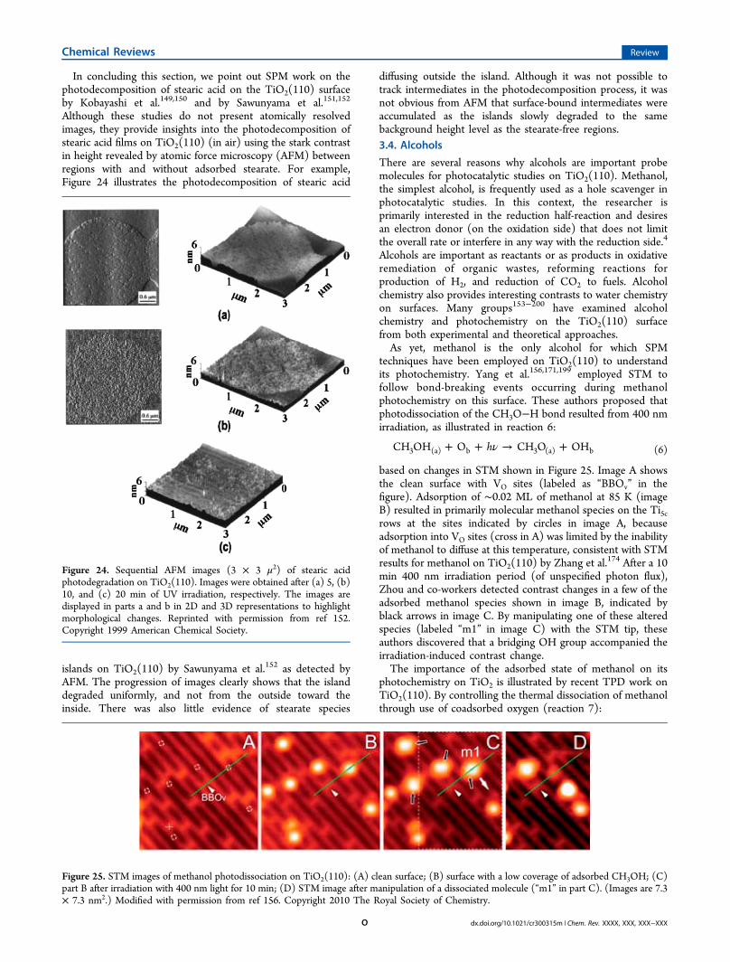
In concluding this section, we point out SPM work on thephotodecomposition of stearic acid on the TiO2(110) surfaceby Kobayashi et al.149,150 and by Sawunyama et al.151,152
Although these studies do not present atomically resolvedimages, they provide insights into the photodecomposition ofstearic acid films on TiO2(110) (in air) using the stark contrastin height revealed by atomic force microscopy (AFM) betweenregions with and without adsorbed stearate. For example,Figure 24 illustrates the photodecomposition of stearic acid
islands on TiO2(110) by Sawunyama et al.152 as detected byAFM. The progression of images clearly shows that the islanddegraded uniformly, and not from the outside toward theinside. There was also little evidence of stearate species
diffusing outside the island. Although it was not possible totrack intermediates in the photodecomposition process, it wasnot obvious from AFM that surface-bound intermediates wereaccumulated as the islands slowly degraded to the samebackground height level as the stearate-free regions.3.4. Alcohols
There are several reasons why alcohols are important probemolecules for photocatalytic studies on TiO2(110). Methanol,the simplest alcohol, is frequently used as a hole scavenger inphotocatalytic studies. In this context, the researcher isprimarily interested in the reduction half-reaction and desiresan electron donor (on the oxidation side) that does not limitthe overall rate or interfere in any way with the reduction side.4
Alcohols are important as reactants or as products in oxidativeremediation of organic wastes, reforming reactions forproduction of H2, and reduction of CO2 to fuels. Alcoholchemistry also provides interesting contrasts to water chemistryon surfaces. Many groups153−200 have examined alcoholchemistry and photochemistry on the TiO2(110) surfacefrom both experimental and theoretical approaches.As yet, methanol is the only alcohol for which SPM
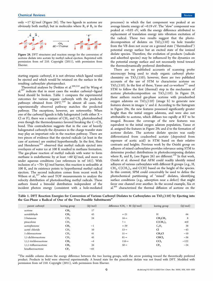
techniques have been employed on TiO2(110) to understandits photochemistry. Yang et al.156,171,199 employed STM tofollow bond-breaking events occurring during methanolphotochemistry on this surface. These authors proposed thatphotodissociation of the CH3O−H bond resulted from 400 nmirradiation, as illustrated in reaction 6:
ν+ + → +hCH OH O CH O OH3 (a) b 3 (a) b (6)
based on changes in STM shown in Figure 25. Image A showsthe clean surface with VO sites (labeled as “BBOv” in thefigure). Adsorption of ∼0.02 ML of methanol at 85 K (imageB) resulted in primarily molecular methanol species on the Ti5crows at the sites indicated by circles in image A, becauseadsorption into VO sites (cross in A) was limited by the inabilityof methanol to diffuse at this temperature, consistent with STMresults for methanol on TiO2(110) by Zhang et al.
174 After a 10min 400 nm irradiation period (of unspecified photon flux),Zhou and co-workers detected contrast changes in a few of theadsorbed methanol species shown in image B, indicated byblack arrows in image C. By manipulating one of these alteredspecies (labeled “m1” in image C) with the STM tip, theseauthors discovered that a bridging OH group accompanied theirradiation-induced contrast change.The importance of the adsorbed state of methanol on its
photochemistry on TiO2 is illustrated by recent TPD work onTiO2(110). By controlling the thermal dissociation of methanolthrough use of coadsorbed oxygen (reaction 7):
Figure 24. Sequential AFM images (3 × 3 μ2) of stearic acidphotodegradation on TiO2(110). Images were obtained after (a) 5, (b)10, and (c) 20 min of UV irradiation, respectively. The images aredisplayed in parts a and b in 2D and 3D representations to highlightmorphological changes. Reprinted with permission from ref 152.Copyright 1999 American Chemical Society.
Figure 25. STM images of methanol photodissociation on TiO2(110): (A) clean surface; (B) surface with a low coverage of adsorbed CH3OH; (C)part B after irradiation with 400 nm light for 10 min; (D) STM image after manipulation of a dissociated molecule (“m1” in part C). (Images are 7.3× 7.3 nm2.) Modified with permission from ref 156. Copyright 2010 The Royal Society of Chemistry.
where OHt is a thermal hydroxyl group, Shen andHenderson162 showed that the rate of molecular methanolphotodecomposition on TiO2(110) was at least an order ofmagnitude less than that for dissociated methanol (i.e.,methoxy). The key hole-mediated reaction appeared to bethe direct oxidation of methoxy, illustrated in reaction 8.
+ + → ++CH O O h CH O OH
(nonthermal reaction)
3 (a) b 2 (a) b
(8)
The relative rates of methanol versus methoxy photooxidationmeasured under conditions in which these two species can bedifferentiated support this combined thermal and nonthermalreaction.162,173 Transient thermal dissociation of methanol tomethoxy (reaction 9) may occur through the influence ofadjacent Ob sites.
+ → +CH OH O CH O OH (thermal reaction)3 (a) b 3 (a) b
(9)
While correlating contrast changes in STM with molecularidentities remains a challenge, as does drawing site-specificconclusions from temperature programmed desorption (TPD)data, a combined nanoscale and ensemble-average approachwill be helpful in resolving the photochemistry of methanol onTiO2(110).Ethanol chemistry and photochemistry on TiO2(110) has
been examined to a lesser extent than the case ofmethanol.160,163−165,168,169,182,185,187,188,192,201 Idriss and co-workers163−165 were perhaps the first to examine the photo-chemistry of ethanol on the TiO2(110) surface. Using XPS andTPD, these authors observed conversion of adsorbed ethanol toacetaldehyde and then to carboxylates, with gas-phase O2playing a key role in these conversions. They also speculatedthat ethoxy, and not molecular ethanol, is the key surfacespecies responsible for ethanol photochemistry on TiO2(110).A recent STM study of ethanol on TiO2(110) by Huo et al.169
illustrates the importance of diffusion in thermal chemistry onthis surface. (This topic will be addressed in greater detail in thelast section.) Another example of alcohol photochemistry onTiO2(110) in the literature for which scanning probetechniques have yet to be applied is 2-proponol. Brinkley andEngel154,155,159 have examined the photodecomposition of 2-propanol on TiO2(110) using TPD and molecular beamtechniques. They found that acetone was the first detectedintermediate in 2-propanol photooxidation on TiO2(110) andthat gas-phase O2 was required for this chemistry to occur.3.5. Organic Carbonyls
The surface photochemistry of a variety of organic carbonylshas been extensively studied on the TiO2(110) surface underUHV conditions.161,177,192,202−226 The most well-studiedorganic carbonyl on TiO2(110) in terms of photochemistry isacetone,161,203−207 but a variety of other carbonyls have alsobeen examined, including butanone,203 pinacolone,207,210
halogen-substituted acetones,208−210 acetaldehyde,202 acetophe-none,210 and acetyl chloride.210 The mechanism by which thisclass of molecules photodecomposes on TiO2(110) isillustrated by the two step process in Figure 26. The firststep is a thermal reaction between an adsorbed oxygen and theadsorbed carbonyl (shown as “(R1)(R2)CO”, where R canrefer to a variety of ligands on the carbonyl) bound to the
surface through the oxygen atom in the so-called η1-configuration to form a diolate species. This process does notrequire light as long as an adsorbed oxygen species (O adatomsor O2 molecules) is available.Such species are generated photochemically in a true
photocatalytic scheme, but they can also be generatedstoichiometrically on vacuum-annealed TiO2(110) throughthe influence of surface oxygen vacancies. With the possibleexception of acetyl chloride, the organic carbonyls listed abovewere all photoinactive on TiO2(110) in the η1-configuration.Once formed, the diolates of these organic carbonyls readilyphotodecomposed on TiO2(110) through hole-mediatedchemistry through ejection of a radical (on the R ligands)and formation of an adsorbed carboxylate species (the secondstep in Figure 26). In the case of acetone, where R1R2CH3, methyl radicals were the photodesorbed product (seeFigure 27) and acetate was left on the surface. Deskins et al.210
used DFT to show that the energy difference between thestarting acetone diolate and the photochemical products(acetate and a gaseous methyl radical) was endothermic by
Figure 26. Schematic illustration of the mechanism of acetone (R1R2=CH3) photodecomposition on TiO2(110). Reprinted withpermission from ref 203. Copyright (2008), with permission fromElsevier.
Figure 27. PSD spectra (masses 15, 18, 29, and 43) obtained during a1 min UV irradiation period of coadsorbed acetone and oxygen onTiO2(110). Spectra are displaced vertically for clarity. Reprinted withpermission from ref 203. Copyright (2008), with permission fromElsevier.
only ∼17 kJ/mol (Figure 28). The two ligands in acetone areobviously both methyl, but in molecules where R1 ≠ R2 in the
starting organic carbonyl, it is not obvious which ligand wouldbe ejected and which would be retained on the surface in theresulting carboxylate photoproduct.Theoretical analyses by Deskins et al.209,210 and by Wang et
al.207 indicate that in most cases the weaker carbonyl−ligandbond should be broken. Table 1 compares the experimentaloutcomes for various organic carbonyls with the predictedpathways obtained from DFT.210 In almost all cases, theexperimentally observed pathway matches the predictedpathway. The exceptions, however, are noteworthy. Whenone of the carbonyl ligands is fully halogenated (with either X =Cl or F), there was a mixture of CH3 and CX3 photodesorbedeven though the thermodynamics favored breaking the C−CX3bond. This contradiction suggests that in the case of heavilyhalogenated carbonyls the dynamics in the charge transfer statemay play an important role in the reaction pathway. There aretwo pieces of evidence that the ejected radicals (at least in thecase of acetone) are emitted hyperthermally. In the first, Shenand Henderson161 observed that methyl radicals ejected intooverlayers of water ice at 100 K resulted in methane formation.The gas-phase reaction of methyl radicals with water to formmethane is endothermic by at least ∼60 kJ/mol, and more sounder aqueous conditions (see references in ref 161). Withinclusion of a ∼70+ kJ/mol barrier, this reaction is unfeasible at100 K, and its existence points to hyperthermal methyl radicalejection. The second indication comes from recent work byWilson et al.,227 who used TOF measurements to analyze thevelocity distribution of photodesorbing methyl radicals. Theseauthors found a bimodal distribution independent of theincident photon energy (consistent with a hole-mediated
processes) in which the fast component was peaked at anaverage kinetic energy of ∼0.19 eV. The “slow” component waspeaked at ∼0.03 eV, with the energy difference attributed toreplacement of translation energy into vibration excitation ofthe radical. These two results suggest that the photo-decomposition of diolates on TiO2(110) via hole transferfrom the VB does not occur on a ground state (“thermalized”)potential energy surface but an excited state of the ionizeddiolate species. Therefore, the evolution of products (radicalsand adsorbed species) may be influenced by the dynamics onthe potential energy surface and not necessarily trend towardthe thermodynamically preferred distribution.There are no published accounts of scanning probe
microscopy being used to study organic carbonyl photo-chemistry on TiO2(110); however, there are two publishedaccounts of the use of STM to characterize acetone onTiO2(110). In the first of these, Yasuo and co-workers211 usedSTM to follow the first (thermal) step in the mechanism ofacetone photodecomposition on TiO2(110). In Figure 29,these authors reacted gas-phase acetone with preadsorbedoxygen adatoms on TiO2(110) (image b) to generate newfeatures shown in images ‘c’ and d. According to the histogramin Figure 29e, the new features possessed significantly greaterheight than the initial oxygen adatoms, and were also notattributable to acetone, which diffuses too rapidly at RT to beimaged. Because the coverage of the new features wasequivalent to the initial oxygen adatom population, Yasuo etal. assigned the features in Figure 29c and d to the formation ofacetone diolate. The acetone diolate species was easilydifferentiated from coadsorbed acetate (deposited fromexposure of acetic acid) in STM based on their relativecontrasts and heights. Previous work by the Onishi group onadlayers of mixed carboxylates provides relevance using STM todetermine product distributions in photodecomposing diolateswhere R1 and R2 (see Figure 26) are different.
133 In that work,Onishi et al. showed that AFM could readily identify mixedadlayers of various carboxylates with different R groups (e.g., H,CH3, C(CH3)3, and CCH) based on the height of the species.In this context, SPM could conceivably be used to define thephotochemical partitioning of “mixed” diolates, identifyingsurface conditions (e.g., adsorption near a defect) that mightfavor one channel over another. In the second example, Xia etal.201 characterized the thermal diffusion of acetone on the
Figure 28. DFT structures and reaction energy for the conversion ofacetone diolate into acetate by methyl radical ejection. Reprinted withpermission from ref 210. Copyright (2011), with permission fromElsevier.
Table 1. DFT Reaction Energies for Conversion of Various Carbonyl Diolates to Carboxylates on TiO2(110) by Ejecting intothe Gas-Phase a Radical of One of the Two Possible Substituentsa
parent carbonyl leaving group (kJ/mol) difference |CH3 − R| (kJ/mol) leaving group (kJ/mol)
aThe middle column shows the energy difference between the two leaving groups, with the arrow pointing toward the theoretically preferredproduct. Products in bold were observed experimentally. A bound state for the pinacolone diolate was not found with DFT. Modified withpermission from ref 210. Copyright (2011), with permission from Elsevier.
surface at RT. These authors observed acetone moleculesdiffusing along Ti5c rows and in/out of Vo sites. They alsoobserved the surprising ability of acetone to assist in vacancydiffusion through the transient formation of a diolate species onthe Ob rows. This behavior lead to a scrambling of O from thesurface into the acetone and the apparent hopping of Vo sitesalong the Ob rows.In another example of ketone chemistry on TiO2(110),
Jensen and co-workers215 used STM to examine the interactionof butyrophenone with the reduced and oxidized surfaces ofTiO2(110). They found that molecularly adsorbed butyrophe-none was mobile on both surfaces even at 55 K. However,butyrophenone molecules reacted with adsorbed oxygen atoms(on the oxidized surface) to form a complex that was immobileat RT. These observations are consistent with the behavior ofacetone and oxygen that sets the stage for photochemistry onTiO2(110).3.6. Water
The chemistry and photochemistry of water on an oxide surfaceis clearly of great importance in the context of solar energyconversion and hydrogen production.13,16,228,229 Water surfacechemistry has been extensively studied on the rutile TiO2(110)surface using SPM techniques;27,35,36,58,59,97−101,230−246 how-ever, very basic issues, such as the extent of molecular versusdissociative adsorption, continue to be poorly understood inthe literature. (Extensive reviews of the surface properties ofwater on TiO2(110) and other surfaces can be foundelsewhere.4,19,20,247) In terms of photochemical studies, therehave been a few examples in which TiO2(110) has been used tostudy water splitting, but mostly under aqueous condi-
tions,248−251 and the Petek group has extensively exploredtransient electron attachment processes for water onT iO 2 ( 1 1 0 ) u s i n g t w o - p h o t o n p h o t o em i s s i o n(2PPE);22,157,158,252−254 however, there have been no observa-tions of bond-forming or bond-breaking events in water surfacephotochemistry on the clean TiO2(110) surface until the recentwork by Tan et al.255 Using STM, these authors detected HO−H bond cleavage after UV irradiation, resulting in well-separated OHt and OHb species on the surface (Figure 30).
In most cases, these authors observed that the resulting waterphotofragments ended up being separated on the surface bymany unit cells or one of the fragments was altogether missingfrom the surface (both cases illustrated in Figure 30). Theauthors also found that the observations of photoactivity scaledwith irradiation time, independent of the photon energy, andnot with the photon flux at a particular energy used or with therelative absorptivity of the TiO2(110) surface at that photonenergy. While these perplexing behaviors clearly require moreexamination, it seems from Tan and co-workers’ images thatwater molecules on TiO2(110) can be activated using UV light,albeit with a rather low probability.3.7. Miscellaneous Molecules
While the above classes of molecules have been the most widelystudied in terms of photochemistry on the TiO2(110) surface,they clearly do not present the extent of potentially interestingmolecular systems accessible to researchers interested instudying heterogeneous photochemistry on TiO2. There areexamples of molecules on TiO2(110) that have been extensivelystudied photochemically with ensemble-average techniques(such as TPD or photoemission) that await examination withan SPM approach. For example, there are several studiesprobing the photochemistry of alkyl hal ides onTiO2(110)
256−267 but, as yet, no SPM studies of thephotochemical properties of this class of adsorbates. Alkylhalides bind weakly to the TiO2(110) surface based on TPDstudies256−259,264,265 and, therefore, will require low temper-atures for imaging. The observed chemistry typically centers onone of two effects: photodesorption of the adsorbed moleculeor photodissociation of the C−X bond. The mechanisms forthese effects are not well-understood. The cross sections forphotodesorption or photodecomposition are generally low(<10−21 cm2), indicating both are low probabilityevents.259,260,264 Higher rates seen under catalytic conditionson high-surface-area TiO2 point to the potential importance of
Figure 29. STM images of a TiO2(110) surface exposed to oxygen andacetone: (a) clean surface; (b) surface from part a exposed to 100 L ofO2; (c) surface from part b exposed to 10 L of acetone; and (d)surface from part b exposed to 100 L of acetone. (Image sizes are 20 ×20 nm2.) The height distributions of the protrusions in the images inparts b and c are shown in part e with gray and dark columns,respectively. The coverage of acetone-induced protrusions is shown inpart f as a function of acetone exposure. Reprinted with permissionfrom ref 211. Copyright 2010 American Chemical Society.
Figure 30. STM images (6.3 × 6.6 nm2) showing photodissociation ofadsorbed water molecules under 400 nm UV irradiation for 1 h. (Blackcircles, adsorbed water on Ti5c sites; white circles, sites forphotodissociated water; squares, OHb; inverted triangles, OHt.)Modified with permission from ref 255. Copyright 2012 AmericanChemical Society.
indirect reaction pathways (such as OH radicals),4 although thisconclusion is not firm.257,258
An interesting example of the use of SPM in characterizingphotochemical reactions on TiO2(110) is found in the work ofIshida et al.268 These authors used AFM to follow thephotooxidation of spherical (∼100 nm diameter) polystyrenelatex (PSL) spheres on TiO2(110) in air. Figure 31 shows
consecutive AFM images during UV irradiation that shownprogressive degradation of the spheres. While the images donot provide atomic resolution, they do provide specificinformation on the rate and characteristics of the photo-degradation process. In particular, Ishida and co-workersanalyzed the height and width of each sphere in typical imagesto determine whether the spheres were degraded uniformlyacross their surfaces (in which case, the heights and widthswould diminish proportionally) or whether the reactionpredominately occurred at the interface between each sphereand the TiO2(110) surface (in which case, the height woulddegrade faster than the width). On the basis of analysis fromimages such as those in Figure 31, Ishida et al. showed that thespheres degraded proportionately in size, as shown in Figure32, which suggests that the rate-limiting step does not involvethe contact area between the spheres and the surface.
4. CONCLUSIONS AND FUTURE CONSIDERATIONSThere are numerous areas in which SPM techniques couldprovide important insights into heterogeneous photocatalyticprocesses occurring on surfaces such as TiO2(110). Below wehighlight a few areas in which we believe SPM can be used tomake specific and immediate impact on the field. Thediscussion below is not restrictive but illustrative of some ofthe opportunities yet to be explored. This statement more thanany other provides the conclusion for this revie: the best daysfor molecular-level studies into photochemical events still lieahead as researchers both stretch the limits of molecularsystems that can be explored on model surfaces such asTiO2(110) and go deeper into molecular-level details that SPMtechniques can provide about photochemical transformationson surfaces.4.1. Charge Trapping Sites
Because STM is sensitive to the electronic structure of asurface, there is promise that this technique can provideinsights into the properties of trapped charge carriers generatedby photochemical excitation of TiO2. In heterogeneousphotocatalysis, the behaviors of both charge carriers (i.e., theexcited electron and the valence band hole) must be taken intoaccount in order to understand the overall catalytic process. Inthe absence of an electrochemical potential that can spatiallyseparate charges (to different electrodes), both charge carrierswill eventually have to be accounted for at the same interface,especially so in the context of UHV photochemical studies onsingle crystal TiO2. Many of the SPM results discussed aboveinvolve situations in which one carrier readily reacts with anadsorbate (e.g., holes with O2
− species resulting in O2photodesorption) but the other charge carrier is not accountedfor. In the case of hole-mediated TMA photodecomposition,there is evidence that the electron is trapped at surface OHgroups when O2 is not present to scavenge.113 There are alsoother examples in which authors have detected with SPM thetrapping of charge carriers at the TiO2(110) surface.
30−33 STM,in particular, could be of great use in characterizing not only thechemistry associated with the more rapidly reacting carrier butalso the trapping and slower reaction of the other carrier.4.2. Surface Modifiers
There are many examples in the literature in which elementaladditives influence photocatalysis on TiO2. A variety ofadditives with potential impact on TiO2 photocatalysis havebeen imaged on the TiO2(110) surface using SPM, includingsupported noble metals,175,269−310 transition metals and metaloxide clusters,311−315 alkali and alkaline earths,316−323 main
Figure 31. Sequential AFM images (3 μm × 3 μm) of the polystyrenelatex spheres on the TiO2(110) taken during UV photolysis in air: (a)before irradiation; after UV irradiation for (b) 10 min; (c) 25 min; (d)45 min; (e) 65 min; (f) 85 min; (g) 105 min; and (h) 165 min.Reprinted with permission from ref 268. Copyright 2011 IOPPublishing, Ltd.
Figure 32. Height and width of the polystyrene latex spheres as afunction of the UV irradiation time. Modified with permission from ref268. Copyright 2011 IOP Publishing, Ltd.
group elements,324−326 and halogens.327−329 An obviousconcern is determining how/where the additive binds to theTiO2(110) surface because this sets the stage for addressing itsinfluence on the thermal chemistry and photochemistry ofvarious adsorbates. In most cases, the additive of interest isbrought to the surface from the gas-phase, but in a fewexamples, it is present as either a bulk impurity or dopant. Inthis section, we will highlight a few examples of how SPM hasbeen used to image the adsorption of a variety of additives onthe TiO2(110) surface. Direct assessments of the impact ofthese species on the photochemistry of TiO2(110) usingscanning probe techniques are more rare. One such example isthe work of Uetsuka et al.330 in which the influence ofsupported Pt clusters on carboxylate photochemistry onTiO2(110) was probed.The presence of coadsorbed surface species or of foreign
atoms in the TiO2 lattice (e.g., dopants) has significantpotential for altering local charge carrier dynamics, for examplethrough band bending and flattening effects.331 Actuallyimaging these influences with SPM is a challenging under-taking. The extremes of electrostatic influence are likely foundat the extremes of the periodic table. On the electropositiveside, numerous groups have imaged alkali or alkaline earthmetals on the TiO2(110) surface.
316−322,332 The example of Kon TiO2(110) is illustrative. Majzik et al.318 have shown that Kcauses surface reconstruction at high coverages and small linearclusters at low coverage. Pang et al.319 observed linear darkregions on TiO2(110) after adsorption of 0.03 ML K on thesurface (see Figure 33). Using a combination of STM withAFM (Figure 33b and c), Pang et al. proposed that the smalllinear clusters corresponded to zigzag rows of K atoms with
each K bound to two bridging and one in-plane O atom for alocal coverage of ∼1/3 ML. These K-rich and K-void regionsprovide a potentially ideal situation to probe with SPM theeffect of local electrostatic field on hole-mediated versuselectron-mediated surface photochemistry.Chloride is a good example of an electronegative additive
that could influence photochemistry on TiO2(110).327−329
Batzill and co-workers329 have imaged chloride adsorption onthe TiO2(110) surface using STM (Figure 34). In contrast to
the case of K on TiO2(110), Cl does not appear to formordered structures or cluster on the surface. Interestinglythough, Batzill et al. observed that areas of bright contrast inSTM extending over several nanometers exerted a repulsiveinfluence on Cl atoms adsorbed on the surface, as shown inFigure 34b. Local regions of higher contrast are not uncommonin published STM images of the TiO2(110) surface and vary infrequency from sample to sample. Batzill et al. proposed thatsuch bright regions likely result from subsurface impurities.Batzill and co-workers proposed that the subsurface impurity intheir case (possibly V cations) competed locally with surface Clfor charge, thus inhibiting Cl binding in those regions. In thebigger picture, such local electronic perturbations resultingfrom surface or subsurface impurities/dopants could play amajor role in (locally) bending bands in ways that favor orinhibit charge carriers from reaching adsorbates in thevicinity.331 Subsurface impurities have been detected in otherSPM studies.233,238,333 Being able to identify the (sub)surfacemodifier and then study its local influence on chemistry andphotochemistry is both a challenge and opportunity for STMpractitioners.
Figure 33. SPM images of 0.03 ML K on TiO2(110): (a) STM image(290 Å × 170 Å); (b) NC-AFM image (350 Å × 165 Å); (c) anexpanded region of part b (170 Å × 100 Å). Reprinted withpermission from ref 319. Copyright (2005), with permission fromElsevier.
Figure 34. STM images of (a) the clean TiO2(110) surface; and (b) alow coverage of adsorbed Cl. (Areas of both images are (28 × 28)nm2.) Circles mark regions presumably associated with subsurfaceimpurities. Reprinted with permission from ref 329. Copyright (2003),with permission from Elsevier.
4.3. Adsorption State, Proximity, and CoverageDependence
Most adsorbates can adopt multiple adsorption states on asurface, with thermodynamics providing the basis for apreference of one confirmation over another. Variations inthe adsorption state can involve simple phenomena as sitevariations, coverage-dependence, or breaking/forming of ahydrogen-bond. In the context of surface photochemistry, theresidence time of an adsorbate in a particular adsorption state,and the relative ability of each state to engage in electrontransfer chemistry, will collectively determine the rates andmechanistic pathways accessible for that adsorbate. Being ableto characterize the accessible adsorption configurations of anadsorbate is therefore an important undertaking in thefundamental study of photocatalysis. In some cases, vibrationalor electronic spectroscopies can be useful in providing anensemble-averaged perspective of adsorbate confirmation;however, SPM techniques are ideally suited for characterizingconfirmation on a site-by-site basis. STM has been verysuccessfully used in characterizing the photochemistry ofadsorbates on TiO2(110) that adopts one preferred adsorptionstate or site. Two examples of this were discussed in sections3.1 and 3.3 for the case of vacancy versus normal sitepreferences of molecular oxygen and bridge-bonded carbox-ylates, respectively.The power of SPM techniques is also found in their ability to
place physical and chemical concepts in the nanoscale frame ofreference. For adsorbates or sites, each can be studied andpotentially understood in the context of the atomic-level detailsof its surroundings. This capability poses the uniqueopportunity to surface photochemistry of knowing whichmolecules show preferences for (or against) photoactivity andhow that activity depends on their proximity to neighboringfeatures, whether they be other molecules of the same (i.e.,coverage dependence), other species (e.g., coadsorbates,impurities, additives, cocatalysts, etc.), or unique structuralfeatures of the surface (e.g., point and extended surface andsubsurface defects). This immense capability of SPM has notyet been fully exploited. There are, however, some isolatedinstances in the literature of site preferences. For example,Uetsuka and co-workers119 found that TMA groups bound atstep edges on the TiO2(110) surface were the least reactive tohole-mediated photodecomposition. Similarly, as mentionedabove, Wang et al.124 found that TMA groups bound at VO siteswere must less photoreactive than those bound at Ti5c sites.
4.4. Adsorbate Dynamics
Scanning probe techniques have been shown to provide uniqueinsights into the dynamics of adsorbates on TiO2(110). Forexample , STM stud ie s o f wa te r chemis t r y onTiO2(110)
19,27,58,97−100 have shown that molecular hoppingand transient chemistry (such as dissociation and recombina-tion or hydronium formation) redistribute reactive species bothalong and across the row structure of this surface. Similardynamical behaviors also likely play a role in alcohol chemistryon TiO2(110) given the similarities between water and alcohols(e.g., −OH functionality and hydrogen-bonding capability).Zhang and co-workers166,167,170 have examined the chemistry ofseveral alcohols on TiO2(110) using STM. These authorsobserved a variety of effects, including dissociation events anddiffusional processes. Zhang et al. also detected more subtledynamical behaviors associated with the alcohol’s organic chain.Figure 35 shows four STM images that illustrate unique chain
dynamics in adsorbed octoxy groups on TiO2(110) thatdepended on the chain length. Images a−d in Figure 35resulted from adsorption of 1-, 2-, 3-, and 4-octanol,respectively, on TiO2(110) at RT. In each case, the octanolmolecule adsorbed at a vacancy site and dissociated to form an“octoxy” species. Because the hydrocarbon chains were ofdifferent lengths and configurations in each case, the resultingoctoxy species adopted different “preferred” orientations.Although somewhat restricted by the symmetry of the surface(or perhaps because of the restrictions), the chains wereobserved to rapidly flip back-and-forth between the preferredorientations, resulting in unique STM images for each alkoxyspecies. The ability of the octoxy chains to reorient at RT onTiO2(110) is clearly evident by the symmetries of the contrastfeatures in Figure 35. More recently, Hansen and co-workers168,169 have examined the thermal chemistry of ethanolon TiO2(110) with STM, laying a foundation for understandingthe photochemistry of this molecule with scanning probemicroscopy. These authors also detected similar diffusionalprocesses observed by Zhang and co-workers.As yet, the dependence of molecular confirmation on
photochemistry has not been explored; however, it is notinconceivable that the reactivity of an alkoxy species, such asoctoxy, could be sensitive to its molecular confirmation,resulting in charge transfer rates that vary with the dynamicalvariations of the adsorbate. One could argue from an electronicstructure perspective that there might be only small differencesin the energies of the highest occupied molecular orbital(HOMO) states of the various octoxy species (shown in Figure35), so that orientation variations should have little impact onelectron transfer reactions from an alkoxy’s HOMO to a VBhole in TiO2. However, if the ability to orient a C−H bondtoward a proton acceptor site (e.g., a Ob) is key in stabilizingthe electron transfer process, as seems to be the case for
Figure 35. STM images of the TiO2(110) surface after adsorption of(a) 1-, (b) 2-, (c) 3-, and (d) 4-octanol at 300 K. The insets showmagnified areas with the octoxy species overlaid by TiO2(110) ballmodels (Ob, blue; Ti5c, magenta). The yellow zigzag lines shows thealkyl chains, and red dots mark the anchoring position on the Ob row.Green lobes forming an “X” shape for 1-, 2-, and 3-octoxy and an “I”shape for 4-octoxy indicate how fast rotation of the octoxy speciesleads to the formation of such features in the STM images. Reprintedwith permission from ref 166. Copyright 2009 American ChemicalSociety.
methanol photooxidation on TiO2(110),156,162,171,173 then
certain octoxy species may show enhanced photoactivitybecause of more favorable chain configurations. Similarly,some octoxy species may situate in orientations that are notpreferred (e.g., for proton transfer), but their chain dynamicsallow for transient sampling of preferred orientations.Conversely, high coverages or low temperature may lock analkoxy’s chain in certain orientations, favorable or not. Zhang etal.166 clearly observed the latter to occur for the 2- and 3-octanol molecules when they conducted STM measurementssimilar to those in Figure 35, except at 150 K. Unfortunately,the interplay between these kinds of adsorbate dynamics andphotochemical activity is largely unexplored in the heteroge-neous photocatalysis literature.As a final note on alcohol dynamics on TiO2(110), Zhang et
al.170 have explored the dynamics of alkoxy species onTiO2(110) at higher temperatures (above RT). At RT, theyfind that butoxy, formed by 2-butanol dissociation at vacancysites, does not diffuse along the bridging oxygen rows as is seenfor OHb groups on TiO2(110).
19,27 This behavior is consistentwith that of other alkoxy groups formed at vacancy sites onTiO2(110). However, Zhang and co-workers found thatadsorbed alkoxy species were able to migrate along the Ob
rows on TiO2(110) at higher temperature, as illustrated inFigure 36. The onset for butoxy diffusion along the Obr rowcorrelated with the onset temperature for vacancy hopping,334
with both occurring with similar activation barriers.The proposed mechanism for butoxy hopping, illustrated in
Figure 36, involves approach of a vacancy to the bridgingbutoxy group, which then transiently occupies an “atop” sitewith an undercoordinated Ti site on either side of it, followedby returning to a bridging configuration as the vacancy passeson. The significance of this diffusion process to photochemistryon TiO2(110) may lie in the residence time that an organicmolecule spends in the vicinity of a vacancy site (and itsaccompanying unpaired electrons). As yet, there is not a clearpicture in the literature of the extent to which occupied states atthe top of the band-gap affect hole-mediated photochemistryon TiO2(110). Conventional wisdom would suggest thatadsorbates bound near these electronic defects would be lesslikely to undergo hole-mediated photochemistry if the electronsin the band-gap state can neutralize the hole arriving from thebulk.
4.5. Beyond the Rutile TiO2(110) Surface
Finally, we note that while the rutile TiO2(110) surface is the
prototypical model oxide surface, the utility of SPM to
characterize surface photochemistry and photocatalysis is
clearly not limited by studies on this single crystal surface.
Other orientations of the rutile phase are being examined
regarding the photochemical properties,45,143,154,160,248,335−340
as have the single crystal surfaces of anatase.107,125,126,160 The
UHV conditions should also not be a restriction to the use of
SPM methods to characterize surface photochemistry. Photo-
chemistry has been reported on a variety of non-TiO2 oxide
single crystal surfaces potentially amenable to analysis with
SPM techniques. These include photochemical studies on
various single crystal metal oxides, including Al2O3,341,342
Cr2O3,343−350 Fe2O3,
351,352 NiO,347,353−359 SrTiO3,360 and
ZnO.361 These, as well as the many potential nonoxide
photocatalytic materials (e.g., CdS and CdSe), provide a wealth
of opportunities for exploring photochemical phenomena on
the atomic scale and advancing the use and capabilities of SPM
techniques in studies important to photocatalysis.
The authors declare no competing financial interest.
Figure 36. (a−d) Sequential STM images recorded at 400 K on the same area of 2-butanol preadsorbed TiO2(110) as a function of time (0, 2, 3, and9 min, respectively). The rectangles mark the area of interest and contain one 2-butoxy (red arrow), two VO’s (green arrows), and one hydroxyl(yellow arrow) species. The corresponding ball models (Ob, blue; Ti5c, magenta; alcohol, green) illustrate the diffusion process. (e) 2-Dimensionalline profiles along the BBO row marked with the dashed lines in images a−d. The vertical dashed lines mark the lattice positions along the Ob row.Reprinted with permission from ref 170. Copyright 2008 American Physical Society.
Michael A. Henderson is a Laboratory Fellow in the Fundamental andComputational Sciences Directorate at the Pacific Northwest NationalLaboratory (PNNL) in Richland, WA. He obtained in Ph.D. in 1988from the Department of Chemistry at the University of Texas, Austin,and did postdoctoral research at the University of Pittsburgh from1989 to 1991 before moving to PNNL in 1991. His expertise is in thesurface science of metal oxides.
Igor Lyubinetsky was born in 1959 in Vinnitsa, Ukraine. He studiedphysics at the Leningrad State University, U.S.S.R., and received hisM.S. in Physics in 1983 and Ph.D. in Condensed Matter Physics in1986 (both in the group of Prof. V. K. Adamchuk), while studyingmetal−semiconductor interface formation with electron spectroscopytechniques. He came to the U.S.A. in 1996 as a visiting scientist fromT. Shevchenko Kiev National University, Ukraine, joining the group ofProf. J. T. Yates, Jr. at the University of Pittsburgh. Since 1999, he wasa research assistant scientist at the University of Maryland, workingwith Prof. E. D. Williams. In 2002, he joined Pacific NorthwestNational Laboratory as a senior research scientist, and he has beenthere ever since. Dr. Lyubinetsky’s research interests are focused oncomplex chemical/physical processes that take place in heterogeneouscatalysis and/or environmental remediation, employing the modelsystem experimental approach. In particular, they include theapplication of ultrahigh vacuum scanning tunneling microscopy andspectroscopy to the fundamental studies of the thermal-induced andphotoinduced reactivities of metal oxide surfaces at the atomic level.
ACKNOWLEDGMENTS
This work was supported by the U.S. Department of Energy,Office of Basic Energy Sciences, Division of Chemical Sciences,Geosciences & Biosciences. I.L. was partially supported by aPacific Northwest National Laboratory (PNNL) Chemical
Imaging Initiative project. PNNL is a multiprogram nationallaboratory operated for DOE by Battelle.
GLOSSARY OF ACRONYMS AND ABBREVIATIONS
AFM atomic force microscopyDFT density functional theorye−/h+ electron−hole pairEELS electron energy loss spectroscopyESD electron stimulated desorptionHOMO highest occupied molecular orbitalHREELS high resolution electron energy loss spectroscopyML monolayerOa adsorbed O adatomOb bridging O2− surface sitePSD photon stimulated desorptionRAIRS reflection−absorption infrared spectroscopyRT room temperatureSPM scanning probe microscopySTM scanning tunneling microscopySTS scanning tunneling spectroscopyTi5c five coordinate Ti4+ surface siteTMA trimethyl acetateTMAA trimethyl acetic acidTOF time-of-flightTPD temperature programmed desorptionUHV ultrahigh vacuumUPS ultraviolet photoemission spectroscopyUV ultravioletVB valence bandVo surface oxygen vacancy siteXPS X-ray photoemission spectroscopy
REFERENCES(1) Fujishima, A.; Honda, K. Nature 1972, 238, 37.(2) Thompson, T. L.; Yates, J. T., Jr. Top. Catal. 2005, 35, 197.(3) Fujishima, A.; Zhang, X. T.; Tryk, D. A. Surf. Sci. Rep. 2008, 63,515.(4) Henderson, M. A. Surf. Sci. Rep. 2011, 66, 185.(5) Thompson, T. L.; Yates, J. T., Jr. Chem. Rev. 2006, 106, 4428.(6) Carp, O.; Huisman, C. L.; Reller, A. Prog. Solid State Chem. 2004,32, 33.(7) Ahmed, S.; Rasul, M. G.; Brown, R.; Hashib, M. A. J. Environ.Manage. 2011, 92, 311.(8) Ardo, S.; Meyer, G. J. Chem. Soc. Rev. 2009, 38, 115.(9) Chen, X.; Mao, S. S. Chem. Rev. 2007, 107, 2891.(10) Duncan, W. R.; Prezhdo, O. V. Annu. Rev. Phys. Chem. 2007, 58,143.(11) Gaya, U. I.; Abdullah, A. H. J. Photochem. Photobiol., C 2008, 9,1.(12) Hoffmann, M. R.; Martin, S. T.; Choi, W. Y.; Bahnemann, D. W.Chem. Rev. 1995, 95, 69.(13) Kudo, A.; Miseki, Y. Chem. Soc. Rev. 2009, 38, 253.(14) Linsebigler, A. L.; Lu, G. Q.; Yates, J. T., Jr. Chem. Rev. 1995, 95,735.(15) Mills, A.; LeHunte, S. J. Photochem. Photobiol., A 1997, 108, 1.(16) Ni, M.; Leung, M. K. H.; Leung, D. Y. C.; Sumathy, K.Renewable Sustainable Energy Rev. 2007, 11, 401.(17) Tachikawa, T.; Fujitsuka, M.; Majima, T. J. Phys. Chem. C 2007,111, 5259.(18) Chen, H.; Nanayakkara, C. E.; Grassian, V. H. Chem. Rev. 2012,112, 5919.(19) Dohnalek, Z.; Lyubinetsky, I.; Rousseau, R. Prog. Surf. Sci. 2010,85, 161.(20) Pang, C. L.; Lindsay, R.; Thornton, G. Chem. Soc. Rev. 2008, 37,2328.
(21) de Lara-Castells, M. P.; Mitrushenkov, A. O.; Roncero, O.;Krause, J. L. Isr. J. Chem. 2005, 45, 59.(22) Petek, H.; Zhao, J. Chem. Rev. 2010, 110, 7082.(23) Diebold, U. Appl. Phys. A: Mater. Sci. Process. 2003, 76, 681.(24) Diebold, U. Surf. Sci. Rep. 2003, 48, 53.(25) Liu, L. M.; Crawford, P.; Hu, P. Prog. Surf. Sci. 2009, 84, 155.(26) Onishi, H. Springer Ser. Chem. Phys. 2003, 70, 75.(27) Zhang, Z.; Bondarchuk, O.; Kay, B. D.; White, J. M.; Dohnalek,Z. J. Phys. Chem. B 2006, 110, 21840.(28) Mezhenny, S.; Maksymovych, P.; Thompson, T. L.; Diwald, O.;Stahl, D.; Walck, S. D.; Yates, J. T., Jr. Chem. Phys. Lett. 2003, 369, 152.(29) Minato, T.; Sainoo, Y.; Kim, Y.; Kato, H.; Aika, K.; Kawai, M.;Zhao, J.; Petek, H.; Huang, T.; He, W. J. Chem. Phys. 2009, 130,124502.(30) Komiyama, M.; Li, Y.-J. Appl. Surf. Sci. 2005, 244, 550.(31) Komiyama, M.; Li, Y. J. Jpn. J. Appl. Phys., Part 1 2004, 43, 4584.(32) Komiyama, M.; Li, Y. J.; Yin, D. H. Jpn. J. Appl. Phys., Part 12002, 41, 4936.(33) Komiyama, M.; Yin, D.; Li, Y. Stud. Surf. Sci. Catal. 2003, 145,153.(34) Li, Y. J.; Matsumoto, T.; Gu, N.; Komiyama, M. Appl. Surf. Sci.2004, 237, 374.(35) Papageorgiou, A. C.; Beglitis, N. S.; Pang, C. L.; Teobaldi, G.;Cabailh, G.; Chen, Q.; Fisher, A. J.; Hofer, W. A.; Thornton, G. Proc.Natl. Acad. Sci. U.S.A. 2010, 107, 2391.(36) Teobaldi, G.; Hofer, W. A.; Bikondoa, O.; Pang, C. L.; Cabailh,G.; Thornton, G. Chem. Phys. Lett. 2007, 437, 73.(37) Tao, J. G.; Luttrell, T.; Batzill, M. Nature Chem. 2011, 3, 296.(38) Bennett, R. A.; Stone, P.; Bowker, M. Faraday Discuss. 1999,114, 267.(39) Bennett, R. A.; Stone, P.; Price, N. J.; Bowker, M. Phys. Rev. Lett.1999, 82, 3831.(40) Li, M.; Hebenstreit, W.; Gross, L.; Diebold, U.; Henderson, M.A.; Jennison, D. R.; Schultz, P. A.; Sears, M. P. Surf. Sci. 1999, 437,173.(41) Li, M.; Hebenstreit, W.; Diebold, U.; Henderson, M. A.;Jennison, D. R. Faraday Discuss. 1999, 114, 245.(42) Li, M.; Hebenstreit, W.; Diebold, U.; Tyryshkin, A. M.;Bowman, M. K.; Dunham, G. G.; Henderson, M. A. J. Phys. Chem. B2000, 104, 4944.(43) Onishi, H.; Iwasawa, Y. Phys. Rev. Lett. 1996, 76, 791.(44) Zhang, Z.; Lee, J.; Yates, J. T., Jr.; Bechstein, R.; Lira, E.;Hansen, J. O.; Wendt, S.; Besenbacher, F. J. Phys. Chem. C 2010, 114,3059.(45) Ariga, H.; Taniike, T.; Morikawa, H.; Tada, M.; Min, B. K.;Watanabe, K.; Matsumoto, Y.; Ikeda, S.; Saiki, K.; Iwasawa, Y. J. Am.Chem. Soc. 2009, 131, 14670.(46) Thompson, T. L.; Yates, J. T., Jr. J. Phys. Chem. B 2006, 110,7431.(47) Fox, M. A.; Dulay, M. T. Chem. Rev. 1993, 93, 341.(48) Dohnalek, Z.; Kim, J.; Bondarchuk, O.; White, J. M.; Kay, B. D.J. Phys. Chem. B 2006, 110, 6229.(49) Kimmel, G. A.; Petrik, N. G. Phys. Rev. Lett. 2008, 100, 196102/1.(50) Ganduglia-Pirovano, M. V.; Hofman, A.; Sauer, J. Surf. Sci. Rep.2007, 62, 219.(51) Henderson, M. A.; Epling, W. S.; Perkins, C. L.; Peden, C. H. F.;Diebold, U. J. Phys. Chem. B 1999, 103, 5328.(52) Wendt, S.; Sprunger, P. T.; Lira, E.; Madsen, G. K. H.; Li, Z.;Hansen, J. O.; Matthiesen, J.; Blekinge-Rasmussen, A.; Laegsgaard, E.;Hammer, B.; et al. Science 2008, 320, 1755.(53) Deskins, N. A.; Dupuis, M. Phys. Rev. B 2007, 75, 195212.(54) Du, Y. G.; Deskins, N. A.; Zhang, Z. R.; Dohnalek, Z.; Dupuis,M.; Lyubinetsky, I. Phys. Chem. Chem. Phys. 2010, 12, 6337.(55) Deskins, N. A.; Rousseau, R.; Dupuis, M. J. Phys. Chem. C 2010,114, 5891.(56) Du, Y. G.; Dohnalek, Z.; Lyubinetsky, I. J. Phys. Chem. C 2008,112, 2649.
(57) Rasmussen, M. D.; Molina, L. M.; Hammer, B. J. Chem. Phys.2004, 120, 988.(58) Wendt, S.; Schaub, R.; Matthiesen, J.; Vestergaard, E. K.;Wahlstrom, E.; Rasmussen, M. D.; Thostrup, P.; Molina, L. M.;Laegsgaard, E.; Stensgaard, I.; et al. Surf. Sci. 2005, 598, 226.(59) Bikondoa, O.; Pang, C. L.; Ithnin, R.; Muryn, C. A.; Onishi, H.;Thornton, G. Nat. Mater. 2006, 5, 189.(60) Lira, E.; Wendt, S.; Huo, P.; Hansen, J. Ø.; Streber, R.;Porsgaard, S.; Wei, Y.; Bechstein, R.; Lægsgaard, E.; Besenbacher, F. J.Am. Chem. Soc. 2011, 133, 6529.(61) Lira, E.; Huo, P. P.; Hansen, J. O.; Rieboldt, F.; Bechstein, R.;Wei, Y. Y.; Streber, R.; Porsgaard, S.; Li, Z. S.; Laegsgaard, E.; et al.Catal. Today 2012, 182, 25.(62) Petrik, N. G.; Zhang, Z.; Du, Y.; Dohnalek, Z.; Lyubinetsky, I.;Kimmel, G. A. J. Phys. Chem. C 2009, 113, 12407.(63) Epling, W. S.; Peden, C. H. F.; Henderson, M. A.; Diebold, U.Surf. Sci. 1998, 413, 333.(64) Henderson, M. A.; Shen, M.; Wang, Z.; Lyubinetsky, I. J. Phys.Chem. C 2013, DOI: 10.1021/jp312161y.(65) de Lara-Castells, M. P.; Krause, J. L. Chem. Phys. Lett. 2002, 354,483.(66) Lu, G. Q.; Linsebigler, A.; Yates, J. T., Jr. J. Chem. Phys. 1995,102, 3005.(67) Wang, Y.; Pillay, D.; Hwang, G. S. Phys. Rev. B 2004, 70,193410/1.(68) Wu, X.; Selloni, A.; Lazzeri, M.; Nayak, S. K. Phys. Rev. B 2003,68, 241402/1.(69) Pillay, D.; Wang, Y.; Hwang, G. S. J. Am. Chem. Soc. 2006, 128,14000.(70) Liu, L. M.; McAllister, B.; Ye, H. Q.; Hu, P. J. Am. Chem. Soc.2006, 128, 4017.(71) Scheiber, P.; Riss, A.; Schmid, M.; Varga, P.; Diebold, U. Phys.Rev. Lett. 2010, 105, 216101.(72) Wang, Z. T.; Du, Y. G.; Dohnalek, Z.; Lyubinetsky, I. J. Phys.Chem. Lett. 2010, 1, 3524.(73) Tan, S. J.; Ji, Y. F.; Zhao, Y.; Zhao, A. D.; Wang, B.; Yang, J. L.;Hou, J. G. J. Am. Chem. Soc. 2011, 133, 2002.(74) Wang, Z.; Zhao, Y.; Cui, X. F.; Tan, S. J.; Zhao, A. D.; Wang, B.;Yang, J. L.; Hou, J. G. J. Phys. Chem. C 2010, 114, 18222.(75) Lira, E.; Hansen, J. O.; Huo, P.; Bechstein, R.; Galliker, P.;Laegsgaard, E.; Hammer, B.; Wendt, S.; Besenbacher, F. Surf. Sci.2010, 604, 1945.(76) Lu, G. Q.; Linsebigler, A.; Yates, J. T., Jr. J. Chem. Phys. 1995,102, 4657.(77) Rusu, C. N.; Yates, J. T., Jr. Langmuir 1997, 13, 4311.(78) Yates, J. T., Jr. Surf. Sci. 2009, 603, 1605.(79) Sporleder, D.; Wilson, D. P.; White, M. G. J. Phys. Chem. C2009, 113, 13180.(80) Zhang, Z.; Yates, J. T., Jr. J. Phys. Chem. C 2010, 114, 3098.(81) Petrik, N. G.; Kimmel, G. A. J. Phys. Chem. Lett. 2010, 1, 1758.(82) Petrik, N. G.; Kimmel, G. A. J. Phys. Chem. C 2011, 115, 152.(83) Wang, Z.-T.; Aaron Deskins, N.; Lyubinetsky, I. J. Phys. Chem.Lett. 2012, 3, 102.(84) Petrik, N. G.; Kimmel, G. A. J. Phys. Chem. Lett. 2011, 2, 2790.(85) Zhang, Z.; Yates, J. T., Jr. J. Phys. Chem. Lett. 2010, 1, 2185.(86) Zhang, Z.; Tang, W. J.; Neurock, M.; Yates, J. T., Jr. J. Phys.Chem. C 2011, 115, 23848.(87) Kobayashi, H.; Yamaguchi, M. Surf. Sci. 1989, 214, 466.(88) Rocker, G.; Gopel, W. Surf. Sci. 1986, 175, L675.(89) Menetrey, M.; Markovits, A.; Minot, C. Surf. Sci. 2003, 524, 49.(90) Linsebigler, A.; Lu, G.; Yates, J. T., Jr. J. Chem. Phys. 1995, 103,9438.(91) Sorescu, D. C.; Yates, J. T., Jr. J. Phys. Chem. B 2002, 106, 6184.(92) Pillay, D.; Hwang, G. S. J. Chem. Phys. 2006, 125, 144706.(93) Zhao, Y.; Wang, Z.; Cui, X.; Huang, T.; Wang, B.; Luo, Y.; Yang,J.; Hou, J. J. Am. Chem. Soc. 2009, 131, 7958.(94) Lee, J. S.; Zhang, Z.; Deng, X. Y.; Sorescu, D. C.; Matranga, C.;Yates, J. T., Jr. J. Phys. Chem. C 2011, 115, 4163.
(95) Mitsuhara, K.; Matsuda, T.; Okumura, H.; Visikovskiy, A.; Kido,Y. Nuclear Instrum. Methods Phys. Res., Sect. B 2011, 269, 1859.(96) Mitsuhara, K.; Okumura, H.; Visikovskiy, A.; Takizawa, M.;Kido, Y. Chem. Phys. Lett. 2011, 513, 84.(97) Du, Y. G.; Deskins, N. A.; Zhang, Z. R.; Dohnalek, Z.; Dupuis,M.; Lyubinetsky, I. J. Phys. Chem. C 2009, 113, 666.(98) Du, Y.; Deskins, N. A.; Zhang, Z.; Dohnalek, Z.; Dupuis, M.;Lyubinetsky, I. Phys. Rev. Lett. 2009, 102, 096102.(99) Matthiesen, J.; Wendt, S.; Hansen, J. O.; Madsen, G. K. H.; Lira,E.; Galliker, P.; Vestergaard, E. K.; Schaub, R.; Laegsgaard, E.;Hammer, B.; et al. ACS Nano 2009, 3, 517.(100) Zhang, Z.; Du, Y.; Petrik, N. G.; Kimmel, G. A.; Lyubinetsky,I.; Dohnalek, Z. J. Phys. Chem. C 2009, 113, 1908.(101) Du, Y. G.; Deskins, N. A.; Zhang, Z. R.; Dohnalek, Z.; Dupuis,M.; Lyubinetsky, I. J. Phys. Chem. C 2010, 114, 17080.(102) Linsebigler, A.; Lu, G. Q.; Yates, J. T., Jr. J. Phys. Chem. 1996,100, 6631.(103) Linsebigler, A.; Rusu, C.; Yates, J. T., Jr. J. Am. Chem. Soc.1996, 118, 5284.(104) Petrik, N. G.; Kimmel, G. A. J. Phys. Chem. Lett. 2010, 1, 2508.(105) Zhang, Z.; Yates, J. T., Jr. J. Am. Chem. Soc. 2010, 132, 12804.(106) Wu, X.; Selloni, A.; Nayak, S. K. J. Chem. Phys. 2004, 120,4512.(107) Xu, M. C.; Gao, Y. K.; Moreno, E. M.; Kunst, M.; Muhler, M.;Wang, Y. M.; Idriss, H.; Woll, C. Phys. Rev. Lett. 2011, 106, 138302.(108) Hebenstreit, W.; Ruzycki, N.; Herman, G. S.; Gao, Y.; Diebold,U. Phys. Rev. B 2000, 62, R16334.(109) Mehring, M.; Kluner, T. Chem. Phys. Lett. 2011, 513, 212.(110) Gratzel, M. Nature 2001, 414, 338.(111) Sherrill, A. B.; Barteau, M. A. In Oxide Surfaces, The ChemicalPhysics of Solid Surfaces; Woodruff, D. P., Ed.; Elsevier: Amsterdam,2001; Vol. 9; p 409.(112) He, M. Y.; White, J. M.; Ekerdt, J. G. J. Mol. Catal. 1985, 30,415.(113) Henderson, M. A.; White, J. M.; Uetsuka, H.; Onishi, H. J. Am.Chem. Soc. 2003, 125, 14974.(114) White, J. M.; Henderson, M. A.; Uetsuka, H.; Onishi, H. Proc.SPIE 2004, 5513, 66.(115) White, J. M.; Szanyi, J.; Henderson, M. A. J. Phys. Chem. B2004, 108, 3592.(116) White, J. M.; Henderson, M. A. J. Phys. Chem. B 2005, 109,12417.(117) Lyubinetsky, I.; Yu, Z. Q.; Henderson, M. A. J. Phys. Chem. C2007, 111, 4342.(118) Lyubinetsky, I.; Deskins, N. A.; Du, Y. G.; Vestergaard, E. K.;Kim, D. J.; Dupuis, M. Phys. Chem. Chem. Phys. 2010, 12, 5986.(119) Uetsuka, H.; Onishi, H.; Henderson, M. A.; White, J. M. J.Phys. Chem. B 2004, 108, 10621.(120) Henderson, M. A.; White, J. M.; Uetsuka, H.; Onishi, H. J.Catal. 2006, 238, 153.(121) Du, Y.; Petrik, N. G.; Deskins, N. A.; Wang, Z.; Henderson, M.A.; Kimmel, G. A.; Lyubinetsky, I. Phys. Chem. Chem. Phys. 2012, 14,3066.(122) Henderson, M. A. Surf. Sci. 1996, 355, 151.(123) Yin, X. L.; Calatayud, M.; Qiu, H.; Wang, Y.; Birkner, A.;Minot, C.; Woll, C. ChemPhysChem 2008, 9, 253.(124) Wang, Z.-T.; Deskins, N. A.; Henderson, M. A.; Lyubinetsky, I.Phys. Rev. Lett. 2012, 109, 266103.(125) Ohsawa, T.; Lyubinetsky, I. V.; Henderson, M. A.; Chambers,S. A. J. Phys. Chem. C 2008, 112, 20050.(126) Ohsawa, T.; Lyubinetsky, I.; Du, Y.; Henderson, M. A.;Shutthanandan, V.; Chambers, S. A. Phys. Rev. B 2009, 79, 085401/1.(127) Foster, A. S.; Gal, A. Y.; Nieminen, R. M.; Shluger, A. L. J. Phys.Chem. B 2005, 109, 4554.(128) Fukui, K.-I.; Iwasawa, Y. Surf. Sci. 2000, 464, L719.(129) Fukui, K.-I.; Onishi, H.; Iwasawa, Y. Chem. Phys. Lett. 1997,280, 296.(130) Fukui, K.-I.; Onishi, H.; Iwasawa, Y. Appl. Surf. Sci. 1999, 140,259.
(131) Onishi, H.; Fukui, K.-I.; Iwasawa, Y. Bull. Chem. Soc. Jpn. 1995,68, 2447.(132) Onishi, H.; Fukui, K.-I.; Iwasawa, Y. Colloid Surf., A 1996, 109,335.(133) Onishi, H.; Sasahara, A.; Uetsuka, H.; Ishibashi, T. Appl. Surf.Sci. 2002, 188, 257.(134) Onishi, H.; Iwasawa, Y. Langmuir 1994, 10, 4414.(135) Onishi, H. Catal. Surv. Jpn. 2002, 6, 1.(136) Onishi, H.; Fukui, K.-I.; Iwasawa, Y. Jpn. J. Appl. Phys., Part 11999, 38, 3830.(137) Onishi, H.; Iwasawa, Y. Surf. Sci. 1996, 357−358, 773.(138) Onishi, H.; Iwasawa, Y. Chem. Phys. Lett. 1994, 226, 111.(139) Onishi, H.; Yamaguchi, Y.; Fukui, K.-I.; Iwasawa, Y. J. Phys.Chem. 1996, 100, 9582.(140) Uetsuka, H.; Sasahara, A.; Yamakata, A.; Onishi, H. J. Phys.Chem. B 2002, 106, 11549.(141) Sasahara, A.; Uetsuka, H.; Ishibashi, T.; Onishi, H. J. Phys.Chem. B 2003, 107, 13925.(142) Sasahara, A.; Uetsuka, H.; Onishi, H. Phys. Rev. B 2001, 64,121406.(143) Quah, E. L.; Wilson, J. N.; Idriss, H. Langmuir 2010, 26, 6411.(144) Idriss, H.; Legare, P.; Maire, G. Surf. Sci. 2002, 515, 413.(145) Ariga, H.; Taniike, T.; Morikawa, H.; Tero, R.; Kondoh, H.;Iwasawa, Y. Chem. Phys. Lett. 2008, 454, 350.(146) Tao, J. G.; Batzill, M. J. Phys. Chem. Lett. 2010, 1, 3200.(147) Tao, J.; Luttrell, T.; Bylsma, J.; Batzill, M. J. Phys. Chem. C2011, 115, 3434.(148) Uetsuka, H.; Henderson, M. A.; Sasahara, A.; Onishi, H. J.Phys. Chem. B 2004, 108, 13706.(149) Kobayashi, K.; Tomita, Y.; Matsuhisa, K.; Doi, Y. Appl. Surf. Sci.2005, 244, 389.(150) Kobayashi, K.; Tomita, Y.; Maeda, Y. J. Nanosci. Nanotechnol.2009, 9, 3382.(151) Sawunyama, P.; Fujishima, A.; Hashimoto, K. Chem. Commun.1998, 2229.(152) Sawunyama, P.; Fujishima, A.; Hashimoto, K. Langmuir 1999,15, 3551.(153) Thompson, T. L.; Yates, J. T., Jr. J. Phys. Chem. B 2005, 109,18230.(154) Brinkley, D.; Engel, T. J. Phys. Chem. B 2000, 104, 9836.(155) Brinkley, D.; Engel, T. J. Phys. Chem. B 1998, 102, 7596.(156) Zhou, C. Y.; Ren, Z. F.; Tan, S. J.; Ma, Z. B.; Mao, X. C.; Dai,D. X.; Fan, H. J.; Yang, X. M.; LaRue, J.; Cooper, R.; et al. Chem. Sci.2010, 1, 575.(157) Li, B.; Zhao, J.; Onda, K.; Jordan, K. D.; Yang, J. L.; Petek, H.Science 2006, 311, 1436.(158) Onda, K.; Li, B.; Zhao, J.; Petek, H. Surf. Sci. 2005, 593, 32.(159) Brinkley, D.; Engel, T. Surf. Sci. 1998, 415, L1001.(160) Ahmed, A. Y.; Kandiel, T. A.; Oekermann, T.; Bahnemann, D.J. Phys. Chem. Lett. 2011, 2, 2461.(161) Shen, M.; Henderson, M. A. J. Phys. Chem. C 2011, 115, 5886.(162) Shen, M. M.; Henderson, M. A. J. Phys. Chem. Lett. 2011, 2,2707.(163) Nadeem, A. M.; Muir, J. M. R.; Waterhouse, G. W. N.; Idriss,H. Proc. SPIE 2011, 8109.(164) Nadeem, A. M.; Muir, J. M. R.; Connelly, K. A.; Adamson, B.T.; Metson, B. J.; Idriss, H. Phys. Chem. Chem. Phys. 2011, 13, 7637.(165) Jayaweera, P. M.; Quah, E. L.; Idriss, H. J. Phys. Chem. C 2007,111, 1764.(166) Zhang, Z. R.; Rousseau, R.; Gong, J. L.; Kay, B. D.; Dohnalek,Z. J. Am. Chem. Soc. 2009, 131, 17926.(167) Zhang, Z. R.; Bondarchuk, E.; Kay, B. D.; White, J. M.;Dohnalek, Z. J. Phys. Chem. C 2007, 111, 3021.(168) Hansen, J. O.; Huo, P.; Martinez, U.; Lira, E.; Wei, Y. Y.;Streber, R.; Laegsgaard, E.; Hammer, B.; Wendt, S.; Besenbacher, F.Phys. Rev. Lett. 2011, 107, 136102.(169) Huo, P.; Hansen, J. O.; Martinez, U.; Lira, E.; Streber, R.; Wei,Y. Y.; Laegsgaard, E.; Hammer, B.; Wendt, S.; Besenbacher, F. J. Phys.Chem. Lett. 2012, 3, 283.
(170) Zhang, Z. R.; Rousseau, R.; Gong, J. L.; Li, S. C.; Kay, B. D.;Ge, Q. F.; Dohnalek, Z. Phys. Rev. Lett. 2008, 101, 156103.(171) Zhou, C. Y.; Ma, Z. B.; Ren, Z. F.; Wodtke, A. M.; Yang, X. M.Energy Environ. Sci. 2012, 5, 6833.(172) Zhou, C. Y.; Ma, Z. B.; Ren, Z. F.; Mao, X. C.; Dai, D. X.;Yang, X. M. Chem. Sci. 2011, 2, 1980.(173) Shen, M.; Henderson, M. A. J. Phys. Chem. C 2012, 116, 18788.(174) Zhang, Z.; Bondarchuk, O.; White, J. M.; Kay, B. D.; Dohnalek,Z. J. Am. Chem. Soc. 2006, 128, 4198.(175) Zhou, J.; Kang, Y. C.; Ma, S.; Chen, D. A. Surf. Sci. 2004, 562,113.(176) Li, Z. J.; Smith, R. S.; Kay, B. D.; Dohnalek, Z. J. Phys. Chem. C2011, 115, 22534.(177) Shen, M.; Henderson, M. A. Langmuir 2011, 27, 9430.(178) Bates, S. P.; Gillan, M. J.; Kresse, G. J. Phys. Chem. B 1998, 102,2017.(179) Bates, S. P.; Kresse, G.; Gillan, M. J. Surf. Sci. 1998, 409, 336.(180) Bondarchuk, O.; Kim, Y. K.; White, J. M.; Kim, J.; Kay, B. D.;Dohnalek, Z. J. Phys. Chem. C 2007, 111, 11059.(181) de Armas, R. S.; Oviedo, J.; San Miguel, M. A.; Sanz, J. F. J.Phys. Chem. C 2007, 111, 10023.(182) Farfan-Arribas, E.; Madix, R. J. J. Phys. Chem. B 2002, 106,10680.(183) Farfan-Arribas, E.; Madix, R. J. Surf. Sci. 2003, 544, 241.(184) Ferris, K. F.; Wang, L. Q. J. Vac. Sci. Technol., A 1998, 16, 956.(185) Gamble, L.; Jung, L. S.; Campbell, C. T. Surf. Sci. 1996, 348, 1.(186) Henderson, M. A.; Otero-Tapia, S.; Castro, M. E. FaradayDiscuss. 1999, 114, 313.(187) Kieu, L.; Boyd, P.; Idriss, H. J. Mol. Catal. A 2002, 188, 153.(188) Kim, Y. K.; Hwang, C. C. Surf. Sci. 2011, 605, 2082.(189) Kim, Y. K.; Kay, B. D.; White, J. M.; Dohnalek, Z. Catal. Lett.2007, 119, 1.(190) Kim, Y. K.; Kay, B. D.; White, J. M.; Dohnalek, Z. J. Phys.Chem. C 2007, 111, 18236.(191) Kim, Y. K.; Kay, B. D.; White, J. M.; Dohnalek, Z. Surf. Sci.2008, 602, 511.(192) Koeppen, S.; Langel, W. Phys. Chem. Chem. Phys. 2008, 10,1907.(193) Martinez, U.; Vilhelmsen, L. B.; Kristoffersen, H. H.;Stausholm-Moller, J.; Hammer, B. Phys. Rev. B 2011, 84, 205434.(194) Oviedo, J.; Sanchez-De-Armas, R.; Miguel, M. A. S.; Sanz, J. F.J. Phys. Chem. C 2008, 112, 17737.(195) Sanchez, V. M.; Cojulun, J. A.; Scherlis, D. A. J. Phys. Chem. C2010, 114, 11522.(196) Sanchez, V. M.; de la Llave, E.; Scherlis, D. A. Langmuir 2011,27, 2411.(197) Sun, K.; Li, W.-X. Prepr. Symp.Am. Chem. Soc., Div. FuelChem. 2010, 55, 534.(198) Zhao, J.; Yang, J. L.; Petek, H. Phys. Rev. B 2009, 80, 235416.(199) Guo, Q.; Xu, C. B.; Ren, Z. F.; Yang, W. S.; Ma, Z. B.; Dai, D.X.; Fan, H. J.; Minton, T. K.; Yang, X. M. J. Am. Chem. Soc. 2012, 134,13366.(200) Shen, M.; Acharya, D. P.; Dohnalek, Z.; Henderson, M. A. J.Phys. Chem. C 2012, 116, 25465.(201) Xia, Y.; Zhang, B.; Ye, J.; Ge, Q.; Zhang, Z. J. Phys. Chem. Lett.2012, 2970.(202) Zehr, R. T.; Henderson, M. A. Surf. Sci. 2008, 602, 2238.(203) Henderson, M. A. Surf. Sci. 2008, 602, 3188.(204) Henderson, M. A. J. Phys. Chem. C 2008, 112, 11433.(205) Henderson, M. A. J. Catal. 2008, 256, 287.(206) Henderson, M. A. J. Phys. Chem. B 2005, 109, 12062.(207) Wang, T. H.; Dixon, D. A.; Henderson, M. A. J. Phys. Chem. C2010, 114, 14083.(208) Zehr, R. T.; Henderson, M. A. Phys. Chem. Chem. Phys. 2010,12, 8084.(209) Zehr, R. T.; Deskins, N. A.; Henderson, M. A. J. Phys. Chem. C2010, 114, 16900.(210) Henderson, M. A.; Deskins, N. A.; Zehr, R. T.; Dupuis, M. J.Catal. 2011, 279, 205.
(211) Yasuo, M.; Sasahara, A.; Onishi, H. J. Phys. Chem. C 2010, 114,14579.(212) Henderson, M. A. J. Phys. Chem. B 2004, 108, 18932.(213) Marquez, A. M.; Plata, J. J.; Sanz, J. F. J. Phys. Chem. C 2009,113, 19973.(214) Plata, J. J.; Collico, V.; Marquez, A. M.; Sanz, J. F. J. Phys.Chem. C 2011, 115, 2819.(215) Jensen, S. C.; Shank, A.; Madix, R. J.; Friend, C. M. ACS Nano2012, 6, 2925.(216) Benz, L.; Haubrich, J.; Jensen, S. C.; Friend, C. M. ACS Nano2011, 5, 834.(217) Benz, L.; Haubrich, J.; Quiller, R. G.; Jensen, S. C.; Friend, C.M. J. Am. Chem. Soc. 2009, 131, 15026.(218) Benz, L.; Haubrich, J.; Quiller, R. G.; Friend, C. M. Surf. Sci.2009, 603, 1010.(219) Haubrich, J.; Kaxiras, E.; Friend, C. M. Chem.Eur. J. 2011,17, 4496.(220) Henderson, M. A. Langmuir 2005, 21, 3451.(221) Henderson, M. A. Langmuir 2005, 21, 3443.(222) Jensen, S. C.; Haubrich, J.; Shank, A.; Friend, C. M. Chem.Eur. J 2011, 17, 8309.(223) Kieu, L.; Boyd, P.; Idriss, H. J. Mol. Catal. A 2001, 176, 117.(224) Kim, J.; Kay, B. D.; Dohnalek, Z. J. Phys. Chem. C 2010, 114,17017.(225) Liu, H. Z.; Wang, X.; Pan, C. X.; Liew, K. M. J. Phys. Chem. C2012, 116, 8044.(226) Qiu, H.; Idriss, H.; Wang, Y. M.; Woell, C. J. Phys. Chem. C2008, 112, 9828.(227) Wilson, D. P.; Sporleder, D.; White, M. G. J. Phys. Chem. C2012, 116, 16541.(228) Matsuoka, M.; Kitano, M.; Takeuchi, M.; Tsujimaru, K.; Anpo,M.; Thomas, J. M. Catal. Today 2007, 122, 51.(229) Rajeshwar, K. J. Appl. Electrochem. 2007, 37, 765.(230) Tilocca, A.; Di Valentin, C.; Selloni, A. J. Phys. Chem. B 2005,109, 20963.(231) Pang, C. L.; Sasahara, A.; Onishi, H.; Chen, Q.; Thornton, G.Phys. Rev. B 2006, 74, 073411.(232) Maksymovych, P.; Mezhenny, S.; Yates, J. T., Jr. Chem. Phys.Lett. 2003, 382, 270.(233) Bechstein, R.; Gonzalez, C.; Schutte, J.; Jelinek, P.; Perez, R.;Kuhnle, A. Nanotechnology 2009, 20, 505703.(234) Brookes, I. M.; Muryn, C. A.; Thornton, G. Phys. Rev. Lett.2001, 87, 266103.(235) Cui, X. F.; Wang, Z.; Tan, S. J.; Wang, B.; Yang, J. L.; Hou, J.G. J. Phys. Chem. C 2009, 113, 13204.(236) Enevoldsen, G. H.; Foster, A. S.; Christensen, M. C.; Lauritsen,J. V.; Besenbacher, F. Phys. Rev. B 2007, 76, 205415.(237) Enevoldsen, G. H.; Pinto, H. P.; Foster, A. S.; Jensen, M. C. R.;Kuhnle, A.; Reichling, M.; Hofer, W. A.; Lauritsen, J. V.; Besenbacher,F. Phys. Rev. B 2008, 78, 045416.(238) Enevoldsen, G. H.; Pinto, H. P.; Foster, A. S.; Jensen, M. C. R.;Hofer, W. A.; Hammer, B.; Lauritsen, J. V.; Besenbacher, F. Phys. Rev.Lett. 2009, 102, 136103.(239) Hammer, B.; Wendt, S.; Besenbacher, F. Top. Catal. 2010, 53,423.(240) Lauritsen, J. V.; Foster, A. S.; Olesen, G. H.; Christensen, M.C.; Kuhnle, A.; Helveg, S.; Rostrup-Nielsen, J. R.; Clausen, B. S.;Reichling, M.; Besenbacher, F. Nanotechnology 2006, 17, 3436.(241) Li, S. C.; Zhang, Z.; Sheppard, D.; Kay, B. D.; White, J. M.; Du,Y.; Lyubinetsky, I.; Henkelman, G.; Dohnalek, Z. J. Am. Chem. Soc.2008, 130, 9080.(242) Matthiesen, J.; Hansen, J. O.; Wendt, S.; Lira, E.; Schaub, R.;Laegsgaard, E.; Besenbacher, F.; Hammer, B. Phys. Rev. Lett. 2009,102, 226101.(243) Namai, Y.; Matsuoka, O. J. Phys. Chem. B 2005, 109, 23948.(244) Namai, Y.; Matsuoka, O. J. Phys. Chem. B 2006, 110, 6451.(245) Schaub, R.; Thostrup, P.; Lopez, N.; Laegsgaard, E.;Stensgaard, I.; Norskov, J. K.; Besenbacher, F. Phys. Rev. Lett. 2001,87, 266104.
(246) Wendt, S.; Matthiesen, J.; Schaub, R.; Vestergaard, E. K.;Laegsgaard, E.; Besenbacher, F.; Hammer, B. Phys. Rev. Lett. 2006, 96,066107.(247) Henderson, M. A. Surf. Sci. Rep. 2002, 46, 5.(248) Imanishi, A.; Okamura, T.; Ohashi, N.; Nakamura, R.; Nakato,Y. J. Am. Chem. Soc. 2007, 129, 11569.(249) Nakamura, R.; Okamura, T.; Ohashi, N.; Imanishi, A.; Nakato,Y. J. Am. Chem. Soc. 2005, 127, 12975.(250) Kobayashi, E.; Matsuda, K.; Mizutani, G.; Ushioda, S. Surf. Sci.1999, 427−428, 294.(251) Kobayashi, E.; Wakasugi, T.; Mizutani, G.; Ushioda, S. Surf. Sci.1998, 402−404, 537.(252) Zhao, J.; Li, B.; Jordan, K. D.; Yang, J. L.; Petek, H. Phys. Rev. B2006, 73, 195309.(253) Onda, K.; Li, B.; Petek, H. Phys. Rev. B 2004, 70, 045415.(254) Onda, K.; Li, B.; Zhao, J.; Jordan, K. D.; Yang, J. L.; Petek, H.Science 2005, 308, 1154.(255) Tan, S.; Feng, H.; Ji, Y.; Wang, Y.; Zhao, J.; Zhao, A.; Wang, B.;Luo, Y.; Yang, J.; Hou, J. G. J. Am. Chem. Soc. 2012, 134, 9978.(256) White, J. M.; Henderson, M. A. J. Phys. Chem. B 2005, 109,14990.(257) Lu, G.; Linsebigler, A.; Yates, J. T., Jr. J. Phys. Chem. 1995, 99,7626.(258) Wong, J. C. S.; Linsebigler, A.; Lu, G. Q.; Fan, J. F.; Yates, J. T.,Jr. J. Phys. Chem. 1995, 99, 335.(259) Garrett, S. J.; Holbert, V. P.; Stair, P. C.; Weitz, E. J. Chem.Phys. 1994, 100, 4615.(260) Garrett, S. J.; Holbert, V. P.; Stair, P. C.; Weitz, E. J. Chem.Phys. 1994, 100, 4626.(261) Holbert, V. P.; Garrett, S. J.; Stair, P. C.; Weitz, E. Surf. Sci.1996, 346, 189.(262) Kim, S. H.; Stair, P. C.; Weitz, E. J. Chem. Phys. 1998, 108,5080.(263) Kim, S. H.; Stair, P. C.; Weitz, E. Chem. Phys. Lett. 1999, 302,511.(264) Henderson, M. A.; Robbins, M. D. Surf. Sci. 2011, 605, 1834.(265) Kim, S. H.; Stair, P. C.; Weitz, E. Langmuir 1998, 14, 4156.(266) Su, C.; Chen, C. C.; Tsai, C. S.; Lin, J. L.; Lin, J. C. J. Chin.Chem. Soc. 2006, 53, 803.(267) Su, C. C.; Chen, C. C.; Tsai, C. S.; Lin, J. L.; Lin, J. C. Jpn. J.Appl. Phys., Part 1 2006, 45, 7845.(268) Ishida, N.; Iwasaki, T.; Fujita, D. Nanotechnology 2011, 22,155705.(269) Stone, P.; Smith, R. D.; Bowker, M. Faraday Discuss. 2004, 125,379.(270) Tong, X.; Benz, L.; Chretien, S.; Metiu, H.; Bowers, M. T.;Buratto, S. K. J. Phys. Chem. C 2010, 114, 3987.(271) Tong, X.; Benz, L.; Kolmakov, A.; Chretien, S.; Metiu, H.;Buratto, S. K. Surf. Sci. 2005, 575, 60.(272) Valden, M.; Lai, X.; Goodman, D. W. Science 1998, 281, 1647.(273) Wahlstrom, E.; Lopez, N.; Schaub, R.; Thostrup, P.; Ronnau,A.; Africh, C.; Laegsgaard, E.; Norskov, J. K.; Besenbacher, F. Phys.Rev. Lett. 2003, 90, 026101.(274) Watanabe, Y.; Wu, X. Y.; Hirata, H.; Isomura, N. Catal. Sci.Technol. 2011, 1, 1490.(275) Xu, C.; Lai, X.; Zajac, G. W.; Goodman, D. W. Phys. Rev. B1997, 56, 13464.(276) Zhou, J.; Kang, Y. C.; Chen, D. A. J. Phys. Chem. B 2003, 107,6664.(277) Lai, X.; St Clair, T. P.; Valden, M.; Goodman, D. W. Prog. Surf.Sci. 1998, 59, 25.(278) Lai, X. F.; Goodman, D. W. J. Mol. Catal. A 2000, 162, 33.(279) Lai, X. F.; St Clair, T. P.; Goodman, D. W. Faraday Discuss.1999, 114, 279.(280) Luo, K.; St Clair, T. P.; Lai, X.; Goodman, D. W. J. Phys. Chem.B 2000, 104, 3050.(281) Maeda, Y.; Fujitani, T.; Tsubota, S.; Haruta, M. Surf. Sci. 2004,562, 1.(282) Maeda, Y.; Kohyama, M. J. Surf. Sci. Nanotechnol. 2011, 9, 234.
(283) Maeda, Y.; Okumura, M.; Tsubota, S.; Kohyama, M.; Haruta,M. Appl. Surf. Sci. 2004, 222, 409.(284) Mutombo, P.; Balazs, N.; Majzik, Z.; Berko, A.; Chab, V. Appl.Surf. Sci. 2012, 258, 4478.(285) Newton, M. A.; Bennett, R. A.; Smith, R. D.; Bowker, M.;Evans, J. Chem. Commun. 2000, 1677.(286) Ovari, L.; Kiss, J. Appl. Surf. Sci. 2006, 252, 8624.(287) Ozturk, O.; Park, J. B.; Ma, S.; Ratliff, J. S.; Zhou, J.; Mullins,D. R.; Chen, D. A. Surf. Sci. 2007, 601, 3099.(288) Santra, A. K.; Kolmakov, A.; Yang, F.; Goodman, D. W. Jpn. J.Appl. Phys., Part 1 2003, 42, 4795.(289) Sasahara, A.; Pang, C. L.; Onishi, H. J. Phys. Chem. B 2006,110, 13453.(290) Spiridis, N.; Haber, J.; Korecki, J. Vacuum 2001, 63, 99.(291) Stone, P.; Bennett, R. A.; Poulston, S.; Bowker, M. Surf. Sci.1999, 433, 501.(292) Stone, P.; Poulston, S.; Bennett, R. A.; Bowker, M. Chem.Commun. 1998, 1369.(293) Fukui, K.-I.; Sugiyama, S.; Iwasawa, Y. Phys. Chem. Chem. Phys.2001, 3, 3871.(294) Gan, S.; Liang, Y.; Baer, D. R.; Grant, A. W. Surf. Sci. 2001,475, 159.(295) Howard, A.; Mitchell, C. E. J.; Egdell, R. G. Surf. Sci. 2002, 515,L504.(296) Isomura, N.; Wu, X. Y.; Watanabe, Y. J. Chem. Phys. 2009, 131,164707.(297) Kielbassa, S.; Kinne, M.; Behm, R. J. J. Phys. Chem. B 2004,108, 19184.(298) Kolmakov, A.; Goodman, D. W. Catal. Lett. 2000, 70, 93.(299) Bowker, M.; Fourre, E. Appl. Surf. Sci. 2008, 254, 4225.(300) Chen, D. A.; Bartelt, M. C.; Seutter, S. M.; McCarty, K. F. Surf.Sci. 2000, 464, L708.(301) Chen, M. S.; Goodman, D. W. Top. Catal 2007, 44, 41.(302) Chung, H. J.; Yurtsever, A.; Sugimoto, Y.; Abe, M.; Morita, S.Appl. Phys. Lett. 2011, 99.(303) Dulub, O.; Hebenstreit, W.; Diebold, U. Phys. Rev. Lett. 2000,84, 3646.(304) Berko, A.; Szoko, J.; Solymosi, F. Solid State Ion 2001, 141,197.(305) Berko, A.; Ulrych, I.; Prince, K. C. J. Phys. Chem. B 1998, 102,3379.(306) Berko, A.; Kiss, A. M.; Szoko, I. Appl. Surf. Sci. 2005, 246, 174.(307) Bechstein, R.; Kitta, M.; Schutte, J.; Kuhnle, A.; Onishi, H. J.Phys. Chem. C 2009, 113, 13199.(308) Bechstein, R.; Kitta, M.; Schutte, J.; Kuhnle, A.; Onishi, H. J.Phys. Chem. C 2009, 113, 3277.(309) Bechstein, R.; Kitta, M.; Schutte, J.; Onishi, H.; Kuhnle, A.Nanotechnology 2009, 20, 264003.(310) Morris, D.; Dou, Y.; Rebane, J.; Mitchell, C. E. J.; Egdell, R. G.;Law, D. S. L.; Vittadini, A.; Casarin, M. Phys. Rev. B 2000, 61, 13445.(311) Petigny, S.; Domenichini, B.; Mostefa-Sba, H.; Lesniewska, E.;Steinbrunn, A.; Bourgeois, S. Appl. Surf. Sci. 1999, 142, 114.(312) Fujikawa, K.; Suzuki, S.; Koike, Y.; Chun, W. J.; Asakura, K.Surf. Sci. 2006, 600, L117.(313) Bondarchuk, O.; Huang, X.; Kim, J.; Kay, B. D.; Wang, L. S.;White, J. M.; Dohnalek, Z. Angew. Chem., Int. Ed. 2006, 45, 4786.(314) Berko, A.; Magony, A.; Szoko, J. Langmuir 2005, 21, 4562.(315) Agnoli, S.; Castellarin-Cudia, C.; Sambi, M.; Surnev, S.;Ramsey, M. G.; Granozzi, G.; Netzer, F. P. Surf. Sci. 2003, 546, 117.(316) Zhang, L. P.; Li, M.; Diebold, U. Surf. Sci. 1998, 412−13, 242.(317) Bikondoa, O.; Pang, C. L.; Muryn, C. A.; Daniels, B. G.;Ferrero, S.; Michelangeli, E.; Thornton, G. J. Phys. Chem. B 2004, 108,16768.(318) Majzik, Z.; Balazs, N.; Berko, A. Catal. Today 2012, 181, 89.(319) Pang, C. L.; Muryn, C. A.; Woodhead, A. P.; Raza, H.;Haycock, S. A.; Dhanak, V. R.; Thornton, G. Surf. Sci. 2005, 583, L147.(320) Norenberg, H.; Harding, J. H. Appl. Surf. Sci. 1999, 142, 174.(321) Norenberg, H.; Harding, J. H. Phys. Rev. B 1999, 59, 9842.(322) Norenberg, H.; Harding, J. H. Surf. Sci. 2001, 473, 151.
(323) Tatsumi, H.; Sasahara, A.; Tomitori, M. J. Phys. Chem. C 2012,116, 13688.(324) Hebenstreit, E. L. D.; Hebenstreit, W.; Diebold, U. Surf. Sci.2000, 461, 87.(325) Hebenstreit, E. L. D.; Hebenstreit, W.; Diebold, U. Surf. Sci.2001, 470, 347.(326) Abad, J.; Rogero, C.; Mendez, J.; Lopez, M. F.; Martin-Gago, J.A.; Roman, E. Appl. Surf. Sci. 2004, 234, 497.(327) Vogtenhuber, D.; Podloucky, R.; Redinger, J.; Hebenstreit, E.L. D.; Hebenstreit, W.; Diebold, U. Phys. Rev. B 2002, 65, 125411.(328) Vogtenhuber, D.; Podloucky, R.; Radinger, J. Surf. Sci. 2000,454, 369.(329) Batzill, M.; Hebenstreit, E. L. D.; Hebenstreit, W.; Diebold, U.Chem. Phys. Lett. 2003, 367, 319.(330) Uetsuka, H.; Pang, C.; Sasahara, A.; Onishi, H. Langmuir 2005,21, 11802.(331) Zhang, Z.; Yates, J. T., Jr. Chem. Rev. 2012, 112, 5520.(332) Tatsumi, H.; Sasahara, A.; Tomitori, M. J. Phys. Chem. C 2012,ASAP.(333) Diebold, U.; Lehman, J.; Mahmoud, T.; Kuhn, M.; Leonardelli,G.; Hebenstreit, W.; Schmid, M.; Varga, P. Surf. Sci. 1998, 411, 137.(334) Zhang, Z.; Ge, Q.; Li, S. C.; Kay, B. D.; White, J. M.; Dohnalek,Z. Phys. Rev. Lett. 2007, 99, 126105.(335) Ohsawa, T.; Henderson, M. A.; Chambers, S. A. J. Phys. Chem.C 2010, 114, 6595.(336) Hotsenpiller, P. A. M.; Bolt, J. D.; Farneth, W. E.; Lowekamp,J. B.; Rohrer, G. S. J. Phys. Chem. B 1998, 102, 3216.(337) Kazuma, E.; Matsubara, K.; Kelly, K. L.; Sakai, N.; Tatsuma, T.J. Phys. Chem. C 2009, 113, 4758.(338) Wilson, J. N.; Idriss, H. J. Catal. 2003, 214, 46.(339) Yamashita, H.; Kamada, N.; He, H.; Tanaka, K.; Ehara, S.;Anpo, M. Chem. Lett. 1994, 855.(340) Salvador, P.; Gonzalez, M. L. G.; Munoz, F. J. Phys. Chem.1992, 96, 10349.(341) Nishimura, S. Y.; Aldrich, D. N.; Hoerth, M. T.; Ralston, C. J.;Tro, N. J. J. Phys. Chem. B 1999, 103, 9717.(342) Sloan, D.; Sun, Y. M.; Ihm, H.; White, J. M. J. Phys. Chem. B1998, 102, 6825.(343) Wilde, M.; Beauport, I.; AlShamery, K.; Freund, H. J. Surf. Sci.1997, 390, 186.(344) Thiel, S.; Kluner, T.; Wilde, M.; Al-Shamery, K.; Freund, H. J.Chem. Phys. 1998, 228, 185.(345) Thiel, S.; Kluner, T.; Lemoine, D.; Freund, H. J. Chem. Phys.2002, 282, 361.(346) Wilde, M.; Seiferth, O.; Al-Shamery, K.; Freund, H. J. J. Chem.Phys. 1999, 111, 1158.(347) Bender, M.; Seiferth, O.; Carley, A. F.; Chambers, A.; Freund,H.; Roberts, M. W. Surf. Sci. 2002, 513, 221.(348) Chambers, S. A.; Williams, J. R.; Henderson, M. A.; Joly, A. G.;Varela, M.; Pennycook, S. J. Surf. Sci. 2005, 587, L197.(349) Henderson, M. A. Surf. Sci. 2010, 604, 1800.(350) Henderson, M. A. Surf. Sci. 2012, 606, 505.(351) Totir, G. G.; Le, Y.; Osgood, R. M. J. Phys. Chem. B 2005, 109,8452.(352) Toledano, D. S.; Henrich, V. E. J. Phys. Chem. B 2001, 105,3872.(353) Zacharias, H.; Eichhorn, G.; Schliesing, R.; Al-Shamery, K.Appl. Phys. B: Laser Opt. 1999, 68, 605.(354) Dittrich, S.; Kluner, T. Chem. Phys. Lett. 2006, 430, 443.(355) Dittrich, S.; Freund, H.-J.; Koch, C. P.; Kosloff, R.; Kluner, T. J.Chem. Phys. 2006, 124, 024702/1.(356) Kluener, T.; Freund, H. J.; Freitag, J.; Staemmler, V. J. Mol.Catal. A 1997, 119, 155.(357) Mehdaoui, I.; Kluener, T. Phys. Chem. Chem. Phys. 2008, 10,4559.(358) Mehdaoui, I.; Kroner, D.; Pykavy, M.; Freund, H. J.; Kluner, T.Phys. Chem. Chem. Phys. 2006, 8, 1584.(359) Zion, B. D.; Sibener, S. J. J. Phys. Chem. C 2008, 112, 5961.
(360) Matsumoto, Y.; Koinuma, H.; Ohsawa, T. J. Phys. Chem. C2007, 111, 10523.(361) Kawano, K.; Komatsu, M.; Yajima, Y.; Haneda, H.; Maki, H.;Yamamoto, T. Appl. Surf. Sci. 2002, 189, 265.