Rev. Brasil. Biol., 58(3): 527-539 MORPHOLOGICAL CHANGES IN THE VAGINAL EPITHELIUM DURING THE OESTROUS CYCLE OF Calomys callosus (Rodentia, Cricetidae) JEAN FÁBIO TORRES RODRIGUES and ELOISA AMÁLIA VIEIRA FERRO Department of Morfhology, Federal University of Uberlândia, Uberlândia, Minas Gerais, Brazil Address correspondence to: Eloisa Amália Vieira Ferro, Departamento de Morfologia, Universidade Federal de Uberlândia, Av. Pará,1720, Campus Umuarama, CEP 38400-902, Uberlândia, MG, Brazil Received October 10, 1996 – Accepted May 05, 1998 – Distributed August 28, 1998 (With 22 figures) ABSTRACT This study describes changes in the pattern of microridges, keratinization, desquamation, secretion, leukocyte infiltration as well as the increasing number of mitotic cells in the vaginal epithelium of Calomys callosus during the oestrous cycle. In proestrus, the epithelium is squamous and stratified with a fine layer of keratin and it is overlain by secretory prismatic cells. In oestrous, the epithelium is squamous, stratified and keratinized. In metoestrus, the epithelium is squamous and stratified with loss of the keratin layer. A leukocyte infiltration, extending from the base to the surface of the epi- thelial layer is also present. At the end of this phase, the surface cells start to become PAS-positive. In dioestrus, the epithelium is stratified. The superficial cells are prismatic, exhibiting the structural and ultrastructural characteristics of glycoprotein secreting cells supported by a layer of squamous cells. At the end of this phase, kerato-hyaline granules appear in the granular layer of the epithelium, indicating the beginning of the keratinization process, present in the next proestrus. Key words: oestrous cycle, vaginal epithelium, Calomys callosus, keratinization, secretion, leukocyte infiltration. RESUMO Alterações morfológicas do epitélio vaginal de Calomys callosus (Rodentia, Cricetidae) durante o ciclo estral Neste estudo são descritas mudanças que ocorrem nos padrões de microrrugas, queratinização, des- camação, secreção, infiltração leucocitária e aumento no número de células em mitose no epitélio vaginal de Calomys callosus durante o ciclo estral. No proestro, o epitélio é estratificado pavimentoso, com uma fina camada de queratina e, sobre esta camada, há células prismáticas secretoras. No estro, o epitélio é estratificado pavimentoso e queratinizado. No metaestro, o epitélio é estratificado pa- vimentoso com poucas camadas de queratina. Infiltrados leucocitários estão presentes desde a base até as camadas superficiais do epitélio. No final desta fase as células da superficie começam a se tornar PAS-positivas. No diestro, o epitélio é estratificado. As células superficiais são prismáticas, exibindo características estruturais e ultra-estruturais de células secretoras de glicoproteínas, as quais estão apoiadas sobre camada de células pavimentosas. No final desta fase, aparecem grânulos de querato- hialina na camada granulosa do epitélio, indicando o começo do processo de queratinização, presente no proestro. Palavras-chave : ciclo estral, epitélio vaginal, Calomys callosus, queratinização, secreção, infiltração leucocitária.
Transcript
Rev. Brasil. Biol., 58(3 ): 527-539
CHANGE IN THE VAGINAL EPITHELIUM OF Colomys callosus 527
MORPHOLOGICAL CHANGES IN THE VAGINALEPITHELIUM DURING THE OESTROUS
CYCLE OF Calomys callosus (Rodentia, Cricetidae)
JEAN FÁBIO TORRES RODRIGUES and ELOISA AMÁLIA VIEIRA FERRODepartment of Morfhology, Federal University of Uberlândia, Uberlândia, Minas Gerais, Brazil
Address correspondence to: Eloisa Amália Vieira Ferro, Departamento de Morfologia,Universidade Federal de Uberlândia, Av. Pará,1720, Campus Umuarama,
CEP 38400-902, Uberlândia, MG, Brazil
Received October 10, 1996 – Accepted May 05, 1998 – Distributed August 28, 1998
(With 22 figures)
ABSTRACT
This study describes changes in the pattern of microridges, keratinization, desquamation, secretion,leukocyte infiltration as well as the increasing number of mitotic cells in the vaginal epithelium ofCalomys callosus during the oestrous cycle. In proestrus, the epithelium is squamous and stratifiedwith a fine layer of keratin and it is overlain by secretory prismatic cells. In oestrous, the epitheliumis squamous, stratified and keratinized. In metoestrus, the epithelium is squamous and stratified withloss of the keratin layer. A leukocyte infiltration, extending from the base to the surface of the epi-thelial layer is also present. At the end of this phase, the surface cells start to become PAS-positive.In dioestrus, the epithelium is stratified. The superficial cells are prismatic, exhibiting the structuraland ultrastructural characteristics of glycoprotein secreting cells supported by a layer of squamouscells. At the end of this phase, kerato-hyaline granules appear in the granular layer of the epithelium,indicating the beginning of the keratinization process, present in the next proestrus.
Alterações morfológicas do epitélio vaginal de Calomys callosus(Rodentia, Cricetidae) durante o ciclo estral
Neste estudo são descritas mudanças que ocorrem nos padrões de microrrugas, queratinização, des-camação, secreção, infiltração leucocitária e aumento no número de células em mitose no epitéliovaginal de Calomys callosus durante o ciclo estral. No proestro, o epitélio é estratificado pavimentoso,com uma fina camada de queratina e, sobre esta camada, há células prismáticas secretoras. No estro,o epitélio é estratificado pavimentoso e queratinizado. No metaestro, o epitélio é estratificado pa-vimentoso com poucas camadas de queratina. Infiltrados leucocitários estão presentes desde a baseaté as camadas superficiais do epitélio. No final desta fase as células da superficie começam a se tornarPAS-positivas. No diestro, o epitélio é estratificado. As células superficiais são prismáticas, exibindocaracterísticas estruturais e ultra-estruturais de células secretoras de glicoproteínas, as quais estãoapoiadas sobre camada de células pavimentosas. No final desta fase, aparecem grânulos de querato-hialina na camada granulosa do epitélio, indicando o começo do processo de queratinização, presenteno proestro.
528 JEAN FÁBIO TORRES RODRIGUES and ELOISA AMÁLIA VIEIRA FERRO
INTRODUCTION
Calomys callosus is a species of South Ameri-can animal recently introduced in laboratory re-search. It is a cricetidae rodent widely found inBrazilian territory and easily adapted to laboratoryconditions (Mello, 1981; Peter et al., 1967; Justines& Johnson, 1970). From a medical point of view C.callosus is the reservoir for Trypanosoma cruzi, theetiologic agent of Chagas’s disease (Ribeiro, 1973),and for the agent of Argentine hemorrhagic fever(Justines & Johnson, 1969). From the reproductivepoint of view, C. callosus is a polyestrus rodent andhas a postpartum oestrus. The species is character-ized by an oestrous cycle of 6.6 days duration,puberty occurs frequently with 40,1 days (± 7,6) infemale and 19,6 days (± 6.6) in male (Mello, 1978).The adult animal has 12 cm long, weighs 30±8 gand the number of youngs born is 5±3. Althoughseveral studies have been published about this spe-cies, morphological changes in the vaginal epithe-lium during the oestrous cycle are not known.Cyclical changes in the vaginal epithelium duringoestrous cycle have been described in many ani-mal species. In general, these changes are charac-terized by keratinization, acquisition of secretoryactivity by the epithelial surface, variation in thenumber of mitotic figures in the germinative stra-tum with consequent increase in thickness of theepithelium, leukocyte infiltration through the epi-thelial cells to the lumen of the organ, and an in-crease in the population of Langerhans cells amongthe keratinocytes.
The purpose of this study was to examine themorphological changes in vaginal epithelium of C.callosus during the oestrous cycle with light micros-copy (LM), transmission electron microscopy(TEM) and scanning electron microscopy (SEM).
MATERIALS AND METHODS
Eighteen, twelve-week-old, female C. callo-sus were obtained from the Tropical Medicine In-stitute of São Paulo. The animals were housed at26 + 2o C under a 12-h light, 12-h dark light pe-riod and received water, granulated ration (Puri-na), sunflower seeds and corn ad libitum .
Every morning vaginal smears were taken,stained using Shorr’s technique (1945) and analyzedunder the LM to define the phase of the cycle. Threefemales were sacrificed to characterize each cycle
phase. The animals were anaesthetized by etherinhalation. After laparotomy, the dissection wasinitiated in the median region of the vagina, di-viding it into 3 fragments. The first fragment wasprocessed for LM, the second fragment for TEMand the last for SEM.
The first fragments were fixed in a solutionof 95% ethanol, formalin, glacial acetic acid anddistilled water (3:1:1:5 v/v) (Finn & McLaren,1967) for 18 hours. The fragments were then rou-tinely processed for Glycol methacrylate embed-ding (Historesin, LKB). Sections of 2 µm thick-ness were stained with 0,25% toluidine blue indistilled water at 40oC. Some sections weretreated using the Periodic-acid-Schiff (PAS) re-action according to McManus (1948).
Second fragments were fixed for about 3hours in a mixture of equal parts of 2% glu-taraldehyde and 2% paraformaldehyde in 0.1 Mphosphate buffer at pH 7.4.
They were then washed in 0.1 M phosphatebuffer at pH 7.4, post-fixed in 1% osmium tetrox-ide in 0.1 M phosphate buffer at pH 7.4 during 1h, embedded in Epon and analyzed in a Zeiss EM-109 Electron Microscope. The third fragmentswere fixed broadly similarly to the second frag-ments and were dehydrated in an ascending seriesof ethanol, dried with C0
2 in a critical-point dryer
(Balzers), mounted on metal stubs with silverpaint, coated with 9 nm gold in a sputter coatedand analyzed in a Zeiss DSM 950 Scanning Elec-tron Microscope.
RESULTS
ProestrusIn this phase, the epithelium was squamous,
stratified and keratinized, exhibiting about 20 lay-ers of cells. PAS-positive, prismatic cells (Fig. l)were observed on the keratinized layer.
In the basal third of the epithelium, the cellswere cylindrical and mitotic figures were seldomobserved. In this layer, the nucleus were predomi-nantly euchromatic with evident nucleolus. Thejunction between the cells and the basal laminawas characterized by hemidesmosomes, andamong adjoining epithelial cells, by typical des-mosomes. In the cytoplasm, there were largequantities of mitochondria with lamellar cristae,cisternae of granular endoplasmic reticulum andnumerous, free polyribosomes. Filaments of
Rev. Brasil. Biol., 58(3 ): 527-539
CHANGE IN THE VAGINAL EPITHELIUM OF Colomys callosus 529
approximately 10 nm diameter, either isolated orin bundles, and lipid droplets were also observed.
Fig. 1 — Vaginal epithelium of Calomys callosus in proestrusstained with PAS and observed by LM. In this phase theepithelium is thick, presenting kerato hyaline granules (ar-rowheads) and a keratinized layer (K). In the vaginal lumen(L) many PAS-positi ve cells (asterisk) are supported on the
keratinized layer. l70X.
In the middle third, most cells were globousor squamous; the other characteristics describedfor the basal layer were present, although thenumber of the desmosomes was greater (Fig. 2).
In the apical portion, two distinct cellularpopulations were observed: an internal layer con-tained squamous cells, and an external layerexhibited prismatic secretory cells which secretedPAS-positive material. In the squamous cell layer,two further regions were distinguished: an infe-rior region, in contact with the cells of the middlethird, and a superior region, in contact with thesecretory cells layer. The inferior region wascomposed by cytoplasms of the cells which werefilled with 10 nm of diameter filaments andkerato-hyaline granules. A reduction in the num-ber of mitochondria and cisternae of granularendoplasmic reticulum compared to the basal andmiddle layers was observed, although the largenumber of free polyribosomes still persisted. Thenucleus presented the same characteristics as seen
in the previously described portions. The supe-rior region exhibited cells containing cytoplasmfilled with an amorphous, homogeneous andelectrondense substance. Nucleus and other or-ganelles were not observed. A linear accumula-tion of electron-dense material near the plasmaticmembrane was observed (Fig. 3).
Fig. 2 — Electronmicrograph of vaginal epithelium ofCalomys callosus during proestrus. Middle region of thevaginal epithelium. Nucleus (N) with prominent nucleolus(asterisk). In the cytoplasm, numerous keratin filaments (ar-rowheads) grouped in bundles or adhering to desmosomes.6000X. Bar – 1µm.
The secretory cells presented predominantlyheterochromatic nucleus. Their cytoplasm revealedorganelles in different degrees of degeneration.There were numerous secretory granules of variableelectron density in the cytoplasm (Fig. 4). Surfaceof epithelium exhibited high cells covered withmicroridges (Figs. 5 and 6). The vaginal lumen wascharacterized by the presence of many PAS-posi-tive, epithelial cells undergoing desquamation.
OestrusIn this phase, the epithelium was stratified,
squamous and keratinizated and constituted byapproximately 35 to 40 layers of cells (Fig. 7).
Rev. Brasil. Biol., 58(3): 527-539
530 JEAN FÁBIO TORRES RODRIGUES and ELOISA AMÁLIA VIEIRA FERRO
In the basal portion, the cells were similar tothose in the same region in the proestrus phase.Mitotic figures were common. In the cytoplasm,well developed granular endoplasmic reticulum,free polyribosomes, 10 nm diameter filamentsand mitochondria presenting either lamellar or tu-bular cristae (Fig. 8) were observed.
Fig. 3 — Vaginal epithelium of Calomys callosus in proestrusobserved by TEM. Interface between keratinized cells (as-terisk) and cells in the process of keratinization (K). In thiscell there are numerous free polyribosomes (arrowheads),kerato-hyaline granules (Q) and bundles of intermediatefilaments (arrow). 29000X. Bar – 1µm.
In the middle portion, the cells have alreadyacquired a squamous form, presenting nuclei withloose chromatin. In the cytoplasm, few organelleswere observed with the exception of free polyri-bosomes. There was an increase in the quantity ofkerato-hyaline granules and 10 nm filamentbundles. At some points in the layer of contact withthe keratinized portion, these filament bundlesadhered to the internal face of the desmosomes,which confered the coupling between the cells ofthis layer and the keratinized layer (Fig. 9).
In the apical portion, the cells were kerati-nized and their cytoplasms were completely filledwith an amorphous, homogeneous, and electron-dense substance; residues of nuclei could be
observed at some points. The adherence of theadjoining keratinized cells was effected by des-mosomes. At these sites, an electron-dense ma-terial was deposited on the internal face of theplasma membrane (Fig. 9). The surface epithe-lium showed flattened cells in process of slough-ing (Fig. 10). At low magnification some surfacecells possessed connected microridges (Fig. 11).
Fig. 4 — Vaginal epithelium of Calomys callosus in proestrusobserved by TEM. Interf ace between keratinized cells (K)and secretory cells (S). The plasma membrane of the secre-tory cells exhibits numerous microvilli. In the cytoplasmthere are secretory granules (g); the nucleus (N) p resents anirregular profile and condensed chromatin. 6000X. Bar – 1µm.
Metoestrus 1In this phase, the epithelium was squamous,
stratified and composed of 9 to 10 cell layers.Infiltrated leukocytes were observed among theepithelial cells (Fig. l2). In the vaginal lumen,keratinized, desquamated, epithelial cells werepresent. The basal and middle portions were simi-lar to those described in the previous phase. Theapical portion presented squamous cells (Fig. 13).In the cytoplasm, mitochondria with lamellarcristae, cisternae of granular endoplasmic reticu-lum, free polyribosomes and 10 nm diameter fila-ment were observed.
Rev. Brasil. Biol., 58(3 ): 527-539
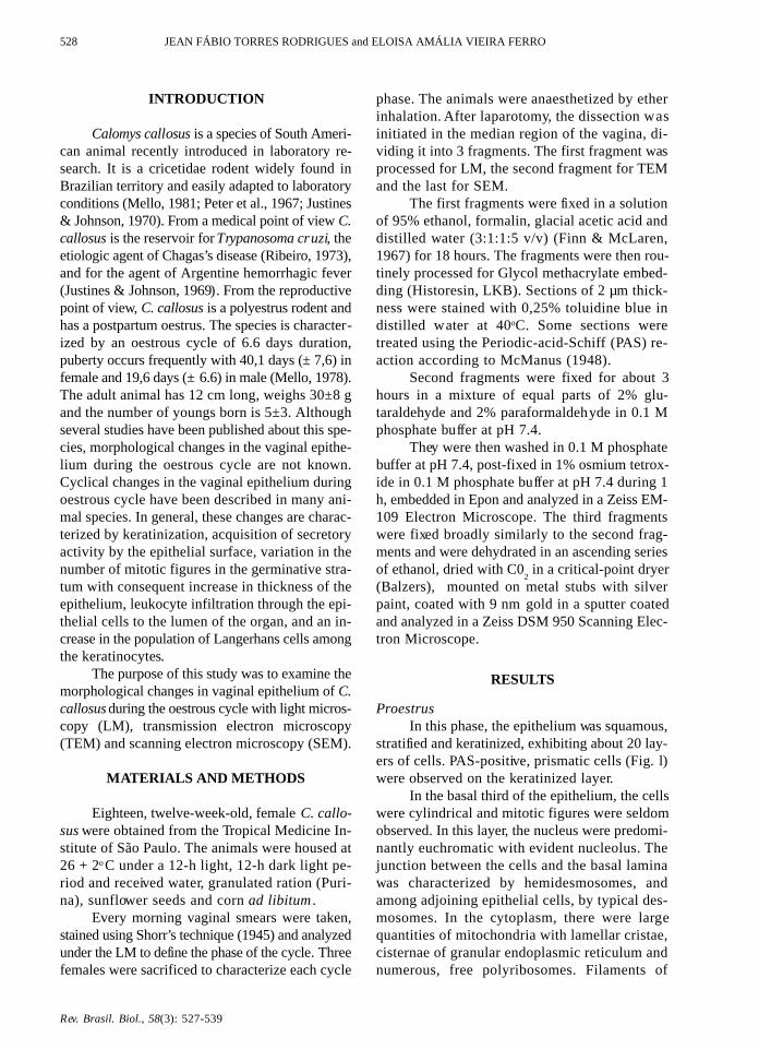
CHANGE IN THE VAGINAL EPITHELIUM OF Colomys callosus 531
Figs. 5 and 6 — Scanning electronmicrographs from the vaginal epithelium of Calomys callosus in proestrus.Fig. 5 — Luminal surface of vaginal epithelium with mucus (arrow) and microridges (arrowheads). l0000X. Bar –1µm. Fig. 6 — Higher magnification the surface of cells covered with microridges (arrow). 17000X. Bar – 1µm.
Rev. Brasil. Biol., 58(3): 527-539
532 JEAN FÁBIO TORRES RODRIGUES and ELOISA AMÁLIA VIEIRA FERRO
Fig. 9 — Electronmicrographs of vaginal epithelium of Calomys callosus during oestrous. Interf ace between keratinized(K) and non-keratinized layer (Q). The upper left insert demonstrates desmosomes seen in the keratinized layer. The twomembranes forming each desmosome are in close juxtaposition and there are deposits of electron dense material on the incytoplasmic f aces (arrows). The lower left insert shows the transition zone between the keratinized and non-keratinized layer.The non-keratinized layer is marked by the presence of numerous desmosomes (asterisk) into which intermediate filamentsare inserted. 14000X. Upper left insert – 40000X. Lower left insert – 20000X. Bar – 1µm.
Fig. 7 — Vaginal epithelium during oestrous obseved by LM.The epithelium is stratified, squamous and keratinized (K).Below this layer there are many kerato-hyaline granules (as-terisk). 310X.
Fig. 8 — Electronmicrograph of vaginal epithelium ofCalomys callosus during oestorus. Middle region of the vagi-nal epithelium where mitochondria (arrowheads) with tubularcristae, bundles of intermediate filaments (asterisk), and freepolyribosomes (arrow) are seen. 48500X.
Rev. Brasil. Biol., 58(3 ): 527-539
CHANGE IN THE VAGINAL EPITHELIUM OF Colomys callosus 533
Figs. 10 and 11— Scanning electronmicrographs of the vaginal epithelium of Calomys callosus in oestrus. Fig. 10 — Surfaceview of a fully keratinizated epithelium showing cells in the process of sloughing (arrow). 2700X. Fig. 11 — At higher mag-nification the surface of these cells have complex microridges (arrow). 13000X. Bar – 1µm.
Rev. Brasil. Biol., 58(3): 527-539
534 JEAN FÁBIO TORRES RODRIGUES and ELOISA AMÁLIA VIEIRA FERRO
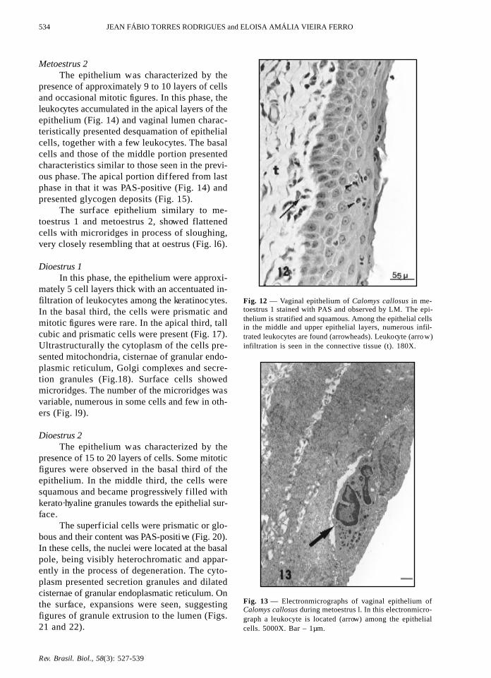
Fig. 12 — Vaginal epithelium of Calomys callosus in me-toestrus 1 stained with PAS and observed by LM. The epi-thelium is stratified and squamous. Among the epithelial cellsin the middle and upper epithelial layers, numerous infil-trated leukocytes are found (arrowheads). Leukocyte (arrow)infiltration is seen in the connective tissue (t). 180X.
Fig. 13 — Electronmicrographs of vaginal epithelium ofCalomys callosus during metoestrus l. In this electronmicro-graph a leukocyte is located (arrow) among the epithelialcells. 5000X. Bar – 1µm.
Metoestrus 2The epithelium was characterized by the
presence of approximately 9 to 10 layers of cellsand occasional mitotic figures. In this phase, theleukocytes accumulated in the apical layers of theepithelium (Fig. 14) and vaginal lumen charac-teristically presented desquamation of epithelialcells, together with a few leukocytes. The basalcells and those of the middle portion presentedcharacteristics similar to those seen in the previ-ous phase. The apical portion differed from lastphase in that it was PAS-positive (Fig. 14) andpresented glycogen deposits (Fig. 15).
The surface epithelium similary to me-toestrus 1 and metoestrus 2, showed flattenedcells with microridges in process of sloughing,very closely resembling that at oestrus (Fig. l6).
Dioestrus 1In this phase, the epithelium were approxi-
mately 5 cell layers thick with an accentuated in-filtration of leukocytes among the keratinocytes.In the basal third, the cells were prismatic andmitotic figures were rare. In the apical third, tallcubic and prismatic cells were present (Fig. 17).Ultrastructurally the cytoplasm of the cells pre-sented mitochondria, cisternae of granular endo-plasmic reticulum, Golgi complexes and secre-tion granules (Fig.18). Surface cells showedmicroridges. The number of the microridges wasvariable, numerous in some cells and few in oth-ers (Fig. l9).
Dioestrus 2The epithelium was characterized by the
presence of 15 to 20 layers of cells. Some mitoticfigures were observed in the basal third of theepithelium. In the middle third, the cells weresquamous and became progressively f illed withkerato·hyaline granules towards the epithelial sur-face.
The superf icial cells were prismatic or glo-bous and their content was PAS-positive (Fig. 20).In these cells, the nuclei were located at the basalpole, being visibly heterochromatic and appar-ently in the process of degeneration. The cyto-plasm presented secretion granules and dilatedcisternae of granular endoplasmatic reticulum. Onthe surface, expansions were seen, suggestingfigures of granule extrusion to the lumen (Figs.21 and 22).
Rev. Brasil. Biol., 58(3 ): 527-539
CHANGE IN THE VAGINAL EPITHELIUM OF Colomys callosus 535
Fig. 14 — Vaginal epithelium of Calomys callosus in me-toestrus 2 stained with PAS and observed by LM. In thisphase, leukocytes predominate in the upper layer of the epi-thelium (arrowheads) which is PAS-positive. 160X.
Fig. 15 — Electronmicrograph of the upper region of theepithelium seen in the previous figure. Observe the nucleus,mitochondria (arrow), desmosomes (arrowheads) and regionof glycogen accumulation (star). 26000X. Bar – 1µm.
Fig. 16 — Scanning electron micrograph of the vaginal epithelium of Calomys callosus in metoestrus. The cells are in processof sloughing and show microridges at their surf aces (arrowhead). 9000X. Bar – 1µm.
Rev. Brasil. Biol., 58(3): 527-539
536 JEAN FÁBIO TORRES RODRIGUES and ELOISA AMÁLIA VIEIRA FERRO
Fig. 19 — Scanning electron micrograph of the luminal surface of the vaginal epithelium in dioestrus l. Cell surface withfew microridges (asterisk) or with many microridges (star). 12000X. Bar – 1µm.
Fig. 17 — Vaginal epithelium of Calomys callosus in di-oestrus 1 stained with PAS and observed by LM. The epi-thelium comprises few cell layers and the superficial layercells are cubic. In the connective tissue, leukocytes (arrow)also insinuate (arrowhead) among the epithelial cells. l50X.
Fig. 18 — Elect ronmicrograph of vaginal epithelium ofCalomys collosus during dioestrous l. Cubic cells seen in theprevious figure. The presence of microvilli on the surface ofthese cells is common (arrow). Secretory granules (G), Golgicomplex (arrowhead ), mitochondria (asterisk) and granularendoplasmic reticule (r) are seen in the cytoplasm. 28000X.Bar – 1µm.
Rev. Brasil. Biol., 58(3 ): 527-539
CHANGE IN THE VAGINAL EPITHELIUM OF Colomys callosus 537
Fig. 20 — Vaginal epithelium of Calomys callosus in di-oestrous 2 stained with PAS an observed by LM. A thickerepithelium is observed; mitotic figures are common in thebasal region (arrowhead). The superficial cells are PAS-posi-tive. Below this layer kerato-hyaline granules (arrow) are alsofound in the cytoplasm of the squamous cells. In the vagi-nal lumen (asterisk) a PAS-positive secretion is seen. l70X.
Fig 21 — Electronmicrograph of vaginal epithelium of Calomyscallosus during dioestrous 2. Prismatic cells seen in Fig. 20.Observe nucleus (n), many secretory granulea (g) are presentin the cytoplasm of these cells. On the apical surface of the cells,certain expansions which may represent the process of gran-ule extrusion (asterisk) are seen. 6000X. Bar – 1µm.
Fig. 22 — Scanning electron micrograph from the vaginal epithelium of Calomys callosus in dioestrus 2. In cell surfacesextrusion of secretion granule (arrow). 11000X. Bar – 1µm.
Rev. Brasil. Biol., 58(3): 527-539
538 JEAN FÁBIO TORRES RODRIGUES and ELOISA AMÁLIA VIEIRA FERRO
DISCUSSION
During the oestrous cycle, the vaginal epi-thelium of C. callosus undergoes profound mor-phological changes. This phenomenon also oc-curs in rat (Parakkal, 1974; Centola, 1978;Vijayasaradhi & Gupta, 1987) and mouse (Allen,1927; Young & Hosking, 1986; Nelson et al,1991; Horvat et al , 1992). The acquisition ofsecretory activity, changes in the pattern of kera-tinization, and an increase in the rate of mitosiswere also seen. As in other animals, these changesprobably occur due to oscillating levels of ova-rian hormones.
In the proestrus phase, the oestrogen levelis maximal and influences the keratinocyteswhich present a greater fluidity of the plasmamembrane (Reddy et al.,1989). In addition to thisindirect effect, oestrogen directly influences theenzyme glucose-6-phosphatase which has animportant role in keratin synthesis (Kang & West,1982). Roop (1987) has demonstrated that oestro-gen modulates the expression of the DNA seg-ment responsible for keratin synthesis. Thesedata, obtained from studies in rats and mice,suggest that in Calomys callosus, the process ofkeratinization, which starts in proestrus and be-comes maximal in oestrus, is related, either di-rectly or indirectly, to high oestrogen levels.
It is known that copulation occurs duringoestrus; thus, greater protection of the vaginalmucous layer is necessary and this is provided bythe keratin layer, by the increased thickness of theother epithelial layers and by the microridges atthe surface of the cells, wich can hold mucus atthe luminal surface of the vagina (Lamb et al .,1978). The increased thickness of epithelial layersis the result of mitosis in a great number of basalcells. A similar fact was observed by Galand etal. (1971) who demonstrated that in mice, dur-ing the oestrogenic phase, there is an increase inthe number of mitotic figures due to a reductionof the S phase of the cellular cycle, thus reduc-ing the duplication time of DNA.
Morphologically, the end of oestrus is mar-ked by the liberation of the keratin to the vaginallumen and a reduction in epithelial thickness. Thismoment marks the transition from oestrus to meto-estrus 1. As the cycle proceeds, the apical portionof the vaginal epithelium is characterized by gly-cogen deposits as noted by ultrastructural evalu-
ation and by the PAS reaction, characterizingmetoestrus 2. During metoestrus, an intense leu-kocyte infiltration is observed among the epithe-lial cells as these migrate towards the vaginal lu-men. This leukocyte infiltration is a well knownphenomenon and has already been described byvarious authors (Busch, 1966; Holtz et al., 1968).Thus, by a still unclear mechanism, the leuko-cytes migrate through the epithelium towards thelumen, assisting in protection from infectiousagents (Corbeil et al., 1985), or to accomplish thephagocytosis of spermatozoa residues still presentin the vaginal lumen (Branscheid & Holtz, 1988).
In dioestrus l, the epithelium of the super-ficial layer is cubic and presents ultrastructuraland histochemical features characteristic of secre-tory cells. This secretory activity may be relatedto increased progesterone levels, since in mice themaximum concentration of progesterone occursin dioestrus 1 (Walmer et al., 1992). As the cyclecontinues, the superficial layer cells become pris-matic or globous with a great number of secre-tory granules; this period is characterized as di-oestrus 2.
The secretion produced by the epithelial cellsis discharged to the vaginal lumen where, togetherwith the secretions from the cervix, seems to per-form an important role in the defense of the epi-thelium, since the leukocytes remain immersed inthis gelatinous net, and thus may exert their ph-agocytic function (Branscheid & Holtz, 1988).
In diestrus 2, kerato-hyaline granules asso-ciated with intermediate filaments of 10 nm diam-eter accumulate below the layer of secretory cellsforming the keratin layer present in the subsequentphase: the proestrus phase. In proestrus, secretorycells were observed in the keratinized layer, re-flecting an advanced degenerative condition whichpersisted since the last dioestrus.
In conclusion, the present results suggest thatsurface cells of vaginal epithelium could be secre-tory or keratinized, probably in response to ovarianhormones. Additionally, the present study givesevidence of wide cellular variety found in vaginalepithelium of C. callosus during oestrous cycle.
Acknowledgements — The authors thank Mr. Hélgio H.Wernech for excellent assistance. Dr. José Roberto Mineokindly reviewed the manuscript. The authors wish to expresstheir appreciation to Dra. Estela Maris Andrade Forell Bevi-lacqua for her interest and helpful suggestions during thisinvestigation and Dr. Hélio Chiarini Garcia for his extensive
Rev. Brasil. Biol., 58(3 ): 527-539
CHANGE IN THE VAGINAL EPITHELIUM OF Colomys callosus 539
instruction in the techniques and principles of scanningelectron microscopy.
REFERENCES
ALLEN, E., 1927, The oestrus cycle in the mouse. Ameri-can Journal of Anatomy, 30(3): 297-371.
BRANSCHEID, W. & HOLTZ, W., 1988, Histochemical ex-amination of the vaginal epithelium of sows at varìousstages of the estrus cycle. Anat. Histol. Embriol., 17 :12-26.
BUSCH, W., 1966, Die periodischen veränderungen desvaginal epithels beim schwein und die möglichkeitenihrer heranziehung zur graviditätsdiagnose. Wiss. Z.Humboldt-Univ. Berlim, Math. Nat. R., 15: 833-865.
CENTOLA, G. M., 1978, Surface features of exfoliated vagi-nal epithelial cells during oestrous cycle of the rat exa-mined by scanning electron microscopy. J. Anat., 127 :553-561.
CORBEIL, L. B., CHATTERJEE, A., FORESMAN, L. &WESTFALL, J. A., 1985, Ultrastruture of cycle changein the murine uterus, cervix and vagina. Tissue & Cell,17 : 53-68.
FINN, C. A. & MC LAREN, A. , 1967, A study of the earlystage of implantation in mice. J. Reprod. Fertil., 13 :259-267.
GALAND, P., LEROY, F. & CHRETIEN, J., 1971, Effect ofvaginal on proliferation and histological changes inthe uterus and vagina of mice. J. Endocr., 49: 243-252.
HOLTZ, W., SMIDT. D., THUME, O. & WESELOH, W.,1968, Veränderugen des scheiden-pH, der rektal undvaginal gemessenen Körpertemperatur und desscheidenepithels in abhängigkeit vom sexualzyklus beimgö ttinger zwergschwein. Zbl. Vet. Med, 15: 329-352.
HORVAT, B., VRCIC, H. & DAMJANOV, I., 1992, Trans-diferentiation of murine squamous vaginal epitheliumin proestrus is associated with changes in the expres-sion of keratin polypeptides. Experimental Cell Re-search, 199: 234-239.
JUSTINES, G. & JOHNSON, K. M., 1969, Imune tolerancein Calomys callosus infected with machupo virus.Nature, 222 : 1090-1091.
JUSTINES, G. & JOHNSON, K. M., 1970, Observations onthe laboratory breeding of the cricetidae rodentCalomys callosus. Lab. Anim. Core, 20 : 57-60.
KANG, Y. & WEST, W. L., 1982, Ultrastructural localiza-tion of glucose-6-phosphatase and alkaline phos-phatase in the vaginal epithelium of rat. Journasl ofMorphology, 171: 1-10.
LAMB, J. C., NEWBOLD, R. R., STUMPF, W. E. &MCLACHLAN, J. A., 1978, Transiotional changes inthe surf ace epithelium of the cycling mouse vagina,cervix and uterus: scanning electron microscopic stud-ies. Biol. reprod., 19: 701-711.
MCMANUS, J. F. A., 1948, Histological and histochemicaluse of periodic acid. Stain Tecnology, 23(3): 99-108.
MELLO, D. A., 1978. Biology of Calomys callosus underlaboratory conditions (Rodentia, Cricetidae). Rev.Bras. Biol., 38 (4): 807-811.
MELLO, D. A., 1981, Studies on reproduction and longevityof Calomys callosus (Renger, 1830) under laboratoryconditions ( Rodentia, Cricetidae ). Rev. Bras. Biol.,41(4): 841-843.
NELSON, K. G., TAKAHASHI, T., BOSSERT, N. L.,WALMER, D. K. & MCLACHLAN, J. A.,1991, Epi-dermal growth factor replaces estrogen in the stimu-lation of female genital-tract growth and differentia-tion. Proc. Natl. Acad. Sci., 88: 21-25.
PARAKKAT, P. F., 1974, Cyclical changes in the vaginalepithelium of the sun by scanning electron micros-copy. Anat. Rec., 178: 529-538.
PETTER, F., KARIMI, Y. & ALMEIDA, C. R., 1967, Unnouvean ranger de laboratoire, lé cricetidé (Roden-tia, Cricetidae). C. R. Acad. Paris, 265: 1974-1976.
RIBEIRO, R. D., 1973, Novo reservatório do Trypanosomacruzi. Rev. Bras. Biol., 33: 429·437.
REDDY, A. G., SHIVAJI, S. & GUPTA, P. D., 1989, Effect ofestradiol on the membrane fluidity of the rat vaginalepithelial cells. J. Steroid. Biochem, 33(6): 1229-1233.
ROOP, D. R., 1987, Regulation of keratìn gene expressionduring differentiation of epidermal and vaginal epi-thelial cells. Current Topics in Developmental Biol-ogy, 22: 195-207.
SHORR, E., 1945, A new technic for staining vaginal smears.A single differentive stain. Science, 84: 545.
VIJAYASARADHI, S. & GIJPTA, P. D., 1987 Keratinizationof rat vaginal epithelium. I - Cell surface study duringnatural and induced keratinization. J. Submicrosc.Cytol., 19 : 595-603.
WALMER, D. K., WRONA, M. A., HLTGHES, C. L. &NELSON, K. G., 1992, Lactoferrin expression in themouse reproductive tract during the natural estrouscycle: Correlation with circulating estradiol andprogesterone. Endocrinodogy, 131 (3): 458-1466.
YOUNG, W. G. & HOSKING, A. R., 1986, Langerhans cellsin murine vaginal epithelium af fected oestrogen andtopical vitamin A. Acta. Anat., 125: 59-64.