REVIEW Mouse models of human TB pathology: roles in the analysis of necrosis and the development of host-directed therapies Igor Kramnik 1 & Gillian Beamer 2 Received: 17 August 2015 /Accepted: 22 October 2015 /Published online: 5 November 2015 # The Author(s) 2015. This article is published with open access at Springerlink.com Abstract A key aspect of TB pathogenesis that maintains Mycobacterium tuberculosis in the human population is the ability to cause necrosis in pulmonary lesions. As co-evolution shaped M. tuberculosis (M.tb) and human responses, the complete TB disease profile and lesion manifestation are not fully reproduced by any animal model. However, animal models are absolutely critical to understand how infection with virulent M.tb generates outcomes necessary for the pathogen transmission and evolutionary success. In humans, a wide spectrum of TB outcomes has been recognized based on clinical and epi- demiological data. In mice, there is clear genetic basis for susceptibility. Although the spectra of human and mouse TB do not completely overlap, comparison of human TB with mouse lesions across genetically diverse strains firm- ly establishes points of convergence. By embracing the genetic heterogeneity of the mouse population, we gain tremendous advantage in the quest for suitable in vivo models. Below, we review genetically defined mouse models that recapitulate a key element of M.tb pathogen- esis—induction of necrotic TB lesions in the lungs—and discuss how these models may reflect TB stratification and pathogenesis in humans. The approach ensures that roles that mouse models play in basic and translational TB research will continue to increase allowing researchers to address fundamental questions of TB pathogenesis and bacterial physiology in vivo using this well-defined, re- producible, and cost-efficient system. Combination of the new generation mouse models with advanced imaging technologies will also allow rapid and inexpensive assess- ment of experimental vaccines and therapies prior to test- ing in larger animals and clinical trials. Keywords Tuberculosis . Granuloma . Necrosis . Animal models . Inbred mice . Mechanisms . Host-directed therapies . sst1 Pulmonary necrosis in M.tb life cycle A long history of human co-evolution with Mycobacterium tuberculosis (M.tb) suggests that unique immune mecha- nisms have evolved explaining substantial resistance of modern humans to the disease [1–3]. However, our species also proved to be an ideal host for M.tb, such that the bac- teria lost the need for any other environmental niche and relied entirely on modifying human body for every stage of its life cycle. It is generally accepted that to establish new infection, M.tb reaches terminal airways in small aero- sol particles generated during cough. Those aerosols are generated from lung cavities where M.tb accumulates in large quantities, perhaps in biofilms, at the air interface, effectively sequestered from host immunity. Thus, from the evolutionary standpoint, M.tb is an obligate lung pathogen, such that substantial M.tb-induced destruction of lung tissue is absolutely required for direct transmission to other humans. Although the bacteria are capable of achieving this This submission is related to Immunopathology of Mycobacterial Diseases - Dr. Stefan Kaufmann * Igor Kramnik [email protected]Gillian Beamer [email protected]1 Pulmonary Center, Department of Medicine, National Emerging Infectious Diseases Laboratories, Boston University School of Medicine, 620 Albany Street, Room 501, Boston, MA 02118, USA 2 Department of Infectious Disease and Global Health, Cummings School of Veterinary Medicine, Tufts University, 200 Westboro Rd, Bldg 20, Grafton, MA 01536, USA Semin Immunopathol (2016) 38:221–237 DOI 10.1007/s00281-015-0538-9
Transcript
REVIEW
Mouse models of human TB pathology: roles in the analysisof necrosis and the development of host-directed therapies
Igor Kramnik1& Gillian Beamer2
Received: 17 August 2015 /Accepted: 22 October 2015 /Published online: 5 November 2015# The Author(s) 2015. This article is published with open access at Springerlink.com
Abstract A key aspect of TB pathogenesis that maintainsMycobacterium tuberculosis in the human population isthe ability to cause necrosis in pulmonary lesions. Asco-evolution shaped M. tuberculosis (M.tb) and humanresponses, the complete TB disease profile and lesionmanifestation are not fully reproduced by any animalmodel. However, animal models are absolutely critical tounderstand how infection with virulent M.tb generatesoutcomes necessary for the pathogen transmission andevolutionary success. In humans, a wide spectrum of TBoutcomes has been recognized based on clinical and epi-demiological data. In mice, there is clear genetic basis forsusceptibility. Although the spectra of human and mouseTB do not completely overlap, comparison of human TBwith mouse lesions across genetically diverse strains firm-ly establishes points of convergence. By embracing thegenetic heterogeneity of the mouse population, we gaintremendous advantage in the quest for suitable in vivomodels. Below, we review genetically defined mousemodels that recapitulate a key element of M.tb pathogen-esis—induction of necrotic TB lesions in the lungs—anddiscuss how these models may reflect TB stratification
and pathogenesis in humans. The approach ensures thatroles that mouse models play in basic and translational TBresearch will continue to increase allowing researchers toaddress fundamental questions of TB pathogenesis andbacterial physiology in vivo using this well-defined, re-producible, and cost-efficient system. Combination of thenew generation mouse models with advanced imagingtechnologies will also allow rapid and inexpensive assess-ment of experimental vaccines and therapies prior to test-ing in larger animals and clinical trials.
A long history of human co-evolution with Mycobacteriumtuberculosis (M.tb) suggests that unique immune mecha-nisms have evolved explaining substantial resistance ofmodern humans to the disease [1–3]. However, our speciesalso proved to be an ideal host for M.tb, such that the bac-teria lost the need for any other environmental niche andrelied entirely on modifying human body for every stageof its life cycle. It is generally accepted that to establishnew infection, M.tb reaches terminal airways in small aero-sol particles generated during cough. Those aerosols aregenerated from lung cavities where M.tb accumulates inlarge quantities, perhaps in biofilms, at the air interface,effectively sequestered from host immunity. Thus, from theevolutionary standpoint, M.tb is an obligate lung pathogen,such that substantial M.tb-induced destruction of lung tissueis absolutely required for direct transmission to otherhumans. Although the bacteria are capable of achieving this
This submission is related to Immunopathology of MycobacterialDiseases - Dr. Stefan Kaufmann
1 Pulmonary Center, Department of Medicine, National EmergingInfectious Diseases Laboratories, Boston University School ofMedicine, 620 Albany Street, Room 501, Boston, MA 02118, USA
2 Department of Infectious Disease and Global Health, CummingsSchool of Veterinary Medicine, Tufts University, 200 Westboro Rd,Bldg 20, Grafton, MA 01536, USA
goal only in a relatively small fraction of the infected hosts,this is sufficient for stable colonization of its unique naturalhabitat, Homo sapiens. Therefore, understanding mecha-nisms of host susceptibility enabling M.tb, transmission isnecessary to counter its evolutionary-refined virulence strat-egy most effectively.
Although animal models have been a lynchpin formechanistic studies of TB since Robert Koch’s discoveryof the pathogen, little progress has been made in under-standing mechanisms of pulmonary destruction caused byM.tb. Recent experimental work has demonstrated thatdespite the uniqueness of human species as a host toM.tb, mice could be reliably used to study mechanismsof necrotization in TB granulomas and formation of lungcavities. Thus, the powerful arsenal of mouse geneticmethods can be utilized to mechanistically dissect thosecrucial transitions during the natural course of TB infec-tion. Below, we review genetically defined mouse modelsthat recapitulate necrotic TB lesions in the lungs and dis-cuss how these models may reflect TB pathogenesis inhumans.
Necrosis occurs at various stages of TB infection
There are at least two distinct stages of M.tb infection atwhich necrosis can occur: (1) at the initial stage of lungcolonization, which leads to necrosis of individual orsmall clusters of macrophages in a primary granuloma,and (2) during advanced disease where large areas of thelung are effaced by coalescing granulomas and tubercu-lous pneumonia. Thus, necrosis delimits the life cycle ofM.tb within an individual and plays an important role inboth establishing persistent infection initially as well as inthe final exit and transmission to a new host. There aremany important mechanistic differences between the twostages.
The initial contact of M.tb with the host occurs in high-ly aerated environment, presumably with alveolar macro-phages in a context of normal lung tissue. The alveolarmacrophages are permissive to M.tb, which establishes itsfirst replicative niche in this cell. Subsequent interactionswith lung epithelium and possibly innate T cells lead torecruitment of inflammatory cells from circulation andestablishing clusters of myeloid cells that contain the bac-teria. These early dynamics cannot be addressed directlyin natural lung environment in humans, but the zebrafishmodel of infection with Mycobacterium marinum pro-vides a detailed view of cellular recruitment and interac-tions that establish nascent microgranulomas [4–6] beforeantigen-specific immunity develops. Although zebrafishdo not have lungs, this model allows detailed cell traffick-ing studies in vivo and elegantly shows that macrophagedeath spreads bacilli to adjacent recruited macrophages
within the same granuloma. Furthermore, when the re-cruitment of myeloid cells fails to contain M.tb, the bacillireplicate extracellularly with conspicuous formation ofcords [7]. Multiple host responses contribute to macro-phage necrosis, including alterations in lipid mediatorsand increased TNFα production or deficient recruitmentof new myeloid cells to the site of infection [8]. Similarly,in the lungs of M.tb-infected C3HeB/FeJ mice (abbreviat-ed HeB), early micronecrotic lesions form 2–3 weekspost-infection, when spread of M.tb to adjacent inflamma-tory cells, as well as robust extracellular replication areobserved [9]. In both the zebrafish model and HeB mice,the microgranulomas undergoing necrosis are composedprimarily of myeloid cells (macrophages and somegranulocytes). In contrast, different host-pathogen dynam-ics are observed in the relatively resistant mouse strainC57BL/6 (B6), where lesions are non-necrotic, containfew neutrophils, and bacilli remain intracellular.
The early granulomas are composed primarily of mye-loid cells that initially act autonomously to restrict thebacterial growth and spread. However, adaptive T cell-mediated immunity is necessary to contain further pro-gression and necrotization of granulomas. The bacterialspread is unstoppable in T cell -deficient mice, wheremycobacteria replicate in unrestricted manner and destroythe infected tissue. Large areas of necrotic inflammation,massive bacterial loads, and extensive neutrophil infiltra-tion are typical for this type of progression, which, how-ever, lacks the characteristics of organized granulomas.Thus, the early TB granulomas can follow necrotic andnon-necrotic trajectories depending on the myeloid cellintrinsic capacity and help of M.tb-induced T lympho-cytes producing IFNγ (Th1-type response). In both cases,however, they serve to constrain M.tb and prevent dissem-ination. In the case of more efficient immune response inresistant hosts, primary granulomas may be sterilized overtime and undergo calcification. In permissive butimmune-competent hosts, however, small necrotic granu-lomas establish a nidus of persistent infection, which canlater reactivate and cause post-primary TB.
The exit and transmission strategy of M.tb at the end ofits life cycle is entirely dependent on granuloma spreadand necrosis leading to formation of lung cavities. Thosenecrotic lesions become M.tb sanctuaries sequestering thepathogen from the host immune system and allowing itsreplication and transmission via aerosols. This transitionoccurs in immune-competent hosts that successfully con-trolled the primary infection. Thus, the local mechanismsand the dynamics of necrosis at the advanced diseasestage are not the same as in primary lesions and theorgan- and organism-scale factors may play bigger or dif-ferent roles. Two different models of necrosis in advancedTB have been proposed:
222 Semin Immunopathol (2016) 38:221–237
Model1
This is the gradual necrotization and local expansionof organized granulomas, including formation andcoalescense of satellite granulomas. Accumulationof dead macrophages that fail to survive intracellularM.tb infection is a primary source of the caseousnecrotic masses.
Model2
This is the rapid dissemination ofM.tb from chroniclesion causing tuberculous pneumonia, wherenecrosis formation may be associated withthrombosed blood vessels and infarcted regions ofthe lungs [10, 11]. Subsequently, M.tb bacilli, deadinflammatory cells, and dead lung tissue may besequestered in a fibrous capsule to re-contain thepathogen.
Clearly mechanisms and consequences of lung necrosisneed to be considered within the genetic and immunologicalcontext of the host and the stage of disease.We alsowould liketo emphasize the distinction between systemic, lung, and cel-lular levels that contribute to necrosis and will discuss hypoth-eses related to necrosis in that order.
Systemic host factors that contribute to necrosis
Arnold Rich pointed out that Btubercule bacilli have very littlepower of producing necrosis of tissue in the normal (non-hypersensitive) body… Injected locally in large amount itdoes not kill tissue… A fraction of the number of bacilli,which could be injected into the tissues without causing ne-crosis in the normal body, produces violent inflammation andextensive necrosis in the hypersensitive one^([12], p. 350).The dual role of inflammation was further investigated byArthur Dannenberg, who distinguished cytotoxic delayed-type hypersensitivity and macrophage-activating cell-mediat-ed immunity and pointed to a therapeutic potential of manip-ulating their balance. He used the rabbit model to experimen-tally reproduce those types [13] and demonstrated that bothtypes of responses were driven by systemic immunity.However, at that stage, it was impossible to identify moleculardeterminants of Bprotective^ versus Bpathogenic^ inflamma-tion as specific therapeutic targets. Those classical experi-ments demonstrating transition from mild protective toBviolent^ destructive inflammation implicated systemic hostimmune response to M.tb in evolution of the host protectivegranulomas into pathogen-favoring ones. However, specificmechanisms of adaptive immunity causing necrosis in TBgranulomas still remain hypothetical [14].
Local granuloma factors that contribute to necrosis
Mechanistically, the formation of necrotic TB lesions andliquefaction of cellular debris were attributed to release ofhydrolytic enzymes by macrophages and neutrophils.
Among them are MMP1 and MMP8 [15–18] and serineproteases [19], which degrade extracellular matrix pro-teins and basement membranes, participating in lung tis-sue destruction and stimulating fibrosis. Although thoseenzymes may be attractive therapeutic targets, they arelikely to be executors but not the root causes of lungtissue necrosis.
Recent studies demonstrate that dysbalance of inflammato-ry pathways may lead to necrotizing inflammation. For exam-ple, activation of type I interferon (IFN-I) pathway byinstilling tlr3 ligand poly(I:C) in the lungs promoted the de-velopment of acute necrotic TB lesions via excessive recruit-ment of myeloid cells [20]. In another model, overexpressionof IL-13 using T cell-specific promoter generated conditionsfor the development of well-organized necrotic lung granulo-mas [21]. Interestingly, the IL-13 overexpression did not re-duce Th1 responses, which would explain the necrotization bysuppression of the essential host resistance pathway.Alternatively, increased sensitivity of alternatively activatedmacrophages to cytotoxic activity of TNFα in granulomasmay be involved, as hypothesized previously by Rook andcoauthors [22]. Another model of necrotic TB lesions hasbeen generated using temporal inactivation of essential mech-anisms of resistance, such as administration of NO inhibitorand neutralization of IFNγ using injections of monoclonalantibodies in mice infected with M.tb intradermally [19].
An important concept emerged recently based on stud-ies of heterogeneous TB granulomas in non-human pri-mates (NHP) using combination of live imaging, analysisof RNA expression patterns, M.tb loads, histopathology,and computational modeling [23–25]. The iNOS andarginase-1 protein expression in granulomas in situ wereused as surrogate markers for the M1 and M2 macrophagephenotypes, respectively. The M1/M2 balance emerged asa best correlate of the granuloma outcome. Double-positive cells were found in granuloma walls, and pheno-typically M1 cells increased towards the necrotic center.In this study, the higher M2 activation did not correlatewith suppression of Th1 responses, as well. In a mousemodel, the expression of arginase-1 was shown to bemodestly protective in granulomas in the absence ofiNOS, and simultaneous Arg1 and iNOS inactivation re-sulted in exuberant necrotic inflammation [26]. Both ofthose observations are more consistent with a possibilitythat the arginase-1 pathway does not suppress the NO-mediated effector pathway, but Arg1 is activated as abackup effector mechanism, when the more efficientNO-mediated effector pathway fails to control the bacte-ria, for example due to hypoxia in large progressive le-sions. Taken together, the NHP provided a unique data setunattainable in the mouse model, meanwhile the mousemodel provided a novel mechanist ic insight forinterpreting the NHP data.
Semin Immunopathol (2016) 38:221–237 223
Factors that contribute to macrophage necrosis
At a cellular level, mechanisms of macrophage death directlycaused byM.tb are under intense investigation. Roughly, theycan be divided into two categories: (1) active mechanisms,whereby virulent mycobacteria or host produce toxic molec-ular mediators that cause macrophage death, and (2) passivemechanisms, where bacillary replication in macrophages re-sults in lysis, essentially a Bload-driven^ death. The formercategory focuses on specific pathways that determine macro-phage death modality. For example, the balance of lipid me-diators PgE2 and lipoxin A4 has been shown to control mem-brane repair of the infected macrophages and apoptotic vsnecrotic cell death [27, 28], as well as production of TNFα[29], whereas the higher concentrations of TNFα inducednecroptotic pathway in macrophages [30]. The necrotic mac-rophage death is perceived as more detrimental for the hostthat might be associated with necrotic granuloma formationin vivo, while apoptotic death has been associated with hostresistance and bacterial control [27, 31, 32].
M.tb produces multiple products that induce macrophagenecrosis in vitro. Cord factor (trehalose 6,6′-dimycolate orTDM) is a mycolic acid-rich membrane glycolipid identifieddecades ago and is a well-known virulence and immunomod-ulatory factor ofM.tb. TDM is particularly interesting becauseit may induce rapid macrophage necrosis. Early secreted anti-genic target-6 (ESAT-6) is a virulence factor encoded withinthe RD1 region [33], which is absent from the vaccine strainMycobacterium bovis BCG. ESAT-6 triggers necrosis directly[34] or indirectly by phagosomal rupture allowingM.tb accessto the cytoplasm [35, 36] and activation of NLRP3inflammasome [37]. Another cytotoxin CpnT has been recentlydiscovered that kills macrophages via hydrolysing macrophageNAD [38, 39]. However, those toxic factors are not sufficient toinduce necrotic granulomas in the genetically resistant mice.Meanwhile, even avirulent (ESAT-6 negative) M. bovis BCGwas capable of inducing granuloma necrosis in immunodefi-cient Mendelian susceptibility to mycobacterial diseases(MSMD) human patients or T cell-deficient mice. The laterfacts are consistent with a burst size hypothesis, where unre-stricted growth of intracellular mycobacteria results in achiev-ing maximal load and eventually death of the infected cell [40].A hybrid hypothesis stipulates that membrane-toxic mycobac-terial products produced intracellularly may allow M.tb escapefrom vacuole to cytoplasm, where it can either replicate morerapidly to achieve the burst size or actively induce macrophagecell death via activation of type I IFN pathway [41, 42].
This brief review of literature suggests the co-existence,and potential cooperation, of multiple pathways leading tonecrosis of granulomas. Here, we would like to emphasizethat causality of necrosis may significantly differ between an-imal hosts and experimental systems, as well as between hu-man patients.
Genetic variation in mice controls necrotizingresponses to M.tb
Many animal models are used to studyM.tb lesions includingmice, guinea pigs, rabbits, non-human primates, bovinecalves, zebrafish, rats, ferrets, mini pigs, fruit flies, nematodes,planarians, and even amoebas [43–56]. Due to the small size,cost-efficiency, and availability of reagents, mice are widelyused and have been crucial for determining immunologicalrequirements that restrict M.tb growth [57, 58]. However,the two most commonly used inbred laboratory mouse strains(C57BL/6 and BALB/c) do not develop necrotizing lesions[56, 59, 60] which, unfortunately, have led to general criticismof the mouse model. When considering responses at themouse population level, however, it becomes apparent thatnecrotizing responses to M.tb commonly occur in inbred(C3HeB/FeJ, DBA/2, CBA/J, I/St), inbred crossed (HET3),and even outbred crossed (Diversity Outbred) mice [61–67].The spectrum of necrosis includes apoptosis and necrosis ofindividual macrophages, necrotizing granulomas with fibroticcapsule, tuberculous pneumonia with intra-alveolar neutro-philic exudates, and fibrin thrombosis of lung alveolar capil-laries (Fig. 1a–d). Similar necrotizing responses are associatedwith and appear to precede cavity formation in humans andlarger animal models (rabbits and NHPs) [68–70]. Althoughcavities were not reported in mice in the past, their formationhas been well documented recently in C3HeB/FeJ [71, 72]and CBA/J mice (Fig. 1e–f) following their typical necrotizingresponses.
Granulomas are often described with stereotypical orga-nization: focal aggregates of central myeloid cells with pe-ripheral lymphocytes and a fibrous capsule that separates theimmune cells from the adjacent normal tissue. This is asimple model to envision; however, in reality, a significantheterogeneity between granulomas is observed even withinthe same individual in the clinic and in experimental animalmodels. Necrotic granulomas may develop necrotic centers(necrotizing granulomas) or be infiltrated by neutrophils(suppurative granulomas), although the conditions whichdrive those transitions are not fully known. Recent studiesin C3HeB/FeJ mice link necrotizing structural and cellularresponses to M.tb with morbidity [72, 73]. Type I, II, and IIIgranulomas were characterized, but the lesions are not quitestereotypical granulomas mentioned above. Regardless ofterminology, highest morbidity was observed in type II le-sions, which were dominated by necrotizing responses:unencapsulated tuberculous pneumonia with macrophages,neutrophils, necrotic cell debris packed within alveolarspaces, and abundant intra- and extracellular bacilli. Type Ilesions were smaller necrotizing lesions with central necrosissurrounded by macrophages, neutrophils, and peripheral fi-brosis with abundant extracellular bacilli in the necrotic cen-ter and intracellular bacilli in foamy macrophages at the
224 Semin Immunopathol (2016) 38:221–237
periphery. Type III were non-necrotic lesions composed ofmononuclear cells and macrophages in alveoli, perivascularand peribronchiolar lymphocytic aggregates, and alveolarseptal interstitial fibrosis. M.tb bacilli were intracellular with-in macrophages and relatively scarce. These lesion types arenot unique to C3HeB/FeJ mice. We observed lesions iden-tical to type I and type III in chronically infected CBA/Jmice but not type II [64]. We also recently reported a similarspectrum in M.tb-infected Diversity Outbred mice. Highestmorbidity occurred in the individuals that developed necro-tizing neutrophilic tuberculous pneumonia (i.e., type II le-sions) and these same mice had the highest bacillary burden[66]. We additionally observed capillary thrombosis in typeII lesions [66] with loss of differential staining suggestive ofinfarction and coagulation necrosis. It is likely that geneti-cally diverse humans and mice share underlying mecha-nisms for these complex necrotizing responses, becausehumans with post-primary TB similarly develop thrombosis,coagulation necrosis, and lung infarction without organizedgranulomas [10, 11, 74]. Thus, the genetically diversemouse models morphologically recapitulate all major formsof pulmonary TB in other species in a reproducible geneti-cally controlled manner.
Mechanisms of necrotizing responses as studiedin mouse models
Neutrophils as mediators of necrotizing responses to M.tb
Neutrophils are suicide bombers of the innate immunity with theprimary function of killing extracellular pathogens. In this pro-cess of eliminating pathogens, neutrophils deploy numerous po-tentially host-damaging molecules such as matrixmetalloproteases from cytoplasmic granules; abundant and po-tent oxidants; and the neutrophils themselves undergo necrosisreleasing formyl peptides, complement C5a, and other mole-cules, which may stimulate Bnecrotaxis^ (a type of neutrophilchemotaxis towards sites of cells and tissue death) [75–84].These molecules from dead and dying bacterial and host cellsare the most potent stimulators of neutrophil recruitment to sitesof infection and cell/tissue death [79, 84]. In TB, large numbersof neutrophils accumulate in airways of pulmonary TB patientsand in airways of mice that develop necrotizing responses [66,72, 85–87] where dead neutrophils are packed in alveoli andterminal bronchioles within and adjacent to necrotizing granulo-mas [66, 72], presumably in response to macrophage death in-duced by rapidly replicating, virulent intracellular bacteria [40].
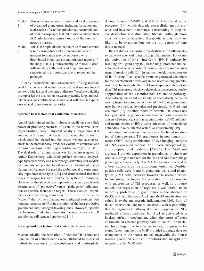
Fig. 1 Manifestation of necrotizing responses and cavitation inducedin vivo in the lungs of mice experimentally infected withMycobacterium tuberculosis. Formalin-fixed, paraffin-embedded lungsections were stained with hematoxylin and eosin (a through d) orstained with a modification of Ziehl-Neelsen’s acid-fast method (f).Death can be observed in individual macrophages as apoptosisrecognized by nuclear fragmentation, magnified 1,000 times, encircled(a). Necrosis of an individual macrophage in an alveolar space isrecognized by cytoplasmic eosinophilia and a pyknotic, condensednucleus, magnified 1,000 times (b). Alveolar septae are present butdifficult to recognize due to accumulation of dead inflammatory cells
and cellular debris within alveolar air spaces, magnified 400 times (c).As the necrotizing process continues, fibrin thrombosis of capillaries isobserved as a transition from a capillary with red blood cells to a capillarycontaining eosinophilic fibrillar material consistent with fibrin (arrows),magnified 400 times (d). Finally, complete destruction of alveolar septaeallows necrotic regions to coalesce and undergo liquefaction and removalof necrotic material, thus contributing to cavity formation, magnified 20times normal (e). Cavities contain variable necrotic debris in whichabundant acid-fast bacilli are detected, magnified 400 times (f). (a–d)necrotic lung lesions in supersusceptible DO mouse; (e–f) cavity in thelung of a CBA/J mouse
Semin Immunopathol (2016) 38:221–237 225
Neutrophils and their chemokines are markers of pulmo-nary TB in humans and in mice [40, 64, 66, 72, 73, 85–92].Some evidence suggests that neutrophils contribute to TB be-cause administration of antioxidants and anti-inflammatories(which may dampen neutrophil responses) decreased TB dis-ease progression [93, 94] and because few neutrophils arepresent when bacillary replication is slowed [40]. However,only a handful of studies definitively link neutrophils [95] ortheir chemoattractants (e.g., CXCL5) [96] with increased sus-ceptibility to M.tb, and others indicate that neutrophils arecellular biomarkers of failed immunity [92]. There are nostudies that definitively prove that neutrophils drive necrotiz-ing responses, and no studies have clearly defined geneticcontrol of neutrophil recruitment. Definite hints exist becauseneutrophil responses vary with the hosts’ genetic architecturein humans and in experimentally infected mice [66, 85, 95,97], and quantitative trait loci (QTLs) for TB susceptibilityoverlap with genes that may control neutrophil responses [91].
There are fundamental knowledge gaps in understandinghow neutrophils contribute to macrophage and granuloma ne-crosis, but there are a number of candidate mechanisms to betested. At the cellular (macrophage) level, neutrophils maycause lipid peroxidation of cell membranes, resulting in mac-rophage necrosis and release of bacilli. At the granuloma andlung tissue level, neutrophil influx could be more even moredetrimental: (1) by causing lipid peroxidation of epithelial andendothelial cell membranes which irreparably damages lungtissue; (2) by proteolytic digestion of collagen-rich matrix andbasement membranes of the lung alveolar septae; and (3) byphysically obstructing airways which disrupt normal pulmo-nary function.We additionally theorize that neutrophils contrib-ute to lung damage, which precedes cavitation by promotingthrombosis and infarction. This is supported by the facts thatneutrophil granules contain pro-thrombotic molecules and thatNETs from dying neutrophils also promote thrombosis [68,98–100]. Additionally, this is supported by in situ observationsof fibrin thrombi within capillaries adjacent to necrotic alveolarseptae (Fig. 1d) as well as thrombosed vessels and necrotic lungtissue in patients with cavitary TB [11, 74].
Current evidence shows that neutrophil roles are complex.The abundance of neutrophil involvement in necrotizing re-sponses and lesion progression does not prove, however, thatneutrophil activity is the mechanistic cause of macrophagenecrosis, granuloma necrosis, or lung tissue necrosis in TBlesions. At the early stage of infection, neutrophils can beprotective by phagocytizing and killing a fraction of M.tbbacilli through oxidative mechanisms [101]. At the laterstages, however, the excessive recruitment of neutrophils toTB lesions in the lung may serve as a common mechanismdownstream of pathological reactions, which are triggered ormaintained by other cell types. As an overall trend, in thesusceptible mouse strains, neutrophils exacerbate susceptibil-ity [95, 97]. However, it remains to be established whether this
occurs because the susceptible mice have a genetic propensityfor increased neutrophil recruitment to the lungs followingM.tb infection, intrinsic functional defects of neutrophils, orit occurs downstream of other cells’ genetic defects.
In part, the paucity of known neutrophil-mediated mecha-nisms reflects scarce methodologies to manipulate and trackneutrophils in vivo. Fortunately, new tools addressing rolesfor neutrophils have recently become available. This includesgenetically diverse populations of mice (Diversity Outbred,Collaborative Cross) [102, 103]; immortalized neutrophil pro-genitor cells that can be manipulated in vitro and in vivo[104]; and neutrophil imaging technologies to track neutro-phils in vivo over time in M.tb-infected mice. Eventually,those and other novel approaches will allow definitive mech-anistic analyses of neutrophil contributions to thenecrotization of TB lesions using mouse models, applicableto other species.
Macrophage roles in necrotizing responses
A scrupulous work by the Kornfeld laboratory provided con-vincing experimental evidence that macrophage necrosis maytrigger neutrophil recruitment to sites of M.tb infection [40,105]. Using relatively resistant C57BL/6 mice infected withM.tb strains of varying virulence, they showed that higherneutrophil recruitment paralleled higher M.tb virulence andthe rates of intracellular bacterial replication. Macrophagedeath in that model was driven by bacterial load (burst size),achieving which was followed by increase in uninfected andM.tb-infected neutrophils in the lungs [40]. This indicates thatmacrophage necrosis and release of M.tb indeed recruit neu-trophils which phagocytize the extracellular M.tb bacilli. Theauthors also hypothesized that the neutrophil recruitment tosites of M.tb infection fundamentally reflects necrotaxis andthat accumulations of dead cells and bacterial products mayestablish a self-sustaining positive feedback loop of neutrophilrecruitment and death, even in the absence of bacterial repli-cation. Thus, initially triggered by bacterial replication andmacrophage death, neutrophil recruitment could accelerate le-sion progression in autonomous manner and create the envi-ronment favorable for further M.tb replication. However, inthe resistance B6 mice, this hypothetical amplification mech-anism did not result in the formation of necrotic granulomas.Additional studies are necessary to determine whethertargeting neutrophil recruitment can prevent or reverse theformation of necrotizing granulomas in those susceptiblemouse strains, in which necrosis is caused by primary geneticdefects of macrophages.
Genetic studies of necrotizing granulomas
We addressed mechanism(s) of necrotizing granulomas usingan unbiased forward genetic approach—Bfrom phenotype to
226 Semin Immunopathol (2016) 38:221–237
gene.^ To map genetic loci controlling the formation of nec-rotizing granulomas in the lungs of C3HeB/FeJ (HeB) mice,we performed classical linkage analysis using crosses with theC57BL/6 (B6) mice [106, 107]. The B6 inbred strain is themost widely used inbred mouse strain in TB research, as awild-type (wt) control for many genetically engineered (most-ly knockout) mice of the same background. The wt B6 miceare permissive to infection with virulent M.tb but efficientlycontrol M.tb replication due to the development of T cell-mediated immunity [57, 58]. Following infection, they typi-cally survive greater than 12 months, and TB disease progres-sion may reflect age-related changes in immune responses orimmunological exhaustion [56] with up to 80 % of their lungoccupied predominantly by macrophages and lymphocyteswith interstitial fibrosis but lacking necrotizing granulomas.
Using the QTL analysis, we demonstrated a complex poly-genic pattern of TB control, with both parental strains, B6 andHeB, carrying resistance and susceptibility alleles [108]. Wefocused our attention on four candidate loci (QTLs) at whichthe B6 mice carried resistance alleles on chromosomes 1, 7, 15,and 17. The chr.17 QTL overlapped with the mouse majorhistocompatibility complex (MHC) locus (H-2). Consistently,congenic mice that carried the C57BL/6-derived H-2b allele onthe C3H background developed more prominent Th1 response,both after M.tb infection and BCG vaccination. However, thiseffect was not sufficient to prevent the formation of necrotizinggranulomas [109, 110]. To study the chromosome 1, 7, and 15loci, we have generated a series of congenic mice by transfer-ring large fragments of C57BL/6-derived chromosomes on theHeB background: each congenic strain received a fragment ofone B6 donor chromosome. We found that only the B6-resistant allele at the chromosome 1 locus was necessary andsufficient to prevent the formation of necrotic granulomas afterinfection with virulentM.tb [61, 111]. The congenic HeB micethat carried the B6-derived allele at that locus lost their extremesusceptibility and resembled other standard substrains of C3H(C3H/HeJ, C3H/HeN, etc.) in terms of their survival and non-necrotic lung pathology [32, 112]. Therefore, we named thechromosome 1 locus sst1, for Bsuper-susceptibility totuberculosis.^ When the sst1 susceptibility allele was trans-ferred from HeB to the resistant B6 background, the resultantcongenic mouse strain B6.C3H-sst1S also developed necrotiz-ing granulomas in the lungs. Together, these results clearlydemonstrated the dominant role of the sst1 locus in controllingthe formation of necrotizing granulomas [61].
Comparing mice that carried the sst1S (susceptible) alleleon two distinct genetic backgrounds, HeB and B6, we ob-served that the necrotizing granulomas developed only in thelungs, regardless of the route of M.tb infection—either thehigh dose intravenous (i.v.) or a low dose aerosol. The i.v.infections resulted in more rapid TB progression and death,as compared to the aerosol model. Nevertheless, there was asignificant difference between the HeB and B6.C3H-sst1S
mice in median survival time (MST), 35 and 86 days, respec-tively, consistent with the overall higher TB resistance of theB6 mice due to the presence of resistance alleles at other, notlinked to sst1, loci. However, the overall outcome was similarin both cases—mice died with large coalescing necrotizinglung lesions dominated by neutrophilic exudate and high my-cobacterial loads, with the bacilli occupying intra- and extra-cellularly compartments [61, 107]. Those lesions closely re-sembled the type II lesions, according to classification ofIrwin et al. discussed above [73].
A different type of lesion was observed in the lungs after alow dose aerosol infection (with a retained dose of M.tb 15–25 CFU at 24 h). Well-organized necrotizing granulomas de-veloped in the lungs of both sst1S mouse strains, HeB andB6.C3H-sst1S, 8–12 weeks post-infection. Remarkably, thedevelopment of necrotic lesions followed different trajectoriesand resulted in necrotic lesions different in their wall structure,as well as in the abundance and localization of the bacterialpopulations. At 5–6 weeks post-infection, the HeB developedcoalescing tuberculous pneumonia. Abundant acid-fast bacilliwere found within myeloid cells occupying alveolar spaces.Large necrotic foci could be found within the pneumonic areasat that stage surrounded by neutrophils that formed demarca-tion zone around necrotic foci. The appearance of organizednecrotic lesions in the HeB mice followed the pneumonia andcoincided with stabilized bacterial growth. In this model, thebacteria resided both extracellularly within the necrotic core,as well as inside macrophages within the granuloma wall,including the most outer layers and outside of the organizedlesions.
Necrosis of TB lesions in B6.C3H-sst1S followed a differentpattern (Fig. 2). Initially, TB lesions in their lungs developedwith kinetics similar to the B6 mice: after a period of rapidreplication for 2 weeks, the bacterial loads were reduced five-to tenfold coinciding with activation of the adaptive immunity.By the sixth week, the bacterial load increased compared to B6mice but remained 20–50 times lower than in the HeB mice(Fig. 2b). No alveolar exudates, pneumonia, necrotizing gran-ulomas, extracellular bacteria, or neutrophil infiltration wereobserved at this stage. The lung lesions were compact, primar-ily composed of mononuclear phagocytes and lymphocytes(Fig. 2a). The extrapulmonary bacterial loads were low, dem-onstrating efficient systemic control of the bacterial replication.However, 9–12 weeks post-infection, large organized granulo-mas several millimeters in diameter, often occupying almost awhole lobe in the mouse lung, were formed. Compared to HeB,their wall was much thicker with more prominent fibrotic cap-sule adjacent to necrotic core. The characteristic concentriclayers composed of macrophages, fibrosis, and tertiary lym-phoid tissue formed around caseous center. The lymphoid layercontained CD4 and CD8-positive cells and CD-19-positive Bcells organized in follicles. Few T cells penetrated the deeperlayers of the lesion, while macrophages were predominant cells
Semin Immunopathol (2016) 38:221–237 227
in contact with the necrotic masses. The bacteria were scarcecompared to HeB and found almost entirely within necroticcore as single cells, suggestive of slow replication or non-replicative state. Occasionally, the intracellular bacteria werefound within granuloma wall inside activated (iNOS-positive)macrophages but not outside the lesions. Obviously, theB6.C3H-sst1S granulomas contained the bacterial spread moreeffectively compared to HeB. Consistent with more efficientcontrol of M. tb, the B6.C3H-sst1S mice survived significantlylonger than the HeB mice—40 and 20 weeks after a low doseaerosol infection, respectively.
The above studies demonstrated complex genetic control ofthe necrotizing phenotype in the mouse model. First, it revealedthe existence of a major genetic locus sst1 specifically control-ling the development of necrotizing granulomas per se, ratherthan necrosis being an outcome of a cumulative effect of manyloci with equally important, but weaker, individual effects.Other genetic loci serve as quantitative modifiers: they deter-mine specific immunological context and stages through whichlesions reach the necrotic phase. Once necrotic foci appear,however, we observe some morphologic convergence on vari-ous genetic backgrounds. Perhaps necrotic products provide amorphogenic trigger for the lesion organization and pathogen
containment via encapsulation. To sum, the genetic analysis ofnecrotizing granulomas in the HeB mouse model reveals acommon underlying mechanism that can be genetically andfunctionally uncoupled from mechanisms of host resistancecontrolling the bacterial loads (Fig. 2c). The sst1-mediatedmechanism is more suitably defined as a mechanism of cellor tissue resilience to inflammatory damage.
The sst1 locus controls intrinsic macrophage function Theavailability of the sst1 congenic mouse strains allowed in-depthanalysis of the cellular basis of the sst1-mediated phenotype.Using reciprocal bone marrow transplantation and adoptivetransfer experiments, we established that sst1 controlled innateimmunitymediated by the bonemarrow-derived non-lymphoidcells [9, 32, 113]. We also established that this mechanismcontrolled progression of infections caused by several taxo-nomically unrelated intracellular bacteria (Listeriamonocytogenes [113], Chlamydia pneumoniae [114], andFrancisella tularensis LVS (in preparation). A candidate geneintracellular pathogen resistance 1 (Ipr1) encoded within sst1locus and expressed in activated macrophages has been identi-fied using positional cloning [32]. We reported that mousemacrophages isolated from Ipr1-deficient mice and infected
6 weeks 12 weeks
1.E+00
1.E+01
1.E+02
1.E+03
1.E+04
1.E+05
1.E+06
1.E+07
1.E+08
Day 1 2 4 6 12
Lung
CFU
Weeks
Lung MTB burden
C3H B6 B6-sst1s
….
MTB bacterial load
Lung
ssue
dam
age
E5 E7 E9
C3H-sst1S
B6(sst1R)
B6.C3H-sst1S
C3H.B6-sst1R
A B
C
Fig. 2 The sst1 locus controls necrosis of TB granulomas. a TBgranulomas in the lungs of B6.C3H-sst1 mice 6 (left panels) and12 weeks (right panels) after a low dose aerosol infection with virulentM.tb Erdman. TB granulomas double stained with hematoxylin and eosin(upper panels) and auramine-rhodamine (lower panels). The acid fastfluorescent staining with auramine-rhodamine identifies intracellular (at6 weeks) and extracellular (at 12 weeks) M.tb. Caseous necrotic center
containing M.tb is surrounded by organized wall at 12 weeks. b TotalM.tb loads in the lungs of the parental C3HeB/FeJ (C3H) and C57BL/6J(B6) and the sst1-congenic (B6-sst1S) mouse strains. c Effects of the sst1locus and the host genetic background on lung tissue damage and M.tblung burdens: the sst1-mediated control of granuloma necrosis (Y-axis) isuncoupled from the bacterial (X-axis)
228 Semin Immunopathol (2016) 38:221–237
with intracellular pathogenic bacteria in vitro die more readilyvia non-apoptotic, presumably necrotic, death. Recent work oncharacterization of the sst1/Ipr1-mediated pathway in macro-phages is beyond the scope of this review and will be publishedelsewhere (Bhattacharya et al., in preparation). Suffice it to saythat Ipr1 is a nuclear protein induced in activated macrophagesand involved in control of macrophage stress responses. Its lossof function makes the sst1 susceptible macrophages, HeB andB6.C3H-sst1S, more sensitive to stress and prone to death uponinfection with intracellular bacteria.
This mechanistic insight provides an explanation for in-complete protection offered byBCGvaccination in HeBmice.On the one hand, the BCG vaccine induces protective ac-quired immune response in HeB mice that reduces M.tbgrowth and extends their survival [9, 110, 115, 116].Obviously, earlier activation of mycobacteria-specific Th1 re-sponse in BCG vaccinated animals produces a significantmodifier effect on the sst1S phenotype. However, it does notremedy the mechanistic root cause of necrosis in macrophagesand the necrotic lesions still develop, albeit with significantdelay in time [9]. Therefore, the sst1 deficiency of macro-phages poses a limit to BCG vaccination efficiency. Becausecurrent vaccine candidates againstM.tb aim at activating mac-rophages via T cell-mediated help, even highly immunogenicvaccines may fail in immune competent hosts whose TB sus-ceptibility is based on certain macrophage defects.
Together, our findings on a specific role of the sst1 pathwayin macrophages extend the burst size hypothesis of Kornfeldet al., which has been established in B6 mice. We suggest thatthe burst size being intrinsic to macrophages is not a fixedproperty but may vary depending on host genetic backgroundand environment, which determine macrophage resilience.Meanwhile, the effector mechanisms of resistance controlthe rates of intracellular bacterial survival and replication,i.e., determine whether and how fast a certain burst size isachieved. Therefore, interactions of resistance and resiliencemechanisms would produce cumulative effects that determinethe lesion dynamics and types of immunopathology. We an-ticipate that this framework is broadly applicable to the anal-ysis of the complex genetic control of host interactions withM.tb and other intracellular pathogens [117].
Awork on dissecting the genetic control is ongoing in othermouse models of necrotic TB inflammation. Using a cross ofI/St and B6 mice, Apt and colleagues have mapped threeQTLs on chromosomes 3, 9, and 17 [118–120]. The chromo-some 17 QTL has been narrowed down to a smaller candidateregion encompassing mouse MHC using a series of interval-specific congenic mice. Functional analysis demonstrated thatthe susceptible I/St-derived MHC allele was associated withhigher bacterial loads, more pronounced inflammation, andlower Th1 cell response in the lungs after aerosol infectionwith virulentM.tb [121–123]. In yet another model of necroticTB inflammation in DBA/2 mice, two QTLs have been
mapped to chromosomes 8 and 19 also in a cross withC57BL/6. Of those, the chromosome 19 QTL (named trl4)specifically controlled the bacterial replication in the lungsafter aerosol infection with M.tb [124, 125]. It remains to beestablished in those models whether lung necrosis isepistatically controlled by a major locus in a manner similarto sst1, or the necrotic phenotype is due to additive effects ofseveral susceptibility loci controlling the bacterial replication.
A more comprehensive dissection of pathways to necrotiz-ing granulomas may be achieved using genetically heteroge-neous populations, such as Diversity Outbred mice, which areas diverse as the human population. A substantial benefit ofthis particular mousemodel is that Diversity Outbred genomesare suited for high-resolution genetic mapping and have al-ready identified novel genetic associations with a variety oftraits [102, 103, 126–129], providing proof of concept thesame will be true for TB research. The genetic heterogeneitycomes from eight distinctive founder strains of which five areinbred laboratory strains and three are wild-derived strains[102, 129]. Only three (C57BL/6, 129, and A/J) have beenused in M.tb research, identifying the importance of Th1-mediated resistance and providing some insight into suscepti-bility [56, 130–132]. Responses of the other five founderstrains (NOD/LtJ, NZO/HlLtJ, CAST/EiJ, PWK/PhJ, andWSB/EiJ) have not yet been published.
DO mice have been used in only three M.tb studies one ofwhich included vaccination responses only, not infection [65,66, 85]. Following aerosol infection, approximately half of DOmice developed necrotizing lung granulomas with neutrophilictuberculosis pneumonia which resembled type II lesions de-scribed in [73] (Fig. 1a–d). All parental mouse strains are im-mune competent. The fact that excessive neutrophil recruitmentand abundant neutrophil chemokines were common TB diseasecorrelates in Diversity Outbred mice [66] suggests that necro-tizing granulomas and neutrophilic tuberculosis pneumoniamay be triggered by multiple genetic loci whose interactionscontrol lung damage caused by TB infection. Some of thoseloci may work upstream of neutrophil recruitment controllingmacrophage death. In this situation, neutrophils may serve asbystanders but it is, more likely, that neutrophils amplify tissuedamage initiated by other causes. Meanwhile, other genetic locimay increase cell-autonomous neutrophil activity [133, 134].Both possibilities would be consistent with the amelioratingeffect by neutrophil blockade, as demonstrated in several stud-ies where positive effects of neutrophil depletion were detectedbut only in strains already prone to developing necrotizinggranulomas and not in resistant B6 mice [94, 95, 97, 135].
We anticipate that high-resolution genetic mapping follow-ed by identifying stages of the disease progression, cell types,and functions controlled by individual genetic loci in diversemouse models may reveal distinct pathways for necrotizinggranuloma formation and progression, as well as mechanisticpoints of their convergence.
Semin Immunopathol (2016) 38:221–237 229
TB spectrum: of mice and men
The fundamental difference between the human and mousespecies as hosts for M.tb is that modern human populationsare a product of long co-evolution with M.tb, while mice arenot. In human populations exposed to M.tb, the highly sus-ceptible individuals are selected against and genetic variantsthat confer high degree of susceptibility to TB remain at lowfrequency. Also, there is a significant proportion of innatelyresistant individuals that remain infection-free even in highexposure settings, ranging between 20 and 70 % in differentstudies. Mice are not naturally infected with M.tb, do nottransmit the bacteria via aerosols, and were not subjected tonatural selection by this pathogen. Not surprisingly, the hu-man and mouse TB spectra do not fully overlap. As currentlyknown, the spectrum of natural genetic variation in TB sus-ceptibility among laboratory mice is shifted towards suscepti-bility as compared to modern human populations. Below, wepropose operational stratification of humans in terms of TBsusceptibility and identify matching genetically definedmouse strains (Table 1).
In the order of decreasing resistance, humans can be clas-sified as innately resistant (IR), permissive resistant (PR), per-missive susceptible (PS), and extremely susceptible (ES).
The IR individuals remain infection free after repeated ex-posure to M.tb. presumably due to effector mechanisms ofinnate immunity that effectively eliminate M.tb before it es-tablishes the primary infection site in the lung. Those mecha-nisms might be evolutionary acquired and refined only inhumans. Macrophage-mediated killing of mycobacteria viavitamin D-stimulated production of bactericidal peptides isone of them. Perhaps other mechanisms also exist, but theirdiscovery is complicated, exactly because of a lack of appro-priate animal models. To date, no IR equivalent has beenidentified among standard laboratory mouse strains. Upon ex-perimental infection, each mouse strain is permissive to somedegree and remains either chronically infected (PR equivalent)or develop primary progressive TB (PS equivalent). The ge-netic analysis of human cohorts that remain infection free inhigh exposure settings, including HIV-infected patients, isperhaps the best approach to discovering human-specific IReffector mechanisms. Subsequently, mouse models can be ge-netically engineered to reproduce those mechanisms usingvarious methods to Bhumanize^ mouse genes.
The ES individuals represent another phenotypic extremein humans—monogenically controlled severe mycobacterialdiseases (MSMD). Inactivating mutations in essential path-ways of antituberculosis immunity in MSMD patients resultsin susceptibility to normally apathogenic environmentalmycobacteria and avirulent vaccine strain of M. bovis BCG[136, 137]. In those individuals, disseminated mycobacterialdiseases develop due to unrestricted replication ofmycobacteria, and necrosis within lesions is driven by high
bacterial loads. Albeit rare, those conditions represent a sig-nificant therapeutic challenge. In this category, the mouse andhuman studies demonstrated remarkable complementarity.First, the devastating effects of inactivating mutations in genesof IL12-IFNγ axis and IRF8 on resistance to M.tb were de-scribed in mice [138–140]. These experimental findings cor-rectly predicted extreme susceptibility to mycobacteria amongthe human mutation carriers. Each mutation inactivating theessential pathway of host resistant to M.tb produces strongindependent effect irrespective of genetic modifiers,explaining the monogenic, Mendelian, pattern of inheritance.The discovery of novel MSMD genes using exome re-sequencing in affected humans [141] can be mechanisticallyfollowed-up using genetically engineered mice with varioustypes of mutations introduced in a corresponding gene. Themutants, either natural or genetically engineered, would serveas accurate models of MSMDs for both mechanistic studiesand therapeutic applications.
The majority of human tuberculosis spectrum rests be-tween the above extremes and is represented by groups ofpeople who are permissive for tuberculosis infection but lesssusceptible than the ES group. Those individuals can be fur-ther subdivided into PR and PS subpopulations. The PR pop-ulation allows implantation of M.tb and establishing chronicasymptomatic infection, while the PS population allows pro-gression of the infection towards overt disease within monthsof the initial exposure.
Most of the mouse strains that develop necrotic lung le-sions match the PS category within the human spectrum.Thus far, the mouse model of necrotic TB lesions in C3HeB/FeJ (HeB) has been evaluated and successfully adopted byseveral research groups. Pathomorphological diversity of TBlesions in HeB mice and their similarity to necrotic lesions inother animal models and humans enabled modeling physio-logically relevant states of the bacteria and allowed the anal-ysis of drug distribution within necrotic lesions [62, 72, 73,142–144]. Other mouse strains that develop necrotic TB le-sions, such as CBA, I/St, A/J, and DBA/2, may be used toexpand the lesion diversity, which is desirable for preclinicaltesting of antibacterial drugs.
From the mechanistic standpoint, perhaps no single inbredmouse strain is sufficient to reproduce the heterogeneity ofpathways leading to the necrotic phenotypes. All of the abovemouse strains possess essential mechanisms of host resistanceto TB, but the genetic basis underlying their susceptibilitymost likely differ between the strains. As discussed above,those mouse phenotypes represent complex genetic traits,where allele combinations shape the phenotypes by producingquantitative and threshold effects. Their discovery via system-atic forward genetic analysis should reveal genes and path-ways that play important roles in controlling progression to-ward necrotic TB lesions in mice. Corresponding mutationsmay not be sufficiently represented in modern human
230 Semin Immunopathol (2016) 38:221–237
Tab
le1
TBspectrum
andcorrespondinggenetic
mouse
models
Strata
M.tb
control
Clin
ical
form
sGeneticcontrolv
senvironm
ent
Immunemechanism
sMouse
model
Necrosisin
TBlesions
Innate
Adaptive
IRRem
ainuninfected
afterrepeated
exposure
toM.tb
N/A
Genes
and
environm
ental
factorsareunknow
n
Sufficientto
eradicateM.tb
Importance
unknow
n,as
Th1
activ
ationis
notd
etectable
Currently
not
available
N/A
Innately
Resistant
PR
Perm
itinitialreplication
ofM.tb
butcontrol
the
diseaseprogression
LatentT
Binfection(LTBI)
ReactivationTB
Genes
thatdifferentiate
from
IRare
unknow
nEnvironmentalfactors
play
dominantroles
inreactiv
ation
Subtledefects:not
sufficient
toeradicateM.tb
Th1
immunity
isadequate
forcontrolT
Bprogression
B6
No
Permissive
Resistance
PS
Perm
itinitialM.tb
replication
andprogressiontowards
thedisease
Prim
aryprogressive
TB,disseminated
andpulm
onaryTB
Com
plex
polygenic
control
Environmental
factorsare
important
Not
sufficient
toeradicateM.tb
and
tocontrolT
Bprogression
Th1
immunity
isnotsufficient
tocontrolT
Bprogression;
noapparent
immunedeficiency
BALB/c
C3H
eB/FeJ
B6-sst1S
CBA
I/St
DBA/2
A/J
No
Yes-N
P,NG
Yes-N
GYes-N
GYes-N
GYes-N
PYes-N
P
Permissive
Susceptible
ES
Perm
itreplicationof
environm
ental
andavirulentm
ycobacteria;
dissem
inated
infectiondue
toinactiv
ationof
anessential
mechanism
Mendelian
susceptib
ility
tomycobacterial
diseases
(MSM
D)
Monogeniccontrol:
Geneticand
environm
ental
modifiersmay
exist,
areunknow
n
Not
sufficient
toeradicateavirulent
mycobacteria
Th1
immunity
isseverely
comprom
ised
dueto
either
defectiveIFNγ
productio
nor
unresponsiveness
toIFNγ
B6-ifn
gtm
B6-stat1tm
B6-irf1tm
B6-prdkctm
Yes-N
PExtremely
Susceptible
NPnecrotizingTBpneumonia,N
GnecrotizingTBgranulom
a
Semin Immunopathol (2016) 38:221–237 231
populations, because of their elimination via negative selec-tion by TB. Due to their low frequency, they may remainundetectable in human genome-wide association studies(GWAS). Nevertheless, those pathways may be compromisedin humans, as well, due to environmental factors, such as co-infections, co-morbidities, and stress among others.Therefore, mechanisms of susceptibility discovered in micemay serve as valid targets for host-directed therapies (HDTs)in humans, while a corresponding mutant mouse strain wouldserve as a genetically defined model for preclinical evaluationof HDTs targeting a specific pathway.
The PR, human hosts control the pathogen, survive throughreproductive age, and are not subjected to intense negativeselection [145]. The PR individuals do not readily eradicateM.tb; however, their immunity is adequate for the diseaseprevention and remains so during lifetime in the majority ofLTBI cases. We believe that this group produces the biggestepidemiological impact for two reasons: first, it represents alarge reservoir of latent M.tb infection (LTBI) and, second,yields individuals that may efficiently transmit the bacteriavia aerosols upon the LTBI reactivation. Therefore, dissectingmechanisms of TB control in this group using animal modelsis especially important.
Perhaps, the best-studied mouse model resembling PRhumans is represented by the commonly used C57BL/6 (B6)mice. These mice control infection withM.tb delivered eitherby aerosol or i.v. routes. After a low dose aerosol infection, aperiod of rapid M.tb replication lasts for approximately2 weeks during whichM.tb doubling time roughly approachesthe doubling time in liquid culture, i.e., the bacterial growthproceeds in unrestrictedmanner. After reaching the peak in thelungs, the bacterial load is reduced five- to tenfold commen-surate with the onset of T cell response in regional lymphnodes and migration of M.tb-specific T cells into the lunglesions. Those stable levels are maintained for several monthsto a year, and no necrotic granulomas have been observed inun-manipulated animals, indicating that pathways that restrictnecrosis in TB lesions remain intact. C57BL/6 mice eventu-ally die as the lungs become filled with degenerating macro-phages and lymphocytes and development of concurrent sep-tal fibrosis. The same type of lesions in humans could corre-spond to latent infection. The BCG vaccination of B6 miceprior to infection further reduces the bacterial loads, but nei-ther prevents the infection nor results in M.tb eradication.
Pathways responsible for necrosis in TB granulomas can beidentified using genetic analysis in crosses of micerepresenting the PR-like (B6) and the PS-like (I/St, CBA,A/J, and DBA/2) mouse strains as well as the DO andCollaborative Cross mice. Genetic mutations identified inthe PS-like mice would point towards pathways, whose inac-tivation result in granuloma necrosis. We suggest that in PRhumans, those pathways may be initially intact but becomecompromised over time due to non-genetic, environmental,
causes, creating phenocopies of mouse mutations. For exam-ple, mice that carry the sst1 susceptible allele on the B6 back-ground may recapitulate phenotypes associated with function-al inactivation of the human homologue of Ipr1, SP110. Theinactivating mutations in Sp110 gene are present in humanpopulations at a very low frequency, because they cause se-vere immune deficiency and susceptibility to opportunisticinfection Pneumocystis jirovecii and CMV [146]. However,a number of studies found that the SP110 protein directlyinteracts with viral proteins and is involved in control of mac-rophage activation and death caused by chronic viral infec-tions, such as EBV and hepatitis C virus [147, 148]. We hy-pothesize that in human macrophages latently infected withcertain viruses, the expression of viral proteins may be upreg-ulated within inflammatory lesions, such as granulomas.Binding and sequestering SP110 by viral proteins would neu-tralize its activity and create a phenocopy of the SP110 defi-ciency in macrophages within granulomas, while systemicimmunity remains intact. This hypothetical scenario emergesfrom the analysis of the Sp110 loss of function at the wholeorganism level in mice and biochemical characterization ofviral protein interactors in isolated human cells in vitro. Itsuggests a novel mechanism of the necrotic granuloma devel-opment in immunocompetent humans, whereby viral co-infections that primarily affect activated macrophages mayplay a leading role. This example demonstrates that the genet-ic dissection of TB in mouse models is capable of identifyingpathways and providing testable hypotheses relevant to TBpathogenesis in real-life human populations.
To conclude, the fact that laboratory mice do not copy allaspects of human disease does not invalidate the mouse mod-el. On the contrary, if researchers stratify human phenotypesand use genetically defined mouse models to match them, thehuman and mouse studies demonstrate remarkable comple-mentarity in the analysis of TB pathogenesis at the cellularand whole organism levels.
Future directions
Because traditional mouse models and basic readouts were notsufficiently aligned with other animal models and human TB,they deemed unreliable for mechanistic and preclinical stud-ies. These disputes during past decade have led to a situationwhere tremendous progress in mouse genetics, genetic engi-neering, and imaging has not been satisfactorily incorporatedinto translational TB research. The situation started to changewith the broader acceptance of the C3HeB/FeJ model of ne-crotic TB lesions for preclinical drug testing and in vivo im-aging [62, 142]. However, as discussed above, no singleBmouse TB^ model can reflect the mechanistically and mor-phologically diverse forms of human TB. Therefore, the ques-tion should not be Bwhether or which mouse is a good model
232 Semin Immunopathol (2016) 38:221–237
for human TB.^ Instead of comparing abstract Bhuman andmouse TB,^ more relevant questions are how to best use theexisting and how to create new mouse models that resemblespecific forms of the human disease more accurately.
A particularly important application of the advancedmousemodels, uncontested by other methods, would be to study thedynamics of human-like TB lesions in the lungs. In mice, as inhumans, advanced TB primarily targets and damages the lung.Currently, the mechanistic basis of this key evolutionary traitremains largely unknown. So far, most of the data on cellinteractions in granulomas was obtained using high-resolution live imaging in zebrafish model and extrapolatedto human TB. These pioneering Bproof of principle^ imagingand genetic studies now can be extended to study specificcontributions of the lung environment at different stages ofthe granuloma progression and regression.
To date, many fundamental questions remain unansweredwith regard to pulmonary TB. For example, no experimentaldata exist on the rate of macrophage survival in TB granulo-mas. What is the time between a monocyte recruitment anddeath in granulomas? Does the balance of cell death, clearanceof dead cells, and monocyte recruitment determine the appear-ance and the dynamics of necrotic lesions? How the macro-phage death modality affects the granuloma dynamicsin vivo? Howmuch replication of immune cells occurs locallyin granulomas? Do antigen-specific T cells directly interactwith infected phagocytes in different granuloma layers?What is the outcome of these interactions? What are specificroles of T lymphocytes in necrotization of TB granulomas?Do T cells induce or prevent the necrotization? Does the neu-trophil recruitment to granulomas precede or does it follow theformation of micro-necrotic lesions? Ultimately, answeringthese questions must reveal why effective systemic immunityis not sufficient to control M.tb in lung granulomas and hownecrosis develops. Determining which factors can shift thegranuloma dynamics towards or away from the necrotic tra-jectory is crucial for developing rational granuloma-directedtherapies to prevent or reverse the necrotization process. Wesuggest that such anti-necrosis therapies may be directed at (i)T cells—to activate or suppress specific subsets; (ii) macro-phages—to increase macrophage longevity and resilience orprime them for activation; and (iii) neutrophils—to block theirrecruitment or enzymatic activities.
New quantitative criteria and tools for the analyses of TBgranuloma progression in mice have to be developed for in-depth mechanistic analyses that also translate well into pre-clinical studies. For example, using novel genetic and geneticengineering tools, new generation mouse models can be cre-ated that not only recapitulate a particular trait of the humandisease but also carry imaging and lineage tracking reportersfor its rapid quantitative analysis. In vivo and ex vivo imagingtechniques, such as MicroCT, PET, SPECT, and optical imag-ing with an expanding array of functional imaging probes
provide a holistic view of pulmonary TB and allow quantita-tive spatio-temporal assessment of individual TB lesionsbased on inflammatory markers, myeloid cell recruitment,and necrosis [144, 149]. Additionally, computer algorithmsare now being developed for automated detection of the cel-lular composition in TB lung lesions at microscopic level[150] and eventual quantification and reconstruction/model-ing. Applying multi-modal imaging approaches will facilitatefunctional analyses of host-pathogen interactions in situ—within dynamic granulomas in lung-specific context.Combination of the new generation mouse models with ad-vanced imaging technologies will also allow rapid and inex-pensive assessment of experimental vaccines and therapiesprior to testing in larger animals and clinical trials.
Bringing the new generation mouse models and readouts tothe field will also bolster the analyses of M.tb virulence andpathogenesis in vivo. M.tbmutants and clinical isolates can besystematically assessed for their ability to induce, and survivewithin, lung necrotic granulomas. Mechanisms of M.tb reac-tivation from latency and translocation from necrotic core out-side granulomas, acquisition of drug tolerance, and evolutionof drug resistance, as well as drug distribution [143], all can bestudied in a context of mouse necrotic lung lesions in combi-nation with bacterial reporters and advanced tools for induc-ible gene expression and silencing.
In conclusion, the role that mouse models play in basic andtranslational TB research will continue to increase allowingresearchers to address fundamental questions of TB pathogen-esis and bacterial physiology in vivo with unparalleled depth.This knowledge is not merely a pursuit of academicians but arational search to identify molecular targets that control necro-sis, a key evolutionary determinant of M.tb propagation inhumans, to develop effective necrosis-directed therapies andto test them in vivo with efficiency, mechanistic depth, andquantitative rigor currently attainable only in a mouse model.
Compliance with ethical standard
Conflict of interest The authors declare that they have no competinginterests.
Open Access This article is distributed under the terms of theCreative Commons Attribution 4.0 International License (http://creativecommons.org/licenses/by/4.0/), which permits unrestricteduse, distribution, and reproduction in any medium, provided you giveappropriate credit to the original author(s) and the source, provide a linkto the Creative Commons license, and indicate if changes were made.
References
1. Modlin RL, Bloom BR (2013) TB or not TB: that is no longer thequestion. Sci Transl Med 5:213sr6
Semin Immunopathol (2016) 38:221–237 233
2. Fabri M, Stenger S, Shin DM, Yuk JM, Liu PT, Realegeno S et al(2011) Vitamin D is required for IFN-mediated antimicrobial activ-ity of human macrophages. Sci Transl Med 3:104ra102–104ra102
3. Montoya D, Inkeles MS, Liu PT, Realegeno S, Teles RMB, VaidyaP et al (2014) IL-32 is a molecular marker of a host defense networkin human tuberculosis. Sci Transl Med 6:250ra114–250ra114
4. Volkman HE, Pozos TC, Zheng J, Davis JM, Rawls JF,Ramakrishnan L (2010) Tuberculous granuloma induction viainteraction of a bacterial secreted protein with host epithelium.Science (New York, NY) 327:466–469
5. Ramakrishnan L (2013) The zebrafish guide to tuberculosisimmunity and treatment. Cold Spring Harb Symp Quant Biol78:179–192
6. Cambier CJ, Takaki KK, Larson RP, Hernandez RE, Tobin DM,Urdahl KB et al (2013) Mycobacteria manipulate macrophagerecruitment through coordinated use of membrane lipids. Nature505:218–222
7. CronanMR, Tobin DM (2014) Fit for consumption: zebrafish as amodel for tuberculosis. Dis Model Mech 7:777–784
8. Ramakrishnan L (2012) Revisiting the role of the granuloma intuberculosis. Nat Rev Immunol 12:352–366
9. Yan BS, Pichugin AV, Jobe O, Helming L, Eruslanov EB,Gutierrez-Pabello JA et al (2007) Progression of pulmonary tu-berculosis and efficiency of bacillus Calmette-Guerin vaccinationare genetically controlled via a common sst1-mediated mecha-nism of innate immunity. J Immunol 179:6919–6932
10. Hunter RL, Actor JK, Hwang SA, Karev V, Jagannath C (2014)Pathogenesis of post primary tuberculosis: immunity and hyper-sensitivity in the development of cavities. Ann Clin Lab Sci 44:365–387
11. Hunter RL (2011) Pathology of post primary tuberculosis of thelung: an illustrated critical review. Tuberculosis 91:497–509
12. Rich A (1951) The pathogenesis of tuberculosis, 2nd edn. CharlesC. Thomas Pulisher, Springfield
13. Dannenberg Jr. A (1994) Roles of cytotoxic delayed-type hyper-sensitivity and macrophage-activating cell-mediated immunity inthe pathogenesis of tuberculosis. Immunobiology 191:461–473
14. Comas I, Chakravartti J, Small PM,Galagan J, Niemann S,Kremer Ket al (2010) Human Tcell epitopes ofMycobacterium tuberculosis areevolutionarily hyperconserved. Nat Genet 42:498–503
15. Kubler A, Luna B, Larsson C, Ammerman NC, Andrade BB,Orandle M et al (2015) Mycobacterium tuberculosis dysregulatesMMP/TIMP balance to drive rapid cavitation and unrestrainedbacterial proliferation. J Pathol 235:431–444
16. Elkington P, Shiomi T, Breen R, Nuttall RK, Ugarte-gil CA,WalkerNF et al (2011) MMP-1 drives immunopathology in human tuber-culosis and transgenic mice. J Clin Invest 121:1827–1833
17. Al Shammari B, Shiomi T, Tezera L, Bielecka MK, Workman V,Sathyamoorthy T et al (2015) The extracellular matrix regulatesgranuloma necrosis in tuberculosis. J Infect Dis 212:463–473
18. Ong CWM, Elkington PT, Brilha S, Ugarte-Gil C, Tome-EstebanMT, Tezera LB et al (2015) Neutrophil-derived MMP-8 drivesAMPK-dependent matrix destruction in human pulmonary tuber-culosis. PLoS Pathog 11, e1004917
19. Reece ST, Loddenkemper C, Askew DJ, Zedler U, Schommer-Leitner S, Stein M et al (2010) Serine protease activity contributesto control of Mycobacterium tuberculosis in hypoxic lung granu-lomas in mice. J Clin Invest 120:3365–3376
20. Antonelli LRV, Rothfuchs AG, Gonçalves R, Roffê E, CheeverAW, Bafica A et al (2010) Intranasal poly-IC treatment exacer-bates tuberculosis in mice through the pulmonary recruitment of apathogen-permissive monocyte/macrophage population. J ClinInvest 120:1674–1682
21. Heitmann L, Abad Dar M, Schreiber T, Erdmann H, Behrends J,Mckenzie AN et al (2014) The IL-13/IL-4R αaxis is involved intuberculosis-associated pathology. J Pathol 234:338–350
22. Hernandez-Pando R, Rook GA (1994) The role of TNF-alpha inT-cell-mediated inflammation depends on the Th1/Th2 cytokinebalance. Immunology 82:591–595
23. Flynn JL, Gideon HP, Mattila JT, Lin PL (2015) Immunologystudies in non-human primate models of tuberculosis. ImmunolRev 264:60–73
24. Gideon HP, Phuah J, Myers AJ, Bryson BD, Rodgers MA,Coleman MT et al (2015) Variability in tuberculosis granulomaT cell responses exists, but a balance of pro- and anti-inflammatory cytokines is associated with sterilization. PLoSPathog 11, e1004603
25. Mattila JT, Ojo OO, Kepka-Lenhart D, Marino S, Kim JH, EumSYet al (2013) Microenvironments in tuberculous granulomas aredelineated by distinct populations of macrophage subsets and ex-pression of nitric oxide synthase and arginase isoforms. J Immunol191:773–784
26. Duque-Correa MA, Kühl AA, Rodriguez PC, Zedler U,Schommer-Leitner S, Rao M et al (2014) Macrophage arginase-1 controls bacterial growth and pathology in hypoxic tuberculosisgranulomas. Proc Natl Acad Sci U S A 111:E4024–4032
27. Divangahi M, Chen M, Gan H, Desjardins D, Hickman TT, LeeDM et al (2009) Mycobacterium tuberculosis evades macrophagedefenses by inhibiting plasma membrane repair. Nat Immunol 10:899–906
28. Chen M, Divangahi M, Gan H, Shin DSJ, Hong S, Lee DM et al(2008) Lipid mediators in innate immunity against tuberculosis:opposing roles of PGE2 and LXA4 in the induction of macro-phage death. J Exp Med 205:2791–2801
29. Tobin DM, Roca FJ, Oh SF, McFarland R, Vickery TW, Ray JPet al (2012) Host genotype-specific therapies can optimize theinflammatory response to mycobacterial infections. Cell 148:434–446
30. Roca FJ, Ramakrishnan L (2013) TNF dually mediates resistanceand susceptibility to mycobacteria via mitochondrial reactive ox-ygen species. Cell 153:521–534
31. DivangahiM, Behar SM, Remold H (2013) Dying to live: how thedeath modality of the infected macrophage affects immunity totuberculosis, vol 783. Springer, New York, pp 103–120
32. PanH, Yan BS, RojasM, Shebzukhov YV, ZhouH, Kobzik L et al(2005) Ipr1 genemediates innate immunity to tuberculosis. Nature434:767–772
33. Junqueira-Kipnis AP, Basaraba RJ, Gruppo V, Palanisamy G,Turner OC, Hsu T et al (2006) Mycobacteria lacking the RD1region do not induce necrosis in the lungs of mice lacking inter-feron-gamma. Immunology 119:224–231
34. Hsu T, Hingley-Wilson SM, Chen B, ChenM, Dai AZ,Morin PMet al (2003) The primary mechanism of attenuation of bacillusCalmette-Guerin is a loss of secreted lytic function required forinvasion of lung interstitial tissue. Proc Natl Acad Sci U S A 100:12420–12425
35. Houben D, Demangel C, van Ingen J, Perez J, Baldeón L,Abdallah AM et al (2012) ESX-1 mediated translocation to thecytosol controls virulence of mycobacteria. Cell Microbiol 14:1287–1298
36. Simeone R, Bobard A, Lippmann J, Bitter W, Majlessi L,Brosch R et al (2012) Phagosomal rupture by Mycobacteriumtuberculosis results in toxicity and host cell death. PLoS Pathog8, e1002507
37. Wong K-W, Jacobs WR Jr (2011) Critical role for NLRP3 innecrotic death triggered by Mycobacterium tuberculosis. CellMicrobiol 13:1371–1384
38. Danilchanka O, Sun J, PavlenokM,Maueröder C, Speer A, SiroyAet al (2014) An outer membrane channel protein ofMycobacteriumtuberculosis with exotoxin activity. Proc Natl Acad Sci U S A 111:6750–6755
234 Semin Immunopathol (2016) 38:221–237
39. Sun J, Siroy A, Lokareddy RK, Speer A, Doornbos KS, CingolaniG et al (2015) The tuberculosis necrotizing toxin kills macro-phages by hydrolyzing NAD. Nat Struct Mol Biol 22:672–678
40. Repasy T, Martinez N, Lee J, West K, Li W, Kornfeld H (2015)Bacillary replication and macrophage necrosis are determinants ofneutrophil recruitment in tuberculosis. Microbes Infect 17:564–574
41. Stanley SA, Johndrow JE, Manzanillo P, Cox JS (2007) The type IIFN response to infection with Mycobacterium tuberculosis re-quires ESX-1-mediated secretion and contributes to pathogenesis.J Immunol 178:3143–3152
43. Dong H, JingW, Yabo Y, XiaokangY,WanW,MinM et al (2014)Establishment of rat model of silicotuberculosis and its patholog-ical characteristic. Pathog Glob Health 108:312–316
44. McCallan L, Corbett D, Andersen PL, Aagaard C, McMurray D,Barry C et al (2011) A New experimental infection model inferrets based on aerosolised Mycobacterium bovis. Vet Med Int2011:981410
45. Renshaw HW, Gessner JW, Woodard LF, Everson DO (1983)Delayed-type skin hypersensitivity and in vitro lymphocyteimmunostimulation responses of swine following inoculationwithMycobacterium avium cell walls and a mycobacterialimmunopotentiating glycolipid. Vet Microbiol 8:281–291
46. Gil O, Diaz I, Vilaplana C, Tapia G, Diaz J, Fort M et al (2010)Granuloma encapsulation is a key factor for containing tuberculo-sis infection in minipigs. PLoS One 5:e10030
47. Pozos TC, Ramakrishnan L (2004) New models for the study ofMycobacterium-host interactions. Curr Opin Immunol 16:499–505
48. Hagedorn M, Rohde KH, Russell DG, Soldati T (2009) Infectionby tubercular mycobacteria is spread by nonlytic ejection fromtheir amoeba hosts. Science 323:1729–1733
49. Dionne MS, Ghori N, Schneider DS (2003) Drosophilamelanogaster is a genetically tractable model host forMycobacterium marinum. Infect Immun 71:3540–3550
50. Petersen CP (2014) Planarian resistance to blades and bugs. CellHost Microbe 16:271–272
51. Scanga CA, Flynn JL (2014) Modeling tuberculosis in nonhumanprimates. Cold Spring Harb Perspect Med 4:a018564
52. Waters WR, Palmer MV, Thacker TC, Davis WC, Sreevatsan S,Coussens P et al (2011) Tuberculosis immunity: opportunitiesfrom studies with cattle. Clin Dev Immunol 2011:768542
53. Clark S, Hall Y, Williams A (2015) Animal models of tuberculo-sis: guinea pigs. Cold Spring Harb Perspect Med 5:a018572
54. Dannenberg AM Jr, Collins FM (2001) Progressive pulmonary tu-berculosis is not due to increasing numbers of viable bacilli in rab-bits, mice and guinea pigs, but is due to a continuous host responseto mycobacterial products. Tuberculosis (Edinb) 81:229–242
55. Ramakrishnan L (2013) The zebrafish guide to tuberculosis immu-nity and treatment. Cold SpringHarb SympQuant Biol 78:179–192
57. Cooper AM (2009) Cell-mediated immune responses in tubercu-losis. Annu Rev Immunol 27:393–422
58. Flynn JL, Chan J (2001) Immunology of tuberculosis. Annu RevImmunol 19:93–129
59. Bharath S, Balasubramanian V (2011) Pulmonary tuberculosis inthe mouse. In: Leong FJ, Dartois V, Dick T (eds) A color atlas ofcomparative pathology of pulmonary tuberculosis. CRC Press,New York, pp 175–193
60. Orme IM (2003) The mouse as a useful model of tuberculosis.Tuberculosis 83:112–115
61. Pichugin AV, Yan B-S, Sloutsky A, Kobzik L, Kramnik I (2009)Dominant role of the sst1 locus in pathogenesis of necrotizing lunggranulomas during chronic tuberculosis infection and reactivationin genetically resistant hosts. Am J Pathol 174:2190–2201
62. Harper J, Skerry C, Davis SL, Tasneen R,Weir M, Kramnik I et al(2012) Mouse model of necrotic tuberculosis granulomas de-velops hypoxic lesions. J Infect Dis 205:595–602
63. Cardona P-J, Gordillo S, Díaz J, Tapia G, Amat I, Pallarés A et al(2003) Widespread bronchogenic dissemination makes DBA/2mice more susceptible than C57BL/6mice to experimental aerosolinfection with Mycobacterium tuberculosis. Infect Immun 71:5845–5854
64. Major S, Turner J, Beamer G (2013) Tuberculosis in CBA/J mice.Vet Pathol 50:1016–1021
65. Harrison DE, Astle CM, Niazi MK, Major S, Beamer GL (2014)Genetically diverse mice are novel and valuable models of age-associated susceptibility to Mycobacterium tuberculosis. ImmunAgeing 11:24
66. Niazi MK, Dhulekar N, Schmidt D, Major S, Cooper R, AbeijonC et al (2015) Lung necrosis and neutrophils reflect commonpathways of susceptibility to Mycobacterium tuberculosis in ge-netically diverse, immune competent mice. Dis Model Mech 8:1141–1153
67. Nikonenko BV, Averbakh MM Jr, Lavebratt C, Schurr E, Apt AS(2000) Comparative analysis of mycobacterial infections in sus-ceptible I/St and resistant A/Sn inbredmice. Bull Int Union TubercLung Dis 80:15–25
68. Luna B, Kubler A, Larsson C, Foster B, Bagci U, Mollura DJ et al(2015) In vivo prediction of tuberculosis-associated cavity forma-tion in rabbits. J Infect Dis 211:481–485
69. Flynn J, Klein E (2011) Pulmonary tuberculosis in monkeys. In:Leong FJ, Dartois V, Dick J (eds) A color atlas of comparatve pathol-ogy of pulmonary tuberculosis. CRC Press, London, pp 83–106
70. Leong FJW-M, Eum S, Via LE, Barry CE (2011) Pathology oftuberculosis in the human lung. In: Leong FJ, Dartois V, Dick T(eds) A color atlas of comparative pathology of pulmonary tuber-culosis. CRC Press, New York, pp 53–81
71. Driver ER, Ryan GJ, Hoff DR, Irwin SM, Basaraba RJ, Kramnik Iet al (2012) Evaluation of a mouse model of necrotic granulomaformation using C3HeB/FeJ mice for testing of drugs against M.tuberculosis drugs. Antimicrob Agents Chemother 56:3181–3195
72. Lanoix JP, Lenaerts AJ, Nuermberger EL (2015) Heterogeneousdisease progression and treatment response in a C3HeB/FeJmouse model of tuberculosis. Dis Model Mech 8:603–610
73. Irwin SM, Driver E, Lyon E, Schrupp C, Ryan G, Gonzalez-Juarrero M et al (2015) Presence of multiple lesion types withvastly different microenvironments in C3HeB/FeJ mice followingaerosol infection with Mycobacterium tuberculosis. Dis ModelMech 8:591–602
74. Hunter RL, Jagannath C, Actor JK (2007) Pathology of postpri-mary tuberculosis in humans and mice: contradiction of long-heldbeliefs. Tuberculosis 87:267–278
75. Jackson SH, Gallin JI, Holland SM (1995) The p47phox mouseknock-out model of chronic granulomatous disease. J Exp Med182:751–758
76. Huang CK, Zhan L, Hannigan MO, Ai Y, Leto TL (2000)P47(phox)-deficient NADPH oxidase defect in neutrophils of di-abetic mouse strains, C57BL/6J-m db/db and db/+. J Leukoc Biol67:210–215
77. Amulic B, Cazalet C, Hayes GL, Metzler KD, Zychlinsky A(2012) Neutrophil function: from mechanisms to disease. AnnuRev Immunol 30:459–489
78. Pillinger M, Sccher J, Abramson S (2008) Neutrophils and eosin-ophils. Firestein: Kelley’s textbook of rheumatology. vol. 8, ed:WB Saunders Company
80. Babior BM, Lambeth JD, Nauseef W (2002) The neutrophilNADPH oxidase. Arch Biochem Biophys 397:342–344
Semin Immunopathol (2016) 38:221–237 235
81. Carey PD, Jenkins JK, Byrne K,Walsh CJ, Fowler AA, SugermanHJ (1992) The neutrophil respiratory burst and tissue injury inseptic acute lung injury: the effect of cyclooxygenase inhibitionin swine. Surgery 112:45–55
82. Myers R, McGavin M (2007) Cellular and tissue responses toinjury. In: McGavin M, Zachary J (eds) Pathologic basis of veter-inary disease, 4th edn. Mosby Elsevier, St. Louis, MO
83. Segel GB, Halterman MW, Lichtman MA (2011) The paradox ofthe neutrophil’s role in tissue injury. J Leukoc Biol 89:359–372
84. Kitayama J, Carr MW, Roth SJ, Buccola J, Springer TA (1997)Contrasting responses to multiple chemotactic stimuli intransendothelial migration: heterologous desensitization in neutro-phils and augmentation of migration in eosinophils. J Immunol158:2340–2349
85. Gopal R, Monin L, Torres D, Slight S, Mehra S, McKenna KCet al (2013) S100A8/A9 proteins mediate neutrophilic inflamma-tion and lung pathology during tuberculosis. Am J Respir CritCare Med 188:1137–1146
86. Eum SY, Kong JH, Hong MS, Lee YJ, Kim JH, Hwang SH et al(2010) Neutrophils are the predominant infected phagocytic cellsin the airways of patients with active pulmonary TB. Chest 137:122–128
87. Cardona PJ (2015) The key role of exudative lesions and theirencapsulation: lessons learned from the pathology of human pul-monary tuberculosis. Front Microbiol 6:612
88. Berry MP, Graham CM, McNab FW, Xu Z, Bloch SA, Oni T et al(2010) An interferon-inducible neutrophil-driven blood transcrip-tional signature in human tuberculosis. Nature 466:973–977
89. Barnes PF, Leedom JM, Chan LS, Wong SF, Shah J, Vachon LAet al (1988) Predictors of short-term prognosis in patients withpulmonary tuberculosis. J Infect Dis 158:366–371
90. Lyadova IV, Tsiganov EN, Kapina MA, Shepelkova GS, SosunovVV, Radaeva TVet al (2010) In mice, tuberculosis progression isassociated with intensive inflammatory response and the accumu-lation of Gr-1dim cells in the lungs. PLoS One 5:e10469
91. Eruslanov EB, Lyadova IV, Kondratieva TK, Majorov KB,Scheglov IV, Orlova MO et al (2005) Neutrophil responses toMycobacterium tuberculosis infection in genetically susceptibleand resistant mice. Infect Immun 73:1744–1753
92. Nandi B, Behar SM (2011) Regulation of neutrophils byinterferon-γ limits lung inflammation during tuberculosis infec-tion. J Exp Med 208:2251–2262
93. Palanisamy GS, Kirk NM, Ackart DF, Shanley CA, Orme IM,Basaraba RJ (2011) Evidence for oxidative stress and defectiveantioxidant response in guinea pigs with tuberculosis. PLoS One6:e26254
94. Vilaplana C, Marzo E, Tapia G, Diaz J, Garcia V, Cardona PJ(2013) Ibuprofen therapy resulted in significantly decreased tissuebacillary loads and increased survival in a newmurine experimen-tal model of active tuberculosis. J Infect Dis 208:199–202
95. Keller C, Hoffmann R, Lang R, Brandau S, Hermann C, Ehlers S(2006) Genetically determined susceptibility to tuberculosis inmice causally involves accelerated and enhanced recruitment ofgranulocytes. Infect Immun 74:4295–4309
96. Nouailles G, Dorhoi A, Koch M, Zerrahn J, Weiner J 3rd, Fae KCet al (2014) CXCL5-secreting pulmonary epithelial cells drivedestructive neutrophilic inflammation in tuberculosis. J ClinInvest 124:1268–1282
97. Yeremeev V, Linge I, Kondratieva T, Apt A (2015) Neutrophilsexacerbate tuberculosis infection in genetically susceptible mice.Tuberculosis (Edinb) 95:447–451
98. Brill A, Fuchs TA, Savchenko AS, Thomas GM, Martinod K, DeMeyer SF et al (2012) Neutrophil extracellular traps promote deepvein thrombosis in mice. J Thromb Haemost 10:136–144
99. von Bruhl ML, Stark K, Steinhart A, Chandraratne S, Konrad I,Lorenz M et al (2012) Monocytes, neutrophils, and platelets
cooperate to initiate and propagate venous thrombosis in micein vivo. J Exp Med 209:819–835
100. Massberg S, Grahl L, von Bruehl ML, Manukyan D, PfeilerS, Goosmann C et al (2010) Reciprocal coupling of coagula-tion and innate immunity via neutrophil serine proteases. NatMed 16:887–896
101. Yang CT, Cambier CJ, Davis JM, Hall CJ, Crosier PS,Ramakrishnan L (2012) Neutrophils exert protection in the earlytuberculous granuloma by oxidative killing of mycobacteriaphagocytosed from infected macrophages. Cell Host Microbe12:301–312
103. FerrisMT, Aylor DL, Bottomly D,Whitmore AC, Aicher LD, BellTA et al (2013) Modeling host genetic regulation of influenzapathogenesis in the collaborative cross. PLoS Pathog 9:e1003196
104. Wang GG, Calvo KR, Pasillas MP, Sykes DB, Hacker H, KampsMP (2006) Quantitative production of macrophages or neutrophilsex vivo using conditional Hoxb8. Nat Methods 3:287–293
105. Repasy T, Lee J, Marino S, Martinez N, Kirschner DE,Hendricks G et al (2013) Intracellular bacillary burden reflectsa burst size forMycobacterium tuberculosis in vivo. PLoS Pathog9:e1003190
106. Kramnik I, Demant P, Bloom BB (1998) Susceptibility to tuber-culosis as a complex genetic trait: analysis using recombinantcongenic strains of mice. Novartis Found Symp 217:120–131,discussion 132–7
107. Kramnik I, Dietrich WF, Demant P, Bloom BR (2000) Geneticcontrol of resistance to experimental infection with virulentMycobacterium tuberculosis. Proc Natl Acad Sci U S A 97:8560–8565
108. Yan BS, Kirby A, Shebzukhov YV, Daly MJ, Kramnik I (2006)Genetic architecture of tuberculosis resistance in amousemodel ofinfection. Genes Immun 7:201–210
109. Kamath AB, Alt J, Debbabi H, Taylor C, Behar SM (2004) Themajor histocompatibility complex haplotype affects T-cell recogni-tion of mycobacterial antigens but not resistance toMycobacteriumtuberculosis in C3H mice. Infect Immun 72:6790–6798
110. Kamath AB, Behar SM (2005) Anamnestic responses of micefollowing Mycobacterium tuberculosis infection. Infect Immun73:6110–6118
111. Sissons J, Yan BS, Pichugin AV, Kirby A, Daly MJ, Kramnik I(2009) Multigenic control of tuberculosis resistance: analysis of aQTL on mouse chromosome 7 and its synergism with sst1. GenesImmun 10:37–46
112. Kramnik I (2008) Genetic dissection of host resistance toMycobacterium tuberculosis: the sst1 locus and the Ipr1 gene.Curr Top Microbiol Immunol 321:123–148
113. Boyartchuk V, Rojas M, Yan BS, Jobe O, Hurt N, Dorfman DMet al (2004) The host resistance locus sst1 controls innate immu-nity to Listeria monocytogenes infection in immunodeficientmice. J Immunol 173:5112–5120
114. He X, Berland R, Mekasha S, Christensen TG, Alroy J, Kramnik Iet al (2013) The sst1 resistance locus regulates evasion of type Iinterferon signaling by Chlamydia pneumoniae as a disease toler-ance mechanism. PLoS Pathog 9:e1003569
115. Dutta NK, Illei PB, Jain SK, Karakousis PC (2014)Characterization of a novel necrotic granuloma model of latenttuberculosis infection and reactivation in mice. Am J Pathol 184:2045–2055
116. Henao-Tamayo M, Obregón-Henao A, Creissen E, Shanley C,Orme I, Ordway DJ (2015) Differential Mycobacterium bovisBCG vaccine-derived efficacy in C3Heb/FeJ and C3H/HeOuJmice exposed to a clinical strain of Mycobacterium tuberculosis.Clin Vaccine Immunol 22:91–98
236 Semin Immunopathol (2016) 38:221–237
117. Kramnik I, Boyartchuk V (2002) Immunity to intracellular patho-gens as a complex genetic trait. Curr Opin Microbiol 5:111–117
118. Lavebratt C, Apt AS, Nikonenko BV, Schalling M, Schurr E(1999) Severity of tuberculosis in mice is linked to distal chromo-some 3 and proximal chromosome 9. J Infect Dis 180:150–155
119. Radaeva TV, Nikonenko BV, Apt AS (2002) Genetic monitoringof tuberculous infection severity in mice with complementary in-heritance of resistance. Probl Tuberk, pp. 28–30
120. Sanchez F, Radaeva TV, Nikonenko BV, Persson AS, Sengul S,Schalling M et al (2003) Multigenic control of disease severityafter virulent Mycobacterium tuberculosis infection in mice.Infect Immun 71:126–131
121. Kahler AK, Persson AS, Sanchez F, Kallstrom H, Apt AS, SchurrE et al (2005) A new coding mutation in the Tnf-alpha leadersequence in tuberculosis-sensitive I/St mice causes higher secre-tion levels of soluble TNF-alpha. Genes Immun 6:620–627
122. Pichugin AV, Petrovskaya SN, Apt AS (2006) H2 complex controlsCD4/CD8 ratio, recurrent responsiveness to repeated stimulations,and resistance to activation-induced apoptosis during T cell re-sponse to mycobacterial antigens. J Leukoc Biol 79:739–746
123. Korotetskaia MV, Kapina MA, AverbakhMM, Evstifeev VV, AptAS, Logunova NN (2011) A locus involved in tuberculosis infec-tion control in mice locates in the proximal part of the H2 com-plex. Mol Biol (Mosk) 45:68–76
124. Mitsos LM, Cardon LR, Fortin A, Ryan L, LaCourse R, North RJet al (2000) Genetic control of susceptibility to infection withMycobacterium tuberculosis in mice. Genes Immun 1:467–477
125. Mitsos LM, Cardon LR, Ryan L, LaCourse R, North RJ, Gros P(2003) Susceptibility to tuberculosis: a locus on mouse chromo-some 19 (Trl-4) regulates Mycobacterium tuberculosis replicationin the lungs. Proc Natl Acad Sci U S A 100:6610–6615
126. Harrison DE, Strong R, Sharp ZD, Nelson JF, Astle CM, FlurkeyK et al (2009) Rapamycin fed late in life extends lifespan in ge-netically heterogeneous mice. Nature 460:392–395
127. Logan RW, Robledo RF, Recla JM, Philip VM, Bubier JA, Jay JJet al (2013) High-precision genetic mapping of behavioral traits inthe diversity outbred mouse population. Genes Brain Behav 12:424–437
129. Svenson KL, Gatti DM, Valdar W, Welsh CE, Cheng R, CheslerEJ et al (2012) High-resolution genetic mapping using the mouseDiversity Outbred population. Genetics 190:437–447
130. Chackerian AA, Behar SM (2003) Susceptibility toMycobacterium tuberculosis: lessons from inbred strains of mice.Tuberculosis 83:279–285
131. Medina E, North RJ (1998) Resistance ranking of some commoninbred mouse strains toMycobacterium tuberculosis and relation-ship to major histocompatibility complex haplotype and Nramp1genotype. Immunology 93:270–274
132. Jagannath C, Hoffmann H, Sepulveda E, Actor JK, Wetsel RA,Hunter RL (2000) Hypersusceptibility of A/J mice to tuberculosisis in part due to a deficiency of the fifth complement component(C5). Scand J Immunol 52:369–379
133. Corleis B, Korbel D, Wilson R, Bylund J, Chee R, Schaible UE(2012) Escape ofMycobacterium tuberculosis from oxidative kill-ing by neutrophils. Cell Microbiol 14:1109–1121
134. Dallenga T, Corleis B, Schaible UE (2015) Infection of humanneutrophils to study virulence properties of Mycobacteriumtuberculosis, vol 1285. Springer, New York, pp 343–355
135. Marzo E, Vilaplana C, Tapia G, Diaz J, Garcia V, Cardona PJ(2014) Damaging role of neutrophilic infiltration in a mouse mod-el of progressive tuberculosis. Tuberculosis (Edinb) 94:55–64
136. Ottenhoff TH, Kumararatne D, Casanova JL (1998) Novel humanimmunodeficiencies reveal the essential role of type-I cytokines inimmunity to intracellular bacteria. Immunol Today 19:491–494
137. Casanova JL, Abel L (2002) Genetic dissection of immunity tomycobacteria: the human model. Annu Rev Immunol 20:581–620
138. Turcotte K, Gauthier S, Malo D, Tam M, Stevenson MM, Gros P(2007) Icsbp1/IRF-8 is required for innate and adaptive immune re-sponses against intracellular pathogens. J Immunol 179:2467–2476
139. Hambleton S, Salem S, Bustamante J, Bigley V, Boisson-DupuisS, Azevedo J et al (2011) IRF8mutations and human dendritic-cellimmunodeficiency. N Engl J Med 365:127–138
140. Salem S, Langlais D, Lefebvre F, Bourque G, Bigley V, Haniffa Met al (2014) Functional characterization of the human dendritic cellimmunodeficiency associated with the IRF8(K108E) mutation.Blood 124:1894–1904
141. Boisson-Dupuis S, Bustamante J, El-Baghdadi J, Camcioglu Y,Parvaneh N, El Azbaoui S et al (2015) Inherited and acquiredimmunodeficiencies underlying tuberculosis in childhood.Immunol Rev 264:103–120
142. Lenaerts A, Barry CE 3rd, Dartois V (2015) Heterogeneity intuberculosis pathology, microenvironments and therapeutic re-sponses. Immunol Rev 264:288–307
143. DeMarco VP, Ordonez AA, Klunk M, Prideaux B,Wang H, ZhuoZ et al (2015) Determination of [11C]rifampin pharmacokineticswithin mycobacterium tuberculosis-infected mice by using dy-namic positron emission tomography bioimaging. AntimicrobAgents Chemother 59:5768–5774
145. Lipsitch M, Sousa AO (2002) Historical intensity of natural selec-tion for resistance to tuberculosis. Genetics 161:1599–1607
146. Cliffe ST, Bloch DB, Suryani S, Kamsteeg E-J, Avery DT,Palendira U et al (2012) Clinical, molecular, and cellular immu-nologic findings in patients with SP110-associated veno-occlusivedisease with immunodeficiency syndrome. J Allergy ClinImmunol 130:735–742.e6
147. Nicewonger J, Suck G, Bloch D, Swaminathan S (2004) Epstein-Barr virus (EBV) SM protein induces and recruits cellular Sp110bto stabilize mRNAs and enhance EBV lytic gene expression. JVirol 78:9412–9422
148. Watashi K, Hijikata M, Tagawa A, Doi T, Marusawa H,Shimotohno K (2003) Modulation of retinoid signaling by a cy-toplasmic viral protein via sequestration of Sp110b, a potent tran-scriptional corepressor of retinoic acid receptor, from the nucleus.Mol Cell Biol 23:7498–7509
149. Xu Z, Bagci U, Mansoor A, Kramer-Marek G, Luna B, Kübler Aet al (2015) Computer-aided pulmonary image analysis in smallanimal models. Med Phys 42:3896–3910
150. Niazi MK, Beamer G, Gurcan MN (2014) Detecting and charac-terizing cellular responses to Mycobacterium tuberculosis fromhistology slides. Cytometry A 85:151–161