Case Report J Korean Orthop Assoc 2014; 49: 74-78 • http://dx.doi.org/10.4055/jkoa.2014.49.1.74 www.jkoa.org
Musculocutaneous Nerve Entrapment after Biceps Long Head Tendon Rupture
Tong Joo Lee, M.D., Ph.D. , and Dong Jin Ryu, M.D.Department of Orthopedic Surgery, Inha University School of Medicine, Incheon, Korea
Biceps long head tendon rupture is relatively common and requires approximately four weeks of splintage as a general treatment. Musculocutaneous nerve entrapment is commonly caused by excessive exercise or direct external force. Musculocutaneous nerve syndrome has barely been reported; however, association of biceps long head tendon rupture and musculocutaneous nerve entrapment syndrome has never been reported. The authors experienced a 70-year-old male patient, who suffered a traffic accident and was hospitalized due to shoulder joint pain caused by direct external force, delayed forearm lateral aspect hypoesthesia and elbow flexion weakness. For identification of the cause, magnetic resonance imaging, electromyography, and surgical opinion were synthesized, resulting in diagnosis of delayed musculocutaneous nerve entrapment syndrome occurring after biceps long head tendon rupture. With surgical treatment, pain, sense, and elbow flexion weakness were recovered, a showing successful treatment result.
Key words: musculocutaneous nerve, entrapment, biceps brachii long head tendon, rupture, nerve symptom
Musculocutaneous nerve arises from the lateral nerve bundle of bra-
chial plexus. After innervating coracobrachialis, musculocutaneous
nerve drives the way between biceps brachii and brachialis diago-
nally and then goes the upper arm laterally on and on. After then, it
pierces through the deep fascia of brachiallis right in the upper part
of elbow joint and becomes lateral antebrachial cutaneous nerve,
driving the forearm part laterally. Because of such pathway, mus-
culocutaneous nerve is rarely damaged alone and can be easily co-
damaged when biceps brachii is damaged.1) When it’s pressed in the
area of coracobrachialis muscle, damages may occur to all the motor
and sensory nerves, but when it’s pressed in the area of terminal
nerve branch, only sensory nerve is usually damaged.2) In general,
such nerve entrapment symptoms are treated with conservative
treatment, and when there is no treatment effect, it can be treated
with surgical method.3)
Musculocutaneous nerve entrapment syndrome has been rarely
reported in the operation of dislocation of shoulder or arthroscopic
shoulder-joint.4) However, musculocutaneous nerve entrapment
syndrome, which was delayed after biceps long head tendon dam-
age, has not been reported yet. The authors get favorable progress
through surgical treatment regarding the delayed musculocutaneous
nerve entrapment syndrome 1 case, occurred in the process of con-
servative treatment of biceps long head tendon rupture after traffic
accident and make a report on it with literature review accordingly.
CASE REPORT
He was a 70-year-old male patient, who had injuries in the left
shoulder side by out-car traffic accident and was hospitalized due
to pain. On the day he was injured, he complained of tenderness
at left shoulder and anterior aspect of upper arm, worsening pain
when doing an exercise in the left shoulder from the physical exam
in emergency room. There was no definite abnormal finding in
simple radiographic examination, but left elbow joint flexion force
and elbow supination force reduced (motor grade 4/5). Hypoesthe-
sia, numbness, and referred pain were not found around the injured
area. Also there was no neurologic abnormality at distal portion of
injured area such as forearm, hand. It was determined that long arm
splint should be applied and then conservative treatment and close
“This is an Open Access article distributed under the terms of the Creative Commons Attribution Non-Commercial License (http://creativecommons.org/licenses/by-nc/3.0/) which permits unrestricted non-commercial use, distribution, and reproduction in any medium, provided the original work is properly cited.”
The Journal of the Korean Orthopaedic Association Volume 49 Number 1 2014
Received October 29, 2013 Revised December 11, 2013 Accepted December 20, 2013Correspondence to: Tong Joo Lee, M.D., Ph.D.Department of Orthopedic Surgery, Inha University Hospital, 27 Inhang-ro, Jung-gu, Incheon 400-711, KoreaTEL: +82-32-890-2380 FAX: +82-32-890-3047 E-mail: [email protected]
75
Musculocutaneous Nerve Entrapment after Biceps Long Head Tendon Rupture
follow-up.5)
On the second day of his injury, he came to outpatient clinic due
to continuous pain in the injured area. A mass as big as the size of
an egg, which wasn’t observed in the emergency room was felt in
the middle of upper arm and ‘Popeye sign’ was shown and more
noticeable when elbow flexion status. Elbow flexion, forearm su-
pination force reduction (grade 4/5) was not changed. From the
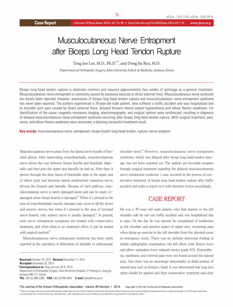
magnetic resonance imaging (MRI) conducted when biceps brachii
rupture was doubted,6) biceps brachii long head tendon was dislo-
cated after rupture and observed in the middle of upper arm: it was
dislocated downward by about 12 cm than the normal location (Fig.
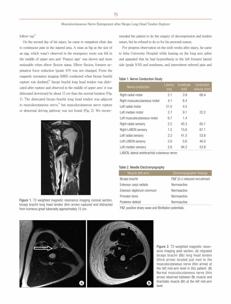
1). The dislocated biceps brachii long head tendon was adjacent
to musculocutaneous nerve,7) but musculocutaneous nerve rupture
or abnormal driving pathway was not found (Fig. 2). We recom-
mended the patient to do the surgery of decompression and tendon
suture, but he refused to do so for his personal reason.
For progress observation on the sixth weeks after injury, he came
to Inha University Hospital while leaning on the long arm splint
and appealed that he had hypoesthesia in the left forearm lateral
side (grade 5/10) and numbness, and intermittent referred pain and
Figure 1. T2-weighted magnetic resonance imaging coronal section, biceps brachii long head tendon (thin arrow) ruptured and distracted from humerus great tuberosity approximately 12 cm.
Table 1. Nerve Conduction Study
Nerve conductionLatency
(ms) Amplitude
(mV)Conduction
velocity (m/s)
Right radial motor 2.1 3.9 68.4
Right musculocutaneous motor 4.1 6.4
Left radial motor 21.0 4.5
Left median motor 2.7 9.1 22.2
Left musculocutaneous motor 6.7 1.4
Right radial sensory 2.2 45.3 65.1
Right LABCN sensory 1.5 15.6 67.1
Left radial sensory 2.2 41.5 53.8
Left LABCN sensory 3.0 5.6 40.0
Left median sensory 2.6 94.3 53.8
LABCN, lateral antebrachial cutaneous nerve.
Figure 2. T2-weighted magnetic reson-ance imaging axial section, (A) migrated biceps brachii (Bb) long head tendon (thick arrow) located just next to the musculocutaneous nerve (thin arrow) at the left mid-arm level in this patient. (B) Normal musculocutaneous nerve (thin arrow) observed between Bb muscle and brachialis muscle (Br) at the left mid-arm level.
Table 2. Needle Electromyography
Muscle (left arm) Electromyographic findings
Biceps brachii P&F (2+) reduced recruitment
Extensor carpi radialis Normoactive
Extensor digitorum communi Normoactive
Pronator teres Normoactive
Posterior deltoid Normoactive
P&F, positive sharp wave and fibrillation potentials.
76
Tong Joo Lee and Dong Jin Ryu
worsened upper arm muscle force decreased (motor grade 3/5).
To detect the musculocutaneous nerve injury, electromyography
was performed and then it was observed that there was a compat-
ible opinion on the temporary paralysis of musculocutaneous nerve
(Table 1, 2).
We assumed that upper arm pain, elbow flexion force reduction
was appeared by biceps brachii long head tendon injury at initial
visit. At MRI exam, ruptured biceps brachii long head tendon was
dislocated downward and positioned just near to musculocutane-
ous nerve. But there was no definite associated symptom. Six weeks
after injury, hypoesthesia was appeared at lateral side of forearm
which was not found at initial exam. We thought that conglutination
between musculocutaneous nerve and ruptured biceps brachii long
head tendon was occurred in healing process. In succession, muscu-
locutaneous entrapment symptom was appeared at last follow-up.
Accordingly, on the 45th day after injury, surgical decompression
was conducted.
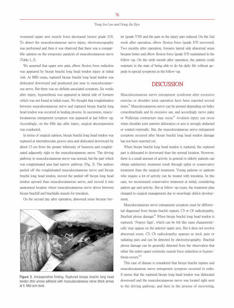
In terms of surgical opinion, biceps brachii long head tendon was
ruptured at intertubercular groove area and dislocated downward by
about 13 cm from the greater tuberosity of humerus and congluti-
nated adjacently right to the musculocutaneous nerve. The driving
pathway to musculocutaneous nerve was normal, but the part which
was conglutinated area had narrow pathway (Fig. 3). The authors
peeled off the conglutinated musculocutaneous nerve and biceps
brachii long head tendon, moved the peeled-off biceps long head
tendon upward than musculocutaneous nerve, and moved it into
anatomical location where musculocutaneous nerve drives between
biceps brachill and brachialis muscle for tenodesis.
On the second day after operation, abnormal sense became bet-
ter (grade 7/10) and the pain in the injury part reduced. On the 2nd
week after operation, elbow flexion force (grade 5/5) recovered.
Two months after operation, forearm lateral side abnormal sense
became better and elbow flexion force (grade 5/5) maintained in the
follow-up. On the sixth month after operation, the patient could
maintain in the state of being able to do his daily life without ap-
peals to special symptoms in the follow-up.
DISCUSSION
Musculocutaneous nerve entrapment syndrome after excessive
exercise or shoulder-joint operation have been reported several
times.5) Musculocutaneous nerve can be pressed depending on bulky
coracobrachialis and its excessive use, and accordingly nerve palsy
or Wallerian contracture may occur.8) Avulsion injury can occur
when shoulder joint anterior dislocation or arm is strongly abducted
or rotated externally. But, the musculocutaneous nerve entrapment
symptom occurred after biceps brachii long head tendon damage
has not been reported yet.
When biceps brachii long head tendon is ruptured, the ruptured
part is dislocated to downward than the normal location. However,
there is a small amount of activity in general or elderly patients can
obtain satisfactory treatment result through splint or conservative
treatment than the surgical treatment. Young patients or patients
who require a lot of activity can be treated with tenodesis. In this
case, we recommend conservative treatment at initial, considering
patient age and activity. But at follow-up exam, the treatment plan
changed to surgical management due to neurologic deficit develop-
ment.
Musculocutaneous nerve entrapment symptom must be differen-
tial diagnosed from biceps brachii rupture, C5 or C6 radiculopathy,
Brachial plexus damage.8) When biceps brachii long head tendon is
ruptured, ‘Popeye Sign’, which can be felt like mass characteristi-
cally may appear on the anterior upper arm. But it does not involve
abnormal sense. C5, C6 radiculopathy appears as neck pain or
radiating pain and can be detected by electromyography. Brachial
plexus damage can be generally detected from the observation that
either the entire upper extremity muscle force reduction or hypoes-
thesia occurs.6,8)
This case of disease is considered that biceps brachii rupture and
musculocutaneous nerve entrapment symptom occurred in order.
It seems that the ruptured biceps long head tendon was dislocated
downward and the musculocutaneous nerve was located right next
to the driving pathway, and then in the process of recovering,
Figure 3. Intraoperative finding. Ruptured biceps brachii long head tendon (thin arrow) adhered with musculocutaneous nerve (thick arrow) at lt. Mid-arm level.
77
Musculocutaneous Nerve Entrapment after Biceps Long Head Tendon Rupture
conglutination between the two tissues occurred. If musculocutane-
ous nerve is damaged in coracobrachialis area, biceps brachii and
brachialis force reduction and anterior/lateral forearm hypoesthesia
may occur, but in this case of disease, it seems that although there
was no damage in the musculocutaneous nerve itself, it was con-
glutinated with surrounding tissues in the process of recovering and
subsequently captured neuropathy occurred. If there is initial traction
injury of musculocutaneous nerve, the patient might be hypoesthe-
sia at initial physical exam. It can be interpreted that the upper arm
muscle force reduction occurred when the initial biceps long head
tendon ruptured and after the occurrence of conglutination, delayed
musculocutaneous nerve entrapment symptom occurred with mus-
cle force reduction and forearm hypoesthesia.
Therefore, if the pain gets better in the patient with biceps bra-
chii rupture, we recommend that early range of motion exercise is
necessary as soon as possible with arm sling protection. We should
avoid long-term splint or immobilization to prevent complication
like above case. In addition, although splint or conservative treat-
ment was performed initially, we should do careful follow-up eval-
uation of patient symptom. If symptom changes like biceps muscle
force reduction and forearm hypoesthesia are seen in a follow-up,
we can consider more evaluation such as MRI, electromyography.
As a result of exam, active surgical treatment would be required.
REFERENCES
1. Yilmaz C, Eskandari MM, Colak M. Traumatic musculocuta-neous neuropathy: a case report. Arch Orthop Trauma Surg. 2005;125:414-6.
2. Dailiana ZH, Roulot E, Le Viet D. Surgical treatment of com-pression of the lateral antebrachial cutaneous nerve. J Bone Joint Surg Br. 2000;82:420-3.
3. Felsenthal G, Mondell DL, Reischer MA, Mack RH. Fore-arm pain secondary to compression syndrome of the lateral cutaneous nerve of the forearm. Arch Phys Med Rehabil. 1984;65:139-41.
4. Ma H, Van Heest A, Glisson C, Patel S. Musculocutaneous nerve entrapment: an unusual complication after biceps teno-desis. Am J Sports Med. 2009;37:2467-9.
5. Krupp RJ, Kevern MA, Gaines MD, Kotara S, Singleton SB. Long head of the biceps tendon pain: differential diagnosis and treatment. J Orthop Sports Phys Ther. 2009;39:55-70.
6. Neal S, Fields KB. Peripheral nerve entrapment and injury in the upper extremity. Am Fam Physician. 2010;81:147-55.
7. Tagliafico AS, Michaud J, Marchetti A, Garello I, Padua L, Martinoli C. US imaging of the musculocutaneous nerve. Skeletal Radiol. 2011;40:609-16.
8. Pećina M, Bojanić I. Musculocutaneous nerve entrapment in the upper arm. Int Orthop. 1993;17:232-4.
78
Tong Joo Lee and Dong Jin Ryu
상완이두근장두건파열후발생한 근피신경포착증후군
이동주 • 류동진
인하대학교 의과대학 정형외과학교실
상완 이두근 장두건 파열은 비교적 흔하게 발생하며, 일반적인 치료로서 약 4주간의 부목고정을 요한다. 근피신경 손상은 일반적으
로 과도한 운동이나 직접적인 외력에 의하여 발생한다. 근피신경 증후군은 드물게 보고되고 있으나, 상완 이두근 파열과 근피신경 포
착 병증이 연관되어 보고된 바는 없다. 저자들이 경험한 환자는 70세 남자로 교통사고로 수상, 직접적인 외력에 의하여 발생한 견관
절 통증 및 지연성으로 발생한 전완부 외측부 감각 저하, 주관절 굴곡력 저하를 주소로 내원하였다. 원인 파악을 위한 자기공명영상,
근전도 검사, 수술 소견 등을 종합하여 상완 이두근 장두건 파열 후 발생한 지연성 근피신경 포착병증으로 진단하였다. 수술적 치료
로 통증과 감각, 주관절 굴곡력 감소는 회복되어 성공적인 치료 결과를 얻을 수 있었다.
색인단어: 근피신경, 포착 증후군, 상완 이두근 장두건, 파열, 신경 증상
접수일 2013년 10월 29일 수정일 2013년 12월 11일 게재확정일 2013년 12월 20일책임저자 이동주인천시 중구 인항로 27, 인하대학교병원 정형외과 TEL 032-890-2380, FAX 032-890-3047, E-mail [email protected]
Case Report J Korean Orthop Assoc 2014; 49: 74-78 • http://dx.doi.org/10.4055/jkoa.2014.49.1.74 www.jkoa.org
“This is an Open Access article distributed under the terms of the Creative Commons Attribution Non-Commercial License (http://creativecommons.org/licenses/by-nc/3.0/) which permits unrestricted non-commercial use, distribution, and reproduction in any medium, provided the original work is properly cited.”