Page 1
MUSE (Microscopy with UV Surface Excitation)
Amir Ghorbani-Aghbolaghi MD, Samuel Balin MD PhD, Tareq Mohammad MD, Yasmine Lahoubi MD Zachary T Harmany PhD, Austin Todd, Farzad Fereidouni PhD
Richard M Levenson MD, Maxwell A Fung MD
ASDP 54th Annual Meeting, Baltimore, MD
Page 2
Disclosure
Dr. Richard Levenson is co-founder and CEO of MUSE Microscopy Inc. Remaining authors declare no conflicts of interest
Page 4
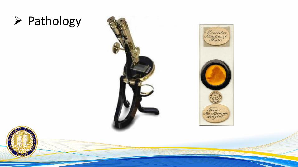
Pathology
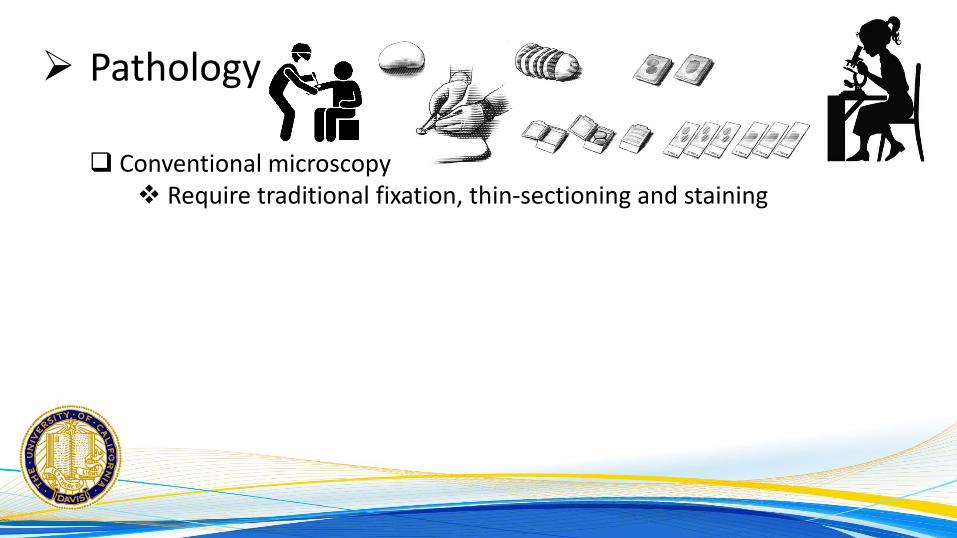
Conventional microscopy Require traditional fixation, thin-sectioning and staining
Page 5
Pathology
Conventional microscopy Require traditional fixation, thin-sectioning and staining
Ex-vivo microscopy (Slide-free) Rapid imaging of biopsy material
Page 6
Pathology
Conventional microscopy Require traditional fixation, thin-sectioning and staining
Ex-vivo microscopy (Slide-free) Rapid imaging of biopsy material
In-Vivo microscopy (Biopsy-free) Evaluation of human tissue microstructure in real time
Page 7
What is MUSE?
A novel Ex-Vivo microscopy Slide-free method developed at UC Davis First in evaluating on human tissue
Microscopy with UV Surface Excitation (MUSE) Using UV-emitting LED with wavelength of 275 to 285 nm Digital camera captures the excitation light
Page 8
How MUSE works?
Ultraviolet (UV) is an electromagnetic radiation Wavelength: 10 nm to 400 nm (Shorter than visible light)
Page 9
How MUSE works?
Ultraviolet (UV) is an electromagnetic radiation Wavelength: 10 nm to 400 nm (Shorter than visible light)
The light penetration depth is depends on the wavelength
Page 10
How MUSE works?
Ultraviolet (UV) is an electromagnetic radiation Wavelength: 10 nm to 400 nm (Shorter than visible light)
The light penetration depth is depends on the wavelength
275 to 285 nm UV light has penetrate depth of 3 microns Approximately the thickness of a conventional tissue section
Page 11
How MUSE works?
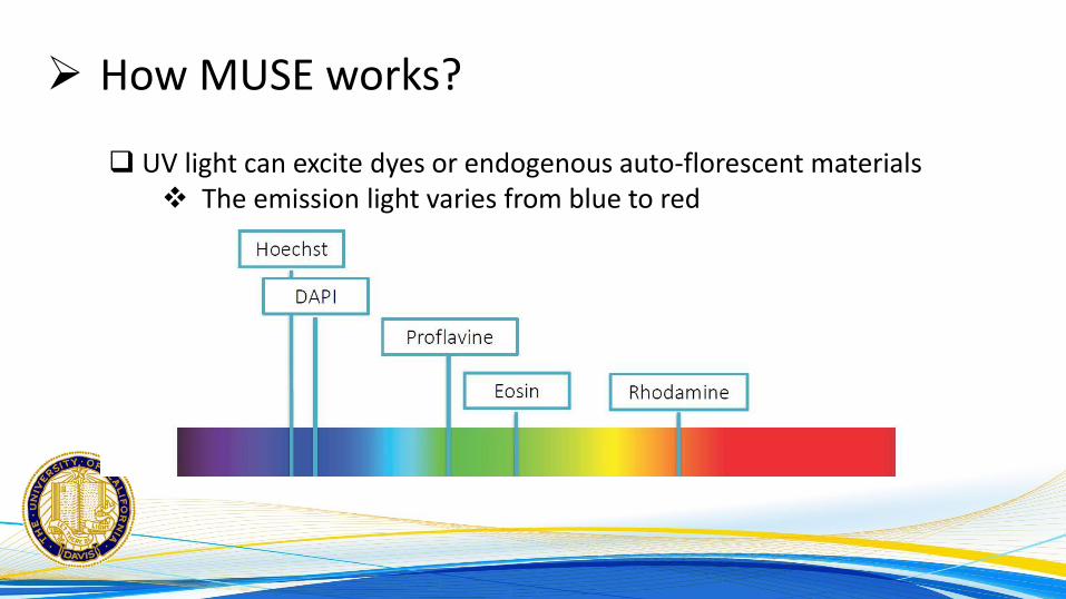
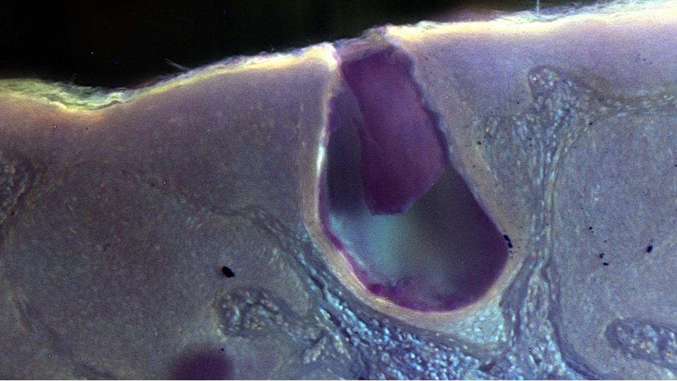
UV light can excite dyes or endogenous auto-florescent materials The emission light varies from blue to red
Page 12
How MUSE works?
UV light can excite dyes or endogenous auto-florescent materials The emission light varies from blue to red
A digital camera can capture the emitted lights
3 microns thickness from the surface of the specimen The images must be similar to H&E but in full color
Page 14
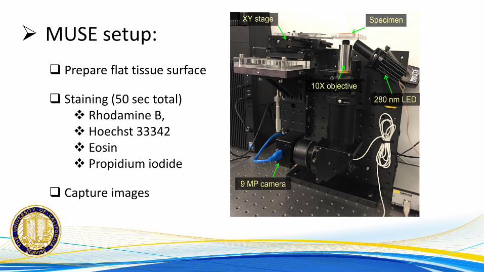
MUSE setup:
Prepare flat tissue surface
Staining (50 sec total) Rhodamine B, Hoechst 33342 Eosin Propidium iodide
Capture images
Page 18
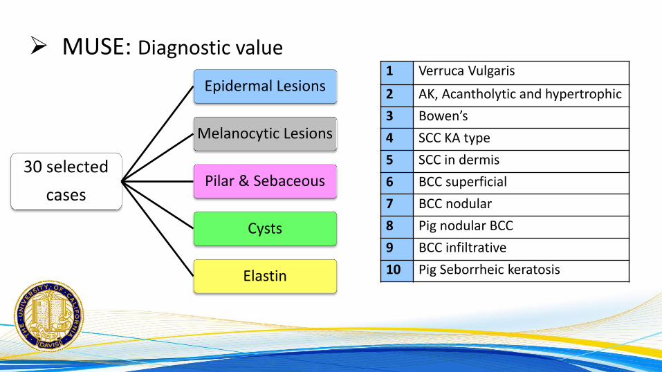
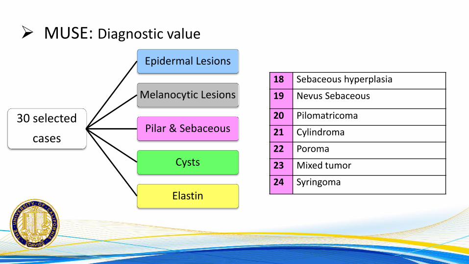
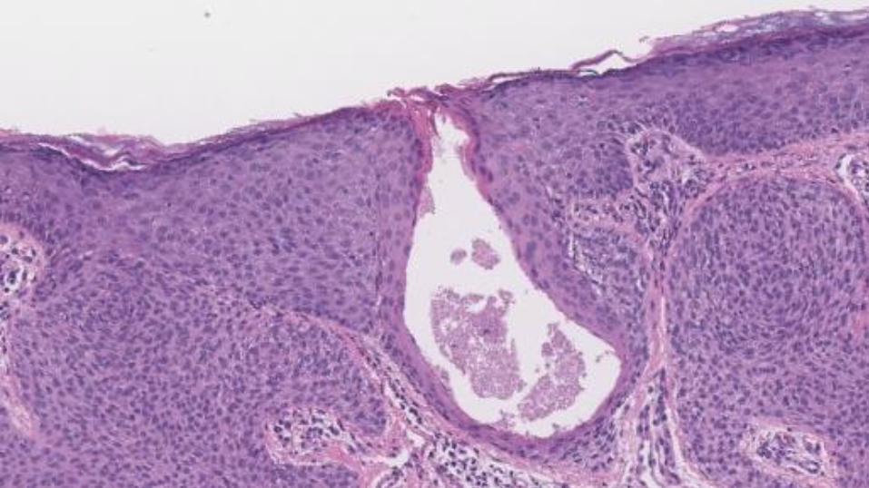
MUSE: Diagnostic value
30 selected
cases
Epidermal Lesions
Melanocytic Lesions
Pilar & Sebaceous
Cysts
Elastin
1 Verruca Vulgaris
2 AK, Acantholytic and hypertrophic
3 Bowen’s
4 SCC KA type
5 SCC in dermis
6 BCC superficial
7 BCC nodular
8 Pig nodular BCC
9 BCC infiltrative
10 Pig Seborrheic keratosis
Page 19
MUSE: Diagnostic value
11 IDN
12 Compound Nevus
13 Lentiginous Nevus
14 Blue Nevus
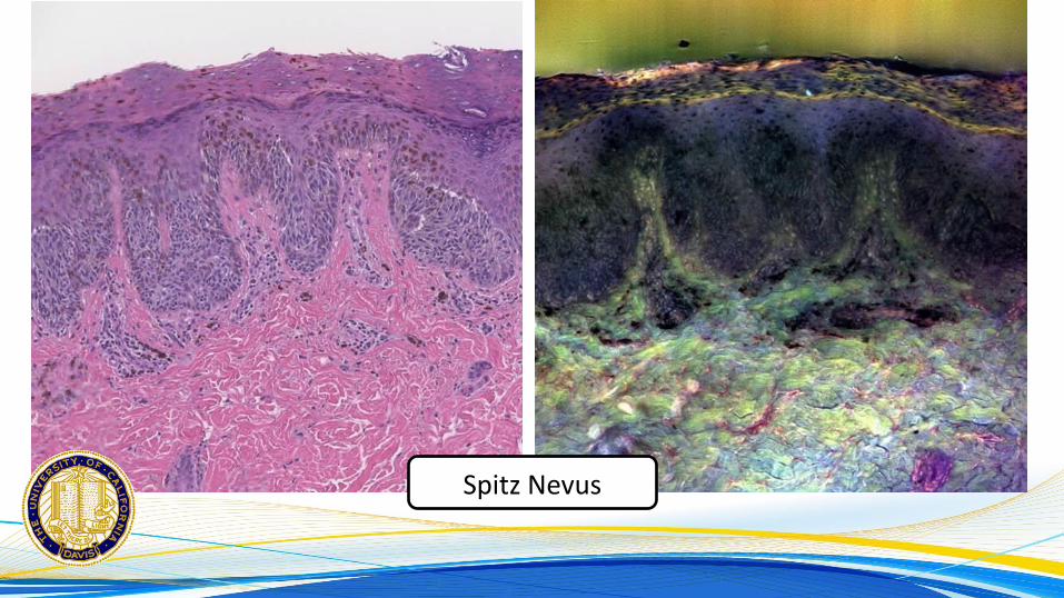
15 Spitz Nevus
16 MIS
17 MM
30 selected
cases
Epidermal Lesions
Melanocytic Lesions
Pilar & Sebaceous
Cysts
Elastin
Page 20
MUSE: Diagnostic value
18 Sebaceous hyperplasia
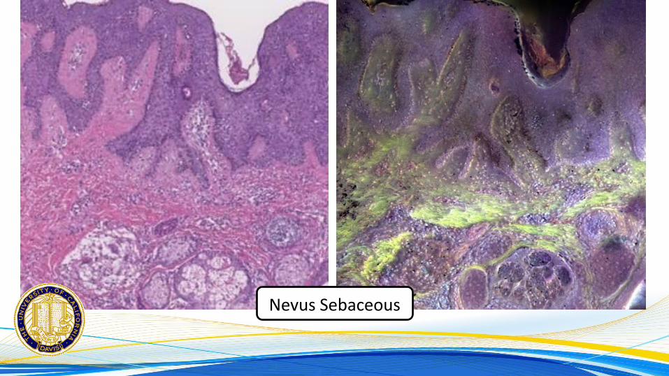
19 Nevus Sebaceous
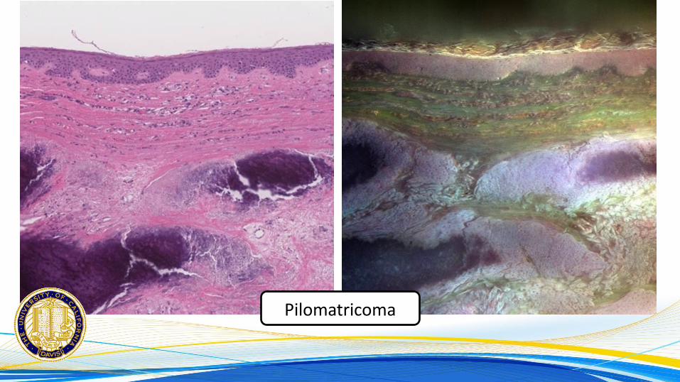
20 Pilomatricoma
21 Cylindroma
22 Poroma
23 Mixed tumor
24 Syringoma
30 selected
cases
Epidermal Lesions
Melanocytic Lesions
Pilar & Sebaceous
Cysts
Elastin
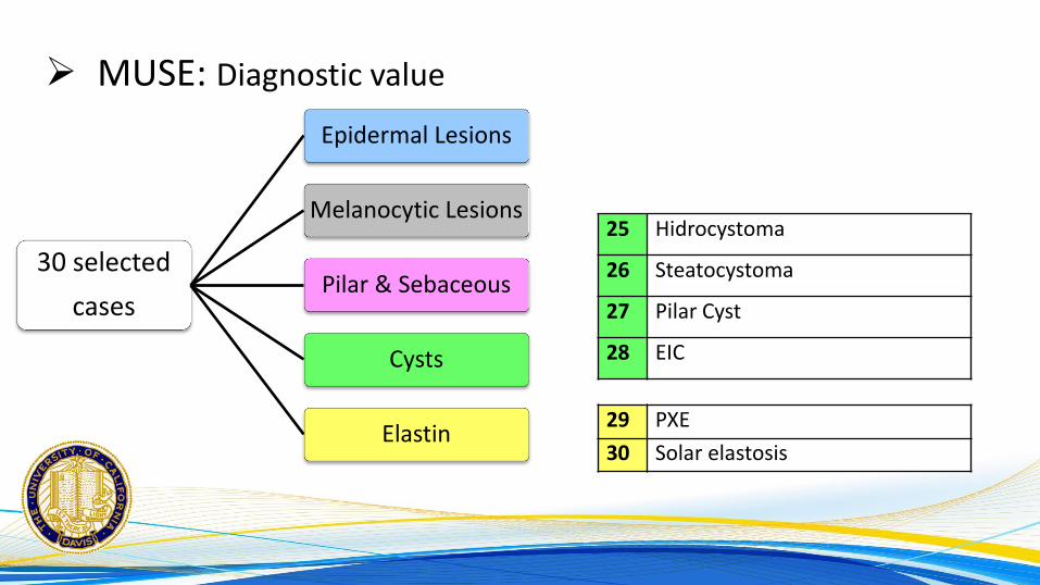
Page 21
MUSE: Diagnostic value
25 Hidrocystoma
26 Steatocystoma
27 Pilar Cyst
28 EIC
29 PXE
30 Solar elastosis
30 selected
cases
Epidermal Lesions
Melanocytic Lesions
Pilar & Sebaceous
Cysts
Elastin
Page 22
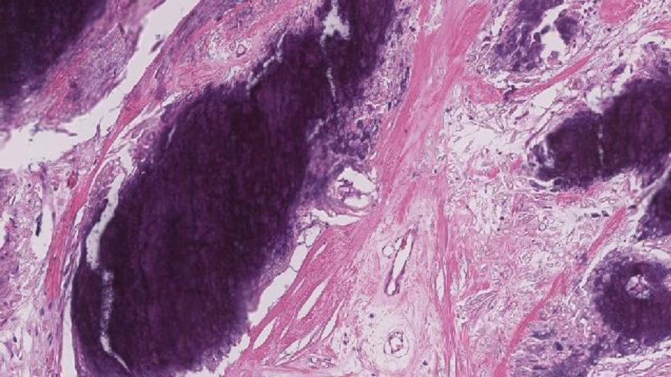
MUSE: Scoring
Diagnostic score: Percentage of correct diagnosis of each MUSE image
Comparison score: Assessed by the concordance between MUSE images and
correlated H&E images generated by whole slide scanner
Page 23
MUSE: Diagnostic score
What is this?
Total Dx score: 70.83%
Cystic lesions: 88% Epidermal lesions: 80% Adnexal lesions: 79% Melanocytic: 46% Elastin lesions: 62%
Page 24
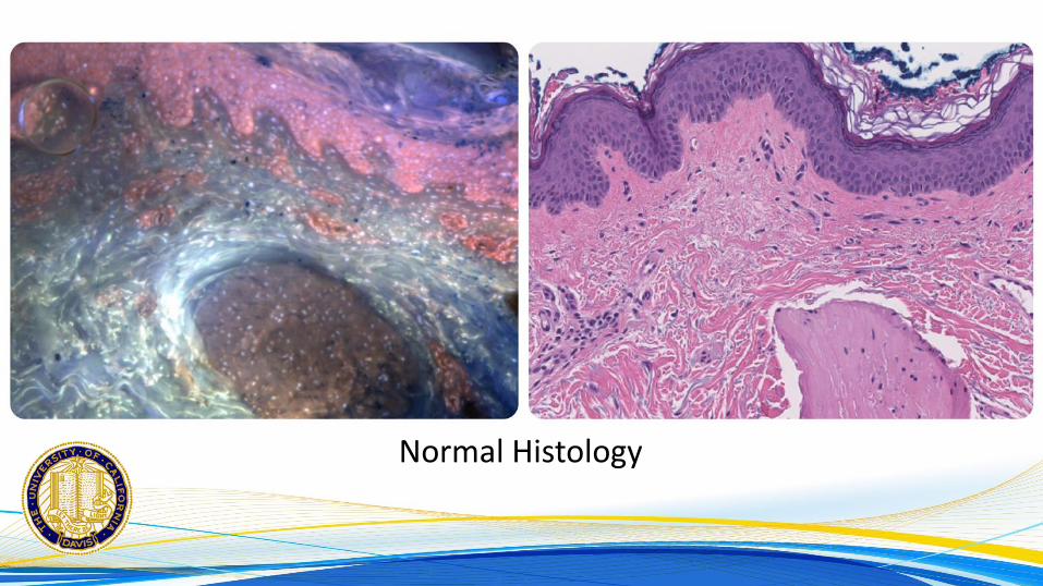
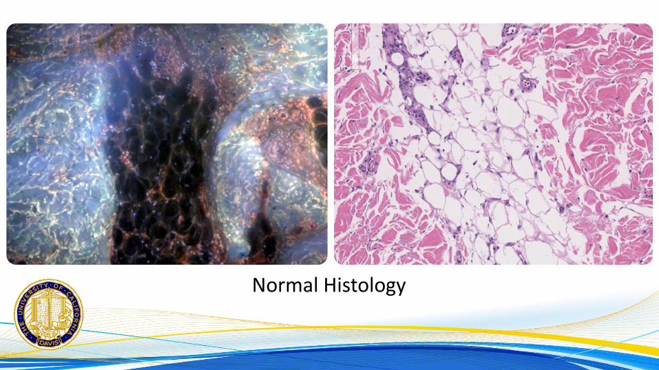
MUSE: Comparison score
Is it better than H&E?
Page 47
MUSE: Comparison score
Is it better than H&E?
Total C. score: 0.8
Cystic lesions: 1.2 Adnexal lesions: 1.0 Elastin lesions: 1.0 Epidermal lesions: 0.7 Melanocytic: 0.6
Page 49
MUSE vs H&E:
Cons: Pre-image:
Unable to changing magnifications Hard to work with very small specimens
Image: Nuclear features (melanocytic, inflammatory) Unfamiliar colors
Post-image: Large data Tissue storage
Page 50
MUSE vs H&E:
Pros: Robust method
Simple physical & chemical principles Fast (2 minutes) Fresh, formalin or alcohol
MUSE images: Multi-color (more informative) 3 Dimensional Similar to H&E (orientation/thickness) High diagnostic value (even for fresh eyes)
Page 51
MUSE vs H&E:
Pros: Ex-vivo microscopy:
Inexpensive (No histology) Preserving tissue (downstream molecular testing) Potential use in intraoperative consultation Can potentially be used as POC
Digital pathology: Provide service to low resource areas
Page 52
MUSE vs H&E:
Pros:
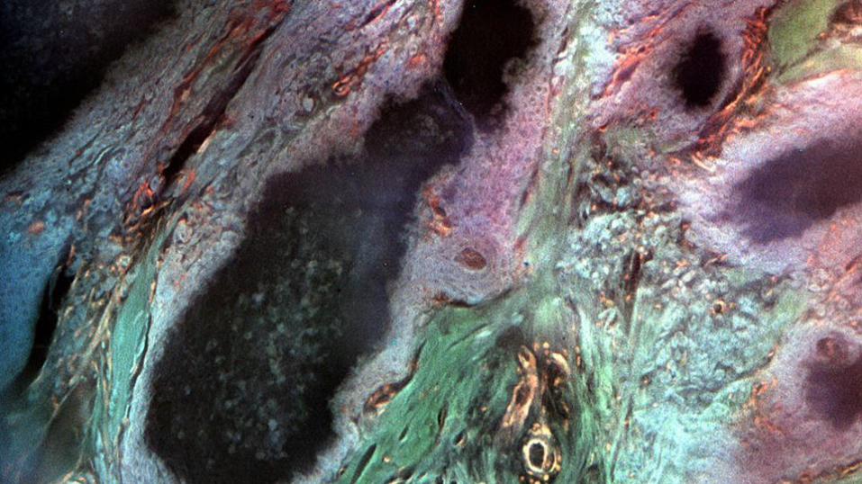
Its BEAUTIFUL
Page 53
MUSE vs H&E:
Pros:
Its BEAUTIFUL
Page 57
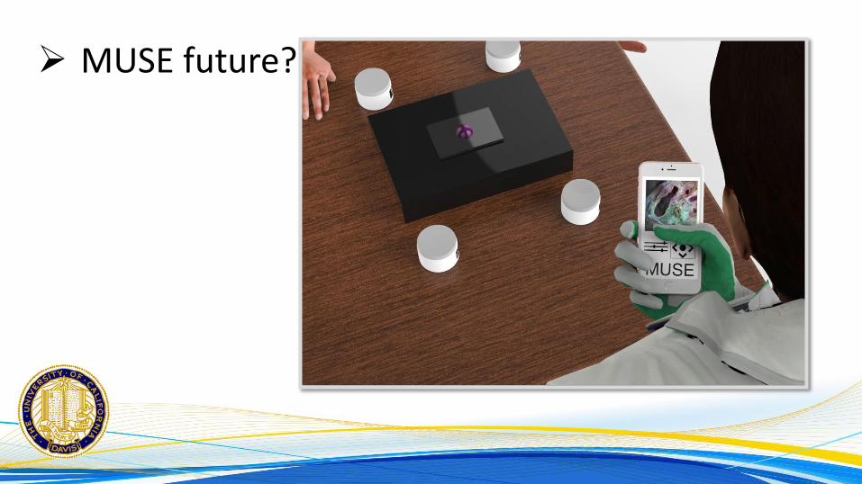
MUSE future?
@BSTPath
@FungMaxwell
Page 58
Our team:
Maxwell A Fung MD Richard Levenson MD Samuel Balin MD PhD Tareq Mohammad MD
Page 59
Our team:
Farzad Fereidouni PhD Yasmine Lahoubi MD Zachary Harmany PhD Austin Todd