+ All Categories
Home > Documents > Myocarditis with normal left ventricular function and troponin of 266

Myocarditis with normal left ventricular function and troponin of 266

Date post: 31-Dec-2016
Category:
Upload: maya
View: 213 times
Download: 0 times
Share this document with a friend
2
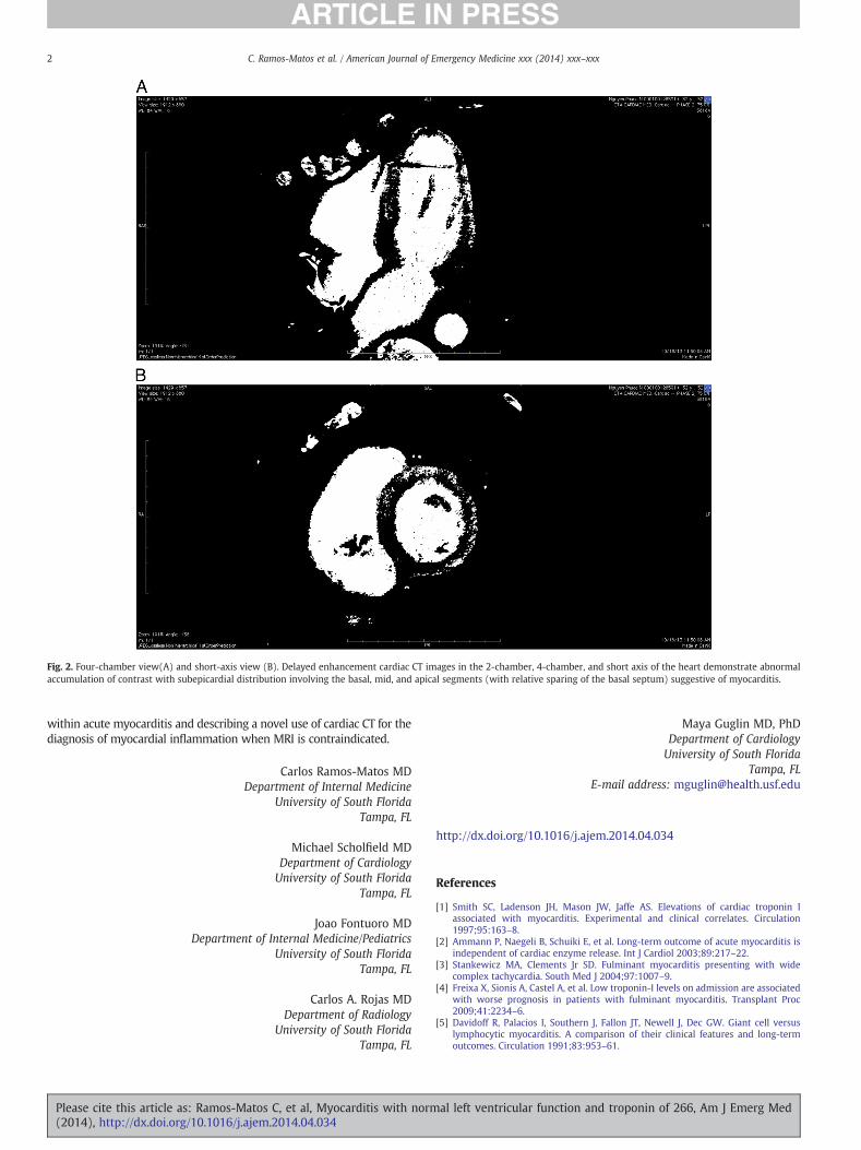
Case Report Myocarditis with normal left ventricular function and troponin of 266 Abstract We are reporting a case, which may represent a new entity within the diagnosis of acute myocarditis. The patient was admitted with new onset atrioventricular block, very high troponin I, peaking at 266 ng/mL, and normal left ventricular function. We also used a novel computer tomography technique with delayed enhancement to diagnose myocarditis, as the patient with a permanent pacemaker could not have magnetic resonance imaging. Since the advent of the troponin I (TnI) assay, markedly elevated levels of this enzyme were largely associated with acute coronary syndromes, with modestly elevated levels seen in myocardial disease and mild abnormalities present with almost every supply/demand mismatch. In myocarditis, TnI is elevated only in 34% of cases [1] . Most patients with acute myocarditis present with new onset heart failure associated with compromised left ventricular ejection fraction (LVEF). We are presenting a case of myocarditis without heart failure or left ventricular (LV) dysfunction but with unusually high TnI. A 52-year-old man with no medical history presented to emergency department with 4 days of night sweats and progressively debilitating fatigue of new onset, culminating in feelings of impending doom. He could not recall any recent infections. On examination, he had heart rate of 38 beats per minute, regular, with distal sounds. There were occasional cannon waves on jugular veins. His blood pressure was 102/82 mm Hg. Lungs were clear; there was no abdominal distension or peripheral edema. He had third-degree heart block on the electrocardiogram. Troponin I was elevated to 25 ng/mL. Emergent coronary angiography showed normal arteries. A temporary pacing wire was placed with immediate improvement of symptoms. A permanent dual chamber pacemaker was implanted on the same day. Bedside echocardiogram revealed normal heart with septal motion abnormalities consistent with right ventricular pacing. The next morning, TnI was elevated to 170 ng/mL. The patient was feeling well but developed paroxysmal atrial utter and transient paresthesias in the toes and ngers. He spiked a fever of 101°F. Cultures were taken; no growth was noted. Troponin I peaked at 266 ng/mL, then trended down (Fig. 1). Lyme titers, human immunodeciency virus testing, and rheumatologic work-up was unremarkable. On the third day, paresthesias resolved, and temperature normalized. Cardiac magnetic resonance imaging (MRI) was contraindicated in this patient due to the presence of a cardiac pacer. Delayed enhancement cardiac computed tomography (CT) was thus performed. Retrospectively gated, delayed images of the heart were obtained after 85 cm 3 intravenous contrast was administered (Isoview 370; Bracco Diagnostics Inc, Princeton, NJ). Delayed enhancement cardiac CT images demonstrated abnormal contrast accu- mulation with a subepicardial distribution predominantly involving the mid and apical LV segments, compatible with myocardial injury/ inammation (Fig. 2 A and B). The distribution of contrast accumulation was suggestive of myocarditis with sarcoid unlikely given the clinical setting. We considered empiric steroids but decided against it. Endomyo- cardial biopsy was performed, with evidence of interstitial edema, perivascular, and interstitial lymphocytic inltration and evolving myocar- dial ber necrosis consistent with lymphocytic myocarditis. The patient remained afebrile and asymptomatic. Repeat transthoracic echocardiogram showed LVEF of 54%. No regional wall motion abnormalities were detected.. The patient was discharged to home. We are presenting a very unusual case of acute myocarditis with normal LVEF and extremely high (peak, 266 ng/mL) TnI. This may represent a distinct clinical entity. Typically, patients with myocarditis present with new onset LV systolic dysfunction and very modestin single digitselevation of TnI [2]. Troponin I reaches double digits and rarely triple digits in fulminant myocarditis with severe hemodynamic compromise and LVEF 5% to 10% [3]. Interestingly, some studies report better outcomes in myocarditis with higher TnI levels [4], although this observation was also made in the setting of severely compromised LVEF. Complete atrioventricular block is not very common in acute lymphocytic myocarditis and occurs in 8.3% of cases [5]. Another highlight of the case is the modi cation of cardiac CT with delayed enhancement, which allowed visualization of typical subepicardial accumulation of contrast indicating inammation and guided the diagnostic biopsy to be sampled from the region with marked inammatory changes. Classically, the standard of care for the diagnosis of myocarditis has been MRI, which was contraindicated in the presence of permanent pacemaker. We demonstrated that retrospectively gated delayed-enhancement cardiac CT with intravenous contrast can be successfully used in this setting. In summary, we are reporting a case of myocarditis with preserved LVEF and massively elevated troponin, which may represent a new entity American Journal of Emergency Medicine xxx (2014) xxxxxx Fig. 1. Troponin I trend from admission to discharge. Contents lists available at ScienceDirect American Journal of Emergency Medicine journal homepage: www.elsevier.com/locate/ajem Please cite this article as: Ramos-Matos C, et al, Myocarditis with normal left ventricular function and troponin of 266, Am J Emerg Med (2014), http://dx.doi.org/10.1016/j.ajem.2014.04.034 0735-6757/© 2014 Elsevier Inc. All rights reserved.
Transcript

American Journal of Emergency Medicine xxx (2014) xxx–xxx

Contents lists available at ScienceDirect

American Journal of Emergency Medicine

j ourna l homepage: www.e lsev ie r .com/ locate /a jem

Case Report

Myocarditis with normal left ventricular function and troponin of 266

Fig. 1. Troponin I trend from admission to discharge.

Abstract

We are reporting a case, which may represent a new entity withinthe diagnosis of acute myocarditis. The patient was admitted withnew onset atrioventricular block, very high troponin I, peaking at 266ng/mL, and normal left ventricular function. We also used a novelcomputer tomography technique with delayed enhancement todiagnose myocarditis, as the patient with a permanent pacemakercould not have magnetic resonance imaging.

Since the advent of the troponin I (TnI) assay,markedly elevated levels ofthis enzyme were largely associated with acute coronary syndromes, withmodestly elevated levels seen in myocardial disease and mild abnormalitiespresent with almost every supply/demand mismatch. In myocarditis, TnI iselevatedonly in34%ofcases [1].Mostpatientswithacutemyocarditispresentwith new onset heart failure associated with compromised left ventricularejection fraction (LVEF). We are presenting a case of myocarditis withoutheart failure or left ventricular (LV) dysfunction but with unusually high TnI.

A 52-year-old man with no medical history presented to emergencydepartment with 4 days of night sweats and progressively debilitatingfatigue of new onset, culminating in feelings of impending doom. He couldnot recall any recent infections. On examination, he had heart rate of 38beatsperminute, regular,withdistal sounds. Therewereoccasional cannonwaves on jugular veins.His bloodpressurewas102/82mmHg. Lungswereclear; there was no abdominal distension or peripheral edema. He hadthird-degreeheart blockon theelectrocardiogram. Troponin Iwas elevatedto 25 ng/mL. Emergent coronary angiography showed normal arteries. Atemporary pacing wire was placed with immediate improvement ofsymptoms. A permanent dual chamber pacemaker was implanted on thesame day. Bedside echocardiogram revealed normal heart with septalmotion abnormalities consistent with right ventricular pacing.

The next morning, TnI was elevated to 170 ng/mL. The patient wasfeeling well but developed paroxysmal atrial flutter and transientparesthesias in the toes and fingers. He spiked a fever of 101°F. Cultureswere taken; no growth was noted. Troponin I peaked at 266 ng/mL, thentrendeddown(Fig. 1). Lyme titers, human immunodeficiency virus testing,and rheumatologic work-up was unremarkable. On the third day,paresthesias resolved, and temperature normalized. Cardiac magneticresonance imaging (MRI) was contraindicated in this patient due to thepresence of a cardiac pacer. Delayed enhancement cardiac computedtomography (CT) was thus performed. Retrospectively gated, delayedimages of the heart were obtained after 85 cm3 intravenous contrast wasadministered (Isoview 370; Bracco Diagnostics Inc, Princeton, NJ). Delayedenhancement cardiac CT images demonstrated abnormal contrast accu-mulation with a subepicardial distribution predominantly involving themid and apical LV segments, compatible with myocardial injury/inflammation (Fig. 2 A and B). The distribution of contrast accumulationwas suggestive of myocarditis with sarcoid unlikely given the clinical

Please cite this article as: Ramos-Matos C, et al, Myocarditis with norm(2014), http://dx.doi.org/10.1016/j.ajem.2014.04.034

0735-6757/© 2014 Elsevier Inc. All rights reserved.

setting. We considered empiric steroids but decided against it. Endomyo-cardial biopsy was performed, with evidence of interstitial edema,perivascular, and interstitial lymphocytic infiltration and evolvingmyocar-dial fiber necrosis consistent with lymphocytic myocarditis. The patientremainedafebrile andasymptomatic.Repeat transthoracic echocardiogramshowedLVEFof54%.Noregionalwallmotionabnormalitiesweredetected..The patient was discharged to home.

We are presenting a very unusual case of acute myocarditis withnormal LVEF and extremely high (peak, 266 ng/mL) TnI. This mayrepresent a distinct clinical entity.

Typically, patientswithmyocarditispresentwithnewonset LVsystolicdysfunction and very modest—in single digits—elevation of TnI [2].Troponin I reaches double digits and rarely triple digits in fulminantmyocarditis with severe hemodynamic compromise and LVEF 5% to 10%[3]. Interestingly, somestudies reportbetteroutcomes inmyocarditiswithhigher TnI levels [4], although this observation was also made in thesetting of severely compromised LVEF.

Complete atrioventricular block is not very common in acutelymphocytic myocarditis and occurs in 8.3% of cases [5].

Another highlight of the case is the modification of cardiac CT withdelayed enhancement, which allowed visualization of typical subepicardialaccumulationof contrast indicating inflammationandguided thediagnosticbiopsy to be sampled from the regionwithmarked inflammatory changes.Classically, the standard of care for the diagnosis of myocarditis has beenMRI, which was contraindicated in the presence of permanent pacemaker.Wedemonstrated that retrospectively gated delayed-enhancement cardiacCT with intravenous contrast can be successfully used in this setting.

In summary, we are reporting a case of myocarditis with preservedLVEF andmassively elevated troponin, whichmay represent a new entity

al left ventricular function and troponin of 266, Am J Emerg Med

Fig. 2. Four-chamber view(A) and short-axis view (B). Delayed enhancement cardiac CT images in the 2-chamber, 4-chamber, and short axis of the heart demonstrate abnormalaccumulation of contrast with subepicardial distribution involving the basal, mid, and apical segments (with relative sparing of the basal septum) suggestive of myocarditis.

2 C. Ramos-Matos et al. / American Journal of Emergency Medicine xxx (2014) xxx–xxx

within acute myocarditis and describing a novel use of cardiac CT for thediagnosis of myocardial inflammation when MRI is contraindicated.

Carlos Ramos-Matos MDDepartment of Internal Medicine

University of South FloridaTampa, FL

Michael Scholfield MDDepartment of CardiologyUniversity of South Florida

Tampa, FL

Joao Fontuoro MDDepartment of Internal Medicine/Pediatrics

University of South FloridaTampa, FL

Carlos A. Rojas MDDepartment of Radiology

University of South FloridaTampa, FL

Please cite this article as: Ramos-Matos C, et al, Myocarditis with norm(2014), http://dx.doi.org/10.1016/j.ajem.2014.04.034

Maya Guglin MD, PhDDepartment of CardiologyUniversity of South Florida

Tampa, FLE-mail address: [email protected]

http://dx.doi.org/10.1016/j.ajem.2014.04.034

References

[1] Smith SC, Ladenson JH, Mason JW, Jaffe AS. Elevations of cardiac troponin Iassociated with myocarditis. Experimental and clinical correlates. Circulation1997;95:163–8.

[2] Ammann P, Naegeli B, Schuiki E, et al. Long-term outcome of acute myocarditis isindependent of cardiac enzyme release. Int J Cardiol 2003;89:217–22.

[3] Stankewicz MA, Clements Jr SD. Fulminant myocarditis presenting with widecomplex tachycardia. South Med J 2004;97:1007–9.

[4] Freixa X, Sionis A, Castel A, et al. Low troponin-I levels on admission are associatedwith worse prognosis in patients with fulminant myocarditis. Transplant Proc2009;41:2234–6.

[5] Davidoff R, Palacios I, Southern J, Fallon JT, Newell J, Dec GW. Giant cell versuslymphocytic myocarditis. A comparison of their clinical features and long-termoutcomes. Circulation 1991;83:953–61.

al left ventricular function and troponin of 266, Am J Emerg Med


Recommended