Provided by the author(s) and NUI Galway in accordance with publisher policies. Please cite the published version when available. Downloaded 2018-02-01T15:05:13Z Some rights reserved. For more information, please see the item record link above. Title N- and O- linked Glycosylation, Developing Mass Spectrometric Strategies for the Characterisation of Glyco- epitopes Author(s) Kenny, Diarmuid T Publication Date 2012-12-10 Item record http://hdl.handle.net/10379/3281
Transcript
Provided by the author(s) and NUI Galway in accordance with publisher policies. Please cite the published
version when available.
Downloaded 2018-02-01T15:05:13Z
Some rights reserved. For more information, please see the item record link above.
TitleN- and O- linked Glycosylation, Developing MassSpectrometric Strategies for the Characterisation of Glyco-epitopes
37. Karlsson, H., et al., High-throughput and high-sensitivity nano-LC/MS and MS/MS for O-glycan
profiling. Methods in Molecular Biology, 2009. 534: p. 117-131.
38. Thomsson, K.A., et al., Enhanced detection of sialylated and sulfated glycans with negative ion mode
nanoliquid chromatography/mass spectrometry at high pH. Analytical Chemistry, 2010. 82(4): p. 1470-
1477.
39. Thomsson, K.A., N.G. Karlsson, and G.C. Hansson, Liquid chromatography-electrospray mass
spectrometry as a tool for the analysis of sulfated oligosaccharides from mucin glycoproteins. Journal
of Chromatography A, 1999. 854(1-2): p. 131-139.
40. Prakobphol, A., et al., Human Low-Molecular-Weight Salivary Mucin Expresses the Sialyl Lewis x
Determinant and Has L-Selectin Ligand Activity. Biochemistry, 1998. 37(14): p. 4916-4927.
41. Xia, B., et al., Altered O-glycosylation and sulfation of airway mucins associated with cystic fibrosis.
Glycobiology, 2005. 15(8): p. 747-775.
42. Kawahira, K., et al., Solid-phase synthesis of O-sulfated glycopeptide by the benzyl-protected glycan
strategy. Tetrahedron, 2009. 65(39): p. 8143-8153.
43. Pabst, M. and F. Altmann, Influence of Electrosorption, Solvent, Temperature, and Ion Polarity on the
Performance of LC-ESI-MS Using Graphitic Carbon for Acidic Oligosaccharides. Analytical
Chemistry, 2008. 80(19): p. 7534-7542.
44. Kawasaki, N., et al., Structural analysis of sulfated N-linked oligosaccharides in erythropoietin.
Glycobiology, 2001. 11(12): p. 1043-1049.
45. Yagi, H., et al., Development of structural analysis of sulfated N-glycans by multidimensional high
performance liquid chromatography mapping methods. Glycobiology, 2005. 15(10): p. 1051-1060.
46. von Witzendorff, D., et al., Characterization of the acidic N-linked glycans of the zona pellucida of
prepuberal pigs by a mass spectrometric approach. Carbohydrate Research, 2009. 344(12): p. 1541-
1549.
47. Murakami, T., et al., Structure determination of a sulfated N-glycans, candidate for a precursor of the
selectin ligand in bovine lung. Glycoconjugate Journal, 2007. 24(4-5): p. 195-206.
Kenny et al Published: Current Proteomics, 2011, 8, 278-296
22
48. Lei, M., Y. Mechref, and M.V. Novotny, Structural Analysis of Sulfated Glycans by Sequential Double-
Permethylation Using Methyl Iodide and Deuteromethyl Iodide. Journal of the American Society for
Mass Spectrometry, 2009. 20(9): p. 1660-1671.
49. Taguchi, T., et al., Occurrence and structural analysis of highly sulfated multiantennary N-linked
glycan chains derived from a fertilization-associated carbohydrate-rich glycoprotein in unfertilized
eggs of Tribolodon hakonensis. European Journal of Biochemistry, 1996. 238(2): p. 357-367.
50. Damen, C.W.N., et al., Electrospray Ionization Quadrupole Ion-Mobility Time-of-Flight Mass
Spectrometry as a Tool to Distinguish the Lot-to-Lot Heterogeneity in N-Glycosylation Profile of the
Therapeutic Monoclonal Antibody Trastuzumab. Journal of the American Society for Mass
Spectrometry, 2009. 20(11): p. 2021-2033.
51. Zhang, Y., et al., Distinguishing Phosphorylation and Sulfation in Carbohydrates and Glycoproteins
Using Ion-Pairing and Mass Spectrometry. Journal of the American Society for Mass Spectrometry,
2006. 17(9): p. 1282-1288.
52. Irungu, J., et al., Method for characterizing sulfated glycoproteins in a glycosylation site-specific
fashion, using ion pairing and tandem mass spectrometry. Analytical Chemistry, 2006. 78(4): p. 1181-
1190.
53. Jiang, H., J. Irungu, and H. Desaire, Enhanced detection of sulfated glycosylation sites in glycoproteins.
Journal of the American Society for Mass Spectrometry, 2005. 16(3): p. 340-348.
54. Toyoda, M., H. Narimatsu, and A. Kameyama, Enrichment Method of Sulfated Glycopeptides by a
Sulfate Emerging and Ion Exchange Chromatography. Analytical Chemistry, 2009. 81(15): p. 6140-
6147.
55. Wakabayashi, H., et al., Novel proteoglycan linkage tetrasaccharides of human urinary soluble
thrombomodulin, SO4-3GlcA beta 1-3Gal beta 1-3(+/- Sia alpha 2-6)Gal beta 1-4Xyl. Journal of
Biological Chemistry, 1999. 274(9): p. 5436-5442.
56. Minamisawa, T. and J. Hirabayashi, Fragmentations of isomeric sulfated monosaccharides using
electrospray ion trap mass spectrometry. Rapid Communications in Mass Spectrometry, 2005. 19(13):
p. 1788-1796.
57. Leach Iii, F.E., et al., Evaluation of the experimental parameters which control electron detachment
dissociation, and their effect on the fragmentation efficiency of glycosaminoglycan carbohydrates.
International Journal of Mass Spectrometry, 2008. 276(2-3): p. 110-115.
58. Estrella, R.P., et al., Graphitized carbon LC-MS characterization of the chondroitin sulfate
oligosaccharides of aggrecan. Anal Chem, 2007. 79(10): p. 3597-606.
59. Staples, G.O., et al., A chip-based amide-HILIC LC/MS platform for glycosaminoglycan glycomics
profiling. Proteomics, 2009. 9(3): p. 686-695.
60. Saad, O.M., et al., Compositional profiling of heparin/heparan sulfate using mass spectrometry: assay
for specificity of a novel extracellular human endosulfatase. Glycobiology, 2005. 15(8): p. 818-826.
61. Wolff, J.J., et al., Electron capture dissociation, electron detachment dissociation and infrared
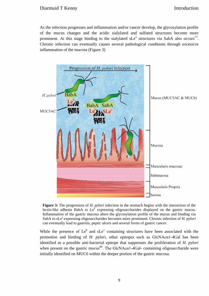
multiphoton dissociation of sucrose octasulfate. European Journal of Mass Spectrometry, 2009. 15(2):
p. 275-281.
62. Wolff, J.J., et al., Electron Detachment Dissociation of Dermatan Sulfate Oligosaccharides. Journal of
the American Society for Mass Spectrometry, 2008. 19(2): p. 294-304.
63. Zaia, J., et al., The Role of Mobile Protons in Negative Ion CID of Oligosaccharides. Journal of the
American Society for Mass Spectrometry, 2007. 18(5): p. 952-960.
64. Imami, K., Y. Ishihama, and S. Terabe, On-line selective enrichment and ion-pair reaction for
structural determination of sulfated glycopeptides by capillary electrophoresis-mass spectrometry.
Journal of Chromatography A, 2008. 1194(2): p. 237-242.
65. Zhang, Y., et al., A novel mass spectrometric method to distinguish isobaric monosaccharides that are
phosphorylated or sulfated using ion-pairing reagents. Journal of the American Society for Mass
Spectrometry, 2005. 16(11): p. 1827-1839.
66. Ohara, K., et al., Matrix-Assisted Laser Desorption/Ionization Mass Spectrometric Analysis of
Polysulfated-Derived Oligosaccharides Using Pyrenemethylguanidine. Journal of the American Society
for Mass Spectrometry, 2009. 20(1): p. 131-137.
67. Fukuyama, Y., et al., Ionic Liquid Matrixes Optimized for MALDI-MS of Sulfated/Sialylated/Neutral
Oligosaccharides and Glycopeptides. Analytical Chemistry, 2008. 80(6): p. 2171-2179.
68. Tissot, B., et al., Towards GAG glycomics: Analysis of highly sulfated heparins by MALDI-TOF mass
spectrometry. Glycobiology, 2007. 17(9): p. 972-982.
69. Przybylski, C., et al., HABA-based ionic liquid matrices for UV-MALDI-MS analysis of heparin and
heparan sulfate oligosaccharides. Glycobiology, 2010. 20(2): p. 224-234.
70. Garenaux, E., et al., A single step method for purification of sulfated oligosaccharides. Glycoconjugate
Journal, 2008. 25(9): p. 903-915.
Kenny et al Published: Current Proteomics, 2011, 8, 278-296
23
71. Sangadala, S., U.R. Bhat, and J. Mendicino, Structures of sulfated oligosaccharides in human trachea
mucin glycoproteins. Mol. Cell. Biochem., 1993. 126(1): p. 37-47.
72. Lei, M., M.V. Novotny, and Y. Mechref, Sequential enrichment of sulfated glycans by strong anion-
exchange chromatography prior to mass spectrometric measurements. J. Am. Soc. Mass Spectrom.,
2010. 21(3): p. 348-357.
73. Larsen, M.R., et al., Highly selective enrichment of phosphorylated peptides from peptide mixtures
using titanium dioxide microcolumns. Molecular and Cellular Proteomics, 2005. 4(7): p. 873-886.
74. Karlsson, N.G., H. Karlsson, and G.C. Hansson, Strategy for the investigation of O-linked
oligosaccharides from mucins based on the separation into neutral, sialic acid- and sulfate-containing
species. Glycoconjugate Journal, 1995. 12(1): p. 69-76.
75. Viseux, N., E. de Hoffmann, and B. Domon, Structural assignment of permethylated oligosaccharide
subunits using sequential tandem mass spectrometry. Analytical Chemistry, 1998. 70(23): p. 4951-
4959.
76. Mank, M., B. Stahl, and G. Boehm, 2,5-Dihydroxybenzoic Acid Butylamine and Other Ionic Liquid
Matrixes for Enhanced MALDI-MS Analysis of Biomolecules. Analytical Chemistry, 2004. 76(10): p.
2938-2950.
77. Chen, P., A.G. Baker, and M.V. Novotny, The use of osazones as matrices for the matrix-assisted laser
desorption/ionization mass spectrometry of carbohydrates. Analytical Biochemistry, 1997. 244(1): p.
144-151.
78. Mank, M., B. Stahl, and G.n. Boehm, 2,5-Dihydroxybenzoic Acid Butylamine and Other Ionic Liquid
Matrixes for Enhanced MALDI-MS Analysis of Biomolecules. Analytical Chemistry, 2004. 76(10): p.
2938-2950.
79. Geyer, H., et al., Core structures of polysialylated glycans present in neural cell adhesion molecule
from newborn mouse brain. Eur J Biochem, 2001. 268(24): p. 6587-99.
80. Yu, S.-Y., et al., Enabling techniques and strategic workflow for sulfoglycomics based on mass
spectrometry mapping and sequencing of permethylated sulfated glycans. Glycobiology, 2009. 19(10):
p. 1136-1149.
81. Laremore, T.N., et al., Matrix-Assisted Laser Desorption/Ionization Mass Spectrometric Analysis of
Uncomplexed Highly Sulfated Oligosaccharides Using Ionic Liquid Matrices. Analytical Chemistry,
2006. 78(6): p. 1774-1779.
82. Bourne, E.J., et al., Studies on trifluoroacetic acid. Part I. Trifluoroacetic anhydride as a promotor of
ester formation between hydroxy-compounds and carboxylic acids. J. Chem. Soc., 1949: p. 2976-2979.
83. Stellner, K., H. Saito, and S.I. Hakomori, DETERMINATION OF AMINOSUGAR LINKAGES IN
GLYCOLIPIDS BY METHYLATION - AMINOSUGAR LINKAGES OF CERAMIDE
PENTASACCHARIDES OF RABBIT ERYTHROCYTES AND OF FORSSMAN ANTIGEN. Archives of
Biochemistry and Biophysics, 1973. 155(2): p. 464-472.
84. Hakomori, S., A rapid permethylation of glycolipid, and polysaccharide catalyzed by methylsulfinyl
carbanion in dimethyl sulfoxide. Journal of Biochemistry, 1964. 55: p. 205-208.
85. Ciucanu, I. and F. Kerek, A simple and rapid method for the permethylation of carbohydrates.
Carbohydrate Research, 1984. 131: p. 209–217.
86. Wuhrer, M., A.R.d. Boer, and A.M. Deelder, Structural glycomics using hydrophilic interaction
chromatography (HILIC) with mass spectrometry. Mass Spectrometry Reviews, 2009. 28(2): p. 192-
206.
87. Ruhaak, L.R., A.M. Deelder, and M. Wuhrer, Oligosaccharide analysis by graphitized carbon liquid
chromatography-mass spectrometry. Analytical and Bioanalytical Chemistry, 2009. 394(1): p. 163-74.
88. Thomsson, K.A., H. Karlsson, and G.C. Hansson, Sequencing of sulfated oligosaccharides from mucins
by liquid chromatography and electrospray ionization tandem mass spectrometry. Anal Chem, 2000.
72(19): p. 4543-9.
89. Schulz, B.L., et al., Identification of two highly sialylated human tear-fluid DMBT1 isoforms: the major
high-molecular-mass glycoproteins in human tears. Biochemical Journal, 2002. 366(Pt 2): p. 511-520.
90. Schulz, B.L., N.H. Packer, and N.G. Karlsson, Small-scale analysis of O-linked oligosaccharides from
glycoproteins and mucins separated by gel electrophoresis. Analytical Chemistry, 2002. 74(23): p.
6088-6097.
91. Staples, G.O., et al., Improved hydrophilic interaction chromatography LC/MS of heparinoids using a
chip with postcolumn makeup flow. Analytical Chemistry, 2010. 82(2): p. 516-522.
92. Williams, T.I., et al., Investigations with O-linked protein glycosylations by matrix-assisted laser
desorption/ionization Fourier transform ion cyclotron resonance mass spectrometry. Journal of Mass
Spectrometry, 2008. 43(9): p. 1215-1223.
93. Adams, J., Charge-remote fragmentations: analytical applications and fundamental studies. Mass
Spectrom. Rev., 1990. 9: p. 141-186.
Kenny et al Published: Current Proteomics, 2011, 8, 278-296
24
94. von Witzendorff, D., et al., Characterization of the acidic N-linked glycans of the zona pellucida of
prepuberal pigs by a mass spectrometric approach. Carbohydr Res, 2009. 344(12): p. 1541-9.
95. Kuster, B., et al., Sequencing of N-linked oligosaccharides directly from protein gels: in-gel
deglycosylation followed by matrix-assisted laser desorption/ionization mass spectrometry and normal-
phase high-performance liquid chromatography. Analytical Biochemistry, 1997. 250(1): p. 82-101.
96. Harvey, D.J., et al., "Internal residue loss": rearrangements occurring during the fragmentation of
carbohydrates derivatized at the reducing terminus. Analytical Chemistry, 2002. 74(4): p. 734-740.
97. Zhang, J., et al., Infrared multiphoton dissociation of O-linked mucin-type oligosaccharides. Anal.
Chem., 2005. 77(1): p. 208-214.
Sulfate migration in oligosaccharides induced by negative ionmode ion trap collision-induced dissociation
Diarmuid T. Kenny1,2, Samah M. A. Issa1,2 and Niclas G. Karlsson2*1School of Chemistry, National University of Ireland, Galway, Ireland2Medical Biochemistry, University of Gothenburg, 405 30 Gothenburg, Sweden
Migration of sulfate groups between hydroxyl groups was identified after collision-induced dissociation (CID) ofsulfated oligosaccharides in an ion trap mass spectrometer in negative ion mode. Analysis of various sulfated oligo-saccharides showed that this was a common phenomenon and was particularly prominent in sulfated oligosacchar-ides also containing sialic acid. It was also shown that the level of migration was increased when the sulfate waspositioned on the flexible areas of the oligosaccharides not involved in the pyranose ring, such as the extra-cyclicC-6 carbon of hexoses or N-acetylhexosamines, or on reduced oligosaccharide. This suggested that migration isdependent on the spatial availability of the sulfate in the ion trap during collision. It is proposed that the migrationis initiated when the negatively charged -SO3
Migration of residues in biopolymers during collision-induced dissociation (CID) fragmentation is one of the mainproblems in structural assignment in mass spectrometry(MS). It has recently been highlighted as a problem for post-translational modification (PTM) identification in phospho-proteomics, where up to 75% of phosphates migrated to anearby phosphate acceptor, causing ambiguity in site assign-ment.[1] Outside the cell, the glycosylation is the dominatingPTM, where over half of all secreted and extracellular pro-teins are glycoproteins.[2] In glycomics, the phenomenon ofmigration is constantly a nuisance for the assignment of fuco-sylation in positive ion mode. The predominate occurrencesof fucose migration involve the transfer of fucose from onemonosaccharide to an adjacent monosaccharide.[3] However,Harvey et al. also reported long-distance migration on carbo-hydrates derivatized at the reducing terminus[4] and Ernstet val. reported fucose migration towards the non-reducingend positioned sialic acid in sialyl Lewis x structures.[5]
In negative ion mode CID, the phenomenon of fucosemigration has not been reported. With the informative frag-mentation generated in negative ion mode, it appears to bean alternative for analyzing glycosylation without the needfor derivatization. Especially for the analysis of sulfated oli-gosaccharides, this is appealing since the protocol for
advanced derivatization, such as permethylation, is not wellestablished and the negative charge of the sulfate group pro-motes the ionization of the oligosaccharides in negative ionmode. Here, we report that the migration problem in struc-tural assignment by CID in glycomics is not exclusive forpositive ion mode, but can also be prominent in negativeion mode, with migration of sulfate residues occurring onnative, reduced and reducing end derivatized oligosacchar-ides. Sulfation, a secondary modification of oligosaccharides,is an important biological modification since it alters the wayglycoproteins interact with other biomolecules. Sulfated gly-coprotein oligosaccharides are involved in many biochemicalpathways, such as the clearance of thyroid stimulating hor-mone and luteinizing hormone to lymphocyte homing.[6–9]
However, sulfation is perhaps best known in the study ofthe glycosaminoglycans, i.e. heparin/heparan sulfate,[10–12]
chondroitin/dermatan sulfate,[13–16] and keratan sulfate.[17–19] Sulfation is also commonly found in the O-linked oligosac-charides of the mucins lining the respiratory and gastrointest-inal tracts[20] and both sulfated N- and O-linkedoligosaccharides are found in the lymphoid, central and per-ipheral nerve tissue.[21]
A mass spectrometer is a rapid and sensitive instrumentfor the study of sulfated oligosaccharides and has emergedas the analytical tool of choice over traditional methodssuch as radioactive isotope labeling[8,22] and nuclear mag-netic resonance.[23,24] However, MS analysis of sulfated oligo-saccharides is not without its challenges. In addition to themigration of the sulfate group presented here, the sulfate isnotorious for its labile nature in positive and negative
* Correspondence to: N. G. Karlsson, University of Gothen-burg, Medical Biochemistry, Box 440, 405 30 Gothenburg,Sweden.E-mail: [email protected]
Received: 10 May 2011 Revised: 16 June 2011 Accepted: 18 June 2011 Published online in Wiley Online Library
Rapid Commun. Mass Spectrom. 2011, 25, 2611–2618(wileyonlinelibrary.com) DOI: 10.1002/rcm.5157
2611
ion mode MS analysis. Dissociation of the sulfate groupcan occur when using matrix-assisted laser desorption/ionization (MALDI) with traditional matrices,[25] whereaselectrospray ionization (ESI) appears to be a softer ionizationmethod and can avoid this problem. Sulfated oligosacchar-ides are typically in low abundance in complex oligosac-charide samples and liquid chromatography (LC) capableof separating oligosaccharides will help to target andsequence sulfated oligosaccharides by CID in the presenceof high abundant neutral and/or sialylated oligosacchar-ides. ESI-MS in conjunction with LC provides a robustand sensitive method for the analysis of native sulfatedoligosaccharides.[12,15,16,26–32]
Interpretation of tandem mass (MS2) spectra from CIDfragmentation can provide full or partial assignment of theoligosaccharide structure as well as sulfate location. The loca-tion of the sulfate has been suggested to be indicated by frag-ment ions in the low-mass region of the MS2 spectracorresponding to a particular sulfated monosaccharide resi-due and even sulfate position.[31] The presence of fragmentions corresponding to more than one sulfated monosacchar-ide residue would then indicate the presence of isomeric com-pounds in the sample. In this report it is shown that this couldalso be due to experimental artifacts introduced by the CIDfragmentation in ion trap MS, inducing structure-dependentsulfate migration.
EXPERIMENTAL
Materials
All chemicals were purchased from Sigma-Aldrich (St. Louis,MO, USA) unless stated otherwise.
Preparation of oligosaccharides
O-Linked oligosaccharides were released from commercialporcine gastric mucin (Sigma-Aldrich) human salivaryMUC5B,[33] chicken ceca samples prepared as per Tierneyet al.[34] (gift from Prof. Steven Carrington, University CollegeDublin, Ireland) in 0.5 M NaBH4 in 50 mM NaOH. Sampleswere desalted with 0.1 mL of AG50WX8 cation-exchangebeads (Bio-Rad, Hercules, CA, USA) packed in C18 zip tips(Millipore, Billerica, MA). Borate complexes were removed byrepeated addition/evaporation with 1% acetic acid in metha-nol (100 mL for each addition). The released oligosaccharideswere dissolved in water for introduction into the LC/MSsystem. SynthesizedNeuAca2-6(HSO3-6)Galb1-3GlcNAcb1 and(HSO3-6)Galb1-3GlcNAcb1- with a +69Da ethyl azide (Az)tag and synthesized NeuAca2-6Galb1-3(HSO3-6)GlcNAcb1-,NeuAca2-3Galb1-3(HSO3-6)GlcNAcb1- and Galb1-3(HSO3-6)GlcNAcb1- with a +43 Da ethylamine (EA) tag weregenerous gifts from Prof James Paulson, Scripps Institute,CA, USA, and were dissolved in 1 mg/mL of water.HSO3-3Galb1-3(Fuca1-4)GlcNAc, HSO3-3Galb1-4(Fuca1-3)GlcNAc, chondroitin-4-sulfate and chondroitin-6-sulfate di-saccharides (Dextra, Reading, UK) were dissolved in waterto a final concentration of 1 mg/mL. These were also reducedwith 0.5 M NaBH4 and desalted as described above.
LC/MS2 and LC/MSn of oligosaccharides by CID and HCD
Oligosaccharides were analyzed by LC/MS2[30] using a 20 cm250 mm i.d. column containing 5 mm porous graphitized car-bon (PGC) particles (Thermo Scientific, Waltham, MA, USA)prepared in-house.[35] Oligosaccharides were eluted using alinear gradient from 0–40% acetonitrile over 40 min at a flowrate of 10 mL/min. The eluted oligosaccharides were detectedin an ESI-IT mass spectrometer (LTQ, Thermo Electron Corp.,San Jose, CA, USA) in negative ion mode with a spray voltageof 3.5 kV. Air was used as sheath gas and the mass range wasset to m/z 380–2000. Ions specified for sulfated oligosacchar-ide structures were isolated for MSn fragmentation by CIDwith the collision energy set to 30%. Sulfated oligosaccharideswere also analyzed with an LC-LTQ Orbitrap mass spectro-meter (Thermo Electron Corp.). The LC setup was the sameas that described for LC/ESI-ITMS. Ions isolated for HCDfragmentation were acquired with a resolution of 30 000and subjected to collision with the collision energy set to60% with an activation time of 30ms.
RESULTS
Identification of CID induced migration of sulfate
Determining the correct position of the sulfate is an importantpiece of information obtainable from mass spectrometric ana-lysis of sulfated oligosaccharides. The low mass range of theMS2 spectra often provides this information with fragmentions corresponding to sulfated monosaccharides.[31] It wasnoticed that fragmenting sulfated N-acetyllactosamine stan-dards with sulfation on either the 6 position on Gal (B1 ionof m/z 241) or the 6 position on GlcNAc (Y1/B2 ion ofm/z 282) gave, in addition to dominating low molecular massfragment ions of the predicted sulfated monosaccharide, alsolow intense fragment ions corresponding to sulfation of thealternate residue. Even after separation, LC/MS2 fragmentscould be detected showing that this was not due to the pre-sence of isomeric impurities (Fig. 1). This triggered the inves-tigation whether the origin of these fragments was due to amigration process of the sulfate induced by CID and initiatedthe search for examples of this phenomenon in order toexplore the mechanism of this migration, looking at varioussulfated glycoconjugates including reducing oligosaccharidesstructures with a reducing end tag and alditols. It was foundthat in the collection of accumulated CID spectra of sulfatedoligosaccharide alditols in negative ion mode (e.g. in the Uni-Carb-DB database[36]) that the migration was often negligible.This is exemplified in Fig. 2(A) with the fragment spectra ofthe monosulfated hexasaccharide Fuca1-2Galb1-3(HSO3-6GlcNAcb1-6)GalNAcol. In this spectrum, only the B1 frag-ment ions of m/z 282 and the C1 fragment ion of m/z 300corresponding to a sulfated GlcNAc residue were present.In other instances, the situation resembled the sulfatedN-acetyllactosamine standards (Fig. 1), where the sulfate mi-gration was indicated to be low (<10% compared to the inten-sity of intact sulfate monosaccharides in the MS2 spectra).This is exemplified by the fragmentation of the [M – H]– ion ofm/z 610 of a 3-sulfated Lewis x alditol (HSO3-3Galb1-3(Fuca1-4)GlcNAcol) (Fig. 2(B)). The B2 fragment ion ofm/z 241 and the C2 fragment ion of m/z 259 are the fragments
of the non-migrated sulfate on the Gal residue, whereas the pre-sence of low intense fragment ions of m/z 284 and 302 corre-spond to the Z1 and Y1 ions with the addition of 80 Da(sulfate). These fragment ions show that the sulfate hasmigrated to the reducing end GlcNAcol residue. The most strik-ing examples of CID-induced sulfate migration were foundwhen structures containing both sialic acid and sulfate werefragmented. In the CID spectrum (Fig. 2(C)) of the [M – H]–
ion of m/z 755 from the structure NeuAca2-6(HSO3-Galb1-3)GalNAcol, the presence of an intense fragment ion at m/z 370shows that the migration is a major event, where the sulfatemigrates quite readily from the Gal residue to the sialic acid(B1a ion of m/z 290) giving the addition of 80 Da (sulfate). Thismigration is so prominent so it makes it difficult to evendetect fragment ions of the original sulfated Gal residue (B1b
and C1b of m/z 241 and 259, respectively). We subsequently
Figure 1. CID-induced migration of sulfate in sulfated N-acetyllactosamines inLC/MS2. Base peak chromatogram of HSO3-6Galb1-4GlcNAcb1- Az and Galb1-4(HSO3-6)GlcNAcb1-EA separated by LC using a PGC column. Precursor ionwas selected for MS2 fragmentation by CID. The low-mass regions of the CIDspectra are displayed to show fragments from sulfated monosaccharidesGlcNAc (m/z 282) and Gal (m/z 241).
Figure 2. CID-induced sulfate migration in oligosaccharide alditols. The MS2 spectrum shows no migration inthe sulfated core 1 fucosylated structure Fuca1-2Galb1-3(HSO3-6GlcNAcb1-6)GalNAcol (A), low migration inthe sulfated Lewis x structure HSO3-3Galb1-3(Fuca1-4)GlcNAcol (B), and high in the sulfated sialylated core 1oligosaccharide NeuAca2-6(HSO3-Galb1-3)GalNAcol (C). Insert shows the full MS2 spectrum from each struc-ture. For key to symbols, see Fig. 1.
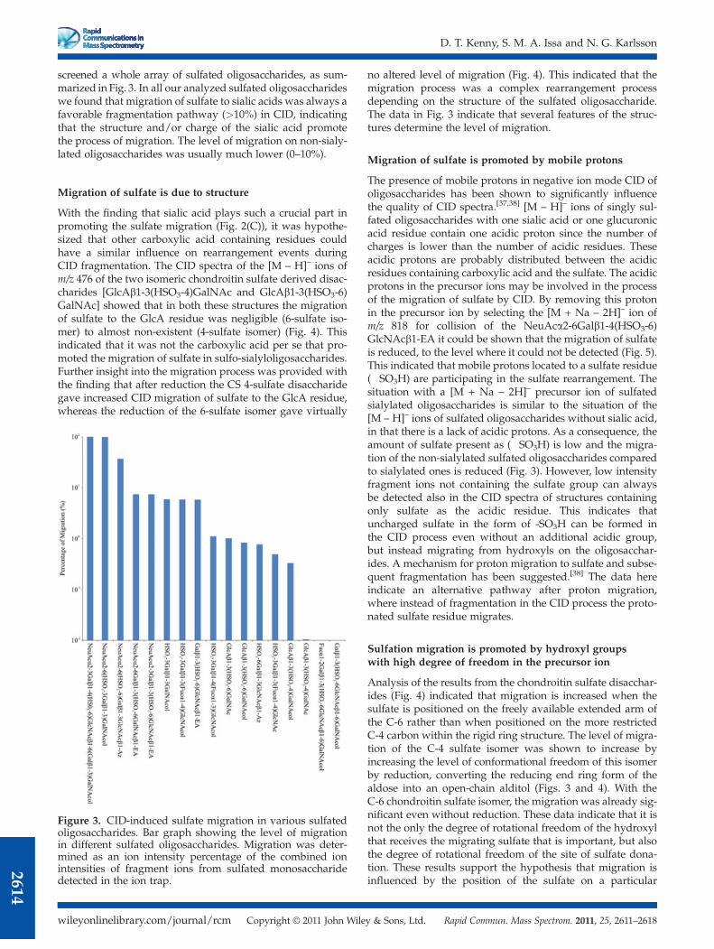
screened a whole array of sulfated oligosaccharides, as sum-marized in Fig. 3. In all our analyzed sulfated oligosaccharideswe found that migration of sulfate to sialic acids was always afavorable fragmentation pathway (>10%) in CID, indicatingthat the structure and/or charge of the sialic acid promotethe process of migration. The level of migration on non-sialy-lated oligosaccharides was usually much lower (0–10%).
Migration of sulfate is due to structure
With the finding that sialic acid plays such a crucial part inpromoting the sulfate migration (Fig. 2(C)), it was hypothe-sized that other carboxylic acid containing residues couldhave a similar influence on rearrangement events duringCID fragmentation. The CID spectra of the [M – H]– ions ofm/z 476 of the two isomeric chondroitin sulfate derived disac-charides [GlcAb1-3(HSO3-4)GalNAc and GlcAb1-3(HSO3-6)GalNAc] showed that in both these structures the migrationof sulfate to the GlcA residue was negligible (6-sulfate iso-mer) to almost non-existent (4-sulfate isomer) (Fig. 4). Thisindicated that it was not the carboxylic acid per se that pro-moted the migration of sulfate in sulfo-sialyloligosaccharides.Further insight into the migration process was provided withthe finding that after reduction the CS 4-sulfate disaccharidegave increased CID migration of sulfate to the GlcA residue,whereas the reduction of the 6-sulfate isomer gave virtually
no altered level of migration (Fig. 4). This indicated that themigration process was a complex rearrangement processdepending on the structure of the sulfated oligosaccharide.The data in Fig. 3 indicate that several features of the struc-tures determine the level of migration.
Migration of sulfate is promoted by mobile protons
The presence of mobile protons in negative ion mode CID ofoligosaccharides has been shown to significantly influencethe quality of CID spectra.[37,38] [M – H]– ions of singly sul-fated oligosaccharides with one sialic acid or one glucuronicacid residue contain one acidic proton since the number ofcharges is lower than the number of acidic residues. Theseacidic protons are probably distributed between the acidicresidues containing carboxylic acid and the sulfate. The acidicprotons in the precursor ions may be involved in the processof the migration of sulfate by CID. By removing this protonin the precursor ion by selecting the [M + Na – 2H]– ion ofm/z 818 for collision of the NeuAca2-6Galb1-4(HSO3-6)GlcNAcb1-EA it could be shown that the migration of sulfateis reduced, to the level where it could not be detected (Fig. 5).This indicated that mobile protons located to a sulfate residue(�SO3H) are participating in the sulfate rearrangement. Thesituation with a [M + Na – 2H]– precursor ion of sulfatedsialylated oligosaccharides is similar to the situation of the[M – H]– ions of sulfated oligosaccharides without sialic acid,in that there is a lack of acidic protons. As a consequence, theamount of sulfate present as (�SO3H) is low and the migra-tion of the non-sialylated sulfated oligosaccharides comparedto sialylated ones is reduced (Fig. 3). However, low intensityfragment ions not containing the sulfate group can alwaysbe detected also in the CID spectra of structures containingonly sulfate as the acidic residue. This indicates thatuncharged sulfate in the form of -SO3H can be formed inthe CID process even without an additional acidic group,but instead migrating from hydroxyls on the oligosacchar-ides. A mechanism for proton migration to sulfate and subse-quent fragmentation has been suggested.[38] The data hereindicate an alternative pathway after proton migration,where instead of fragmentation in the CID process the proto-nated sulfate residue migrates.
Sulfation migration is promoted by hydroxyl groupswith high degree of freedom in the precursor ion
Analysis of the results from the chondroitin sulfate disacchar-ides (Fig. 4) indicated that migration is increased when thesulfate is positioned on the freely available extended arm ofthe C-6 rather than when positioned on the more restrictedC-4 carbon within the rigid ring structure. The level of migra-tion of the C-4 sulfate isomer was shown to increase byincreasing the level of conformational freedom of this isomerby reduction, converting the reducing end ring form of thealdose into an open-chain alditol (Figs. 3 and 4). With theC-6 chondroitin sulfate isomer, the migration was already sig-nificant even without reduction. These data indicate that it isnot the only the degree of rotational freedom of the hydroxylthat receives the migrating sulfate that is important, but alsothe degree of rotational freedom of the site of sulfate dona-tion. These results support the hypothesis that migration isinfluenced by the position of the sulfate on a particular
Figure 3. CID-induced sulfate migration in various sulfatedoligosaccharides. Bar graph showing the level of migrationin different sulfated oligosaccharides. Migration was deter-mined as an ion intensity percentage of the combined ionintensities of fragment ions from sulfated monosaccharidedetected in the ion trap.
residue of an oligosaccharide and that the more sterically'flexible' of the sulfate oligosaccharide molecules are increas-ingly more likely to support migration of sulfate. This is whatcan be identified in Fig. 3, with migration of sulfate to theflexible side chain of sialic acid and to the flexible linear formof a reduced monosaccharide residue on the reducing end ofan oligosaccharide chain.With the indication that the tertiary structure of the precur-
sor ion of the sulfated oligosaccharide is involved in promotingCID-induced sulfate migration, the process of the rearrange-ment was further investigated by identification of the locationof the sulfate after its migration. The MS3 spectrum of theY1/B2 + S fragment ion of m/z 282 (sulfate migrating toGlcNAc) fromthe [M –H]– ion of HSO3-6Galb1-4GlcNAcb1-EA(Fig. 1) showed an intense diagnostic fragment of m/z 139(Fig. 6(A)). This type of 0,4A fragmentation has previouslybeen shown to be a diagnostic fragment for C-6-substitutedHex or HexNAc.[32,39] An intense 0,4A fragment of m/z 139was also detected in the reverse MS3 experiment of the B1 + Sfragment ion of m/z 241 (sulfate migrating to Gal) from the[M – H]– ion of Galb1-4(HSO3-6)GlcNAcb1-Az (Fig. 6(B)).These data further strengthen the argument of CID-inducedmigration of sulfate between less restricted hydroxyls, inthese cases between the C-6 positions of adjacent Gal andGlcNAc residues. These data also give further insight into
the particular feature of increased migration of the struc-ture containing both sialic acid and sulfate. The extendedflexible glycerol side chain of sialic acid would be an excel-lent receiving area for a migrating sulfate. The MS3 spec-trum of the B1 + S fragment ion of m/z 370 from NeuAca2-6(HSO3-Galb1-3)GalNAcol (Fig. 2(C)) showed in addition tothe prominent ions of sialic acid (B1 ion of m/z 290) also lowerintense fragments locating the sulfate to the glycerol sidechain, with fragments cleaving between C-6 and C-7 andbetween C-7 and C-8, corresponding to m/z 139 and 169,respectively (Fig. 6(C)). Whether all hydroxyls of the glycerolchain are the target for migration or only one (e.g. C-9) couldnot be determined.
Selection of MS fragmentation conditions to preventsulfate migration
The addition of a sodium salt to generate Na+ adducts (Fig. 5)as a concept for preventing migration of sulfate in oligosac-charides containing both sulfate and sialic acid is not withoutproblems. In ESI-MS the addition of non-volatile salts is at itsbest only lowering the ionization efficiency and at its worstbeing detrimental for the instrument. Selection of multiplycharged ions as precursor ions is an attractive alternative toremove these acidic protons in sulfo-sialooligosaccharides
Figure 4. CID fragmentation of chondroitin sulfate disaccharide aldoses and alditols. The CIDspectra of the [M – H]– ion monosulfated chondroitin-4-sulfate (A) and chondroitin-6-sulfatedisaccharides (B) showing migration of the sulfate from the GalNAc residue to the GlcA. The spec-tra of the aldose (grey) and alditol (black) derivatives. For key to symbols, see Fig. 1.
Figure 5. Inhibition of sulfate migration in CID in sulfo-sialooligosaccharides.Comparison of the level of CID-induced sulfate migration of the NeuAca2-6Galb1-4(HSO3-6)GlcNAcb1-EA oligosaccharide showing the MS2 spectra ofthe [M – H]– ion (black) and the [M + Na – 2H]– ion (grey) in the low-massregion. For key to symbols, see Fig. 1.
and prevent migration. Also, with two charges available on asulfo-sialylated structure (one from the sialic acid carboxylgroup and one from the sulfate), the doubly charged speciesis easy to generate in the mass spectrometer. The singly anddoubly charged species of NeuAca2-3Galb1-4(HSO3-6GlcNAcb1-6(Gal b1-3)GalNAcol ([M – H]– ion of m/z 1120and [M – 2H]2– ion of m/z 560, respectively) were isolatedfor fragmentation by CID (Fig. 7). The presence of a B1a + Sfragment ion of the m/z 370 fragment ion in the singlycharged precursor indicates migration of the sulfate to theNeuAc residue. Fragmentation of the doubly charged precur-sor did not show any evidence of sulfate migration (Fig. 7(B)),and the spectrum quality was improved compared to the sin-gly charged CID spectrum, showing more sequence ions.For structures only containing one sulfate group as the only
negative residue, the approach of using multicharged negativeprecursors in CID is not possible. Since CID in the ion trap ison a time frame where rearrangement is quite frequentlyobserved, we investigated Higher-energy C-trap dissociation(HCD) in an Orbitrap as a possible alternative to CID for frag-mentation of sulfated oligosaccharides in an effort to overcomethe difficulties associated with sulfate migration. Using anOrbitrap would also provide increased resolution and accuracyto identify larger oligosaccharides and would be an excellent
complement to our current approach, where we only cansequence <3000Da oligosaccharides on a regular basis due tothe mass accuracy of the ion trap. Optimization of the HCD
Figure 6. MS3 spectra to identify the target hydroxyl site forCID-induced migration of sulfate. (A) MS3 spectrum of them/z 282 Y1/B2 + S fragment ion of sulfate migrating fromHSO3-6Gal to GlcNAc (Fig. 1). (B) MS3 spectrum of them/z 241 B1 + S fragment ion of sulfate migrating fromHSO3-6GlcNAc to Gal (Fig. 1). MS3 spectrum of the m/z 370B1 + S fragment ion of sulfate migrating from the 3 positionof Gal to NeuAc (Fig. 2(C)). The right-hand side shows pro-posed sulfation attachment after migration and the support-ing fragment pathway.
Figure 7. Comparison of the CID-induced sulfate migrationin sulfo-sialooligosaccharides in different charge states. TheMS2 spectrum after CID of the structure NeuAca2-3Galb1-4(HSO3-6GlcNAcb1-6(Gal b1-3)GalNAcol of the [M – H]– ion(A) and the [M – 2H]2– ion (B). Insert shows the scaled-upregion of the mass range where the B1 + S fragment ion forsialic acid is detected. For key to symbols, see Fig. 1.
Figure 8. HCD fragmentation of sulfo-sialo-N-acetyllactosa-mine and sulfo-N-acetyllactosamine derivatives. Fragmentspectra of the [M – H]– ions of (A) NeuAca2-3Galb1-4(HSO3-6)GlcNAcb1-EA and (B) Galb1-4(HSO3-6)GlcNAcb1-EA with the detection of the sulfate site on the GlcNAc resi-due (fragment ion of m/z 343) and the fragment spectraof the [M – H]– ions of (C) NeuAca2-3(HSO3-6)Galb1-4GlcNAcb1-Az and (D) HSO3-6Galb1-4GlcNAcb1-Az withthe detection of the sulfate site on the Gal residue (fragmention of m/z 241). For key to symbols, see Fig. 1.
showed that a collision energy of 60% provided informativefragmentation of [M – H]– ions of both unsialylated and sialy-lated sulfated oligosaccharides. Figure 8 shows the HCDspectra from [M – H]– ions of both sialylated and non-sialylated N-acetyllactosamine derivates with the sulfate posi-tioned on either the Gal or the GlcNAc. Migration of sulfate,especially towards the sialic acid residue which is the mostprominent migration pathway in CID, was completely absentin HCD. In CID, the reducing end tags (EA) are cleaved fromthe GlcNAc residue (Fig. 1), whereas in the HCD experimentthese tags remain so that a modified sulfated GlcNAc showsa Z1 ion of m/z 343 with an EA tag in both the non-sialylatedand sialylated species (Figs. 8(A) and 8(B)). Sulfated Gal wasalso identified by HCD by the presence of the fragment ion ofm/z 241 (B1/Y1 in Fig. 8(C) and B1 in Fig. 8(D), respectively).With the higher radiofrequency voltage in the C-trap that isa feature of HCD, it is possible that the uncharged -SO3Hdoes not have time to form, thus stabilizing the sulfate frommigration. The results suggest that HCD may be a preferentialfragmentation pathway for the analysis of sulfated oligosacchar-ides providing correct, unambiguous assignment of sulfate to aparticular monosaccharide residue.
DISCUSSION
The presented data shows the nature of the sulfate migrationinduced by CID in ITMS. Despite efforts to minimize themigration by increasing or decreasing the activation time inthe ion trap, our experience was that it was difficult to control.Two molecular properties were identified as important for
promoting the migration. The first prerequisite was that anacidic proton had to be present to initiate sulfate migration. Instructures containing both sialic acid and sulfate, the presenceof acidic protons made migration significant in the CID of their[M – H]– ions but was lower when multiply charged ionswere selected for fragmentation. Consequently, the absenceof acidic protons in non-sialylated singly sulfated structuresmade the level of sulfate migration in their CID spectra lower(Fig. 3). A second prerequisite for the sulfate to migrate wasthat the precursor ion provided the molecular environmentto allow migration from one hydroxyl to another. In this con-text the flexibility of hydroxyls is important to allow them tobridge between the donor and the acceptor site. We proposethat the areas of high degrees of conformational freedomwithin the oligosaccharides are triggering this migration,based on the data presented. This makes the C-6 hydroxylgroups of Hex and HexNAc residues excellent donors andacceptors of a migrating sulfate, as well as the flexible glyc-erol side chain of sialic acid (Fig. 9). Reduction from aldosesto alditol, thus increasing the flexibility of the reducing endsugar residue by changing it from the rigid ring structure tothe increasingly flexible extended structure, also increasedsulfate migration in CID. We found that sulfate on the C-6branch of GalNAcol appeared to be quite stable, while sulfateon the C-3 branch was more prone to migrate to the reducingend GalNAcol. This was probably due to the flexible C-4 toC-6 tail of a GalNAcol to provide spatial closeness to a C-3-branch-linked sulfate (Fig. 9), while C-6-branch-linked sulfatewas constrained in this interaction (Fig. 2(A)).It was identified that the migration was a phenomenon
exclusive to the time frame of the CID in an ion trap, where
rearrangement has time to occur. The experiment with HCDshows that the sulfate migration can be controlled. We alsohave indications that CID in a Q-TOF also allows a better con-trol of the sulfate migration.
Analysis of non-derivatized oligosaccharides in negativeion mode MS is an appealing concept, especially for sulfatedoligosaccharides where derivatization can be problematic dueto the labile nature of the sulfate residue. With the low con-sumption of valuable samples due to sensitive detection ofnative oligosaccharides by ESI-MS, there is always the possi-bility of subsequent analysis by derivatization, such as per-methylation or reducing end derivatization, in order to directfragmentation for further structural analysis. With the abilityto control the sulfate migration and to generate informativefragmentation spectra as indicated here, MSn provides struc-tural information of sulfated oligosaccharides that allow thisapproach to highlight and address biological questions ofthe role of sulfated oligosaccharides.
AcknowledgementsProfessors James Paulson, Scripps Institute, and Steven Carring-ton, University College Dublin, are gratefully acknowledged fortheir gifts of sulfated glycoconjugates. Dr Kristina Thomsson(University of Gothenburg) is acknowledged for her valuableinput during the preparation of this manuscript. This workwas supported by the Swedish Research Council (621-2010-5322). The mass spectrometers were obtained by grants fromthe Swedish Research Council (342-2004-4434) and from theKnut and Alice Wallenberg Foundation (KAW2007.0118).
REFERENCES
[1] J. N. Heck, D. L. Mellman, K. Ling, Y. Sun, M. R. Wagoner,N. J. Schill, R. A. Anderson. Crit. Rev. Biochem. Mol. Biol.2007, 42, 15.
[2] M. Toyoda, H. Narimatsu, A. Kameyama. Anal. Chem. 2009,81, 6140.
Figure 9. Summary of pathways for migration of sulfate inCID. Proposed migration of sulfate to and from areas of highdegree of rotational freedom in oligosaccharides containingsialic acid, alditols and hexoses with a free rotational C-6.
[3] M. Wuhrer, C. A. M. Koeleman, C. H. Hokke, A. M. Deelder.Rapid Commun. Mass Spectrom. 2006, 20, 1747.
[4] D. J. Harvey, T. S. Mattu, M. R. Wormald, L. Royle, R. A.Dwek, P. M. Rudd. Anal. Chem. 2002, 74, 734.
[5] B. Ernst, D. R. Muller, W. J. Richter. Int. J. Mass Spectrom. IonProcesses 1997, 160, 283.
[6] H. Kawashima. Biol. Pharm. Bull. 2006, 29, 2343.[7] J. U. Baenziger, S. Kumar, R. M. Brodbeck, P. L. Smith, M. C.
Beranek. Proc. Natl. Acad. Sci. USA 1992, 89, 334.[8] J. U. Baenziger, E. D. Green. Biochim. Biophys. Acta 1988, 947,
287.[9] Y. Imai, M. S. Singer, C. Fennie, L. A. Lasky, S. D. Rosen. J.
Cell Biol. 1991, 113, 1213.[10] N. Jastrebova, M. Vanwildemeersch, U. Lindahl, D. Spillmann.
J. Biol. Chem. 2010, 285, 26842.[11] M. Viola, D. Vigetti, E. Karousou, B. Bartolini, A. Genasetti,
M. Rizzi, M. Clerici, F. Pallotti, G. De Luca, A. Passi. Electro-phoresis 2008, 29, 3168.
[12] J. E. Turnbull, R. L. Miller, Y. Ahmed, T. M. Puvirajesinghe,S. E. Guimond. Methods Enzymol. 2010, 480, 65.
[13] A. Muresan, M. Galusca, D. G. Seidler, N. Dinca, A. D. Zamfir,in NATO Advanced Research Workshop on Applications of MassSpectrometry in Life Safety, (Eds: C. Popescu, A. D. Zamfir,N. Dinca), Springer, Arad, Romania, 2007, p. 85.
[14] J. Zaia, J. E. McClellan, C. E. Costello. Anal. Chem. 2001, 73,6030.
[15] N. Volpi. Curr. Pharm. Des. 2006, 12, 639.[16] J. Zaia, X. Q. Li, S. Y. Chan, C. E. Costello. J. Am. Soc. Mass
Spectrom. 2003, 14, 1270.[17] N. G. Karlsson, B. L. Schulz, N. H. Packer, J. M. Whitelock. J.
Chromatogr. B Analyt. Technol. Biomed. Life Sci. 2005, 824, 139.[18] Y. Zhang, T. Iwamoto, G. Radke, Y. Kariya, K. Suzuki, A. H.
Conrad, J. M. Tomich, G. W. Conrad. J. Mass Spectrom. 2008,43, 765.
[19] T. Oguma, S. Tomatsu, A. M. Montano, O. Okazaki. Anal.Biochem. 2007, 368, 79.
[20] S. K. Linden, P. Sutton, N. G. Karlsson, V. Korolik, M. A.McGuckin. Mucosal Immunol. 2008, 1, 183.
[22] K. G. Bowman, B. N. Cook, C. L. de Graffenried, C. R.Bertozzi. Biochemistry 2001, 40, 5382.
[23] K. Hård, G. Van Zadelhoff, P. Moonen, J. P. Kamerling, F. G.Vliegenthart. Eur. J. Biochem. 1992, 209, 895.
[24] J. J. M. Van Rooijen, J. P. Kamerling, J. F. G. Vliegenthart. Eur.J. Biochem. 1998, 256, 471.
[25] Y. Fukuyama, S. Nakaya, Y. Yamazaki, K. Tanaka. Anal.Chem. 2008, 80, 2171.
[26] K. A. Thomsson, M. Backstrom, J. M. Holmen Larsson, G. C.Hansson, H. Karlsson. Anal. Chem. 2010, 82, 1470.
[27] R. P. Estrella, J. M. Whitelock, N. H. Packer, N. G. Karlsson.Anal. Chem. 2007, 79, 3597.
[28] A. Antonopoulos, P. Favetta, W. Helbert, M. Lafosse. J. Chro-matogr. A 2007, 1147, 37.
[29] M. Pabst, F. Altmann. Anal. Chem. 2008, 80, 7534.[30] H. Karlsson, J. M. Larsson, K. A. Thomsson, I. Härd, M.
Bäckström, G. C. Hansson. Methods Mol. Biol. 2009, 534,117.
[31] K. A. Thomsson, N. G. Karlsson, G. C. Hansson. J. Chroma-togr. A 1999, 854, 131.
[32] K. A. Thomsson, H. Karlsson, G. C. Hansson. Anal. Chem.2000, 72, 4543.
[33] K. A. Thomsson, B. L. Schulz, N. H. Packer, N. G. Karlsson.Glycobiology 2005, 15, 791.
[34] J. B. Tierney, E. Matthews, S. D. Carrington, G. Mulcahy. J.Parasitol. 2007, 93, 634.
[35] D. T. Kenny, L. Ali, S. Issa, N. G. Karlsson, in Sample Prepara-tion in Biological Mass Spectrometry, (1st edn.), (Eds: A. R. Iva-nov, A. V. Lazarev), Springer, 2011, p. 498.
[36] Available: www.unicarb-DB.com.[37] J. L. Seymour, C. E. Costello, J. Zaia. J. Am. Soc. Mass Spec-
trom. 2006, 17, 844.[38] J. Zaia, M. J. Miller, J. L. Seymour, C. E. Costello. J. Am. Soc.
Mass Spectrom. 2007, 18, 952.[39] N. G. Karlsson, H. Karlsson, G. C. Hansson. J. Mass Spec-
Semi-Quantitative Data Analysis for Membrane Associated N-linked Oligosaccharides Highlights Differences in the Glycosylation of Two Enriched Membranes SamplesDiarmuid T. Kenny1,2, Kristina A. Thomsson1, Niclas G. Karlsson1
1Department of Medical Biochemistry, Göteborg University, Sweden and 2School of Chemistry, National University Ireland Galway, Ireland
Glycomic data is difficult to display due to the fact that the oligosaccharides are diverse, and structural and functional aspects overlap, where an attribute cannot be explained by only one individual structure. We set out to investigate how we could transform glycomic MS data derived from the analysis of N-linked oligosaccharides into easily accessible information representing differ-ences in glycosylation. In order to do this we adopted a method for the global enrichment of cellular membrane using carbonate extraction and analysed two samples with differing levels of sialylation. We could identify 41 different oligosaccharides based on their composition. By measuring the MS intensities of the compositions that were identified, we could determine the relative abundance of the individual oligosaccharides. By sort-ing the identified oligosaccharides by their structural characteristics (high-mannose, high and low sialylation content) and applying mass spectrometric average composition (MSAC) analysis, we could identify particu-lar traits about the enriched membranes. Using this ap-proach it was possible to highlight differences in glyco-sylation profile in order to make the data from complex MS-spectra accessible for structural comparison.
The cellular membrane proteins account for up to 30 per-cent of the total cellular protein content and are usually
found to be N-linked glycosylated 1. N-linked oligosaccah-rides are, for instance, present on the plasma membrane where it has been identified as a mediator of extracellular signalling and involved in immunological events 2. Therefore, characterising the structural features of the oligosaccharides that mediates this signalling is important. Glycomics of the immunologically important cell surface oligosaccharide epit-opes can aid in our understanding of their role in immunolog-ical signalling. Mass spectrometry (MS) has revolutionised the field of glycomics and has become the primary instru-ment of choice for a glycomic platform 3-5. When coupled to an LC-system, it is ideally suited to characterising the mix-ture of oligosaccharides structures typically present on a gly-coprotein 6. The data generated by glycomic investigation are usually quite complex and confusing, since similar structural features are present on several structures and the informa-tion about their biological importance is usually quite difficult to digest. The communication between the glycobiologist and the glycoanalysist would be greatly enhanced if they conformed to using a common language and terminology of important epitopes such as high-mannose, sialylation, blood group antigens, sulfation, core types, N-acetyllactosamine
Correspondence: Niclas G. Karlsson, Dept. Medical Biochemistry, Univer-sity of Gothenburg, Box 440, 405 30 Gothenburg, SWEDEN; Phone: +46 31 786 6528. Email: [email protected]
elongation. This would aid in the amalgamation of reports and also provide a platform for better communication regard-ing MS specific information that is not obtainable by other methods such as antibody binding and lectin epitopes includ-ing details about antenna distribution that serves as scaffold for the terminal epitopes and potentially semi-quantitative data relating to the abundance of oligosaccharides. In this report we investigate how to display the glycomic information obtained from MS data when comparing the glycosylation from two different mammalian membranes.
N-linked | glycosylation | LC-MS | Sialylation
Materials and MethodsAll materials were obtained from Sigma Aldrich (St Louis, MO) unless otherwise stated. The 18 MΩ water was pro-duced using the MilliQ water purification system (Millipore, Billerica, MA)
Enrichment of Membrane proteins from mammalian cells. Membrane proteins were enriched by precipitation in sodium carbonate 7, 8. Cellular material (100 mg) of was suspended in 100 mM sodium carbonate. The solution was sonicated on ice at 40% maximum amplitude four times for 15 seconds and cooled on ice in between each sonication for 1 minute. After sonication, the solution was transferred to a beaker and 100 mM of sodium carbonate was added to a final volume of 100 mL and stirred for 5 minutes at 4 ºC. Cell de-bris was removed by centrifugation at 5,000 g for 10 minutes. After ultracentrifugation, the supernatant was transferred to a new centrifuge tubes and ultracentrifuged at 115,000 g for 75 minutes. The supernatant was discarded and the pellet was washed in 50 mM tris(hydroxymethyl)aminomethane-HCl (Tris-HCl) pH 7.3 and ultracentrifuged at 115,000 g for 75 minutes. The wash step was repeated once. The washed membrane containing pellets were solubilised in 7.0 M urea, 2.0 M thiourea, 40 mM Tris base and 4% w/v 3-[(3 cholami-dopropyl)dimethylammonio]-1-propanesulfonate (CHAPS).
Release of N-linked oligosaccharides for LC-MS analy-sis. The solubilised membrane extracts were blotted onto Immobilon-P PVDF membranes (Millipore, Billerica, MA) and stained with DB71 and de-stained in 10% acetic acid in 40% ethanol. The membranes were blocked with 1% polyvinylpyr-rolidone (PVP) in methanol. The N-linked oligosaccharides were enzymatically released from the protein by PNGase F. The enzyme (5 µL, 5 units) was added to each blot and incu-
1
D. T Kenny et al.
bated for 10 minutes at 37ºC. After 10 minutes incubation a further 5 µL of PNGase F and 10 µl of water was added and the blots were incubated overnight at 37ºC. The released oli-gosaccharides were reduced by the addition of 0.5 M NaBH4 in 50 mM NaOH and incubated at 60ºC overnight. The reac-tion was suspended with acetic acid and the samples were desalted with 60 µl of AG 50W-X8 cation exchange beads (Bio-Rad, Hercules, CA) packed in C18 zip tips (Millipore, Billerica, MA). Borate complexes were removed by repeated addition/evaporation with 1% acetic acid in methanol (100 µL for each addition). The released oligosaccharides were dis-solved in water prior analysis by LC-MS.
LC-MS of released N-linked oligosaccharides. Released membrane associated oligosaccharides were analysed by LC-MS using a 10 cm × 250 μm I.D. column containing 5-μm porous graphitized carbon (PGC) particles (Thermo Scien-tific, Waltham, MA) prepared in-house 9. Oligosaccharides were eluted using a linear 8mM aqueous ammonium bicar-bonate/acetonitrile gradient from 0-40% in 40 minutes at a flow rate of 10 μL/minute. The eluted oligosaccharides were analysed using an ESI-IT MS (LTQ, Thermo Electron Corp., San Jose, CA) operating in negative ion mode with a spray voltage of -3.5 kV. Oligosaccharides were detected as [M-H]-
, [M-2H]-2 and [M-3H]-3 over a scan range of m/z 380-2000. Individual oligosaccharides were isolated for fragmentation by collision induced dissociation (CID) with the collision ener-gy of 35% and a dynamic integration time. The oligosaccha-ride composition was determined by manual interpretation of the MS and MS/MS spectral data. For relative abundance determination, compositions were identified by their m/z val-ue and retention time. An extracted ion chromatogram was generated for each oligosaccharide and the MS intensity for each MS scan was summed to determine the relative abun-dance of the particular oligosaccharide. The MSAC analysis was performed as described by Hayes et al 10. Briefly, the intensities for each composition identified were determined. The compositions were reduced to their monosaccharide content by determining the number GlcNAc, Man, Gal, Fuc and NeuAc residues. The monosaccharide content for each composition was multiplied by the relative abundance of the particular structure and then each parameter summed over the entire sample and the percentage of each summed monosaccharide was calculated to give the MSAC data. An example of this calculation is given in Supplementary Table 1.
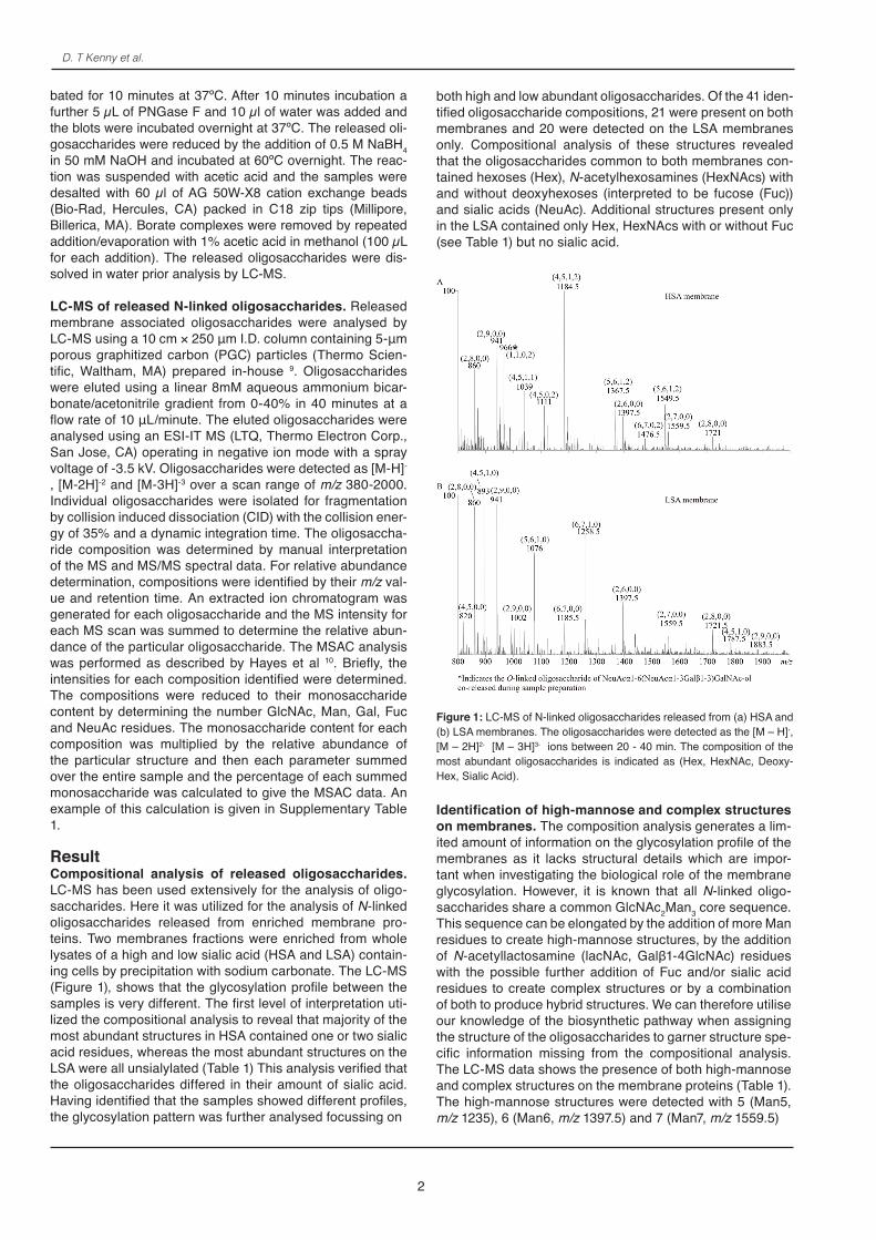
ResultCompositional analysis of released oligosaccharides.LC-MS has been used extensively for the analysis of oligo-saccharides. Here it was utilized for the analysis of N-linked oligosaccharides released from enriched membrane pro-teins. Two membranes fractions were enriched from whole lysates of a high and low sialic acid (HSA and LSA) contain-ing cells by precipitation with sodium carbonate. The LC-MS (Figure 1), shows that the glycosylation profile between the samples is very different. The first level of interpretation uti-lized the compositional analysis to reveal that majority of the most abundant structures in HSA contained one or two sialic acid residues, whereas the most abundant structures on the LSA were all unsialylated (Table 1) This analysis verified that the oligosaccharides differed in their amount of sialic acid. Having identified that the samples showed different profiles, the glycosylation pattern was further analysed focussing on
Figure 1: LC-MS of N-linked oligosaccharides released from (a) HSA and (b) LSA membranes. The oligosaccharides were detected as the [M – H]-, [M – 2H]2- [M – 3H]3- ions between 20 - 40 min. The composition of the most abundant oligosaccharides is indicated as (Hex, HexNAc, Deoxy-Hex, Sialic Acid).
Identification of high-mannose and complex structures on membranes. The composition analysis generates a lim-ited amount of information on the glycosylation profile of the membranes as it lacks structural details which are impor-tant when investigating the biological role of the membrane glycosylation. However, it is known that all N-linked oligo-saccharides share a common GlcNAc2Man3 core sequence. This sequence can be elongated by the addition of more Man residues to create high-mannose structures, by the addition of N-acetyllactosamine (lacNAc, Galβ1-4GlcNAc) residues with the possible further addition of Fuc and/or sialic acid residues to create complex structures or by a combination of both to produce hybrid structures. We can therefore utilise our knowledge of the biosynthetic pathway when assigning the structure of the oligosaccharides to garner structure spe-cific information missing from the compositional analysis. The LC-MS data shows the presence of both high-mannose and complex structures on the membrane proteins (Table 1). The high-mannose structures were detected with 5 (Man5, m/z 1235), 6 (Man6, m/z 1397.5) and 7 (Man7, m/z 1559.5)
2
both high and low abundant oligosaccharides. Of the 41 iden-tified oligosaccharide compositions, 21 were present on both membranes and 20 were detected on the LSA membranes only. Compositional analysis of these structures revealed that the oligosaccharides common to both membranes con-tained hexoses (Hex), N-acetylhexosamines (HexNAcs) with and without deoxyhexoses (interpreted to be fucose (Fuc)) and sialic acids (NeuAc). Additional structures present only in the LSA contained only Hex, HexNAcs with or without Fuc (see Table 1) but no sialic acid.
Semi-Quantitative Data Analysis for Membrane Associated N-linked Oligosaccharides
Man residues as their [M-H]- ions and with 8 (Man8, m/z 1721.5 and 860) and 9 (Man9, m/z 1883.5 and 941) Man residues detected as the [M-H]- and/or [M-2H]2- ions. The complex structures were detected as the [M-H]-, [M-2H]2- and [M-3H]3- ions (Table 1). Of the complex structures identified, 16 were detected on both membranes. These were predomi-nantly core fucosylated with or without terminal sialylation. The additional complex structures identified on the LSA membrane were all unsialylated extended with up to 12 lac-NAc residues. The data obtained by interpretation of masses into composition and biosynthetic pathways are providing the first level of interpretation and is required for stringent docu-mentation of the raw data. This is also the minimal amount of data that is required for submission to structural databases or knowledgebases and for recording and future references.
Oligosaccharides Identified on HSA and LSA
[M-nH]-n Composition Structure
1 12351- (2,5,0,0) } x2
2 1397.51- (2,6,0,0) } x3
3 1559.51- (2,7,0,0) } x4
4 1721.51- 8602-
(2,8,0,0) } x5
5 1883.51- 9412-
(2,9,0,0) } x6
6 10392- (4,5,1,1) } x1
7 12202- (5,5,1,1) } x1x1
8 14042- (6,7,1,1) } x2x1
9 11112- (4,5,0,2) } x2
10 1184.52- (4,5,1,2) } x2
11 1367.52- (5,6,1,2) } x1x2
12 1476.52- (6,7,0,2) } x2x2
13 1549.52- (6,7,1,2) } x2x2
14 1732.52- (7,8,1,2) } x3x2
15 19152- (8,9,1,2) } x4x2
16 10571- (2,3,1,0)
17 1642.5-1
8202- (4,5,0,0)
18 1788.5-1
8932- (4,5,1,0)
19 1002.52- (5,6,0,0) } 20 10762- (5,6,1,0) }
21 1258.52- (6,7,1,0) } x2
Oligosaccharides Identified on LSA only
22 9111- (2,3,0,0)
23 12761- (3,4,0,0) }
24 1480.5-1
7392- (4,4,0,0) }
25 11042- (6,6,0,0) }
26 1185.52- (6,7,0,0) } x2
27 13682- (7,8,0,0) } x3
28 1550.52- (8,9,0,0) } x4
29 14221- (3,4,1,0) }30 9952- (5,5,1,0) }
31 11772- (6,6,1,0) }
32 13602- (8,7,1,0) x2}
33 14412- (7,8,1,0) x3}
34 1542.52- (8,8,1,0) x3}
35 1623.52- (8,9,1,0) x4}
36 1806.52- (9,10,1,0) x5}
37 19882- (10,11,1,0) x6}
38 14473- (11,12,1,0) x7}
39 15683- (12,13,1,0) x8}
40 1690.53- (13,14,1,0) x9}
41 18123- (14,15,1,0) x10}
[M-nH]-n Composition Structure
Table 1: Composition analysis of the oligosaccharides identified on the High and Low SA membrane. The composition is pre-sented as (HexNAc, Hex, Deoxyhex, Sialic Acid). The structures for each oligosaccharides is presented using CFG annotation.
It is then the glycoanalysists obligation to do further inter-pretation of the data in order to communicate the result to a wider community.
Semi-quantitative display of individual structures with high-mannose and complex type glycosylation. To better reflect the biological aspects of the membrane glycosylation, the oligosaccharides can also be displayed according to their biological characteristics. The lack of quantitative data in Ta-ble 1 is limiting the amount of biologically relevant information that can be ascertained from it. Using the predicted oligo-saccharide structures from LC-MS and the intensity of each composition, it is possible generate semi-quantitative data that shows there is a significant variation in the abundance of the oligosaccharides between HSA and LSA membranes (Figure 2). Further information about the glycosylation can be obtained by sorting the oligosaccharides into high-mannose and complex type and this reveals that the main difference
3
D. T Kenny et al.
between the samples lie in the amount of sialylated and unsi-alylated structures However, the MS based information is still obscuring the picture by sorting the x-axis into features that are only accessible for a trained glycobiologist, and would be difficult even for researchers in the field to digest and make the connection between the identified structures and their function.
Average based MS data to display glycosylation dif-ferences. We have described a method for how to use the structural information from MS and the intensities to calculate a mass spectrometry average composition (MSAC) 10 (Figure 3A). The monosaccharide content of the membranes, based on the semi-quantitative data, can be used to display the gly-cosylation within a global context. This approach used for displaying O-linked semi-quantitative data was found to be superior in highlighting the difference in the amount of N-linked sialic acid present on both membranes. Furthermore, from the data we can see a higher amount of HexNAc and Hex in the complex oligosaccharides residues, indicating a higher abundance of lacNAc residues. For O-linked data there are a number of cores that could be terminated or ex-tended, but N-linked oligosaccharides all have a common core. In figure 3Ai the monosaccharide content was deter-mined with the inclusion of the GlcNAc2Man3 core sequence. However, since all structures contain this core structures, the MS based monosaccharide content data was normalized by excluding the core sequence to stress the differences in the antennas (Fig 3Aii). Combining this MSAC method with info about N-linked glycosylation (complex, high-mannose) pro-vides an efficient way to grasp the MS data. In figure 3B we
can see that the abundance of high-mannose and complex structures for both membranes is similar and that the majority of the oligosaccharides in both samples are complex struc-tures. Of those complex structures, the sialylated structures are the major component on the HSA membrane, whereas they are only a minor component on the LSA membrane (Figure 3C). The relative abundance of the individual high-mannose oligosaccharides did not vary greatly between the samples, the complex oligosaccharides differed significantly with the sialylated structures having greater abundance on HSA membranes than LSA membranes. Conversely the most abundance structures on the LSA membrane were unsialylated. The display of the data in Figure 3 allows dis-cussion about the results obtained by MS. While character-ising the two membranes, the figure highlights the differ-ences in the amount of sialic acid. The initial investigation also revealed that the LSA membrane had a large amount of additional unsialylated polylacNAc extended structures. When investigating these unsialylated structures further we could see that it was not just the quantitative differences of sialylation between the samples but also qualitative. The ad-dition of sialic acid typically acts as a termination event which ceases the elongation by halting further addition of lacNAc’s. Hence, the presence of polylacNAc in the LSA sample may not only indicate a higher galactosyltransferase activity, but also lower sialyltransferase activity.
DiscussionWe have previously described a protocol for the global en-richment of membrane proteins from mammalian cells as a method for global analysis of membrane associated oligo-saccharides 9. Here, we have used this method and LC-MS to characterise the N-linked oligosaccharides released from two enriched membrane fractions, which are shown to dif-fer in their amounts of sialic acid. The LC-MS profile of both membranes were shown to differ considerably and analysis
Figure 2: The relative abundance of the N-linked oligosaccharides de-tected on the HSA (back) and LSA (grey) membranes. The high mannose, complex sialylated structures and complex unsialylated structures are in-dicated. The number of each structure corresponds to their numbering in Table 1.
4
Semi-Quantitative Data Analysis for Membrane Associated N-linked Oligosaccharides
of the released oligosaccharides revealed the presence of high-mannose and complex structures present on the cel-lular membranes. The presence of high-mannose structures on various cell membranes has previously been reported 11-15 and their presence here suggests that they are normal feature of membrane glycosylation. However, since plasma membranes containing the fully processed oligosaccharides are only contributing to 2-5% of the total membrane fraction 16, there is the possibility of co-enrichment of premature na-scent N-linked oligosaccharides that contain the N-linked intermediary and the trimmed versions of core structures present in secretory pathway in ER and Golgi or in the endo-cytosis and degradative pathways. This would contribute to
a portion of high-mannose structures present in both mem-branes samples. The role of complex oligosaccharides on membranes 15, 17 is dependent on their structural features. Fucosylation is important for glycoprotein processing 18
whereas sialylation is involved in the binding and transport of cationic molecules 19 and serves as a self-recognition factors preventing activation of the immune system 20.
Highlighting biological importance using relative quan-tification. Changes in the abundance of particular oligo-saccharides have been shown to have adverse effects and altered glycosylation profiles have been associated with dif-ferent diseases states; for example a decrease in the abun-dance of high-mannose oligosaccharides on the membranes of peripheral blood mononuclear cells in patients with liver cirrhosis has been correlated with a decrease in natural killer cell activity 21 whereas increased levels of sialylation have been associated with various tumours. The semi-quantita-tive data provides an overview into the glycosylation beyond merely determining the presence or absence of particular structures. In figure 2 we present the relative abundance of
Figure 3: A) The monosaccharide composition based on the relative in-tensity the oligosaccharides detected on the HSA (black) and LSA (grey) membranes with the GlcNAc2Man3 core (i) included and (ii) excluded. Pie charts indicating the ratio of (B) high mannose and complex structures and (C) sialylated and unsialylated structures present on the HSA and LSA membranes based on relative quantification of the oligosaccharides detected.
5
D. T Kenny et al.
12. Kim, Y.G., et al., The identification and characterization of xenoan-tigenic nonhuman carbohydrate sequences in membrane proteins from porcine kidney. Proteomics, 2006. 6(4): p. 1133-42.13. An, H.J., et al., Extensive Determination of Glycan Heterogeneity Reveals an Unusual Abundance of High Mannose Glycans in Enriched Plasma Membranes of Human Embryonic Stem Cells. Molecular & Cel-lular Proteomics, 2012. 11(4).14. Penuela, S., et al., Glycosylation Regulates Pannexin Intermixing and Cellular Localization. Molecular Biology of the Cell, 2009. 20(20): p. 4313-4323.15. Clark, R.A., et al., Characterisation of tissue-specific oligosaccha-rides from rat brain and kidney membrane preparations enriched in Na+,K+-ATPase. Glycoconj J, 1999. 16(8): p. 437-56.16. Alberts, B., et al., eds. Molecular Biology of the Cell. 5th ed. 2008, Garland Science.17. Li, J., et al., Processing of N-linked oligosaccharide depends on its location in the anion exchanger, AE1, membrane glycoprotein. Biochem-ical Journal, 2000. 349: p. 51-57.18. Becker, D.J. and J.B. Lowe, Fucose: biosynthesis and biological function in mammals. Glycobiology, 2003. 13(7): p. 41R-53R.19. Hu, Q.L., et al., Intracellular pathways and nuclear localization signal peptide-mediated gene transfection by cationic polymeric nanovectors. Biomaterials, 2012. 33(4): p. 1135-1145.20. Pilatte, Y., J. Bignon, and C.R. Lambré, Sialic acids as important molecules in the regulation of the immune system: pathophysiological implications of sialidases in immunity. Glycobiology, 1993. 3(3): p. 201-218.21. Miyaguchi, S., et al., Changes in high mannose-type glycoproteins of peripheral blood mononuclear cells in cirrhosis patients. International Hepatology Communications, 1995. 3(1): p. 41-
6
the individual oligosaccharides and treat each oligosaccha-ride as a distinct structure. In reality, the oligosaccharides have common epitopes and thus share common biological purposes. Very rarely can a biological role be attributed to one particular oligosaccharide structure. With a lack of data representing the biological function, we have to resort to sort-ing the structures based on their common structural features. This approach could make more sense when communicating the glycomic results to researchers unfamiliar with MS based glycomic analysis. With the belief that structure and function are associated, sorting based on structural families is rea-sonable and in this case highlights the difference between samples that would require further investigation. Since inter-pretation of the semi-quantitative data would reveal the bio-logical aspects of the glycosylation, we found that the type of data representation as in figure 3B and 3C as appealing, together with the MSAC metric that has been shown to be well suited for comparison of O-linked oligosaccharides 10. With carbohydrate signalling being more analogue then digi-tal as described above, the representation provides informa-tion of increased or decreased biological ability, rather than absence or presence of this ability
AcknowledgementsProfessor Hans-Joachim Gabius, University of Munich is gratefully ac-knowledged for his gift cellular material. The mass spectrometers were obtained by grants from the Swedish Research Council (342-2004-4434) and from the Knut and Alice Wallenberg Foundation (KAW2007.0118).
Reference1. Stevens, T.J. and I.T. Arkin, Do more complex organisms have a greater proportion of membrane proteins in their genomes? Proteins: Structure, Function, and Bioinformatics, 2000. 39(4): p. 417-420.2. Gabius, H.J., Glycans: bioactive signals decoded by lectins. Biochem-ical Society transactions, 2008. 36(Pt 6): p. 1491-6.3. Dell, A. and H.R. Morris, Glycoprotein structure determination mass spectrometry. Science, 2001. 291(5512): p. 2351-2356.4. Mechref, Y. and M.V. Novotny, Structural Investigations of Glycocon-jugates at High Sensitivity. Chemical Reviews, 2002. 102(2): p. 321-370.5. Harvey, D.J., Matrix-assisted laser desorption/ionization mass spec-trometry of carbohydrates and glycoconjugates. International Journal of Mass Spectrometry, 2003. 226(1): p. 1-35.6. Mechref, Y. and M.V. Novotny, Miniaturized separation techniques in glycomic investigations. Journal of Chromatography B, 2006. 841(1-2): p. 65-78.7. Robinson, L.J., et al., Proteomic analysis of the genetic premature aging disease Hutchinson Gilford progeria syndrome reveals differential protein expression and glycosylation. J Proteome Res, 2003. 2(5): p. 556-7.8. Karlsson, N.G., et al., Negative ion graphitised carbon nano-liquid chromatography/mass spectrometry increases sensitivity for glycopro-tein oligosaccharide analysis. Rapid Commun Mass Spectrom, 2004. 18(19): p. 2282-92.9. Kenny, D.T., et al., Glycomic Analysis of Membrane-Associated Pro-teins, in Sample Preparation in Biological Mass Spectrometry, A.R. Iva-nov and A.V. Lazarev, Editors. 2011, Springer. p. 498-513.10. Hayes, C.A., S. Nemes, and N.G. Karlsson, Statistical analysis of glycosylation profiles to compare tissue type and inflammatory disease state. Bioinformatics, 2012.11. Nuck, R., et al., Comparative study of high-mannose-type oligosac-charides in membrane glycoproteins of rat hepatocytes and different rat hepatoma cell lines. European Journal of Biochemistry, 1993. 216(1): p. 215-221.
Semi-Quantitative Data Analysis for Membrane Associated N-linked Oligosaccharides Highlights Differences in the Glycosylation of Two Enriched Membranes SamplesDiarmuid T. Kenny1,2, Kristina A. Thomsson1, Niclas G. Karlsson1
1Department of Medical Biochemistry, Göteborg University, Sweden and 2School of Chemistry, National University Ireland Galway, Ireland
Glycomic data is difficult to display due to the fact that the oligosaccharides are diverse, and structural and functional aspects overlap, where an attribute cannot be explained by only one individual structure. We set out to investigate how we could transform glycomic MS data derived from the analysis of N-linked oligosaccharides into easily accessible information representing differ-ences in glycosylation. In order to do this we adopted a method for the global enrichment of cellular membrane using carbonate extraction and analysed two samples with differing levels of sialylation. We could identify 41 different oligosaccharides based on their composition. By measuring the MS intensities of the compositions that were identified, we could determine the relative abundance of the individual oligosaccharides. By sort-ing the identified oligosaccharides by their structural characteristics (high-mannose, high and low sialylation content) and applying mass spectrometric average composition (MSAC) analysis, we could identify particu-lar traits about the enriched membranes. Using this ap-proach it was possible to highlight differences in glyco-sylation profile in order to make the data from complex MS-spectra accessible for structural comparison.
The cellular membrane proteins account for up to 30 per-cent of the total cellular protein content and are usually
found to be N-linked glycosylated 1. N-linked oligosaccah-rides are, for instance, present on the plasma membrane where it has been identified as a mediator of extracellular signalling and involved in immunological events 2. Therefore, characterising the structural features of the oligosaccharides that mediates this signalling is important. Glycomics of the immunologically important cell surface oligosaccharide epit-opes can aid in our understanding of their role in immunolog-ical signalling. Mass spectrometry (MS) has revolutionised the field of glycomics and has become the primary instru-ment of choice for a glycomic platform 3-5. When coupled to an LC-system, it is ideally suited to characterising the mix-ture of oligosaccharides structures typically present on a gly-coprotein 6. The data generated by glycomic investigation are usually quite complex and confusing, since similar structural features are present on several structures and the informa-tion about their biological importance is usually quite difficult to digest. The communication between the glycobiologist and the glycoanalysist would be greatly enhanced if they conformed to using a common language and terminology of important epitopes such as high-mannose, sialylation, blood group antigens, sulfation, core types, N-acetyllactosamine
Correspondence: Niclas G. Karlsson, Dept. Medical Biochemistry, Univer-sity of Gothenburg, Box 440, 405 30 Gothenburg, SWEDEN; Phone: +46 31 786 6528. Email: [email protected]
elongation. This would aid in the amalgamation of reports and also provide a platform for better communication regard-ing MS specific information that is not obtainable by other methods such as antibody binding and lectin epitopes includ-ing details about antenna distribution that serves as scaffold for the terminal epitopes and potentially semi-quantitative data relating to the abundance of oligosaccharides. In this report we investigate how to display the glycomic information obtained from MS data when comparing the glycosylation from two different mammalian membranes.
N-linked | glycosylation | LC-MS | Sialylation
Materials and MethodsAll materials were obtained from Sigma Aldrich (St Louis, MO) unless otherwise stated. The 18 MΩ water was pro-duced using the MilliQ water purification system (Millipore, Billerica, MA)
Enrichment of Membrane proteins from mammalian cells. Membrane proteins were enriched by precipitation in sodium carbonate 7, 8. Cellular material (100 mg) of was suspended in 100 mM sodium carbonate. The solution was sonicated on ice at 40% maximum amplitude four times for 15 seconds and cooled on ice in between each sonication for 1 minute. After sonication, the solution was transferred to a beaker and 100 mM of sodium carbonate was added to a final volume of 100 mL and stirred for 5 minutes at 4 ºC. Cell de-bris was removed by centrifugation at 5,000 g for 10 minutes. After ultracentrifugation, the supernatant was transferred to a new centrifuge tubes and ultracentrifuged at 115,000 g for 75 minutes. The supernatant was discarded and the pellet was washed in 50 mM tris(hydroxymethyl)aminomethane-HCl (Tris-HCl) pH 7.3 and ultracentrifuged at 115,000 g for 75 minutes. The wash step was repeated once. The washed membrane containing pellets were solubilised in 7.0 M urea, 2.0 M thiourea, 40 mM Tris base and 4% w/v 3-[(3 cholami-dopropyl)dimethylammonio]-1-propanesulfonate (CHAPS).
Release of N-linked oligosaccharides for LC-MS analy-sis. The solubilised membrane extracts were blotted onto Immobilon-P PVDF membranes (Millipore, Billerica, MA) and stained with DB71 and de-stained in 10% acetic acid in 40% ethanol. The membranes were blocked with 1% polyvinylpyr-rolidone (PVP) in methanol. The N-linked oligosaccharides were enzymatically released from the protein by PNGase F. The enzyme (5 µL, 5 units) was added to each blot and incu-
1
D. T Kenny et al.
bated for 10 minutes at 37ºC. After 10 minutes incubation a further 5 µL of PNGase F and 10 µl of water was added and the blots were incubated overnight at 37ºC. The released oli-gosaccharides were reduced by the addition of 0.5 M NaBH4 in 50 mM NaOH and incubated at 60ºC overnight. The reac-tion was suspended with acetic acid and the samples were desalted with 60 µl of AG 50W-X8 cation exchange beads (Bio-Rad, Hercules, CA) packed in C18 zip tips (Millipore, Billerica, MA). Borate complexes were removed by repeated addition/evaporation with 1% acetic acid in methanol (100 µL for each addition). The released oligosaccharides were dis-solved in water prior analysis by LC-MS.
LC-MS of released N-linked oligosaccharides. Released membrane associated oligosaccharides were analysed by LC-MS using a 10 cm × 250 μm I.D. column containing 5-μm porous graphitized carbon (PGC) particles (Thermo Scien-tific, Waltham, MA) prepared in-house 9. Oligosaccharides were eluted using a linear 8mM aqueous ammonium bicar-bonate/acetonitrile gradient from 0-40% in 40 minutes at a flow rate of 10 μL/minute. The eluted oligosaccharides were analysed using an ESI-IT MS (LTQ, Thermo Electron Corp., San Jose, CA) operating in negative ion mode with a spray voltage of -3.5 kV. Oligosaccharides were detected as [M-H]-
, [M-2H]-2 and [M-3H]-3 over a scan range of m/z 380-2000. Individual oligosaccharides were isolated for fragmentation by collision induced dissociation (CID) with the collision ener-gy of 35% and a dynamic integration time. The oligosaccha-ride composition was determined by manual interpretation of the MS and MS/MS spectral data. For relative abundance determination, compositions were identified by their m/z val-ue and retention time. An extracted ion chromatogram was generated for each oligosaccharide and the MS intensity for each MS scan was summed to determine the relative abun-dance of the particular oligosaccharide. The MSAC analysis was performed as described by Hayes et al 10. Briefly, the intensities for each composition identified were determined. The compositions were reduced to their monosaccharide content by determining the number GlcNAc, Man, Gal, Fuc and NeuAc residues. The monosaccharide content for each composition was multiplied by the relative abundance of the particular structure and then each parameter summed over the entire sample and the percentage of each summed monosaccharide was calculated to give the MSAC data. An example of this calculation is given in Supplementary Table 1.
ResultCompositional analysis of released oligosaccharides.LC-MS has been used extensively for the analysis of oligo-saccharides. Here it was utilized for the analysis of N-linked oligosaccharides released from enriched membrane pro-teins. Two membranes fractions were enriched from whole lysates of a high and low sialic acid (HSA and LSA) contain-ing cells by precipitation with sodium carbonate. The LC-MS (Figure 1), shows that the glycosylation profile between the samples is very different. The first level of interpretation uti-lized the compositional analysis to reveal that majority of the most abundant structures in HSA contained one or two sialic acid residues, whereas the most abundant structures on the LSA were all unsialylated (Table 1) This analysis verified that the oligosaccharides differed in their amount of sialic acid. Having identified that the samples showed different profiles, the glycosylation pattern was further analysed focussing on
Figure 1: LC-MS of N-linked oligosaccharides released from (a) HSA and (b) LSA membranes. The oligosaccharides were detected as the [M – H]-, [M – 2H]2- [M – 3H]3- ions between 20 - 40 min. The composition of the most abundant oligosaccharides is indicated as (Hex, HexNAc, Deoxy-Hex, Sialic Acid).
Identification of high-mannose and complex structures on membranes. The composition analysis generates a lim-ited amount of information on the glycosylation profile of the membranes as it lacks structural details which are impor-tant when investigating the biological role of the membrane glycosylation. However, it is known that all N-linked oligo-saccharides share a common GlcNAc2Man3 core sequence. This sequence can be elongated by the addition of more Man residues to create high-mannose structures, by the addition of N-acetyllactosamine (lacNAc, Galβ1-4GlcNAc) residues with the possible further addition of Fuc and/or sialic acid residues to create complex structures or by a combination of both to produce hybrid structures. We can therefore utilise our knowledge of the biosynthetic pathway when assigning the structure of the oligosaccharides to garner structure spe-cific information missing from the compositional analysis. The LC-MS data shows the presence of both high-mannose and complex structures on the membrane proteins (Table 1). The high-mannose structures were detected with 5 (Man5, m/z 1235), 6 (Man6, m/z 1397.5) and 7 (Man7, m/z 1559.5)
2
both high and low abundant oligosaccharides. Of the 41 iden-tified oligosaccharide compositions, 21 were present on both membranes and 20 were detected on the LSA membranes only. Compositional analysis of these structures revealed that the oligosaccharides common to both membranes con-tained hexoses (Hex), N-acetylhexosamines (HexNAcs) with and without deoxyhexoses (interpreted to be fucose (Fuc)) and sialic acids (NeuAc). Additional structures present only in the LSA contained only Hex, HexNAcs with or without Fuc (see Table 1) but no sialic acid.
Semi-Quantitative Data Analysis for Membrane Associated N-linked Oligosaccharides
Man residues as their [M-H]- ions and with 8 (Man8, m/z 1721.5 and 860) and 9 (Man9, m/z 1883.5 and 941) Man residues detected as the [M-H]- and/or [M-2H]2- ions. The complex structures were detected as the [M-H]-, [M-2H]2- and [M-3H]3- ions (Table 1). Of the complex structures identified, 16 were detected on both membranes. These were predomi-nantly core fucosylated with or without terminal sialylation. The additional complex structures identified on the LSA membrane were all unsialylated extended with up to 12 lac-NAc residues. The data obtained by interpretation of masses into composition and biosynthetic pathways are providing the first level of interpretation and is required for stringent docu-mentation of the raw data. This is also the minimal amount of data that is required for submission to structural databases or knowledgebases and for recording and future references.
Oligosaccharides Identified on HSA and LSA
[M-nH]-n Composition Structure
1 12351- (2,5,0,0) } x2
2 1397.51- (2,6,0,0) } x3
3 1559.51- (2,7,0,0) } x4
4 1721.51- 8602-
(2,8,0,0) } x5
5 1883.51- 9412-
(2,9,0,0) } x6
6 10392- (4,5,1,1) } x1
7 12202- (5,5,1,1) } x1x1
8 14042- (6,7,1,1) } x2x1
9 11112- (4,5,0,2) } x2
10 1184.52- (4,5,1,2) } x2
11 1367.52- (5,6,1,2) } x1x2
12 1476.52- (6,7,0,2) } x2x2
13 1549.52- (6,7,1,2) } x2x2
14 1732.52- (7,8,1,2) } x3x2
15 19152- (8,9,1,2) } x4x2
16 10571- (2,3,1,0)
17 1642.5-1
8202- (4,5,0,0)
18 1788.5-1
8932- (4,5,1,0)
19 1002.52- (5,6,0,0) } 20 10762- (5,6,1,0) }
21 1258.52- (6,7,1,0) } x2
Oligosaccharides Identified on LSA only
22 9111- (2,3,0,0)
23 12761- (3,4,0,0) }
24 1480.5-1
7392- (4,4,0,0) }
25 11042- (6,6,0,0) }
26 1185.52- (6,7,0,0) } x2
27 13682- (7,8,0,0) } x3
28 1550.52- (8,9,0,0) } x4
29 14221- (3,4,1,0) }30 9952- (5,5,1,0) }
31 11772- (6,6,1,0) }
32 13602- (8,7,1,0) x2}
33 14412- (7,8,1,0) x3}
34 1542.52- (8,8,1,0) x3}
35 1623.52- (8,9,1,0) x4}
36 1806.52- (9,10,1,0) x5}
37 19882- (10,11,1,0) x6}
38 14473- (11,12,1,0) x7}
39 15683- (12,13,1,0) x8}
40 1690.53- (13,14,1,0) x9}
41 18123- (14,15,1,0) x10}
[M-nH]-n Composition Structure
Table 1: Composition analysis of the oligosaccharides identified on the High and Low SA membrane. The composition is pre-sented as (HexNAc, Hex, Deoxyhex, Sialic Acid). The structures for each oligosaccharides is presented using CFG annotation.
It is then the glycoanalysists obligation to do further inter-pretation of the data in order to communicate the result to a wider community.
Semi-quantitative display of individual structures with high-mannose and complex type glycosylation. To better reflect the biological aspects of the membrane glycosylation, the oligosaccharides can also be displayed according to their biological characteristics. The lack of quantitative data in Ta-ble 1 is limiting the amount of biologically relevant information that can be ascertained from it. Using the predicted oligo-saccharide structures from LC-MS and the intensity of each composition, it is possible generate semi-quantitative data that shows there is a significant variation in the abundance of the oligosaccharides between HSA and LSA membranes (Figure 2). Further information about the glycosylation can be obtained by sorting the oligosaccharides into high-mannose and complex type and this reveals that the main difference
3
D. T Kenny et al.
between the samples lie in the amount of sialylated and unsi-alylated structures However, the MS based information is still obscuring the picture by sorting the x-axis into features that are only accessible for a trained glycobiologist, and would be difficult even for researchers in the field to digest and make the connection between the identified structures and their function.
Average based MS data to display glycosylation dif-ferences. We have described a method for how to use the structural information from MS and the intensities to calculate a mass spectrometry average composition (MSAC) 10 (Figure 3A). The monosaccharide content of the membranes, based on the semi-quantitative data, can be used to display the gly-cosylation within a global context. This approach used for displaying O-linked semi-quantitative data was found to be superior in highlighting the difference in the amount of N-linked sialic acid present on both membranes. Furthermore, from the data we can see a higher amount of HexNAc and Hex in the complex oligosaccharides residues, indicating a higher abundance of lacNAc residues. For O-linked data there are a number of cores that could be terminated or ex-tended, but N-linked oligosaccharides all have a common core. In figure 3Ai the monosaccharide content was deter-mined with the inclusion of the GlcNAc2Man3 core sequence. However, since all structures contain this core structures, the MS based monosaccharide content data was normalized by excluding the core sequence to stress the differences in the antennas (Fig 3Aii). Combining this MSAC method with info about N-linked glycosylation (complex, high-mannose) pro-vides an efficient way to grasp the MS data. In figure 3B we
can see that the abundance of high-mannose and complex structures for both membranes is similar and that the majority of the oligosaccharides in both samples are complex struc-tures. Of those complex structures, the sialylated structures are the major component on the HSA membrane, whereas they are only a minor component on the LSA membrane (Figure 3C). The relative abundance of the individual high-mannose oligosaccharides did not vary greatly between the samples, the complex oligosaccharides differed significantly with the sialylated structures having greater abundance on HSA membranes than LSA membranes. Conversely the most abundance structures on the LSA membrane were unsialylated. The display of the data in Figure 3 allows dis-cussion about the results obtained by MS. While character-ising the two membranes, the figure highlights the differ-ences in the amount of sialic acid. The initial investigation also revealed that the LSA membrane had a large amount of additional unsialylated polylacNAc extended structures. When investigating these unsialylated structures further we could see that it was not just the quantitative differences of sialylation between the samples but also qualitative. The ad-dition of sialic acid typically acts as a termination event which ceases the elongation by halting further addition of lacNAc’s. Hence, the presence of polylacNAc in the LSA sample may not only indicate a higher galactosyltransferase activity by promoting the elongation, but also lower sialyltransferase activity.
DiscussionWe have previously described a protocol for the global en-richment of membrane proteins from mammalian cells as a method for global analysis of membrane associated oligo-saccharides 9. Here, we have used this method and LC-MS to characterise the N-linked oligosaccharides released from two enriched membrane fractions, which are shown to dif-fer in their amounts of sialic acid. The LC-MS profile of both membranes were shown to differ considerably and analysis
Figure 2: The relative abundance of the N-linked oligosaccharides de-tected on the HSA (back) and LSA (grey) membranes. The high mannose, complex sialylated structures and complex unsialylated structures are in-dicated. The number of each structure corresponds to their numbering in Table 1.
4
Semi-Quantitative Data Analysis for Membrane Associated N-linked Oligosaccharides
of the released oligosaccharides revealed the presence of high-mannose and complex structures present on the cel-lular membranes. The presence of high-mannose structures on various cell membranes has previously been reported 11-15 and their presence here suggests that they are normal feature of membrane glycosylation. However, since plasma membranes containing the fully processed oligosaccharides are only contributing to 2-5% of the total membrane fraction 16, there is the possibility of co-enrichment of premature na-scent N-linked oligosaccharides that contain the N-linked intermediary and the trimmed versions of core structures present in secretory pathway in ER and Golgi or in the endo-cytosis and degradative pathways. This would contribute to
a portion of high-mannose structures present in both mem-branes samples. The role of complex oligosaccharides on membranes 15, 17 is dependent on their structural features. Fucosylation is important for glycoprotein processing 18
whereas sialylation is involved in the binding and transport of cationic molecules 19 and serves as a self-recognition factors preventing activation of the immune system 20.
Highlighting biological importance using relative quan-tification. Changes in the abundance of particular oligo-saccharides have been shown to have adverse effects and altered glycosylation profiles have been associated with dif-ferent diseases states; for example a decrease in the abun-dance of high-mannose oligosaccharides on the membranes of peripheral blood mononuclear cells in patients with liver cirrhosis has been correlated with a decrease in natural killer cell activity 21 whereas increased levels of sialylation have been associated with various tumours. The semi-quantita-tive data provides an overview into the glycosylation beyond merely determining the presence or absence of particular structures. In figure 2 we present the relative abundance of
Figure 3: A) The monosaccharide composition based on the relative in-tensity the oligosaccharides detected on the HSA (black) and LSA (grey) membranes with the GlcNAc2Man3 core (i) included and (ii) excluded. Pie charts indicating the ratio of (B) high mannose and complex structures and (C) sialylated and unsialylated structures present on the HSA and LSA membranes based on relative quantification of the oligosaccharides detected.
5
D. T Kenny et al.
12. Kim, Y.G., et al., The identification and characterization of xenoan-tigenic nonhuman carbohydrate sequences in membrane proteins from porcine kidney. Proteomics, 2006. 6(4): p. 1133-42.13. An, H.J., et al., Extensive Determination of Glycan Heterogeneity Reveals an Unusual Abundance of High Mannose Glycans in Enriched Plasma Membranes of Human Embryonic Stem Cells. Molecular & Cel-lular Proteomics, 2012. 11(4).14. Penuela, S., et al., Glycosylation Regulates Pannexin Intermixing and Cellular Localization. Molecular Biology of the Cell, 2009. 20(20): p. 4313-4323.15. Clark, R.A., et al., Characterisation of tissue-specific oligosaccha-rides from rat brain and kidney membrane preparations enriched in Na+,K+-ATPase. Glycoconj J, 1999. 16(8): p. 437-56.16. Alberts, B., et al., eds. Molecular Biology of the Cell. 5th ed. 2008, Garland Science.17. Li, J., et al., Processing of N-linked oligosaccharide depends on its location in the anion exchanger, AE1, membrane glycoprotein. Biochem-ical Journal, 2000. 349: p. 51-57.18. Becker, D.J. and J.B. Lowe, Fucose: biosynthesis and biological function in mammals. Glycobiology, 2003. 13(7): p. 41R-53R.19. Hu, Q.L., et al., Intracellular pathways and nuclear localization signal peptide-mediated gene transfection by cationic polymeric nanovectors. Biomaterials, 2012. 33(4): p. 1135-1145.20. Pilatte, Y., J. Bignon, and C.R. Lambré, Sialic acids as important molecules in the regulation of the immune system: pathophysiological implications of sialidases in immunity. Glycobiology, 1993. 3(3): p. 201-218.21. Miyaguchi, S., et al., Changes in high mannose-type glycoproteins of peripheral blood mononuclear cells in cirrhosis patients. International Hepatology Communications, 1995. 3(1): p. 41-
6
the individual oligosaccharides and treat each oligosaccha-ride as a distinct structure. In reality, the oligosaccharides have common epitopes and thus share common biological purposes. Very rarely can a biological role be attributed to one particular oligosaccharide structure. With a lack of data representing the biological function, we have to resort to sort-ing the structures based on their common structural features. This approach could make more sense when communicating the glycomic results to researchers unfamiliar with MS based glycomic analysis. With the belief that structure and function are associated, sorting based on structural families is rea-sonable and in this case highlights the difference between samples that would require further investigation. Since inter-pretation of the semi-quantitative data would reveal the bio-logical aspects of the glycosylation, we found that the type of data representation as in figure 3B and 3C as appealing, together with the MSAC metric that has been shown to be well suited for comparison of O-linked oligosaccharides 10. With carbohydrate signalling being more analogue then digi-tal as described above, the representation provides informa-tion of increased or decreased biological ability, rather than absence or presence of this ability
AcknowledgementsProfessor Hans-Joachim Gabius, University of Munich is gratefully ac-knowledged for his gift cellular material. The mass spectrometers were obtained by grants from the Swedish Research Council (342-2004-4434) and from the Knut and Alice Wallenberg Foundation (KAW2007.0118).
Reference1. Stevens, T.J. and I.T. Arkin, Do more complex organisms have a greater proportion of membrane proteins in their genomes? Proteins: Structure, Function, and Bioinformatics, 2000. 39(4): p. 417-420.2. Gabius, H.J., Glycans: bioactive signals decoded by lectins. Biochem-ical Society transactions, 2008. 36(Pt 6): p. 1491-6.3. Dell, A. and H.R. Morris, Glycoprotein structure determination mass spectrometry. Science, 2001. 291(5512): p. 2351-2356.4. Mechref, Y. and M.V. Novotny, Structural Investigations of Glycocon-jugates at High Sensitivity. Chemical Reviews, 2002. 102(2): p. 321-370.5. Harvey, D.J., Matrix-assisted laser desorption/ionization mass spec-trometry of carbohydrates and glycoconjugates. International Journal of Mass Spectrometry, 2003. 226(1): p. 1-35.6. Mechref, Y. and M.V. Novotny, Miniaturized separation techniques in glycomic investigations. Journal of Chromatography B, 2006. 841(1-2): p. 65-78.7. Robinson, L.J., et al., Proteomic analysis of the genetic premature aging disease Hutchinson Gilford progeria syndrome reveals differential protein expression and glycosylation. J Proteome Res, 2003. 2(5): p. 556-7.8. Karlsson, N.G., et al., Negative ion graphitised carbon nano-liquid chromatography/mass spectrometry increases sensitivity for glycopro-tein oligosaccharide analysis. Rapid Commun Mass Spectrom, 2004. 18(19): p. 2282-92.9. Kenny, D.T., et al., Glycomic Analysis of Membrane-Associated Pro-teins, in Sample Preparation in Biological Mass Spectrometry, A.R. Iva-nov and A.V. Lazarev, Editors. 2011, Springer. p. 498-513.10. Hayes, C.A., S. Nemes, and N.G. Karlsson, Statistical analysis of glycosylation profiles to compare tissue type and inflammatory disease state. Bioinformatics, 2012.11. Nuck, R., et al., Comparative study of high-mannose-type oligosac-charides in membrane glycoproteins of rat hepatocytes and different rat hepatoma cell lines. European Journal of Biochemistry, 1993. 216(1): p. 215-221.
Supplementary Data 1: Determination of MSAC for HSA from the cumulative intensities of
all the structures detected. The MSAC analysis reduces the samples to their monosaccahride
content.
Presence of terminal N-acetylgalactosamineβ1-4N-acetylglucosamine residues on O-linkedoligosaccharides from gastric MUC5AC:Involvement in Helicobacter pylori colonization?
Diarmuid T Kenny2,3, Emma C Skoog3,4, SaraK Lindén3,4, Weston B Struwe5, Pauline M Rudd5,and Niclas G Karlsson1,3
2School of Chemistry, National University of Ireland, Galway, Ireland;3Medical Biochemistry, University of Gothenburg, PO Box 440,405 30 Gothenburg, Sweden; 4Mucosal Immunobiology and Vaccine Center,Sahlgrenska Academy, University of Gothenburg, 405 30 Gothenburg,Sweden; and 5National Institute for Bioprocessing Research and Training,University College Dublin, Dublin, Ireland
Received on October 21, 2011; revised on March 5, 2012; accepted onApril 21, 2012
Isolation of MUC5AC mucins from the gastric mucosafrom two secretor individuals (one from normal mucosafrom a patient with gastric cancer and one from a control)showed different abilities to bind and induce the prolifer-ation of the Helicobacter pylori strain J99. Analysis of thereleased O-linked oligosaccharides by LC-MS from theseindividuals showed a very heterogeneous mixture ofspecies from the cancer patient containing both neutraland sialylated structures, whereas the normal sampleshowed dominating neutral blood group H terminatingstructures as well as neutral structures containing thedi-N-acetyllactosamine (lacdiNAc) unit GalNAcβ1-4GlcNAcβ1- on the C-6 branch of the reducing endGalNAc. The linkage configuration of these epitopes weredetermined using C-4-specific fragmentation for theGalNAcβ1-4GlcNAcβ1- glycosidic linkage, comparison ofthe MS3 fragmentation with standards for linkage config-uration and N-acetylhexosamine type as well as exoglycosi-dase treatment. It was also shown that the lacdiNAcepitope is present in both human and porcine gastricmucins, indicating that this is an epitope preservedbetween species. We hypothesize that the termination ongastric MUC5AC with lacdiNAc is in competition withcomplex glycosylation such as the Leb and H type 1 aswell as complex sialylated structures. These are epitopesknown to bind the H. pylori BabA and SabA adhesins.
Keywords: BabA / gastric cancer /Helicobacter / lacdiNAc /mucin / SabA
Introduction
The gastro intestinal tract is covered in a semi-permeablemucus layer that primarily consists of secreted mucins. Thislayer serves to protect the gastric epithelial cells from poten-tial damage that could occur due to mechanical and chemicalstress or enzymatic activity within the tract. The mucus layeris composed of oligomeric mucin glycoproteins that serve asa scaffold for an abundance of anti-microbial epitopes(McGuckin et al. 2011). The mucin layer can also act as afunctional binding site for certain pathogens such asH. pylori. H. pylori are a spiral-shaped gram-negative bacteriathat colonizes the mucus layer covering the gastric epithelium.The bacteria were first isolated and cultured from the gastricmucosa by Marshall and Warren in 1983 and their discoveryrevealed the H. pylori’s association with gastritis, pepticulcers and several forms of gastric cancer (Marshall andWarren 1984). H. pylori infection is the most common bacter-ial infection worldwide and it is estimated to affect over 50%of the world’s population (Kobayashi et al. 2009). Infectionusually begins in early childhood and infection and its asso-ciated symptoms such as chronic gastritis and peptic ulcerscan persist throughout an individual’s life. H. pylori infectioncan dramatically increase the risk of gastric cancer and hasbeen attributed as a key mitigating factor for up to 65% ofgastric cancers, equivalent to 5.5% of all cancers globally(Menaker et al. 2004).Gastric mucins contain highly diverse carbohydrates that
can be utilized by H. pylori for adhesion. The attachment ofH. pylori is mediated by outer membrane proteins (OMPs)with lectin-like properties. The adhesion of H. pylori to thegastric surface is mediated by both the presence of these adhe-sion OMPs on the surface of the H. pylori cell and the pres-ence of particular glyco-epitopes on the mucin. Of the OMPsthat are involved in attachment, two have been identified asparticularly important for the attachment of H. pylori to themucosal layer: blood group antigen-binding adhesin (BabA)and the sialic acid-binding adhesin (SabA). BabA recognizes
1To whom correspondence should be addressed: Tel: +46-31-786-6528;Fax: +46-31-41-6108; e-mail: [email protected]
Glycobiology vol. 22 no. 8 pp. 1077–1085, 2012doi:10.1093/glycob/cws076Advance Access publication on April 26, 2012
the Leb and H type-1 epitopes (Ilver et al. 1998) and SabArecognizes sialylated oligosaccharides (Mahdavi et al. 2002).In the adult stomach, the primary secreted mucins on the
gastric wall are MUC5AC and MUC6, which harbor a vastnumber of different glyco-structures which are predominantlyneutral in healthy tissue (Nordman et al. 2002; McGuckinet al. 2011). Therefore, BabA-mediated binding to MUC5ACvia Leb containing mucin structures is the dominating modeof adhesion (Lindén et al. 2002; Styer et al. 2010). During thecourse of H. pylori infection, inflammation and cancer devel-opment, the mucin glycosylation can change, displaying moresialylated and sulfated structures on the mucins (Sakamotoet al. 1989; Cooke et al. 2009). Binding through SabA, there-fore, also occur after colonization and subsequent inflamma-tion (Mahdavi et al. 2002). The glands of the gastric mucosahave also been shown to express the oligosaccharide epitopeGlcNAcα1-4Gal. The presence of this epitope has been indi-cated to suppress the growth of H. pylori (Kawakubo et al.2004). In contrast, binding to MUC5AC increase H. pyloriproliferation (Skoog et al. 2011).We have previously used MUC5AC and MUC6 isolated
from the stomachs with and without cancer to investigate therelationship between the mucin glycosylation and H. pyloribinding and proliferation (Skoog et al. 2011). Binding assaysusing a wild-type J99 strain of H. pylori was used to assessBabA mediated for binding to mucins from the healthy andcancerous stomachs. From this sample set, we identified oneinteresting characteristic secretor sample that neither boundH. pylori nor promoted the proliferation of the bacteria.We were interested in identifying key glyco-epitopes that werepresent on this healthy tissue differentiating it from thenormal tissue from patients with gastric cancer. By identifyingkey glycosylation characteristics, it may be possible to usethis information to predict an individual susceptibility toH. pylori infection and thus develop preventative measuresto avoid chronic infection.
Results
We compared the glycosylation of MUC5AC from two secre-tor individuals, which interacted differently with H. pylori.
One human gastric mucin (HGM) sample (HGM-1,MUC5AC isolated from the normal mucosa of a patient withgastric cancer) was found to bind to, and promote, the prolif-eration of the H. pylori strain J99 in a BabA/Leb-dependentmanner, whereas the other sample (HGM-2, MUC5AC iso-lated from a healthy individual) was negative for both charac-teristics and did not have any apparent Leb activity, despiteexpressing blood group H (Figure 1 and Tables I and II). Weaimed to identify particular features of the glycosylation ofHGM-2 that could explain this negative behavior.
LC-MS analysis of mucin oligosaccharidesTo look at the structural differences between both samples,the O-linked oligosaccharides from MUC5AC isolated fromstomach biopsies were released by reductive β-eliminationanalyzed by LC-MS. The two MS spectra were shown to bevery different (Figure 2). The MS spectrum showed that theglycosylation of HGM-1 (Figure 2A) was very heterogeneous,dominated by neutral and sialylated structures containing bothLewis-type fucosylation and blood group A, B and H epitopes(Supplementary data, Table SI). This is contrasted to theHGM-2 glycosylation (Figure 2B) that was dominated by asmall number of neutral structures and low levels of sialyla-tion. The LC-MS2 analysis of this samples verified that thiswas indeed a secretor individual, i.e. the [M–H]− ions of m/z530, 733 and 1041 gave LC-MS2 spectra identical to the MS2
spectra of blood group H containing structures reported in theMS2 database UniCarb-DB (Hayes et al. 2011; Table II). Thespectra showed confirmative A-fragment ions for blood groupH type 2 and H type 3 (Karlsson et al. 2004) and LC-MS re-tention times (RTs) consistent with the biological standards ofthe assigned blood group H structures. In addition to thesestructure, we also found a prominent signal from an [M–H]−
ion of m/z 936 corresponding to a composition of threeHexNAc residues, one Hex residue and one Fuc residue. InHGM-2, this composition was mainly made up of one domin-ating isomer as shown by the selected ion chromatogram(SIC) showing a peak at RT 24.1 min corresponding to 95%of the signal from the m/z 936 positive peaks throughout theSIC. This isomer was also present in the HGM-1 sample butit only corresponded to 30% of the isomers. With a
Fig. 1. Immunodetection of Leb and terminal GalNAc on gastric MUC5AC. MUC5AC containing HGM-1 and HGM-2 was blotted onto PVDF membranes andprobed with biotinylated lectin from W. floribunda and antibody against Leb. Bovine fetuin used as a negative control showed no staining with either of theprobes. Table I shows the characteristics of the mucins and their effect on H. pylori strain J99 as published (Skoog et al. 2011).
DT Kenny et al.
1078
at Goteborgs U
niversitet on July 9, 2012http://glycob.oxfordjournals.org/
differential expression of these isomers between the samplesand with the high overall abundance of the RT 24.1 minisomer in the HGM-2 sample that did not bind to H. pyloriJ99 or promote its proliferation, we decided to further investi-gate the m/z 936 isomer corresponding to this composition,considering that it was unusually high in HexNAc residuescompared with most O-linked oligosaccharides.
Identification of the di-N-acetyllactosamine epitopeon human gastric MUC5ACIn order to further investigate the features of the HGM-2 gly-cosylation and in particular the abundant isomer found to bedifferentially expressed, LC-MS2 was performed and thesequences interpreted. The LC-MS2 spectrum of the RT 24.1min m/z 936 isomer is shown in Figure 3A and the MS2 ofthis structure showed the dominating Z fragment of m/z 610and a lower intense ion corresponding to the Y fragment ofm/z 628. An intense m/z 610 fragment ion in the MS2 spec-trum was also detected for the less intense [M–H]− ion of m/z790 in the MS spectrum of oligosaccharides released fromHGM-2 (data not shown). It has previously been shown thatthe loss of the C-3 extension of the reducing end GalNAcolprovides the most prominent fragmentation ion (Karlssonet al. 2004). In the case of the m/z 936 isomer shown inFigure 3, this loss to the fragment ion m/z 610 corresponds tothe mass of a Fuc (m/z 146) and a Hex (m/z 162) residue. Thelow intense Y fragment ion of m/z 790 in Figure 3A showedthat the C-3 branch of the GalNAcol was arranged in asequence corresponding to the blood group H sequenceFucα1-2Galβ1-. The remaining oligosaccharide after the lossof blood group H type provided a composition correspondingto two HexNAcs and the reducing end GalNAcol. In conclu-sion, the analysis of the MS2 spectrum indicated that theoverall structure has a core 2 type sequence Galβ1-3(GlcNAcβ1-6)GalNAc that is terminating on the C-3 branchwith a Fuc residue giving a blood group H type 3 sequence(Fucα1-2Galβ1-3GalNAc) and the C-6 branch terminatingwith the addition of a HexNAc residue on the core 2 GlcNAc(Table II). Further insight into the nature of this terminatingresidue was provided by cross ring 0,2A fragments of the core2 GlcNAc residue, where extension to the C-4 provides adiagnostic ion of m/z 304 after loss of water, whereas theextension of C-3 does not give this fragment (Karlsson et al.2004). This shows that the structure is Fucα1-2Galβ1-3(HexNAc1-4GlcNAβ1-6)GalNAcol. The 1-4 linkage was also
Table I. Characteristics of isolated HGMs
Sample name Tissue status Mucin type Leb Blood group Helicobacter pylori
MUC5AC MUC6 Prolif. Binding
HGM-1 Normal tissue, cancer patient + Trace + AB + +HGM-2 Normal tissue, healthy patient + Trace − O,H − −
Table II. Structures identified on the HGM-2 samples neither binding to norinducing the proliferation of the H. pylori strain J99
aSequences identified by comparison of MSn spectra from UniCarb-DB(www.unicarb-DB.org). Linkage configuration and positions are based oncore 1, 2 and 3 series of extension. Type 2 configuration Galβ1-4GlcNAcβ1-is based on the detection of C-4 linkage-specific 0,2A fragment ions.Terminal epitope GlcNAcβ1-4GlcNAc is assumed based on evidence in thisreport and the presence of the 0,2A fragment ion of m/z 304 inHexNAc-HexNAc sequences confirming the linkage position. Fucconfiguration is assumed to be α1-2 if present in a blood group H sequenceand α1-3/4 if linked to GlcNAc. GalNAcα1-3 and Galα1-3 is assumed inblood group AB sequences. Hex always assumed to be Gal and HexNAc tobe GlcNAc except in blood group A. Reducing end HexNAc-ol assumed tobe GalNAc-ol. Bolded and underlined sequences correspond to C-6terminating lacdiNAc.
The lacdiNAc gastric mucin epitope
1079
at Goteborgs U
niversitet on July 9, 2012http://glycob.oxfordjournals.org/
Fig. 2. LC-MS of oligosaccharides released from human gastric Muc5AC. Oligosaccharides detected as [M–H]− ions between 10 and 30 min from HGM-1(non-tumor tissue from gastric cancer patient) (A) and HGM-2 (normal tissue) (B), from two secretor individuals. Oligosaccharide composition is labeled as[HexNAc, Hex, Fuc, NeuAc]. Inserted is the SIC of m/z 936, where the lacdiNAc containing component Fucα1-2Galβ1-3(GalNAcβ1-4GlcNAcβ1-6)GalNAcoleluting at 24.1 min is indicated (black). The confirmation of the lacdiNAc epitope as described in the text and in Figures 3 and 4. Other isomers detected in theSIC are labeled with RT. These peaks are also numbered, with the number corresponding to their respective structures from Supplementary data, Table SI.Unlabelled peaks correspond to signals from non-isomeric oligosaccharides.
DT Kenny et al.
1080
at Goteborgs U
niversitet on July 9, 2012http://glycob.oxfordjournals.org/
confirmed using MS2 fragmentation after permethylation (datanot shown).Having identified that HGM can be terminated by a
di-HexNAc epitope, we set out to uncover the identity of theterminal 4-linked HexNAc of the structure. Further investiga-tions, using both structural databases and literature, showed that
certain structural motifs with two HexNAcs have been identifiedin the gastric system. These included the GlcNAcβ1-4GlcNAcchitobiose (Newman and Kabat 1976) and the GalNAcβ1-4GlcNAc di-N-acetyllactosamine (lacdiNAc) motifs (Ikeharaet al. 2006). An MS3 approach was adopted, whereby thefragmentation pattern of the known standards containing
Fig. 3. Identification of the lacdiNAc sequence by MSn. The LC-MS2 fragmentation of the component Fucα1-2Galβ1-3(GalNAcβ1-4GlcNAcβ1-6)GalNAcol inFigure 1 to identify the C-6 branch of the GalNAcol (Z1β fragment ion of m/z 610) and the linkage position of the GalNAcβ1-4GlcNAcβ1- moiety (0,2A1α–H2OC-4-specific fragment ion of m/z 304) (A). (B) The MS3 fragmentation of the 0,2A1α–H2O ion is seen containing the GalNAcβ1-4 moiety plus partof the cleaved GlcNAc. Inserts show the MS3 fragmentation of the 0,2A1α–H2O fragment ion of m/z 304 isolated from GalNAcβ1-4Gal (left) andGlcNAcβ1-4GlcNAcβ1-4GlcNAc, showing with the R2 values that the linkage of configuration of the sample correspond to the GalNAc1-4 linkage of thestandard. Confirmation of the β-linkage of the lacdiNAc moiety as described in Figure 4. Key for symbols, see Figure 2.
The lacdiNAc gastric mucin epitope
1081
at Goteborgs U
niversitet on July 9, 2012http://glycob.oxfordjournals.org/
GlcNAcβ1-4 and GalNAcβ1-4 was compared with the frag-mentation of the di-HexNAc structure identified in the domin-ating m/z 936 isomer. The cross ring 0,2A fragment with an m/z304 characteristic for the C-4 extension of the core 2 GlcNAc(Figure 3B) was selected for MS3 fragmentation for both thesample and the standards. This particular fragment retainedthe full terminal HexNAc as well as the linkage position to thesecond outermost monosaccharide residue on the reducing end.Figure 3B shows the spectra for the MS3 of HGM-2 with them/z 936 parent and also the subsequent collision of the daugh-ter ion m/z 304 and the MS3 spectra of the daughter ion m/z304 after the collision of the [M–H]− ion for both standards.Correlation of the MS3 fragments and their intensities from thegastric sample with the standard showed that the standardoligosaccharide with the β1-4 GlcNAc had an R2 value of0.49, whereas the β1-4 GalNAc had an R2 value of 0.95. Thesedata confirm that the di-HexNAc terminal epitope is terminat-ing in a terminal 4-linked GalNAc. However, due to the loss ofthe anomeric configuration in the 0,2A–H2O fragment ion(Doohan et al. 2011), the lacdiNAc β-configuration needed tobe confirmed using exoglycosidase digestion (see below). Wecould also verify that both HGM-1 and HGM-2 containedterminal GalNAc residues, using the lectin from Wisteria flori-bunda (Table I), previously used to detect lacdiNAc epitopes inHGMs. The amount of lectin binding to the HGM-2 was higher
compared with HGM-1 as indicated also in the LC-MS chro-matogram (Figure 2) of the lacdiNAc type oligosaccharides.In order to confirm that the linkage configuration of the
terminal GalNAc, we used exo-N-acetylhexosaminidase diges-tion. With limitations on the amount of HGM available foranalysis, we resorted using porcine gastric mucins (PGMs) asan alternative as we could identify the identical structure withan [M–H]− ion of m/z 936 and RT 24.1 min (Supplementarydata, Figures S1 and S2). Extensive investigation of exoglyco-sidase digestions showed that the structure was unaffectedby β-hexosaminidases that remove terminal β2,4- and6-linked GlcNAc (Saccharomyces cerevisiae) and α-N-acetylgalactosaminidase (Figure 4). Treatment with β-N-acetylhexosaminidase from jack bean (JBH), which removesboth β-linked GlcNAc and GalNAc residues, showed that astructure with a terminal β1-6-linked GlcNAc was significant-ly digested within hours after initiating the digestion, whereasthe [M–H]− ion of m/z 936 isomer containing the 4-linkedGalNAc required extended digestion, consistent with a β1-4configuration (Figure 4). The preferred removal of 6-linkedover to 4-linked N-acetylhexosamine has been reported forthis enzyme (Peracaula et al. 2003) and confirms, togetherwith the evidence above, that the m/z 936 isomer contains alacdiNAc moiety. Increased amount of the productFucα1-2Galβ1-3GalNAcol was detected in the LC-MS after
Fig. 4. Confirmation of the β-linkage configuration of the lacdiNAc moiety in the gastric Fucα1-2Galβ1-3(GalNAcβ1-4GlcNAcβ1-6)GalNAcol structure byN-acetylhexosaminidases and LC-MS. Effect on the LC-MS intensity of the lacdiNAc containing structure Fucα1-2Galβ1-3(GalNAcβ1-4GlcNAcβ1-6)GalNAcolisolated from PGM and HGM-2 (A) and the control Fucα1-2Galβ1-3(GlcNAcβ1-6)GalNAcol from PGM and HGM-2 (B) as measured by LC-MS using variousN-acetylhexosaminidases. Intensities are normalized against internal standard Fucα1-2Galβ1-3(Fucα1-2Galβ1-4GlcNAcβ1-6)GalNAcol. Key for symbols,see Figure 2.
DT Kenny et al.
1082
at Goteborgs U
niversitet on July 9, 2012http://glycob.oxfordjournals.org/
the treatment having removed both N-acetylhexosamines ofthe C-6 branch on the GalNAcol (data not shown). The oligo-saccharides from HGM-2 was also subjected to extended JBHtreatment, and the lacdiNAc containing structure fromHGM-2 with the [M–H]− ion of m/z 936 was also digestedcompletely (Figure 4), as was its unfucosylated structure[M–H]− ion of m/z 790. These data confirm that the structuredescribed in Figure 3 is indeed Fucα1-2Galβ1-3(GalNAcβ1-4GlcNAcβ1-6)GalNAcol and is present in both human andpig gastric mucins.
Discussion
It has been shown that a GalNAc transferase (β1,4-N-acetylgalactosaminyltransferase III) is present in humanstomach capable of making the lacdiNAc epitope, with a spe-cificity toward the core 2 type structures of O-linked oligosac-charides. The data from staining with the lectin from W.floribunda, binding to terminal GalNAc, indicated in the pre-vious report that lacdiNAc is found on a secreted mucinpresent on the gastric mucosal lining (Ikehara et al. 2006). Webelieve that the identified lacdiNAc structures on MUC5ACdetected in this report are the biological products from thisenzyme. LacdiNAc as a terminal structure is a rare modifica-tion in mammals and is most frequently described in N-linkedoligosaccharides (Green et al. 1985), but more recently it hasalso been identified on O-linked oligosaccharides from zonapellucida 3 (Dell et al. 2003). The only structural differencebetween lacdiNAc and lacNAc (Galβ1-4GlcNAcβ1-) is theexchange of a C-2–OH to a C-2–NHAc on the outermost Galresidue. Although this looks like a minor difference, the con-sequences in the biosynthesis are dramatic. Whereas thelacNAc motif is used for the elongation of the oligosacchar-ides as well as decoration with fucose, sialic acid and sulfateresidues, the lacdiNAc epitope appears to be a terminal motifresulting in fewer complex structures with limited decoration,possibly only with sulfate or sialic acid (Green et al. 1985;Marino et al. 2011). This novel terminal epitope may provideadditional functionality into the interplay between gastricmucosa and gastric bacteria, where other terminal gastric epi-topes such as the Leb structure and sialyl Lex/a being thetarget for H. pylori binding via BabA (Ilver et al. 1998) orSabA (Mahdavi et al. 2002) and the GlcNAcα1- epitope(Kawakubo et al. 2004) that has been shown to have antibac-terial properties. Additional lectin adhesins have been indi-cated to be present in the H. pylori genome, in the phylogenictree of OMPs containing BabA, BabB, BabC and SabA (Almet al. 2000). Additional function and specificity of membersof this family is unknown. Interaction with an epitope such aslacdiNAc is complementary to the Leb epitope, since it wouldnot be secretor-dependent and is present in the normal gastrictissue, while binding to SabA is dependent on the inductionof sialylation due to H. pylori-induced gastritis. The signifi-cance of the presence of the lacdiNAc epitope on the gastricmucosal layer will need further investigation. The data in thisreport indicate that it may be involved in suppressing thebinding to and the proliferation of H. pylori. This could eitherbe via an active interaction of the epitope and the bacteria oran indirect effect, where the termination of lacdiNAc
influence the expression of Leb and complex sialylated struc-tures that are known to influence H. pylori bacterium’s abilityto establish and thrive in the gastric area.
Materials and methods
All materials were obtained from Sigma Aldrich (St Louis,MO) unless otherwise stated. The 18-mΩ water was producedusing the MilliQ water purification system (Millipore,Billerica, MA).
Biological sample preparationOligosaccharides from PGM for hexosaminidase digestionand LC-MS were obtained from Sigma Aldrich or preparedfrom the antrum of the porcine stomach as per the methoddescribed earlier (Nordman et al. 1998). Human gastric speci-mens were obtained after informed consent and the approvalof local ethics committee (Lund University Hospital, Lund,Sweden). The mucin sample HGM-1 was isolated from theantrum of the normal (normal as determined by a clinicalpathologist) part of the stomach of a patient with gastriccancer, and HGM-2 was isolated from the junction betweenantrum and corpus from a patient undergoing obesity surgery.Frozen specimens were thawed in the presence of di-isopropylphosphorofluoridate. Briefly, mucins were extracted in 6 Mguanidinium chloride/5 mM Na2EDTA/5 mM N-ethylmaleimide/10 mM sodium phosphate buffer, pH 6.5, and purified bydensity gradient centrifugation in CsCl/4 M guanidiniumchloride as per the method described previously (Lindén et al.2004). Samples were confirmed to contain MUC5AC withonly small contribution of MUC6 (Skoog et al. 2011).
Binding and proliferation of H. pylori J99 to HGMBinding of the H. pylori strain J99 to mucins was investigatedby a microtiter-based assay. The 96-well polysorb plates(NUNC A/S, Roskilde, Denmark) were coated with the iso-lated HGM diluted in 4 M guanidine hydrochloride. Themicrotiter plates were washed three times in washing bufferbetween all ensuing steps. Unbound sites were blocked with ablocking reagent (Roche, Basel, Switzerland) containing0.05% Tween. Biotinylated H. pylori J99 was added to themucins and incubated in a bacterial shaker at 37°C for 2 h.The plates were washed three times and incubated for 1 h atroom temperature with horseradish peroxidase-conjugatedstreptavidin. After further washings, the tetramethylbenzidinesubstrate was added. The reaction was stopped after 25 minwith H2SO4 and the plates were read using a microplatereader at 450 nm.The proliferation of H. pylori J99 when cultured on
HGM-1 and HGM-2 was tested over a 60-h period of time.J99 was cultured in 96-well plates with either HGM-1 orHGM-2 in brain heart infusion broth supplemented with 10%fetal bovine serum. OD560 measurements were recorded atregular intervals over a 60-h period. Any significant differ-ences in the proliferation level compared with the J99 prolifer-ation in the absence of mucins after the 60-h incubationperiod were recorded. Significantly lower proliferation com-pared with the control was considered as a negative result.
The lacdiNAc gastric mucin epitope
1083
at Goteborgs U
niversitet on July 9, 2012http://glycob.oxfordjournals.org/
Release of O-linked oligosaccharides for LC-MS andN-acetylhexosaminidase digestionThe glyco-variant of MUC5AC isolated as HGM-1, HGM-2and PGMs was blotted onto Immobillin P PVDF membranes(Millipore) and stained with DB71and destained in 10%acetic acid in 40% ethanol. The O-linked oligosaccharideswere released in 0.5 M NaBH4 in 50 mM NaOH (Karlssonet al. 2004). The reaction was suspended with acetic acid andthe samples were desalted with 60 µL of AG50WX8 cationexchange beads (Bio-Rad, Hercules, CA) packed in C18 ziptips (Millipore). Borate complexes were removed by repeatedaddition/evaporation with 1% acetic acid in methanol (100 μLfor each addition). The released oligosaccharides were dis-solved in water for introduction to the LC-MS.
LC-MS and LC-MSn by CID of released oligosaccharidesfrom HGM and PGMThe isolated MUC5AC oligosaccharides were analyzed byLC-MS and LC-MSn using a 10 cm × 250 μm I.D. columncontaining 5-μm porous graphitized carbon (PGC) particles(Thermo Scientific, Waltham, MA) prepared in-house (Kennyet al. 2011). Oligosaccharides were eluted using a linear gradi-ent from 0-40% acetonitrile over 40 min at a flow rate of10 μL/min. The eluted oligosaccharides were detected in anESI-IT MS (LTQ, Thermo Electron Corp., San Jose, CA) inthe negative ion mode with a spray voltage of 3.5 kV. Air wasused as a sheath gas and mass ranges were defined as per thespecific structure to be analyzed. Specified ions were isolatedfor MSn fragmentation by CID with the collision energy setto 30%. The data were manually interpreted following theguidelines as described (Karlsson et al. 2004) for the inter-pretation of O-linked oligosaccharides. Structural assignmentwas performed by the comparison of MS2 spectra from iso-lated chromatographic peaks to structures identified in theUniCarb-DB glycomic database (Hayes et al. 2011). Identifiedstructures in Table II and Supplementary data, Table SI havebeen submitted to UniCarb-DB (www.unicarb.org).A GlcNAcβ1-4GlcNAc β1-4GlcNAc standard (Sigma
Aldrich) and a GalNAcβ1-4-Gal standard (DextraUK,Reading, UK) were used to obtain the fragmentation spectraof a terminal β1-4-linked GalNAc and a β1-4-linked GlcNAc.The standards were prepared in water to a concentration of10 µg/µL and analyzed by LC-MS with an in-source fragmen-tation of 30%. The 0,2A2 fragment from chitotriose and 0,2A2
fragment from GalNAc-β1-4-Gal were isolated with an isola-tion width of ±3 mass units for MS3 fragmentation withcollision energy of 30%. Specific structures from HGM-2,identified as having an unknown terminal HexNAc structureby the presence of the diagnostic fragment ion m/z 610, werealso isolated for MS3 fragmentation.
Hexosaminidase treatment of PGM and HGM-2The LC-MS of oligosaccharides from PGM (Nordman et al.1998) after reductive β-elimination showed a selective reactionmonitoring chromatogram of three parent ions that fragmentedinto the specific Z-fragment of m/z 610 (Supplementary data,Figure S1). This fragment ion is indicative for a HexNAc-HexNAc- HexNAcol species and the nature is described inFigure 3A. These parent ions also showed the diagnostic 0,2A
fragment of m/z 304 from 4-linked HexNAc (Supplementarydata, Figure S2 for the [M–H]− ion of m/z 936). The [M–H]−
ion of the m/z 936 structure was shown by RT and LC-MS2 tobe identical to the human lacdiNAc containing species inHGM-2 and was used for exoglycosidase digestion andLC-MS to confirm the linkage of the terminal HexNAc.O-Linked oligosaccharides (10 µg) released from PGM and
HGM-2 were treated with 0.1 U of a JBH (Prozyme, Hayward,CA), β-N-acetylglucosaminidase (Prozyme from S. pneumonia)or α-N-acetylgalactosaminidase from Clostridium perfringens(R&D Systems, Minneapolis, MN) in 10 µL of the enzymesbuffer. The reaction was incubated at 37°C for 1 h (or 24 h forJBH) and stopped by the addition of 1.0 M HCl. Digested andnon-digested oligosaccharides were cleaned with 0.6 mL ofHyperCarb® PGC (ThermoFisher Scientific, Waltham, MA)packed in C18 zip tips and analyzed by LC-MS and LC-MS2.The relative intensities of oligosaccharides corresponding to aGlcNAcβ1-6(Fucα1-2Galβ1-3)GalNAc-ol ([M–H]− ion of m/z733) and GlcNAcβ1-GlcNAcβ1-6(Fucα1-2Galβ1-3)GalNAc-ol(m/z 936) were compared with the relative intensity of aninternal standard of Fucα1-2Galβ1-4GlcNAcβ1-6(Fucα1-2Galβ1-3)GalNAc-ol (m/z 1041). The intensity of this par-ticular oligosaccharide remains unchanged after enzymaticdigestion).
Lectin and antibody probing of HGM-1, HGM-2 and PGMHGM and PGM were transferred to PVDF membrane withthe amount of mucin corresponding to 5 µg of carbohydratecontent. Bovine fetuin was also transferred and used as anegative control. The membranes were blocked with 1%bovine albumin in Tris-buffered saline with 0.05% Tween-20(TBST). Blots were probed with a biotinylated lectin from W.floribunda (Vector Laboratories, Burlingame, CA) diluted to1:10,000 in TBST or mouse anti-Leb mono-clonal antibodies(2-25LE, Abcam, Cambridge) diluted to 1:1000 in TBST for3 h incubation at room temperature. After probing, the blotwas washed five times with TBST for 5 min. The blots wereincubated in horseradish peroxidase conjugated to streptavidindiluted to 1:8000 in blocking solution for blots probed withlectin from W. floribunda and with horseradish peroxidaseconjugated to goat anti-mouse IgG (P0161, Dako, Glostrup,Denmark) diluted to 1:20,000 in blocking solution for blotsprobed with the Leb antibody for 60 min at room temperature.The blots were further washed for five times with TBST andonce with TBS. The blot was developed using SuperSignalWest Pico Chemiluminescent Substrate (Thermo Scientific).Signal intensity was measured using ImageJ software(National Institute of Health, Bethesda, MA).
Supplementary data
Supplementary data for this article is available online at http://glycob.oxfordjournals.org/.
Acknowledgements
The antral mucin from porcine stomach was a gift from Prof.Ingemar Carlstedt and Dr Henrik Nordman, Lunds University.
DT Kenny et al.
1084
at Goteborgs U
niversitet on July 9, 2012http://glycob.oxfordjournals.org/
Dr Catherine A Hayes is acknowledged for her help inuploading the MS data to UniCarb-DB.
Funding
This work was supported by the Swedish Research Council(621-2010-5322 and K20008-58X-20693-01-4), The SwedishFoundation for International Cooperation in Research andHigher Education, Åke Vibergs Foundation and the SwedishCancer Foundation. The mass spectrometer was obtained by agrant from the Swedish Research Council (342-2004-4434).
Alm RA, Bina J, Andrews BM, Doig P, Hancock REW, Trust TJ. 2000.Comparative genomics of Helicobacter pylori: Analysis of the outer mem-brane protein families. Infect Immun. 68(7):4155–4168.
Cooke CL, An HJ, Kim J, Canfield DR, Torres J, Lebrilla CB, Solnick JV.2009. Modification of gastric mucin oligosaccharide expression inrhesus macaques after infection with Helicobacter pylori. Gastroenterol.137(3):1061–1071.
Dell A, Chalabi S, Easton RL, Haslam SM, Sutton-Smith M, Patankar MS,Lattanzio F, Panico M, Morris HR, Clark GF. 2003. Murine and humanzona pellucida 3 derived from mouse eggs express identical O-glycans.Proc Natl Acad Sci USA. 100(26):15631–15636.
Doohan RA, Hayes CA, Harhen B, Karlsson NG. 2011. Negative ion CIDfragmentation of O-linked oligosaccharide aldoses—charge induced andcharge remote fragmentation. J Am Soc Mass Spectrom. 22(6):1052–1062.
Green ED, van Halbeek H, Boime I, Baenziger JU. 1985. Structural elucida-tion of the disulfated oligosaccharide from bovine lutropin. J Biol Chem.260(29):15623–15630.
Hayes CA, Karlsson NG, Struwe WB, Lisacek F, Rudd PM, Packer NH,Campbell MP. 2011. UniCarb-DB: A database resource for glycomicdiscovery. Bioinformatics. 27(9):1343–1344.
Ikehara Y, Sato T, Niwa T, Nakamura S, Gotoh M, Ikehara SK, KiyoharaK, Aoki C, Iwai T, Nakanishi H, et al. 2006. Apical Golgi localizationof N,N′-diacetyllactosediamine synthase, β4GalNAc-T3, is responsible forLacdiNAc expression on gastric mucosa. Glycobiology. 16(9):777–785.
Ilver D, Arnqvist A, Ögren J, Frick IM, Kersulyte D, Incecik ET, Berg DE,Covacci A, Engstrand L, Borén T. 1998. Helicobacter pylori adhesin
binding fucosylated histo-blood group antigens revealed by retagging.Science. 279(5349):373–377.
Karlsson NG, Schulz BL, Packer NH. 2004. Structural determination ofneutral O-linked oligosaccharide alditols by negative ion LC-electrospray-MSn. J Am Soc Mass Spectrom. 15(5):659–672.
Kawakubo M, Ito Y, Okimura Y, Kobayashi M, Sakura K, Kasama S, FukudaMN, Fukuda M, Katsuyama T, Nakayama J. 2004. Natural antibiotic func-tion of a human gastric mucin against Helicobacter pylori infection.Science. 305(5686):1003–1006.
Kenny DT, Issa SMA, Karlsson NG. 2011. Sulfate migration in oligosacchar-ides induced by negative ion mode ion trap collision-induced dissociation.Rapid Commun Mass Spectrom. 25(18):2611–2618.
Kobayashi M, Lee H, Nakayama J, Fukuda M. 2009. Roles of gastric mucin-type O-glycans in the pathogenesis of Helicobacter pylori infection.Glycobiology. 19(5):453–461.
Lindén S, Borén T, Dubois A, Carlstedt I. 2004. Rhesus monkey gastricmucins: Oligomeric structure, glycoforms and Helicobacter pylori binding.Biochem J. 379:765–775.
Lindén S, Nordman H, Hedenbro J, Hurtig M, Borén T, Carlstedt I. 2002.Strain- and blood group-dependent binding of Helicobacter pylorito human gastric MUC5AC glycoforms. Gastroenterology. 123(6):1923–1930.
Mahdavi J, Sondén B, Hurtig M, Olfat FO, Forsberg L, Roche N, ÅngströmJ, Larsson T, Teneberg S, Karlsson K-A, et al. 2002. Helicobacter pyloriSabA adhesin in persistent infection and chronic inflammation. Science.297(5581):573–578.
Marino K, Lane JA, Abrahams JL, Struwe WB, Harvey DJ, Marotta M,Hickey RM, Rudd PM. 2011. Method for milk oligosaccharide profiling by2-aminobenzamide labeling and hydrophilic interaction chromatography.Glycobiology. 21(10):1317–1330.
Marshall BJ, Warren JR. 1984. Unidentified curved bacilli in the stomachof patients with gastritis and peptic-ulceration. Lancet. 1(8390):1311–1315.
Menaker RJ, Sharaf AA, Jones NL. 2004. Helicobacter pylori infection andgastric cancer: Host, bug, environment, or all three? Curr GastroenterologyRep. 6(6):429–435.
Newman W, Kabat EA. 1976. Immunochemical studies on blood groups.Structures and immunochemical properties of nine oligosaccharides fromB-active and non-B-active blood group substances of horse gastricmucosae. Arch Biochem Biophys. 172(2):535–550.
Nordman H, Davies JR, Carlstedt I. 1998. Mucus glycoproteins from piggastric mucosa: Different mucins are produced by the surface epitheliumand the glands. Biochem J. 331(3):687–694.
Nordman H, Davies JR, Lindell G, de Bolos C, Real F, Carlstedt I. 2002.Gastric MUC5AC and MUC6 are large oligomeric mucins that differ insize, glycosylation and tissue distribution. Biochem J. 364:191–200.
Peracaula R, Tabarés G, Royle L, Harvey DJ, Dwek RA, Rudd PM, deLlorens R. 2003. Altered glycosylation pattern allows the distinctionbetween prostate-specific antigen (PSA) from normal and tumor origins.Glycobiology 13(6):457–470.
Sakamoto J, Watanabe T, Tokumaru T, Takagi H, Nakazato H, Lloyd KO.1989. Expression of Lewis-A, Lewis-B, Lewis-X, Lewis-Y, sialyl-Lewis-A,and sialyl–Lewis-X blood-group antigens in human gastric-carcinoma andin normal gastric tissue. Cancer Res. 49(3):745–752.
Skoog EC, Lindberg M, Linden SK. 2011. Strain-dependent proliferation inresponse to human gastric mucin and adhesion properties of Helicobacterpylori are not affected by co-isolated Lactobacillus sp. Helicobacter.16(1):9–19.
Styer CM, Hansen LM, Cooke CL, Gundersen AM, Choi SS, Berg DE,Benghezal M, Marshall BJ, Peek RM, Boren T, et al. 2010. Expression ofthe BabA adhesin during experimental infection with Helicobacter pylori.Infect Immun. 78(4):1593–1600.
The lacdiNAc gastric mucin epitope
1085
at Goteborgs U
niversitet on July 9, 2012http://glycob.oxfordjournals.org/