42
Naming Skeletal Muscle
| Date post: | 24-Dec-2015 |
| Category: |
Documents |
| Upload: | heather-stokes |
| View: | 214 times |
| Download: | 0 times |

Naming Skeletal Muscle

Types of Muscles
• Most often body movements are the result of the activity of pairs or teams of muscles acting together or against each other
• Muscles that oppose or reverse a movement are antagonists
E.g. the biceps is antagonized by the triceps• The muscle that has the major responsibility for
causing a particular movement is called the prime mover

Types of Muscle
• The muscles that help prime movers by producing the same movement or by reducing undesirable or unnecessary movement are called synergist (syn= together, erg = work)
• Fixators are specialized synergists. They act to hold a bone still or to stabilize the origin of a prime mover so all the tension can be used to move the insertion bone
e.g. the muscles that anchor the scapula

Naming Skeletal Muscles• Muscles are named on the basis of several
criteria, each of which focuses on a particular structural or functional characteristic

Naming Skeletal Muscles1. Direction of the muscle
fibers: muscles are named in reference to some imaginary line, usually the midline of the body or the long axis of a limb bone
- Rectus: (straight) its fibers run parallel to that imaginary line
- Oblique: (slanted) its fiber run obliquely to
the imaginary lineRectusOblique

Naming Skeletal Muscles
2. Relative size of the muscle:- Maximus: largest- Minimus: smallest- Longus: longe.g. gluteus Maximus3. Location of the muscle: some muscles are named for the bone with which they are associatede.g. temporalis and frontalis

Naming Skeletal Muscles4. Number of origins: When the term biceps, triceps or quadriceps forms part of a muscle name, one can assume that the muscle has two, three, or four origins 5. Location of the muscle’s origin and insertion: Muscles are named for their attachment sites.e.g. sternocleidomastoid muscle: has its origin on the sternum and clavicle and inserts on the mastoid process of the temporal bone

Naming Skeletal Muscles
6. Shape of the muscle:- Deltoid: triangular7. Action of the muscle: When muscles are named for their actions, terms such as flexor, extensor and adductor appear in their names

Head Muscles

Facial Muscles• Frontalis: covers the frontal
bone. This muscle allows you to raise your eyebrows
• Orbicularis Oculi: fibers that run in circles around the eyes. It allows you to close your eyes, squint, blink and wink
- Orbicularis: circling - Oculi: eyelike
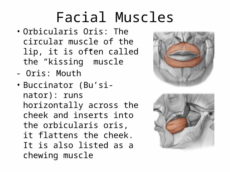
Facial Muscles• Orbicularis Oris: The
circular muscle of the lip, it is often called the “kissing” muscle
- Oris: Mouth• Buccinator (Bu’si-nator):
runs horizontally across the cheek and inserts into the orbicularis oris, it flattens the cheek. It is also listed as a chewing muscle

Facial Muscles
• Zygomaticus: extends from the corner of the mouth to the cheek bone. It is often referred to as the smiling muscle
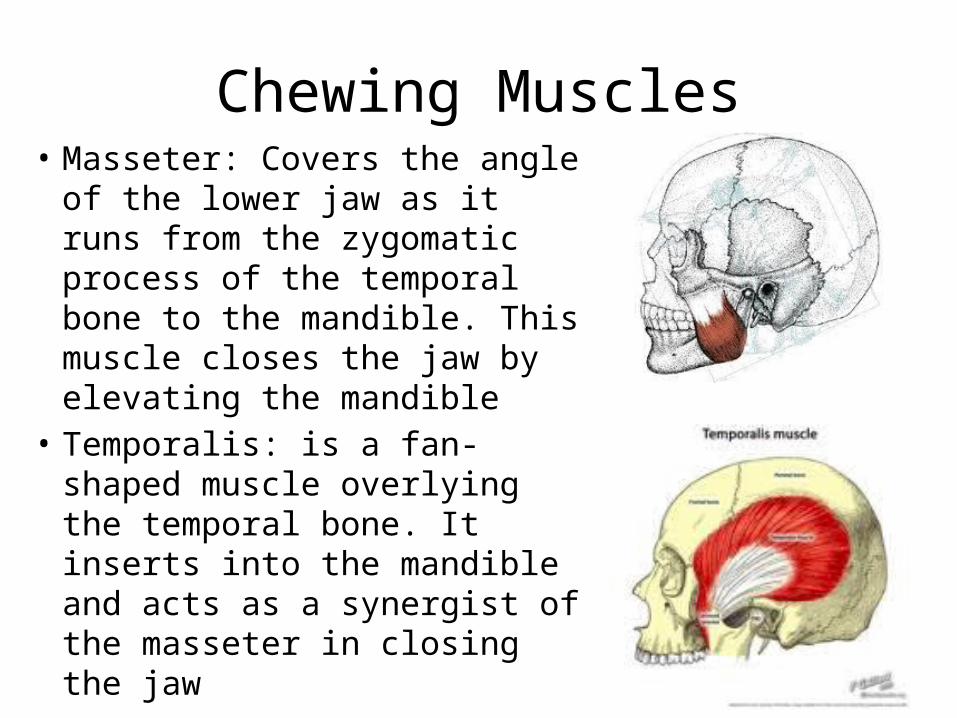
Chewing Muscles• Masseter: Covers the angle of
the lower jaw as it runs from the zygomatic process of the temporal bone to the mandible. This muscle closes the jaw by elevating the mandible
• Temporalis: is a fan-shaped muscle overlying the temporal bone. It inserts into the mandible and acts as a synergist of the masseter in closing the jaw

Trunk and Neck Muscles
Anterior Muscle
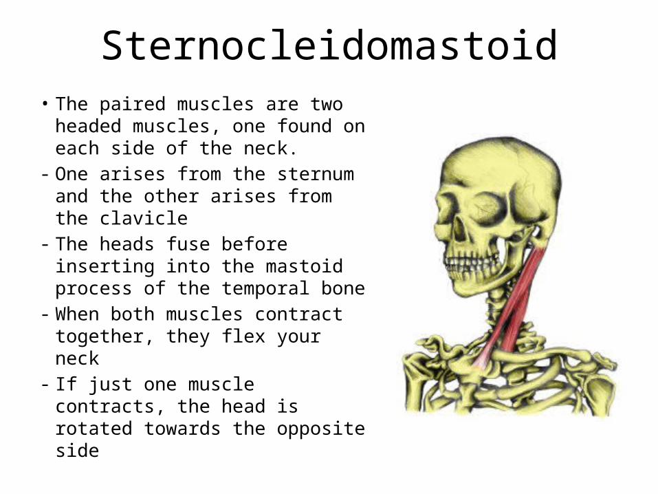
Sternocleidomastoid• The paired muscles are two
headed muscles, one found on each side of the neck.
- One arises from the sternum and the other arises from the clavicle
- The heads fuse before inserting into the mastoid process of the temporal bone
- When both muscles contract together, they flex your neck
- If just one muscle contracts, the head is rotated towards the opposite side

Pectoralis Major
• A large fan-shaped muscle covering the upper part of the chest
• Its origin is from the shoulder girdle and the first six ribs and inserts on the proximal end of the humerus
• It is the prime mover for shoulder flexion and adduction

Intercostal Muscles• Deep muscles found
between the ribs• The external intercostals
are important in breathing because they help to raise the rib cage for breathing air in
• The internal intercostals depress the rib cage, which helps to move air out of the lungs when you exhale

Muscles of the Abdominal Girdle• Rectus abdominis: the paired
strap-like muscles are the most superficial muscles of the abdomen.
• They run from the pubis to the rib cage
• Their main function is to flex the vertebral column
• They also compress the abdominal contents and are involved in forced breathing

• External oblique: are paired superficial muscles that make up the lateral walls of the abdomen
• Their fibers run downward and medially from the last eight ribs and insert into the ilium
• They flex the vertebral column but they also rotate the trunk and bend it laterally
Muscles of the Abdominal Girdle

• Internal oblique: Paired muscles deep to the external obliques
• Their fiber run at right angles to those of the external obliques
• They raise from the iliac crest and insert into the last three ribs
• Their functions are the same as those of the external obliques
Muscles of the Abdominal Girdle

• Transversus abdominis: The deepest muscle of the abdominal wall and has fibers that run horizontally across the abdomen
• It raises from the lower ribs and iliac crest and inserts into the pubis
• This muscle compresses the abdominal contents
Muscles of the Abdominal Girdle

Which one is which?
Internal Oblique
Transversus abdominis
External Oblique

Trunk and Neck Muscles
Posterior Muscle

Trapezius (Trah-pee-ze-us)• These muscles are the most
superficial muscles of the posterior neck and upper trunk
• When seen together, they form a diamond or kite-shaped muscle mass
• These muscles are antagonists of the sternocleidomastoids
• They also can elevate, depress, adduct and stabilize the scapula

Latissimus Dorsi• The large, flat muscle pair
that covers the lower back• It originates on the lower
spine and ilium and then sweeps superiorly to insert into the proximal end of the humerus
• It extends and adducts the shoulder and help maintain upright posture

Erector Spinae (spi’na)• It is a prime mover of back
extension.• Each erector spinae is a
composite muscle consisting of three muscle columns that collectively span the entire length of the vertebral column
• They act as powerful back extensors and also provide resistance that helps control the action of bending over at the waist

Deltoid
• They are fleshy, triangle-shaped muscles that form the rounded shape of your shoulders
• The deltoids are the prime movers of arm abduction
• It is the antagonist of latissimus dorsi
Muscles of the upper limb
Upper Limb Muscles
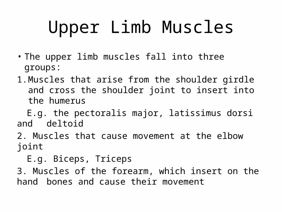
• The upper limb muscles fall into three groups:1. Muscles that arise from the shoulder girdle and
cross the shoulder joint to insert into the humerusE.g. the pectoralis major, latissimus dorsi and deltoid
2. Muscles that cause movement at the elbow joint E.g. Biceps, Triceps
3. Muscles of the forearm, which insert on the hand bones and cause their movement

Biceps Brachii (bra’ke-i)
• It originates by two heads from the shoulder girdle and inserts into the radial tuberosity.
• It is the powerful prime mover for flexion of the forearm and acts to supinate the forearm

• The only muscle fleshing out the posterior humerus.
• Its three heads arise from the shoulder girdle and proximal humerus and it inserts into the olecranon process of the ulna
Triceps Brachii

Muscles of the lower limb

Iliopsoas (il’e-o-so’us)• A fused muscle composed of
two muscles• It runs from the iliac bone and
lower vertebrae deep inside the pelvis to insert on the lesser trochanter of the femur
• It is a prime mover of hip flexion
• It also acts as a postural muscle to keep the upper body from falling backward when standing up

Adductor Muscles
• The muscle of the adductor group form the muscle mass at the medial side of each thigh
• They adduct the thighs together
• The adductors have their origin on the pelvis and insert on the femur

Gluteus Maximus
• It is a superficial muscle of the hip that forms most of the flesh of the buttock
• It is a powerful hip extensor that acts to bring the thigh in a straight line with the pelvis
• It originates from the sacrum and iliac bones and runs to insert on the gluteal tuberosity of the femur

Gluteus Medius
• It runs from the ilium to the femur, beneath the gluteus maximus for most of its length
• It is a hip abductor and is important in steadying the pelvis during walking
• It is an important site for giving intramuscular injections

Sartorius
• A thin, strap-like muscle• It runs obliquely across the
thigh from the anterior iliac crest to the medial side of the tibia
• It is a weak thigh flexor
Quadriceps Group
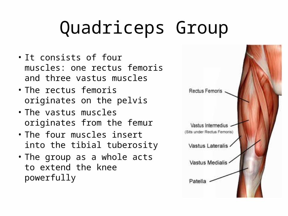
• It consists of four muscles: one rectus femoris and three vastus muscles
• The rectus femoris originates on the pelvis
• The vastus muscles originates from the femur
• The four muscles insert into the tibial tuberosity
• The group as a whole acts to extend the knee powerfully

Hamstring Group• The muscles forming the
muscle mass of the posterior thigh are the hamstrings
• It extends the thigh and flexes the knee
• The group consists of three muscles, the biceps femoris, semimembranosus and semitendinosus which originate on the ischial tuberosity and run down the thigh to insert on both sides of the proximal tibia

Tibialis Anterior
• It is a superficial muscle on the anterior leg. It arises from the upper tibia and then parallels the anterior crest as it runs to the tarsal bones where it inserts by a long tendon
• It acts to dorsiflex and invert the foot

Peroneus Muscles
• The peroneus muscles of this group are found on the lateral part of the leg
• They arise from the fibula and insert into the metatarsal bones of the foot
• The group as a whole plantar flexes and everts the foot

Gastrocnemius (gas’trok-ne’meus)
• It is a two bellied muscle that forms the curved calf of the posterior leg
• It arises by two heads, one from each side of the distal femur, and inserts through the large Achilles tendon into the heel of the foot
• It is also known as the toe dancers muscle


