Nanoparticles for Cancer Detection and Treatment Wole Soboyejo US/Africa Materials Institute (USAMI) Princeton Institute of Science and Technology of Materials (PRISM) and Department of Mechanical and Aerospace Engineering Princeton University
Transcript
Nanoparticles for Cancer Detection and Treatment
Wole Soboyejo
US/Africa Materials Institute (USAMI)Princeton Institute of Science and Technology of
Materials (PRISM) and
Department of Mechanical and Aerospace EngineeringPrinceton University
The Structure of USAMI• NSF funded virtual institute operated from Princeton• Princeton setting (usami.princeton.edu) includes
– PRISM (Jim Sturm, Kim Hegelbach, BarbaraVarga, Dan Steinberg, Shannon Swilley)
– MAE (Maureen Hickey, Jenny Kokini, Vocaturo, Whitehead)– Institute for Advanced Studies (Arlen Hastings & Phillip
Griffiths)– Carl Fields Center/Third World Center (Makeba Clay)
• Administrative/technical staff members– Dale Grieb (Administrator)– Laura Ceritto (Administrative Assistant)– Betty Adam (Secretary)– Eric Paul (Web Support)
U.S. Institutions/Collaborators• Ohio State University Soboyejo • Harvard University Vlassak, Hutchinson• Howard Mitchell• Brown Needleman • Yale University Ramirez• Columbia Kysar• Duke Warren• University of Michigan Forrest• LSU/CAMD Hormes, Kumar• UIUC Paulino• Rutgers University Cuitino• Sandia Buchheit, Boyce• Pennington Biomedical Leuschner
The Americas Program
• A complementary initiative of the NSF and other American agencies (Holmer Savastano et al.)
• Emerging U.S./South America collaboration in infrastructure materials– Focus on natural fiber reinforcement– Supports 5 South American researchers (per year) to
visit the U.S. for 9 weeks per year– Current collaborators from Brazil, Argentina and
Guadeloupe
International Collaborators• Eastern Africa – Kenya, Uganda, Ethiopia, Tanzania• Northern Africa – Egypt, Tunisia, Algeria, Morocco• Western Africa – Senegal, Nigeria, Ghana, Burkina Faso• Central Africa – Rwanda• Southern Africa – Zambia, Botswana, Mozambique,
South Africa, Namibia• South America - Brazil• Caribbean - Guadeloupe
Approach of The IMI Program
• 16 international researchers visit the U.S. to work with U.S. collaborators for 9 weeks
• They then return to their home countries to continue their work
• Many return over the next few years to do a complete piece of work
• A systems based approach - must work in one of the four areas of focus
The Areas of Focus of the IMI
• Advanced Materials/Small Structures– MEMS/thin films and organic electronics– Biomaterials
• Materials for Societal Development– Materials for affordable infrastructure– Thermostructural materials
Acknowledgments
• Post-docs – Jikou Zhou, Craig Steeves• Students - C. Milburn, S. Mwenifumbo, R. Weissbard, L.
Ionescu, L. Hayward, O. Bravo, J. Meng, C. Thieraux• Colleagues - Challa Kumar (CAMD), Josef Hormes
(CAMD), Carolla Leuschner (Pennington), Jeff Schwartz (Princeton), Julie Young (Princeton), Aboubaker Beye(Cheikh Anta Diop), Tom Otiti (Makerere), Warren Warren (Princeton), Bob Prud’homme (Princeton)
• MRI Technician - Silvia Cenzano (Princeton)• Financial Support - Carmen Huber (NSF) and Princeton
University
Background and Introduction
• Several people could benefit from implantable or injectable systems for disease detection and treatment
• This class examines nanotechnology for disease detection and treatment (with a focus on cancer)– Breast cancer– Prostate cancer
Our Approach to Early Cancer Detection and Treatment!
LP conjugates
LP conjugates
LP conjugatesLP conjugates
LP conjugates
LP conjugates LP conjugates
LP conjugates
LHRH
LHRHLHRH
LHRH
LHRH
LHRH LHRH
LHRH
Magnetic core
Polymer shellwith lytic peptideconjugates
Wet Chemical Synthesis of Nano-particles
Metallic, polymeric and metal-polymer Nano-particles using bottom-up approaches
Novel Micro reactor technology for scale-up and controlled synthesis
Synchrotron radiation based X-ray absorption Spectroscopic characterization
Capability to attach bio-molecules
In-Vitro Experiments
• Studied attachment of nano-particles in cell culture experiments
• Studied effects of temperature and time• Imaging done using TEM after fixing• Studies conducted on breast cancer cells
with LHRH receptors– Unconjugated nanoparticles– LHRH-coated nanoparticles
Confocal Movie of Cell Intake• Experimental process
① Cells grow in round flask overnight ② Stain cells with Cell Tracker Orange (Color Cytoplasm) ③ Settle cells in confocal microscope and add nanoparticles ④ Observation
• Nanoparticles start to enter cell at about 10 mins after being
added into the flask
⇒
⇒
5 mins 10 mins
2 hours
1 hour
2.5 hours
⇒
SubstrateCell
In-Vivo Experiments
• Mice injected in 4 different ways:1. LHRH nanoparticles2. saline solution3. nanoparticles4. LHRH nanoparticles but with
mice that do not contain breast tumor
Materials Characterization of Organs (TEM and Histology)
Organs obtained:– breast or prostate tumor– Kidney– Lung– Liver
Ensure that the nanoparticles do not accumulate in Ensure that the nanoparticles do not accumulate in other major organs.other major organs.
SPION/SPION-LHRH in Breast TumorSPION in Tumor LHRH-SPION in Tumor
SPION/SPION-LHRH in Breast Tumor
LHRH-SPION in Tumor LHRH-SPION in Tumor
SPION in Lung
SPION in Lung LHRH-SPION in Lung
SPION in LungLHRH-SPION in Lung LHRH-SPION in Lung
SPION/SPION-LHRH in LiverSPION in Liver LHRH-SPION in Liver
LHRH-SPION in Kidney
SPION in Kidney LHRH-SPION in Kidney
0
10
20
30
40
50
60
70
Tumor Lung Liver Kidney
Iron
Cont
ent (
%)
0
10
20
30
40
50
60
Tumor Lung Liver Kidney
Iron
Cont
ent (
%)
Biological Distribution of SPIONs
LHRH-SPION in Mouse SPION in Mouse
Targeted Destruction of Prostate Cancer in Balb/c athymic nude mice
CAMD
PC-3.luc Xenograft bearing male nude mice were used
LHRH bound nanoparticles effectively bind to tumor
Use of Nano-LHRH results in accumulation68% of nanoparticles in tumor
Distribution of iron in other tissues is being mapped
Introduction to MRI• How does MRI work?
– Interaction between external magnetic field and spins of protons in hydrogen
– Spins align due to the external field (z axis)
– RF pulse tips spins to x-y plane– After this pulse, spins relax back
• How does MRI get contrast? – Different tissue – hydrogen density
/ different relaxation property
• How do Contrast Agents work?– Change the relaxation property of
tissueJ. Phys. D: Appl. Phys. 36 (2003) R167–R181
Initial MRI Experiments: Cherry Tomato and Grape
• Injected grapes with saturated saline solution of nanoparticles
• Observed contrast at the location of the injection (nanoparticles)
The iron creates a magnetic field in the water, thus creating a blind spot (dark) for the MRI
T2 Images of Tumors – Contrast Enhancement Due to LHRH-MNPs
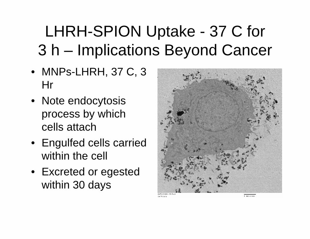
LHRH-SPION Uptake - 37 C for 3 h – Implications Beyond Cancer
• MNPs-LHRH, 37 C, 3 Hr
• Note endocytosisprocess by which cells attach
• Engulfed cells carried within the cell
• Excreted or egested within 30 days
Antagonistic Virus Control with Nanoparticles
Antagonistic Virus Control with Nanoparticles
Antagonistic Virus Control with Nanoparticles
• Tumor Reduction Due to Localized Drug Delivery
• Work of Langer et al., 2006
• USAMI goal is to use materials science approaches to explore ways of shrinking the tumor size to zero
• The other goal is to use localized delivery to reduce the side effects of chemotherapy
• Collaboration with Prud’homme
Triggered Drug Release & Hyperthermia
Combined chemotherapy + Heating of certain organs or tissues to temperatures between 41 and 43OC as a treatment of cancer