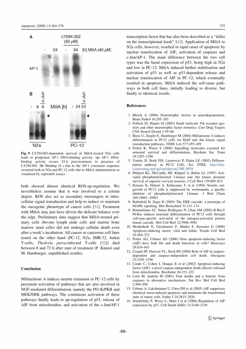
Natural Products AKTing on Signal Transduction Pathways- Molecules and Methods Inauguraldissertation zur Erlangung der Würde eines Doktors der Philosophie vorgelegt der Philosophisch-Naturwissenschaftlichen Fakultät der Universität Basel von Sabine Kiefer aus Starrkirch-Wil/ SO Zürich, 2009
Transcript
Natural Products AKTing on Signal Transduction Pathways-
Molecules and Methods
Inauguraldissertation
zur
Erlangung der Würde eines Doktors der Philosophie
vorgelegt der
Philosophisch-Naturwissenschaftlichen Fakultät
der Universität Basel
von
Sabine Kiefer
aus Starrkirch-Wil/ SO
Zürich, 2009
Genehmigt von der Philosophisch-Naturwissenschaftlichen Fakultät
das Werk vervielfältigen, verbreiten und öffentlich zugänglich machen
Zu den folgenden Bedingungen:
Namensnennung. Sie müssen den Namen des Autors/Rechteinhabers in der von ihm festgelegten Weise nennen (wodurch aber nicht der Eindruck entstehen darf, Sie oder die Nutzung des Werkes durch Sie würden entlohnt).
Keine kommerzielle Nutzung. Dieses Werk darf nicht für kommerzielle Zwecke verwendet werden.
Keine Bearbeitung. Dieses Werk darf nicht bearbeitet oder in anderer Weise verändert werden.
• Im Falle einer Verbreitung müssen Sie anderen die Lizenzbedingungen, unter welche dieses Werk fällt, mitteilen. Am Einfachsten ist es, einen Link auf diese Seite einzubinden.
• Jede der vorgenannten Bedingungen kann aufgehoben werden, sofern Sie die Einwilligung des Rechteinhabers dazu erhalten.
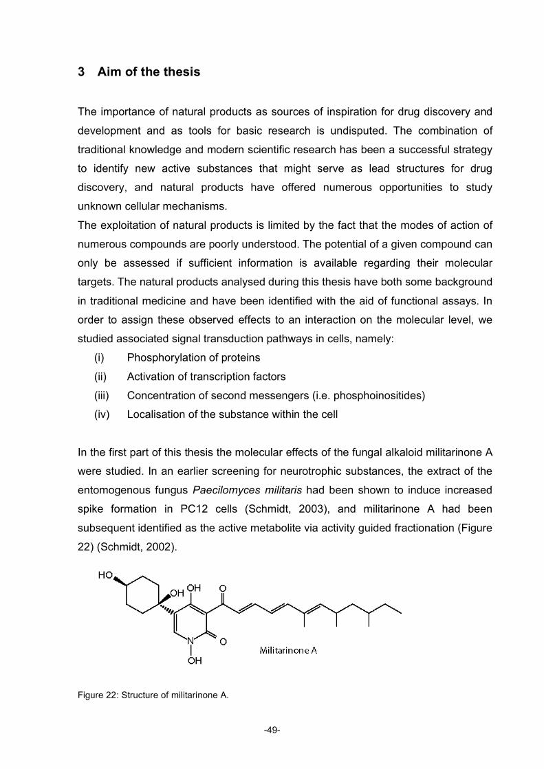
• Diese Lizenz lässt die Urheberpersönlichkeitsrechte unberührt.
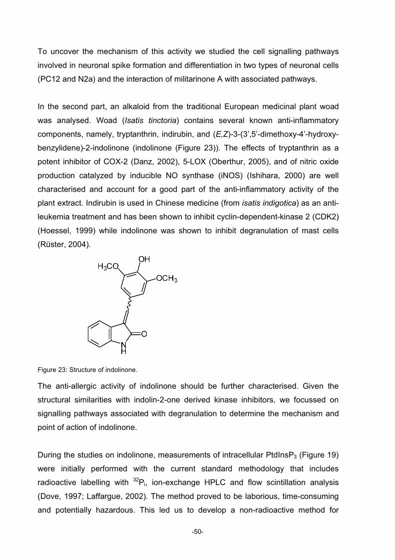
Die gesetzlichen Schranken des Urheberrechts bleiben hiervon unberührt.
Die Commons Deed ist eine Zusammenfassung des Lizenzvertrags in allgemeinverständlicher Sprache: http://creativecommons.org/licenses/by-nc-nd/2.5/ch/legalcode.de
Haftungsausschluss:Die Commons Deed ist kein Lizenzvertrag. Sie ist lediglich ein Referenztext, der den zugrundeliegenden Lizenzvertrag übersichtlich und in allgemeinverständlicher Sprache wiedergibt. Die Deed selbst entfaltet keine juristische Wirkung und erscheint im eigentlichen Lizenzvertrag nicht. Creative Commons ist keine Rechtsanwaltsgesellschaft und leistet keine Rechtsberatung. Die Weitergabe und Verlinkung des Commons Deeds führt zu keinem Mandatsverhältnis.
3 Separation and Detection of all Phosphoinositide Isomers.............................. 87
C. CONCLUSIONS .............................................................................................. 105
D. ACKNOWLEDGEMENTS................................................................................ 109
E. CURRICULUM VITAE ..................................................................................... 111
iv
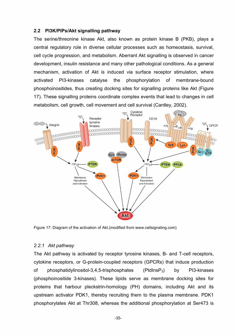
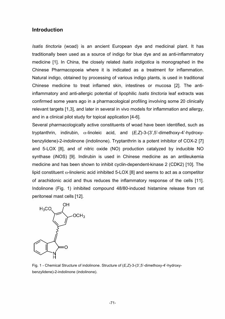
Table of Figures Figure 1: Diagram describing the roles of natural products in drug discovery and development ............2 Figure 2: Structures of compactin, mevilonin and simvastatin...................................................................5 Figure 3: Structure of paclitaxel ....................................................................................................................6 Figure 4: Structure of rapamycin...................................................................................................................8 Figure 5: Structures of artemisinin, artemether, and artesunate ................................................................8 Figure 6: Structures of sphingosine, myriocin and fingolimod ..................................................................10 Figure 7: Structures of gossypol .................................................................................................................11 Figure 8: Structure of bryostatin-1 and its analogue..................................................................................12 Figure 9: Structures of wortmannin (left) and staurosporine (right) ..........................................................14 Figure 10: Structure of the phorbol ester 4β-12-O-tetradecanoylphorbol-13-acetate (TPA) ..................15 Figure 11: Structure of the marine phosphatase inhibitor okadaic acid ...................................................16 Figure 12: Simplified diagram of apoptosis pathways ...............................................................................25 Figure 13: Mitochondrial pathway of apoptosis..........................................................................................26 Figure 14: Extrinsic pathway and possible points of inhibition..................................................................28 Figure 15: Regulation of p53 .......................................................................................................................30 Figure 16: Regulation of NFκB....................................................................................................................31 Figure 17: Diagram of the activation of Akt ................................................................................................34 Figure 18: Effects of Akt activation on different cellular functions ............................................................35 Figure 19: Structure of PIs...........................................................................................................................36 Figure 20: Interconversions of all the PIs ...................................................................................................37 Figure 21: Localisation of the specific PtdIns at different membranes in the cell....................................39 Figure 22: Structure of militarinone A .........................................................................................................48 Figure 23: Structure of indolinone...............................................................................................................49
v
Abbreviations A3AR A3 adenosine receptor AD Alzheimer's disease Ade Adenosine AIF Apoptosis inducing factor AP-1 Activator protein 1 Apaf-1 Apoptotic protease-activating factor-1 ARE Antioxidant responsive element AS160 Akt substrate of 160 kDa ATP Adenosine-5'-triphosphate BH Bcl-2 homology BMMC Murine bone marrow derived mast cells BPC Base peak chromatogram BSA Bovine serum albumin BTK Bruton’s tyrosine kinase CDK2 Cyclin-dependent-kinase 2 c-FLIP Cellular caspase-8 (FLICE)-like inhibitory protein CMT Charcot-Marie-Tooth CNS Central nervous system DAG Diacylglycerol DIABLO Direct IAP binding protein with low pI DISC Death inducing signalling complex DMEM Dulbecco's Modified Eagle Medium DMHA N,N-dimethy-hexylamine DNA Deoxyribonucleic acid DNP Dinitrophenyl EIC Extracted ion chromatogram ELSD Evaporative light scattering EMSA Electrophoretic mobility shift assay ESI Electrospray ionisation FACS Flow cytometry (fluorescence activated cell sorting) FADD Fas-associated death domain FasL Fas-ligand FCS Foetal calf serum FDA U.S. Food and Drug Administration FITC Fluorescein isothiocyanate FLICE FADD-like interleukin-1 beta-converting enzyme fMLP N-formyl-methionyl-leucyl-phenylalanine FoxO1 Forkhead box O1 FRAP FKBP12-rapamycin-associated protein GPCR G-protein-coupled receptor GSK3 Glycogen synthase kinase 3 GTP Guanosine-5'-triphosphate HIV Human immunodeficiency virus HMG-CoA 3-hydroxy-3-methylglutaryl-coenzyme A HPLC High performance liquid chromatography
vi
HS Horse serum HTS High throughput screening IAP Inhibitor of apoptosis IκB Inhibitor of NFκB IKK IκB-kinase IL2 Interleukin 2 IL3 Interleukin 3 IMDM Iscove's modified Dulbecco's Medium IP3 Inositol-(1,4,5)-trisphosphate ITAM Immunoreceptor tyrosine-based activation motif JNK c-Jun N-terminal kinase LC Liquid chromatography LDL Low-density lipoprotein MAPK Mitogen-activated protein kinase MiliA Militarinone A MOMP Mitochondrial outer membrane permeabilisation MS Mass spectrometry mTOR Mammalian target of rapamycin NAC N-acetyl-L-cysteine NFκB Nuclear factor kappa B NGF Nerve growth factor OCRL Occulocerebrorenal syndrome of Lowe OTC Over-the-counter PDK1 3-phosphoinositide dependent protein kinase-1 PFTa Pifithrin a PH Pleckstrin-homology PI Phosphoinositide PI3-K Phosphoinositide 3-kinase PKB Protein kinase B PKC Protein kinase C PLC Phospholipase C PMA Phorbol 12-myristate-13-acetate PP Protein serine/threoinine phosphatase PtdIns Phosphatidylinositol PtdIns(3,4)P2 Phosphatidylinositol-3,4-bisphosphate PtdIns(3,5)P2 Phosphatidylinositol-3,5-bisphosphate PtdIns(4,5)P2 Phosphatidylinositol-4,5-bisphosphate PtdIns3P Phosphatidylinositol-3-phosphate PtdIns4P Phosphatidylinositol-4-phosphate PtdIns5P Phosphatidylinositol-5-phosphate PtdInsP3 Phosphatidylinositol-3,4,5-trisphosphates PTEN Phosphatase and tensin homologue deleted on chromosome ten ROS Reactive oxygen species RP Reversed phase S1P Sphingosine-1-phosphate SERCA Sarco/endoplasmatic reticulum Ca2+ -ATPase SHIP SH2-containing inositol 5-phosphatase
vii
Smac Second mitochondrial-derived activator of caspases SphK Sphingosine kinase TNF Tumour necrosis factor TNFR TNF-receptor TOR Target of rapamycin TORC1 mTOR complex 1 TORC2 mTOR complex 2 TPA 4β-12-O-tetradecanoylphorbol-13-acetate TRAIL TNF-related apoptosis ligand v-FLIP Viral caspase-8 (FLICE)-like inhibitory protein wort Wortmannin
viii
A. INTRODUCTION
1 Natural Products in Research and Drug Discovery
˝Embrace the Challenge˝ -Teachings of Anusara Yoga
The therapeutic use of plants is as old as human civilisation. Even today plants
remain the primary sources of health care for most people in the world. It is estimated
that around 80% of the world’s population rely mainly on traditional medicine for their
primary health care (Wang, 2008). Over the centuries, natural products have
provided a tremendous amount of substances that serve as medicine or as lead
structure for drug development. Around one third of the currently marketed drugs
have structures that are related or derived from natural products (Onaga, 2001).
1.1 History Natural products have been the first and, for a long time, sole source of medicine.
The exploitation of traditional medicines for the development of modern drugs
produced the first commercially available pure drug substances. The isolation of
morphine from the opium latex by the German Pharmacist Sertürner in 1805 could be
seen as the start of pharmaceutical natural product research. Shortly thereafter the
isolation of many other substances followed, atropine in 1819, quinine and caffeine in
1820, and digitoxin in 1841 (Potterat and Hamburger, 2008). Quinine was isolated in
1820 from the bark of several Cinchona species that have been used by Peruvian
Indians to treat shivering and malarial fevers (Corson and Crews, 2007; Greenwood,
1992). In 1826, quinine and morphine became the first commercially available pure
natural compounds produced by Caventou and Merck, respectively (Newman, 2000;
Potterat and Hamburger, 2008).
The synthetic modification of salicylic acid to acetylsalicylic acid in 1897, and the
modification of morphine into diacetylmorphine (heroin) in 1898 by Hoffmann at
Bayer AG produced the first semi-synthetic natural-product derived drugs (Schmidt,
2008; Sneader, 2000).
-1-
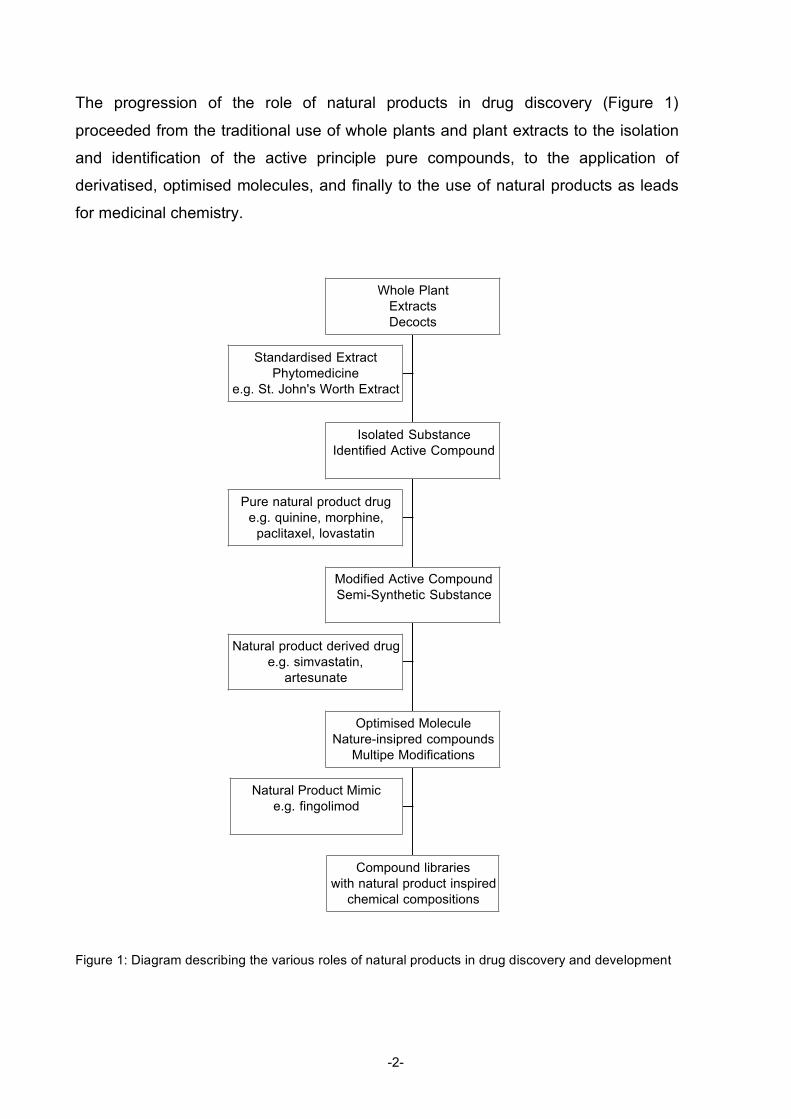
The progression of the role of natural products in drug discovery (Figure 1)
proceeded from the traditional use of whole plants and plant extracts to the isolation
and identification of the active principle pure compounds, to the application of
derivatised, optimised molecules, and finally to the use of natural products as leads
for medicinal chemistry.
Figure 1: Diagram describing the various roles of natural products in drug discovery and development
Standardised Extract
Phytomedicine
e.g. St. John's Worth Extract
Pure natural product drug
e.g. quinine, morphine,
paclitaxel, lovastatin
Natural product derived drug
e.g. simvastatin,
artesunate
Natural Product Mimic
e.g. fingolimod
Compound libraries
with natural product inspired
chemical compositions
Optimised Molecule
Nature-insipred compounds
Multipe Modifications
Modified Active Compound
Semi-Synthetic Substance
Isolated Substance
Identified Active Compound
Whole Plant
Extracts
Decocts
-2-
1.2 Today
After decades of very successful drug discovery and development, the
pharmaceutical industry downscaled natural product research in the late 1990s in
favour of automated high throughput screening (HTS) of compound libraries (Baker,
2007). Compound libraries assembled with the aid of combinatorial chemistry were
thought to produce more hits than ‘old fashioned’ natural products. Despite this
decline in the use of natural products in drug discovery, newly marketed drugs
derived from natural compounds hold about the same share as before (Newman,
2003).
The numerous and successful discoveries of compounds in the early times of
modern drug discovery were quite exclusively based on the traditional use of the
plant (Rishton, 2008). Of all known organic molecules, only 1% are natural products,
99% are synthetic (von Nussbaum, 2006), but more than one third of all drug sales
are based on natural products (Newman, 2003). How can these striking numbers be
explained? Evolutionary selection is the answer; nature’s own high-throughput
screening has optimized these biologically active compounds (Paterson and
Anderson, 2005). Especially the numerous compounds with antibacterial activity do
not surprise, as fighting for space and resources, and against other organisms, plays
a pivotal role in survival.
Early attempts to apply HTS to botanical extracts were faced with many difficulties.
With the introduction of biochemical assays in the 90s, the screening process had
shifted from functional cellular assays to cell-free biochemical assay formats, which
are very sensitive and prone to artefacts. The typically coloured plant extracts are not
compatible with such screening assays due to interference with detection caused by
colour, fluorescence, or quenching effects of components in the extract. Moreover
the complexity of extracts potentially induces aggregation of components, chemical
reactions within the assay or difficulties of solubility in assay buffer (Rishton, 2008).
Pre-fractionation or purification to reduce the chemical complexity of the extracts
needs to be implemented before HTS can be performed. This is time-consuming and
laborious and generally reduces the attractiveness of screening of natural
compounds. Another approach is to use the power of combinatorial chemistry in
combination with knowledge on active natural products and create a library that
extends upon the structural properties of known natural compounds. This strategy
-3-
generates libraries with enhanced specificity and selectivity (Koehn and Carter,
2005).
1.3 Marketed Drugs Here I briefly describe selected examples of successfully marketed drugs that are
natural compounds, derivatives of natural compounds, or synthetic molecules for
which the lead was a natural product. The focus here is on some more recent drugs
that had a major impact on human lives, while the more historical and well-known
examples such as morphine, penicillin, quinine, streptomycin and others are left out.
1.3.1 Lovastatin (FDA Approval 1987)
Since the discovery of a correlation between high cholesterol levels and coronary
heart disease in the 1950s, the lowering of high cholesterol levels with drugs has
been pursued (Kannel, 1995; Keys, 1984). The cholesterol biosynthesis is a complex
process involving more than 30 enzymes and was discovered during the 1950s and
60s (Russell, 1992). HMG-CoA (3-hydroxy-3-methylglutaryl-coenzyme A) reductase
is the rate-limiting enzyme of the biosynthetic pathway and hence most suitable for
inhibition. Furthermore, when HMG-CoA reductase is inhibited an alternative
pathway for degradation of the substrate is available which prevents accumulation of
HMG-CoA.
A potent inhibitor of HMG-CoA reductase, compactin (later additionally named
mevastatin (Figure 2)), was found in a broth of Penicillium citrinum (Endo, 1976).
Soon after, in 1978, another HMG-CoA reductase inhibitor was discovered in the
Merck laboratories from Aspergillus terreus and named mevilonin (lovastatin (Figure
2)) (Alberts, 1980). Compactin was highly effective in lowering plasma cholesterol in
animal models as well as patients with hypercholesterolaemia (Kuroda, 1979;
Mabuchi, 1981; Mabuchi, 1983; Tsujita, 1979; Watanabe, 1981). However,
compactin was withdrawn from clinical trials in 1980 due to unpublished reasons
(Tobert, 2003). Because of the structural similarity between compactin and lovastatin,
clinical studies with lovastatin had to be stopped as well. After additional animal
studies and some investigation in small-scale high-risk patient studies, clinical
development was re-launched in 1983 until, finally, in 1987 the FDA (U.S. Food and
Drug Administration) approval for lovastatin was obtained (Illingworth and Sexton,
1984; Thompson, 1986). Soon after, other statins from microbial sources were
-4-
released. Simvastatin (Figure 2) entered the market in 1988, it is semi-synthetically
derived from lovastatin introducing a minor side chain modification. In 1991
pravastatin followed, with a modification in the side chain ring. Later, synthetically
designed products with different chemical structures followed; fluvastatin in 1994,
atrovastatin in 1997, cerivastatin in 1998 and rosuvastatin in 2003.
Figure 2: Structures of compactin, mevilonin and simvastatin
The mechanism of action of statins goes beyond blocking cholesterol biosynthesis.
Inhibition of HMG-CoA reductase reduces levels of mevalonate, which in turn leads
to upregulation of low-density lipoprotein (LDL) receptors on hepatocytes. The
upregulation of LDL receptors increases the uptake of LDL from blood, the major
marker of elevated cholesterol levels (Brown and Goldstein, 1980; Reihner, 1990).
With the discovery of lovastatin it was for the first time possible to achieve large
reductions in plasma cholesterol of up to 40% (Tobert, 1982). The treatments
formerly available were all of limited efficacy or tolerability. The bile acid sequestrants
are moderately effective and poorly tolerated due to gastrointestinal side effects
whereas fibrates produce a rather small reduction in LDL-cholesterol but are well
tolerated and widely used. The statins revolutionised the treatment of
hypercholesterinaemia and annual sales are > 15 billion US $ (Downton and Clark,
2003). In 2001, however, cerivastatin, only introduced in 1998, had to be withdrawn
from the market due to severe side effects of rhabdomyolysis which occurred in
concomitant use with gemfibrozil (Furberg and Pitt, 2001). The mechanism for this
side effect still remains elusive but further studies demonstrated the safety of other
statins. Nevertheless myalgia as a side effect under statin therapy occurs but seldom
develops into severe myolysis.
-5-
The statins became the most effective drugs so far for preventing and halting
arteriosclerosis. Despite the wide use of these drugs it is believed that they are
underutilised in patients who are free of symptoms with only moderately elevated
cholesterol levels. This led to the decision to approve simvastatin as an ‘over-the-
counter’ (OTC) medicine, available without prescription, in the UK in July 2004 (Link,
2004; Roberts, 2004).
1.3.2 Paclitaxel (FDA Approval 1992)
Plants have a long history in the use of cancer treatment. The first plant derived drug
to treat cancer was the Vinca alkaloid vincristine, which was approved for clinical use
in 1963. A more recent discovery of a plant-derived chemotherapeutic agent was
paclitaxel (Figure 3) from Taxus brevifolia bark (Wani, 1971). Paclitaxel was shown to
stabilise microtubule assembly, whereas Vinca alkaloids and colchicin prevent the
assembly of microtubules (Schiff, 1979; Schiff and Horwitz, 1981). Even in absence
of essential GTP, paclitaxel promotes microtubule assembly. Although paclitaxel
shows no structural resemblance to GTP, it is able to interact specifically with the β-
subunit of microtubules, a region that is associated with GTP binding and hydrolysis
(Snyder, 2001). The stabilisation of microtubules by paclitaxel forces the tumour cell
into multiple DNA replication cycles that eventually initiate apoptosis (Stewart, 1999).
Figure 3: Structure of paclitaxel
Clinical trials with paclitaxel were started in the early 1980s, and FDA approval for
treatment of refractory ovarian cancer was granted in 1992. Since the introduction of
paclitaxel to the treatment of ovarian cancer the survival rate has more than doubled
(Crown and O'Leary, 2000), and further applications have been approved since.
-6-
Today, paclitaxel is also used in the treatment of breast and colon cancers as well as
Kaposi’s sarcomas of HIV infected patients (Oberlies and Kroll, 2004).
Preparation of sufficient amounts of paclitaxel to launch clinical studies was nearly
impossible, as isolation from the bark results in very low yields and excoriation
causes the trees to die. In 1986, the precursor deacetyl baccatin III was isolated from
the needles of Taxus baccata, and the semi-synthetic approach lead to the
production of sufficient amount of paclitaxel from renewable sources (Gueritte-
Voegelein, 1991). Furthermore, semisynthesis enabled the creation of an analogue
of paclitaxel, docetaxel (Bissery, 1991), which entered the market in 1996.
From the perspective of both basic science and clinics, paclitaxel has led to
significant progress in understanding and treating cancer.
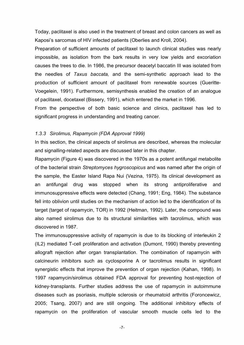
1.3.3 Sirolimus, Rapamycin (FDA Approval 1999)
In this section, the clinical aspects of sirolimus are described, whereas the molecular
and signalling-related aspects are discussed later in this chapter.
Rapamycin (Figure 4) was discovered in the 1970s as a potent antifungal metabolite
of the bacterial strain Streptomyces hygroscopicus and was named after the origin of
the sample, the Easter Island Rapa Nui (Vezina, 1975). Its clinical development as
an antifungal drug was stopped when its strong antiproliferative and
immunosuppressive effects were detected (Chang, 1991; Eng, 1984). The substance
fell into oblivion until studies on the mechanism of action led to the identification of its
target (target of rapamycin, TOR) in 1992 (Heitman, 1992). Later, the compound was
also named sirolimus due to its structural similarities with tacrolimus, which was
discovered in 1987.
The immunosuppressive activity of rapamycin is due to its blocking of interleukin 2
(IL2) mediated T-cell proliferation and activation (Dumont, 1990) thereby preventing
allograft rejection after organ transplantation. The combination of rapamycin with
calcineurin inhibitors such as cyclosporine A or tacrolimus results in significant
synergistic effects that improve the prevention of organ rejection (Kahan, 1998). In
1997 rapamycin/sirolimus obtained FDA approval for preventing host-rejection of
kidney-transplants. Further studies address the use of rapamycin in autoimmune
diseases such as psoriasis, multiple sclerosis or rheumatoid arthritis (Foroncewicz,
2005; Tsang, 2007) and are still ongoing. The additional inhibitory effects of
rapamycin on the proliferation of vascular smooth muscle cells led to the
-7-
development of rapamycin as antirestenosis drug, and coronary-artery stents
releasing rapamycin are approved in surgery since 2003 (Morice, 2002).
Figure 4: Structure of rapamycin
1.3.4 Artemisin (Swissmedic Approval 2000, no FDA Approval)
The Chinese medicinal herb qing hao (Artemisia annua) was traditionally used to
reduce fever and, in 1596, was mentioned for the first time to treat malaria (Klayman,
1985). In 1972, Chinese scientists managed to isolate the active principle of the herb
and called it qinghaosu, meaning ‘the active principle of qing hao’ (1979), named
artemisinin (Figure 5) for the Western world. The structure was elucidated in 1980
and revealed to be a sesquiterpene structure with an unusual endoperoxide group
(Acton and Klayman, 1985). The high lipophilicity of artemisinin made administration
as a drug difficult; therefore, various derivatives were synthesised, including
arthemether, arteether and artesunate (Figure 5).
Figure 5: Structures of artemisinin, artemether, and artesunate
-8-
The biological activity of the artemisinins depends on the cleavage of the peroxide
bond after contact with iron-II-hem within the parasite. The generated free radical
alkylates the hem molecule or parasite proteins (Bhisutthibhan, 1998; Olliaro, 2001).
Inhibition of the sarco/endoplasmatic reticulum Ca2+ -ATPase (SERCA) has been
proposed as an additional target (Eckstein-Ludwig, 2003). The active metabolite
dihydroartemisinin kills nearly all asexual stages of parasite lifecycle in the blood, and
also affects the gametocytes, which are responsible for the infection of the
Anopheles mosquito and transmit the disease. Furthermore the artemisinins act
faster than any other antimalarial drug with a fever and parasite clearance time of
less than two days (Wiesner, 2003). However, due to the short plasma half-life of
these drugs therapy needs to be continued for 5-7 days, or needs to be combined
with other antimalarial drugs (White, 2008). The combination usually applied is
artesunate-lumefantrine. In 2000 Swissmedic approved the drug for sale under the
name Riamet; in other countries it is sold as Coartem. At the moment, Novartis is still
awaiting FDA approval for Coartem.
1.4 Promising Research The examples discussed here were selected for their uniqueness, as they all
represent the first substances in clinical development with their respective mode of
action. Mostly the understanding of their molecular target has evolved concurrently
with the discovery of the substance and the subsequent studies of the
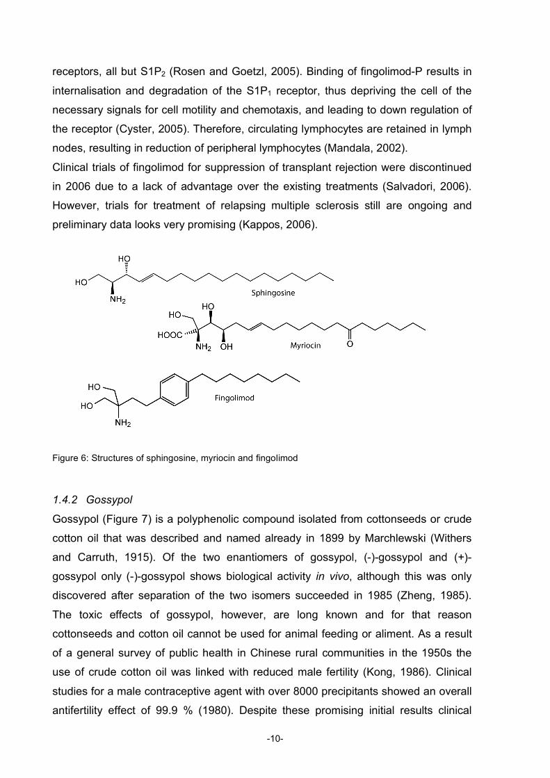
pharmacological and clinical effects. 1.4.1 Fingolimod
Fingolimod is a synthetic compound inspired by the structure of myriocin (Figure 6),
which is produced by the fungus Isaria sinclarii (Fujita, 1994). Myriocin is a structural
analogue of sphingosine, a C18 amino alcohol, which is a part of sphingolipids. In
vivo myriocin caused severe intoxication resulting in death of the animals. Because
of potent immunosuppressive activity in vitro, synthetic modifications were introduced
to reduce toxicity (Chiba, 1996). Fingolimod (FTY720 (Figure 6)) is such a synthetic
analogue that only obtains its immunosuppressive activity after phosphorylation by
sphingosine kinase 2 in vivo (Suzuki, 1996; Zemann, 2006). The biologically active
fingolimod-phosphate binds to four of the five known sphingosine-1-phosphate (S1P)
-9-
receptors, all but S1P2 (Rosen and Goetzl, 2005). Binding of fingolimod-P results in
internalisation and degradation of the S1P1 receptor, thus depriving the cell of the
necessary signals for cell motility and chemotaxis, and leading to down regulation of
the receptor (Cyster, 2005). Therefore, circulating lymphocytes are retained in lymph
nodes, resulting in reduction of peripheral lymphocytes (Mandala, 2002).
Clinical trials of fingolimod for suppression of transplant rejection were discontinued
in 2006 due to a lack of advantage over the existing treatments (Salvadori, 2006).
However, trials for treatment of relapsing multiple sclerosis still are ongoing and
preliminary data looks very promising (Kappos, 2006).
Figure 6: Structures of sphingosine, myriocin and fingolimod
1.4.2 Gossypol
Gossypol (Figure 7) is a polyphenolic compound isolated from cottonseeds or crude
cotton oil that was described and named already in 1899 by Marchlewski (Withers
and Carruth, 1915). Of the two enantiomers of gossypol, (-)-gossypol and (+)-
gossypol only (-)-gossypol shows biological activity in vivo, although this was only
discovered after separation of the two isomers succeeded in 1985 (Zheng, 1985).
The toxic effects of gossypol, however, are long known and for that reason
cottonseeds and cotton oil cannot be used for animal feeding or aliment. As a result
of a general survey of public health in Chinese rural communities in the 1950s the
use of crude cotton oil was linked with reduced male fertility (Kong, 1986). Clinical
studies for a male contraceptive agent with over 8000 precipitants showed an overall
antifertility effect of 99.9 % (1980). Despite these promising initial results clinical
-10-
studies as male contraceptive were discontinued in 1998 due to sustained infertility
after prolonged use of gossypol (Porat, 1990). One possible explanation for the
inhibition of spermatogenesis is the suppression of telomerase activity by gossypol
(Moon, 2008). The reverse transcriptase telomerase is only found in embryonal,
gonadal and cancer cells, as it enables unlimited cell division (Mego, 2002).
Telomerase inhibition, however, is an interesting target in cancer therapy and
gossypol was therefore tested in several cancer models as well as in vivo against
diffuse large cell lymphoma, head and neck squamous cell carcinoma, and breast
cancer (Mohammad, 2005; Van Poznak, 2001; Wolter, 2006).
Figure 7: Structure of gossypol
In a screening of a small natural product library gossypol showed potent inhibition of
the antiapoptotic proteins Bcl-2 and Bcl-xL (Kitada, 2003). It binds to the BH3 binding
domain of the antiapoptotic Bcl-2 family members, where the pro-apoptotic Bcl-2
family member Bid would bind to induce apoptosis. Gossypol is the first substance
found to mimic BH3-binding to Bcl-2, Bcl-xL, Bcl-w, and Mcl-1 and serves now as
lead substance for further development of small inhibitory molecules (Marzo and
Naval, 2008). Currently, gossypol is evaluated as the first Bcl-2 inhibitor in clinical
trials against various cancers.
1.4.3 Bryostatin-1
The macrocyclic lactone bryostatin-1 (Figure 8) was isolated and identified in 1982 by
Pettit et al. (Pettit, 1982) from the marine ‘moss-animal’ (Ectoprocta or Bryozoa)
Bugula neritina, which was collected in the Gulf of Mexico in 1968. Bryozoans are
aquatic colonial animals that are abundant in marine enviroment. Already in 1970 the
antineoplastic effect of Bugula neritina extract was shown to lead to prolonged
survival in a leukaemia mouse model (Pettit, 1970). Like the phorbol esters,
-11-
bryostatin-1 can be a potent activator of protein kinase C (PKC) (Berkow and Kraft,
1985). Prolonged exposure to bryostatin-1, however, induces PKC inhibition by
degradation and subsequent downregulation of PKC (Isakov, 1993). Furthermore,
bryostatin-1 inhibits phorbol ester induced tumourgenesis and differentiation of
promyelocytic leukaemia cells (Hennings, 1987; Kraft, 1987). Bryostatin-1 was shown
to bind to the ‘phorbol ester receptor’, which means the diacylglycerol (DAG)- binding
pocket of PKC (de Vries, 1988) and, when bound to PKC, induces PKC-degradation
by ubiquitination (Lee, 1996a; Lee, 1996b).
Figure 8: Structures of bryostatin-1 and its analogue, synthesised by Wender 2004.
Clinical phase II studies with single-agent bryostatin-1 have been conducted for
melanoma, renal cell carcinoma, colorectal cancer, and non-Hodgkin’s lymphoma,
mostly with disappointing results (Kortmansky and Schwartz, 2003). But co-
administration of bryostatin-1 with other cytotoxic agents produced promising results;
particularly the combinations of bryostatin-1/paclitaxel and bryostatin-1/temsirolimus
proved to be effective and are currently evaluated in clinical trials (Ku, 2008).
Development of analogues of bryostatin-1 (Figure 8) by Wender et al. (Wender,
2004) led to simplifications of parts of the structure that allow large-scale synthesis
but preserve the crucial parts for bioactivity (Paterson and Anderson, 2005).
Additionally, these studies created a better understanding of the structure-activity
relationship of PKC inhibition and most likely will generate bryostatin analogues for
clinical development.
Besides development as an anticancer drug bryostatin-1 is also under investigation
as a central nervous system (CNS) drug (Sun and Alkon, 2006). Numerous reports
imply a critical role of PKC malfunctions in the development of Alzheimer’s disease
(AD) (Cole, 1988; Favit, 1998; Lee, 2004). The PKC modulating effects of bryostatin-
-12-
1 seems to promote memory-enhancing and mood regulation effects (Sun and Alkon,
2005). Although passage of Bryostatin-1 through the blood-brain barrier was not
determined in this study, previous studies with mice indicate that bryostatin-1 can
pass across the blood brain barrier, but the brain levels of the drug were much lower
than plasma levels (Zhang, 1996). Development of analogues of bryostatin-1 may
result in compounds with improved CNS permeability and that could produce the
desired effects at much lower doses.
1.5 Natural products as research tool
The elucidation of signal transduction pathways uses several tools of molecular
biology, such as gene knockdown, overexpression of proteins, and the use of specific
inhibitors of certain signalling molecules. All have been helpful to trace the function of
pathways in vivo (Levine, 2007). Especially inhibitors of specific signal transduction
molecules have offered opportunities for studying the signal transduction
mechanisms. The example of rapamycin, its biological activity, the detection of its
target mTOR, the mammalian target of rapamycin, and the following identification of
a novel signalling cascade involved in fundamental processes of growth and
development, shows the value of natural products like rapamycin as research tool.
1.5.1 Rapamycin
Rapamycin (also named sirolimus) was discussed as an immunosuppressive
treatment earlier in this chapter (Chapter 1.3.3; Figure 4). Here the impact of the
discovery of rapamycin on biological research shall be highlighted.
The target of rapamycin (TOR) was identified in the budding yeast Saccharomyces
cervisiae in the 1990s (Heitman, 1992), and subsequent studies in mammalian cells
led to the identification of the mammalian TOR (mTOR) (Sabers, 1995). Since
several groups cloned the gene at about the same time, TOR is also known as FRAP
(FKBP12-rapamycin-associated protein), RAFT (rapamycin and FKBP12 target),
RAPT (rapamycin target), and SEP (sirolimus effector protein) (Fingar and Blenis,
2004). TOR is a 290 kDa large member of the PI3K-kinase-related-kinase (PIKK)
superfamily and is 40-60% identical amongst mammals, flies, worms and yeast
(Wullschleger, 2006). Two different TOR complexes are formed, in yeast containing
two different TORs, TOR1 and TOR2. In mammals mTOR is associated with raptor
forming the mTORC1 complex or with rictor, forming the mTORC2 complex. In cells
-13-
rapamycin forms a complex with a cofactor, FKBP12, and binds to TOR resulting in
the inhibition of TOR. In mammalian cells rapamycin only inhibits the mTORC1
complex, but not the mTORC2.
Rapamycin treatment results in cell cycle arrest in late G1 phase (Dumont and Su,
1996), because mTOR initiates the signal for translation of key mRNAs required for
cell cycle progression from G1 to S phase. In addition, rapamycin blocks cyclin-
dependent activation and accelerates the turnover of cyclin D, resulting in growth
arrest in G1 phase of the cell cycle (Rowinsky, 2004).
1.5.2 Wortmannin
The fungal metabolite wortmannin (Figure 9) was isolated from Talaromyces
wortmanni, a Penicillium strain, in 1957 by Brian and Norris (Brian, 1957) and the
structural determination as a furanosteroid followed in 1968 (MacMillan, 1968).
Wortmannin was found to inhibit phosphoinositide 3-kinases (PI3-kinases), where it
binds colvalently to the p110α subunit of the PI3-kinase into the ATP-binding site and
blocks it with an IC50 of 5 nM (Wipf and Halter, 2005; Wymann, 1996). Other potential
targets as protein kinases remain fairly unaffected in these concentrations (Bain,
2007). Therefore, the compound has been a useful tool for investigations of signal-
transduction pathways involving PI3-kinase activity (Cardenas, 1998). Due to the
high toxicity the clinical use of wortmannin never became possible, and due to lack of
isoform specificity all essential PI3-kinase isoforms are equally inhibited. Isoform
specific inhibitors of PI3-kinase γ are in clinical development.
Figure 9: Structures of wortmannin (left) and staurosporine (right)
-14-
1.5.3 Staurosporine
Staurosporine (Figure 9) is an alkaloid isolated from a Streptomyces strain that was
discovered in 1977 in a screening for PKC inhibitors (Omura, 1977). The compound
turned out to be a potent, but not selective, inhibitor of protein kinases, competing
with ATP for binding (Lamers, 1999). Staurosporin shows nanomolar activity against
many protein kinases and has become the ‘lead’ inhibitor for the design of protein
kinase inhibitors. Various analogues have been synthesised to obtain better
selectivity, but the precise mechanisms to achieve selectivity remains elusive. The
recently synthetically derived analogue of staurosporine, Enzastaurin (LY 315615)
has now entered Phase III clinical trials for prevention of relapse in patients with
some specific tumours (Butler and Newman, 2008; Graff, 2005). Staurosporine was
also found to be a potent inducer of apoptosis through caspase-dependent, as well
as independent pathways (Belmokhtar, 2001). It has been shown to induce apoptosis
in all cell types tested to date and therefore became a widely employed inducer of
mitochondria-dependent apoptosis in research (Kruman, 1998; Leist and Jaattela,
2001).
1.5.4 Phorbol Esters
Phorbol esters (Figure 10) activate PKC in a DAG-mimicking manner. The tumour
promoting activity of Croton oil from Croton tiglium has been observed by Berenblum
in 1941 (Berenblum, 1941) and was linked to PKC in 1988 (Nishizuka, 1988). The
widely used active phorbol ester TPA (4β-12-O-tetradecanoylphorbol-13-acetate,
also known as PMA (phorbol 12-myristate-13-acetate)) acts as an analogue of the
natural PKC substrate, DAG, but is a much more potent activator of PKC. Prolonged
incubation with phorbols, however, results in down-regulation of PKC (Silinsky and
Searl, 2003).
Figure 10: Structure of the phorbol ester 4β-12-O-tetradecanoylphorbol-13-acetate (TPA)
-15-
In vivo the phorbols do not induce tumour formation but promote tumour growth
following exposure to carcinogens. Thus, they can be characterised as co-
carcinogenics (Goel, 2007).
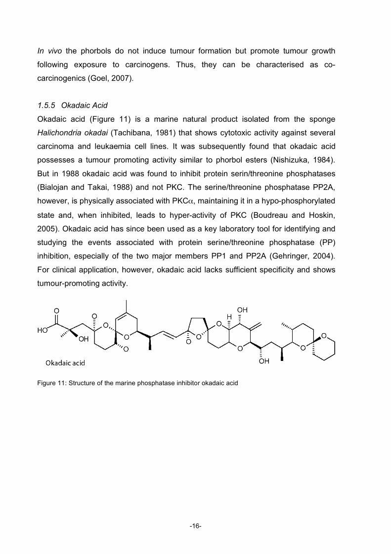
1.5.5 Okadaic Acid
Okadaic acid (Figure 11) is a marine natural product isolated from the sponge
Halichondria okadai (Tachibana, 1981) that shows cytotoxic activity against several
carcinoma and leukaemia cell lines. It was subsequently found that okadaic acid
possesses a tumour promoting activity similar to phorbol esters (Nishizuka, 1984).
But in 1988 okadaic acid was found to inhibit protein serin/threonine phosphatases
(Bialojan and Takai, 1988) and not PKC. The serine/threonine phosphatase PP2A,
however, is physically associated with PKCα, maintaining it in a hypo-phosphorylated
state and, when inhibited, leads to hyper-activity of PKC (Boudreau and Hoskin,
2005). Okadaic acid has since been used as a key laboratory tool for identifying and
studying the events associated with protein serine/threonine phosphatase (PP)
inhibition, especially of the two major members PP1 and PP2A (Gehringer, 2004).
For clinical application, however, okadaic acid lacks sufficient specificity and shows
tumour-promoting activity.
Figure 11: Structure of the marine phosphatase inhibitor okadaic acid
-16-
1.6 References
Acton, N., and Klayman, D.L. (1985). Artemisitene, a new sesquiterpene lactone endoperoxide from Artemisia annua. Planta Med 51, 441-442.
Alberts, A.W., Chen, J., Kuron, G., Hunt, V., Huff, J., Hoffman, C., Rothrock, J., Lopez, M., Joshua, H., Harris, E., et al. (1980). Mevinolin: a highly potent competitive inhibitor of hydroxymethylglutaryl-coenzyme A reductase and a cholesterol-lowering agent. Proc Natl Acad Sci U S A 77, 3957-3961.
Anonymous (1979). Antimalaria studies on Qinghaosu. Chin Med J (Engl) 92, 811-816.
Anonymous (1980). Gossypol as an oral contraceptive for men. J Med Soc N J 77, 50.
Bain, J., Plater, L., Elliott, M., Shpiro, N., Hastie, C.J., McLauchlan, H., Klevernic, I., Arthur, J.S., Alessi, D.R., and Cohen, P. (2007). The selectivity of protein kinase inhibitors: a further update. Biochem J 408, 297-315.
Baker, D.D., Chu, M., Oza, U., and Rajgarhia, V. (2007). The value of natural products to future pharmaceutical discovery. Nat Prod Rep 24, 1225-1244.
Belmokhtar, C.A., Hillion, J., and Segal-Bendirdjian, E. (2001). Staurosporine induces apoptosis through both caspase-dependent and caspase-independent mechanisms. Oncogene 20, 3354-3362.
Berenblum, I. (1941). The Cocarcinogenic Action of Croton Resin. Cancer Research 1, 44-48.
Berkow, R.L., and Kraft, A.S. (1985). Bryostatin, a non-phorbol macrocyclic lactone, activates intact human polymorphonuclear leukocytes and binds to the phorbol ester receptor. Biochem Biophys Res Commun 131, 1109-1116.
Bhisutthibhan, J., Pan, X.Q., Hossler, P.A., Walker, D.J., Yowell, C.A., Carlton, J., Dame, J.B., and Meshnick, S.R. (1998). The Plasmodium falciparum translationally controlled tumor protein homolog and its reaction with the antimalarial drug artemisinin. J Biol Chem 273, 16192-16198.
Bialojan, C., and Takai, A. (1988). Inhibitory effect of a marine-sponge toxin, okadaic acid, on protein phosphatases. Specificity and kinetics. Biochem J 256, 283-290.
Bissery, M.C., Guenard, D., Gueritte-Voegelein, F., and Lavelle, F. (1991). Experimental antitumor activity of taxotere (RP 56976, NSC 628503), a taxol analogue. Cancer Res 51, 4845-4852.
Boudreau, R.T., and Hoskin, D.W. (2005). The use of okadaic acid to elucidate the intracellular role(s) of protein phosphatase 2A: lessons from the mast cell model system. Int Immunopharmacol 5, 1507-1518.
Brian, P.W. (1957). The effects of some microbial metabolic products on plant growth. Symp Soc Exp Biol 54, 166-182.
Brown, M.S., and Goldstein, J.L. (1980). Multivalent feedback regulation of HMG CoA reductase, a control mechanism coordinating isoprenoid synthesis and cell growth. J Lipid Res 21, 505-517.
Butler, M.S., and Newman, D.J. (2008). Mother Nature's gifts to diseases of man: the impact of natural products on anti-infective, anticholestemics and anticancer drug discovery. Prog Drug Res 65, 1, 3-44.
Cardenas, M.E., Sanfridson, A., Cutler, N.S., and Heitman, J. (1998). Signal-transduction cascades as targets for therapeutic intervention by natural products. Trends Biotechnol 16, 427-433.
Chang, J.Y., Sehgal, S.N., and Bansbach, C.C. (1991). FK506 and rapamycin: novel pharmacological probes of the immune response. Trends Pharmacol Sci 12, 218-223.
Chiba, K., Hoshino, Y., Suzuki, C., Masubuchi, Y., Yanagawa, Y., Ohtsuki, M., Sasaki, S., and Fujita, T. (1996). FTY720, a novel immunosuppressant possessing unique mechanisms. I. Prolongation of
-17-
skin allograft survival and synergistic effect in combination with cyclosporine in rats. Transplant Proc 28, 1056-1059.
Cole, G., Dobkins, K.R., Hansen, L.A., Terry, R.D., and Saitoh, T. (1988). Decreased levels of protein kinase C in Alzheimer brain. Brain Res 452, 165-174.
Corson, T.W., and Crews, C.M. (2007). Molecular understanding and modern application of traditional medicines: triumphs and trials. Cell 130, 769-774.
Crown, J., and O'Leary, M. (2000). The taxanes: an update. Lancet 355, 1176-1178.
Cyster, J.G. (2005). Chemokines, sphingosine-1-phosphate, and cell migration in secondary lymphoid organs. Annu Rev Immunol 23, 127-159.
de Vries, D.J., Herald, C.L., Pettit, G.R., and Blumberg, P.M. (1988). Demonstration of sub-nanomolar affinity of bryostatin 1 for the phorbol ester receptor in rat brain. Biochem Pharmacol 37, 4069-4073.
Downton, C., and Clark, I. (2003). Statins--the heart of the matter. Nat Rev Drug Discov 2, 343-344.
Dumont, F.J., Staruch, M.J., Koprak, S.L., Melino, M.R., and Sigal, N.H. (1990). Distinct mechanisms of suppression of murine T cell activation by the related macrolides FK-506 and rapamycin. J Immunol 144, 251-258.
Dumont, F.J., and Su, Q. (1996). Mechanism of action of the immunosuppressant rapamycin. Life Sci 58, 373-395.
Eckstein-Ludwig, U., Webb, R.J., Van Goethem, I.D., East, J.M., Lee, A.G., Kimura, M., O'Neill, P.M., Bray, P.G., Ward, S.A., and Krishna, S. (2003). Artemisinins target the SERCA of Plasmodium falciparum. Nature 424, 957-961.
Endo, A., Kuroda, M., and Tanzawa, K. (1976). Competitive inhibition of 3-hydroxy-3-methylglutaryl coenzyme A reductase by ML-236A and ML-236B fungal metabolites, having hypocholesterolemic activity. FEBS Lett 72, 323-326.
Eng, C.P., Sehgal, S.N., and Vezina, C. (1984). Activity of rapamycin (AY-22,989) against transplanted tumors. J Antibiot (Tokyo) 37, 1231-1237.
Favit, A., Grimaldi, M., Nelson, T.J., and Alkon, D.L. (1998). Alzheimer's-specific effects of soluble beta-amyloid on protein kinase C-alpha and -gamma degradation in human fibroblasts. Proc Natl Acad Sci U S A 95, 5562-5567.
Fingar, D.C., and Blenis, J. (2004). Target of rapamycin (TOR): an integrator of nutrient and growth factor signals and coordinator of cell growth and cell cycle progression. Oncogene 23, 3151-3171.
Foroncewicz, B., Mucha, K., Paczek, L., Chmura, A., and Rowinski, W. (2005). Efficacy of rapamycin in patient with juvenile rheumatoid arthritis. Transpl Int 18, 366-368.
Fujita, T., Inoue, K., Yamamoto, S., Ikumoto, T., Sasaki, S., Toyama, R., Chiba, K., Hoshino, Y., and Okumoto, T. (1994). Fungal metabolites. Part 11. A potent immunosuppressive activity found in Isaria sinclairii metabolite. J Antibiot (Tokyo) 47, 208-215.
Furberg, C.D., and Pitt, B. (2001). Withdrawal of cerivastatin from the world market. Curr Control Trials Cardiovasc Med 2, 205-207.
Gehringer, M.M. (2004). Microcystin-LR and okadaic acid-induced cellular effects: a dualistic response. FEBS Lett 557, 1-8.
Goel, G., Makkar, H.P., Francis, G., and Becker, K. (2007). Phorbol esters: structure, biological activity, and toxicity in animals. Int J Toxicol 26, 279-288.
-18-
Graff, J.R., McNulty, A.M., Hanna, K.R., Konicek, B.W., Lynch, R.L., Bailey, S.N., Banks, C., Capen, A., Goode, R., Lewis, J.E., et al. (2005). The protein kinase Cbeta-selective inhibitor, Enzastaurin (LY317615.HCl), suppresses signaling through the AKT pathway, induces apoptosis, and suppresses growth of human colon cancer and glioblastoma xenografts. Cancer Res 65, 7462-7469.
Greenwood, D. (1992). The quinine connection. J Antimicrob Chemother 30, 417-427.
Gueritte-Voegelein, F., Guenard, D., Lavelle, F., Le Goff, M.T., Mangatal, L., and Potier, P. (1991). Relationships between the structure of taxol analogues and their antimitotic activity. J Med Chem 34, 992-998.
Heitman, J., Movva, N.R., and Hall, M.N. (1992). Proline isomerases at the crossroads of protein folding, signal transduction, and immunosuppression. New Biol 4, 448-460.
Hennings, H., Blumberg, P.M., Pettit, G.R., Herald, C.L., Shores, R., and Yuspa, S.H. (1987). Bryostatin 1, an activator of protein kinase C, inhibits tumor promotion by phorbol esters in SENCAR mouse skin. Carcinogenesis 8, 1343-1346.
Illingworth, D.R., and Sexton, G.J. (1984). Hypocholesterolemic effects of mevinolin in patients with heterozygous familial hypercholesterolemia. J Clin Invest 74, 1972-1978.
Isakov, N., Galron, D., Mustelin, T., Pettit, G.R., and Altman, A. (1993). Inhibition of phorbol ester-induced T cell proliferation by bryostatin is associated with rapid degradation of protein kinase C. J Immunol 150, 1195-1204.
Kahan, B.D., Podbielski, J., Napoli, K.L., Katz, S.M., Meier-Kriesche, H.U., and Van Buren, C.T. (1998). Immunosuppressive effects and safety of a sirolimus/cyclosporine combination regimen for renal transplantation. Transplantation 66, 1040-1046.
Kannel, W.B. (1995). Range of serum cholesterol values in the population developing coronary artery disease. Am J Cardiol 76, 69C-77C.
Kappos, L., Antel, J., Comi, G., Montalban, X., O'Connor, P., Polman, C.H., Haas, T., Korn, A.A., Karlsson, G., and Radue, E.W. (2006). Oral fingolimod (FTY720) for relapsing multiple sclerosis. N Engl J Med 355, 1124-1140.
Keys, A., Menotti, A., Aravanis, C., Blackburn, H., Djordevic, B.S., Buzina, R., Dontas, A.S., Fidanza, F., Karvonen, M.J., Kimura, N., et al. (1984). The seven countries study: 2,289 deaths in 15 years. Prev Med 13, 141-154.
Kitada, S., Leone, M., Sareth, S., Zhai, D., Reed, J.C., and Pellecchia, M. (2003). Discovery, characterization, and structure-activity relationships studies of proapoptotic polyphenols targeting B-cell lymphocyte/leukemia-2 proteins. J Med Chem 46, 4259-4264.
Klayman, D.L. (1985). Qinghaosu (artemisinin): an antimalarial drug from China. Science 228, 1049-1055.
Koehn, F.E., and Carter, G.T. (2005). The evolving role of natural products in drug discovery. Nat Rev Drug Discov 4, 206-220.
Kong, Y.C., Xie, J.X., and But, P.P. (1986). Fertility regulating agents from traditional Chinese medicines. J Ethnopharmacol 15, 1-44.
Kortmansky, J., and Schwartz, G.K. (2003). Bryostatin-1: a novel PKC inhibitor in clinical development. Cancer Invest 21, 924-936.
Kraft, A.S., Appling, C., and Berkow, R.L. (1987). Specific binding of phorbol esters to nuclei of human promyelocytic leukemia cells. Biochem Biophys Res Commun 144, 393-401.
-19-
Kruman, I., Guo, Q., and Mattson, M.P. (1998). Calcium and reactive oxygen species mediate staurosporine-induced mitochondrial dysfunction and apoptosis in PC12 cells. J Neurosci Res 51, 293-308.
Ku, G.Y., Ilson, D.H., Schwartz, L.H., Capanu, M., O'Reilly, E., Shah, M.A., Kelsen, D.P., and Schwartz, G.K. (2008). Phase II trial of sequential paclitaxel and 1 h infusion of bryostatin-1 in patients with advanced esophageal cancer. Cancer Chemother Pharmacol 62, 875-880.
Kuroda, M., Tsujita, Y., Tanzawa, K., and Endo, A. (1979). Hypolipidemic effects in monkeys of ML-236B, a competitive inhibitor of 3-hydroxy-3-methylglutaryl coenzyme A reductase. Lipids 14, 585-589.
Lamers, M.B., Antson, A.A., Hubbard, R.E., Scott, R.K., and Williams, D.H. (1999). Structure of the protein tyrosine kinase domain of C-terminal Src kinase (CSK) in complex with staurosporine. J Mol Biol 285, 713-725.
Lee, H.W., Smith, L., Pettit, G.R., and Bingham Smith, J. (1996a). Dephosphorylation of activated protein kinase C contributes to downregulation by bryostatin. Am J Physiol 271, C304-311.
Lee, H.W., Smith, L., Pettit, G.R., Vinitsky, A., and Smith, J.B. (1996b). Ubiquitination of protein kinase C-alpha and degradation by the proteasome. J Biol Chem 271, 20973-20976.
Lee, W., Boo, J.H., Jung, M.W., Park, S.D., Kim, Y.H., Kim, S.U., and Mook-Jung, I. (2004). Amyloid beta peptide directly inhibits PKC activation. Mol Cell Neurosci 26, 222-231.
Leist, M., and Jaattela, M. (2001). Four deaths and a funeral: from caspases to alternative mechanisms. Nat Rev Mol Cell Biol 2, 589-598.
Levine, A.J., Hu, W., Feng, Z., and Gil, G. (2007). Reconstructing signal transduction pathways: challenges and opportunities. Ann N Y Acad Sci 1115, 32-50.
Link, M. (2004). Statin switch to OTC. Nat Rev Drug Discov 3, 641-642.
Mabuchi, H., Haba, T., Tatami, R., Miyamoto, S., Sakai, Y., Wakasugi, T., Watanabe, A., Koizumi, J., and Takeda, R. (1981). Effect of an inhibitor of 3-hydroxy-3-methyglutaryl coenzyme A reductase on serum lipoproteins and ubiquinone-10-levels in patients with familial hypercholesterolemia. N Engl J Med 305, 478-482.
Mabuchi, H., Sakai, T., Sakai, Y., Yoshimura, A., Watanabe, A., Wakasugi, T., Koizumi, J., and Takeda, R. (1983). Reduction of serum cholesterol in heterozygous patients with familial hypercholesterolemia. Additive effects of compactin and cholestyramine. N Engl J Med 308, 609-613.
MacMillan, J., Vanstone, A.E., and Yeboah, S.K. (1968). The structure of wortmannin, a steroidal fungal metabolite. Chemical Communications (London), 613-614.
Mandala, S., Hajdu, R., Bergstrom, J., Quackenbush, E., Xie, J., Milligan, J., Thornton, R., Shei, G.J., Card, D., Keohane, C., et al. (2002). Alteration of lymphocyte trafficking by sphingosine-1-phosphate receptor agonists. Science 296, 346-349.
Marzo, I., and Naval, J. (2008). Bcl-2 family members as molecular targets in cancer therapy. Biochem Pharmacol 76, 939-946.
Mego, M. (2002). Telomerase inhibitors in anticancer therapy: gossypol as a potential telomerase inhibitor. Bratisl Lek Listy 103, 378-381.
Mohammad, R.M., Wang, S., Aboukameel, A., Chen, B., Wu, X., Chen, J., and Al-Katib, A. (2005). Preclinical studies of a nonpeptidic small-molecule inhibitor of Bcl-2 and Bcl-X(L) [(-)-gossypol] against diffuse large cell lymphoma. Mol Cancer Ther 4, 13-21.
Moon, D.O., Kim, M.O., Choi, Y.H., Lee, H.G., Kim, N.D., and Kim, G.Y. (2008). Gossypol suppresses telomerase activity in human leukemia cells via regulating hTERT. FEBS Lett 582, 3367-3373.
-20-
Morice, M.C., Serruys, P.W., Sousa, J.E., Fajadet, J., Ban Hayashi, E., Perin, M., Colombo, A., Schuler, G., Barragan, P., Guagliumi, G., et al. (2002). A randomized comparison of a sirolimus-eluting stent with a standard stent for coronary revascularization. N Engl J Med 346, 1773-1780.
Newman, D.J., Cragg, G.M., and Snader, K.M. (2000). The influence of natural products upon drug discovery. Nat Prod Rep 17, 215-234.
Newman, D.J., Cragg, G.M., and Snader, K.M. (2003). Natural products as sources of new drugs over the period 1981-2002. J Nat Prod 66, 1022-1037.
Nishizuka, Y. (1984). The role of protein kinase C in cell surface signal transduction and tumour promotion. Nature 308, 693-698.
Nishizuka, Y. (1988). The molecular heterogeneity of protein kinase C and its implications for cellular regulation. Nature 334, 661-665.
Oberlies, N.H., and Kroll, D.J. (2004). Camptothecin and taxol: historic achievements in natural products research. J Nat Prod 67, 129-135.
Olliaro, P.L., Haynes, R.K., Meunier, B., and Yuthavong, Y. (2001). Possible modes of action of the artemisinin-type compounds. Trends Parasitol 17, 122-126.
Omura, S., Iwai, Y., Hirano, A., Nakagawa, A., Awaya, J., Tsuchya, H., Takahashi, Y., and Masuma, R. (1977). A new alkaloid AM-2282 OF Streptomyces origin. Taxonomy, fermentation, isolation and preliminary characterization. J Antibiot (Tokyo) 30, 275-282.
Onaga, L. (2001). Cashing in on nature's pharmacy: Bioprospecting and protection of biodiversity could go hand in hand. EMBO Rep 2, 263-265.
Paterson, I., and Anderson, E.A. (2005). Chemistry. The renaissance of natural products as drug candidates. Science 310, 451-453.
Pettit, G.R., Day, J.F., Hartwell, J.L., and Wood, H.B. (1970). Antineoplastic components of marine animals. Nature 227, 962-963.
Pettit, G.R., Herald, C.L., Doubek, D.L., Herald, D.L., Arnold, E., and Clardy, J. (1982). Isolation and structure of bryostatin 1. Journal of the American Chemical Society 104, 6846-6848.
Porat, O. (1990). Effects of gossypol on the motility of mammalian sperm. Mol Reprod Dev 25, 400-408.
Potterat, O., and Hamburger, M. (2008). Drug discovery and development with plant-derived compounds. Prog Drug Res 65, 45, 47-118.
Reihner, E., Rudling, M., Stahlberg, D., Berglund, L., Ewerth, S., Bjorkhem, I., Einarsson, K., and Angelin, B. (1990). Influence of pravastatin, a specific inhibitor of HMG-CoA reductase, on hepatic metabolism of cholesterol. N Engl J Med 323, 224-228.
Rishton, G.M. (2008). Natural products as a robust source of new drugs and drug leads: past successes and present day issues. Am J Cardiol 101, 43D-49D.
Sabers, C.J., Martin, M.M., Brunn, G.J., Williams, J.M., Dumont, F.J., Wiederrecht, G., and Abraham, R.T. (1995). Isolation of a protein target of the FKBP12-rapamycin complex in mammalian cells. J Biol Chem 270, 815-822.
Salvadori, M., Budde, K., Charpentier, B., Klempnauer, J., Nashan, B., Pallardo, L.M., Eris, J., Schena, F.P., Eisenberger, U., Rostaing, L., et al. (2006). FTY720 versus MMF with cyclosporine in de novo renal transplantation: a 1-year, randomized controlled trial in Europe and Australasia. Am J Transplant 6, 2912-2921.
Schiff, P.B., Fant, J., and Horwitz, S.B. (1979). Promotion of microtubule assembly in vitro by taxol. Nature 277, 665-667.
Schiff, P.B., and Horwitz, S.B. (1981). Taxol assembles tubulin in the absence of exogenous guanosine 5'-triphosphate or microtubule-associated proteins. Biochemistry 20, 3247-3252.
Schmidt, B., Ribnicky, D.M., Poulev, A., Logendra, S., Cefalu, W.T., and Raskin, I. (2008). A natural history of botanical therapeutics. Metabolism 57, S3-9.
Silinsky, E.M., and Searl, T.J. (2003). Phorbol esters and neurotransmitter release: more than just protein kinase C? Br J Pharmacol 138, 1191-1201.
Sneader, W. (2000). The discovery of aspirin: a reappraisal. Bmj 321, 1591-1594.
Snyder, J.P., Nettles, J.H., Cornett, B., Downing, K.H., and Nogales, E. (2001). The binding conformation of Taxol in beta-tubulin: a model based on electron crystallographic density. Proc Natl Acad Sci U S A 98, 5312-5316.
Stewart, Z.A., Mays, D., and Pietenpol, J.A. (1999). Defective G1-S cell cycle checkpoint function sensitizes cells to microtubule inhibitor-induced apoptosis. Cancer Res 59, 3831-3837.
Sun, M.K., and Alkon, D.L. (2005). Dual effects of bryostatin-1 on spatial memory and depression. Eur J Pharmacol 512, 43-51.
Sun, M.K., and Alkon, D.L. (2006). Bryostatin-1: pharmacology and therapeutic potential as a CNS drug. CNS Drug Rev 12, 1-8.
Suzuki, S., Enosawa, S., Kakefuda, T., Amemiya, H., Hoshino, Y., and Chiba, K. (1996). Long-term graft acceptance in allografted rats and dogs by treatment with a novel immunosuppressant, FTY720. Transplant Proc 28, 1375-1376.
Tachibana, K., Scheuer, P.J., Tsukitani, Y., Kikuchi, H., Van Engen, D., Clardy, J., Gopichand, Y., and Schmitz, F.J. (1981). Okadaic acid, a cytotoxic polyether from two marine sponges of the genus Halichondria. Journal of the American Chemical Society 103, 2469-2471.
Thompson, G.R., Ford, J., Jenkinson, M., and Trayner, I. (1986). Efficacy of mevinolin as adjuvant therapy for refractory familial hypercholesterolaemia. Q J Med 60, 803-811.
Tobert, J.A. (2003). Lovastatin and beyond: the history of the HMG-CoA reductase inhibitors. Nat Rev Drug Discov 2, 517-526.
Tobert, J.A., Hitzenberger, G., Kukovetz, W.R., Holmes, I.B., and Jones, K.H. (1982). Rapid and substantial lowering of human serum cholesterol by mevinolin (MK-803), an inhibitor of hydroxymethylglutaryl-coenzyme A reductase. Atherosclerosis 41, 61-65.
Tsang, C.K., Qi, H., Liu, L.F., and Zheng, X.F. (2007). Targeting mammalian target of rapamycin (mTOR) for health and diseases. Drug Discov Today 12, 112-124.
Tsujita, Y., Kuroda, M., Tanzawa, K., Kitano, N., and Endo, A. (1979). Hypolipidemic effects in dogs of ML-236B, a competitive inhibitor of 3-hydroxy-3-methylglutaryl coenzyme A reductase. Atherosclerosis 32, 307-313.
-22-
Van Poznak, C., Seidman, A.D., Reidenberg, M.M., Moasser, M.M., Sklarin, N., Van Zee, K., Borgen, P., Gollub, M., Bacotti, D., Yao, T.J., et al. (2001). Oral gossypol in the treatment of patients with refractory metastatic breast cancer: a phase I/II clinical trial. Breast Cancer Res Treat 66, 239-248.
Vezina, C., Kudelski, A., and Sehgal, S.N. (1975). Rapamycin (AY-22,989), a new antifungal antibiotic. I. Taxonomy of the producing streptomycete and isolation of the active principle. J Antibiot (Tokyo) 28, 721-726.
von Nussbaum, F., Brands, M., Hinzen, B., Weigand, S., and Häbich, D. (2006). Antibacterial Natural Products in Medicinal Chemistry - Exodus or Revival? Angewandte Chemie International Edition 45, 5072-5129.
Wang, Y. (2008). Needs for new plant-derived pharmaceuticals in the post-genome era: an industrial view in drug research and development. Phytochemistry Reviews 7, 395-406.
Wani, M.C., Taylor, H.L., Wall, M.E., Coggon, P., and McPhail, A.T. (1971). Plant antitumor agents. VI. The isolation and structure of taxol, a novel antileukemic and antitumor agent from Taxus brevifolia. J Am Chem Soc 93, 2325-2327.
Watanabe, Y., Ito, T., Saeki, M., Kuroda, M., Tanzawa, K., Mochizuki, M., Tsujita, Y., and Arai, M. (1981). Hypolipidemic effects of CS-500 (ML-236B) in WHHL-rabbit, a heritable animal model for hyperlipidemia. Atherosclerosis 38, 27-31.
Wender, P.A., Baryza, J.L., Brenner, S.E., Clarke, M.O., Craske, M.L., Horan, J.C., and Meyer, T. (2004). Function oriented synthesis: the design, synthesis, PKC binding and translocation activity of a new bryostatin analog. Curr Drug Discov Technol 1, 1-11.
White, N.J. (2008). Qinghaosu (artemisinin): the price of success. Science 320, 330-334.
Wiesner, J., Ortmann, R., Jomaa, H., and Schlitzer, M. (2003). New antimalarial drugs. Angew Chem Int Ed Engl 42, 5274-5293.
Wipf, P., and Halter, R.J. (2005). Chemistry and biology of wortmannin. Org Biomol Chem 3, 2053-2061.
Withers, W.A., and Carruth, F.E. (1915). Gossypol--a Toxic Substance in Cottonseed. a Preliminary Note. Science 41, 324.
Wolter, K.G., Wang, S.J., Henson, B.S., Wang, S., Griffith, K.A., Kumar, B., Chen, J., Carey, T.E., Bradford, C.R., and D'Silva, N.J. (2006). (-)-gossypol inhibits growth and promotes apoptosis of human head and neck squamous cell carcinoma in vivo. Neoplasia 8, 163-172.
Wullschleger, S., Loewith, R., and Hall, M.N. (2006). TOR signaling in growth and metabolism. Cell 124, 471-484.
Wymann, M.P., Bulgarelli-Leva, G., Zvelebil, M.J., Pirola, L., Vanhaesebroeck, B., Waterfield, M.D., and Panayotou, G. (1996). Wortmannin inactivates phosphoinositide 3-kinase by covalent modification of Lys-802, a residue involved in the phosphate transfer reaction. Mol Cell Biol 16, 1722-1733.
Zemann, B., Kinzel, B., Muller, M., Reuschel, R., Mechtcheriakova, D., Urtz, N., Bornancin, F., Baumruker, T., and Billich, A. (2006). Sphingosine kinase type 2 is essential for lymphopenia induced by the immunomodulatory drug FTY720. Blood 107, 1454-1458.
Zhang, X., Zhang, R., Zhao, H., Cai, H., Gush, K.A., Kerr, R.G., Pettit, G.R., and Kraft, A.S. (1996). Preclinical pharmacology of the natural product anticancer agent bryostatin 1, an activator of protein kinase C. Cancer Res 56, 802-808.
Zheng, D.K., Si, Y.K., Meng, J.K., Zhou, J., and Huang, L. (1985). Resolution of racemic gossypol. J Chem Soc Chem Commun, 168-169.
-23-
-24-
2 Signal transduction pathways
˝Flowing with Grace˝ -Teachings of Anusara Yoga
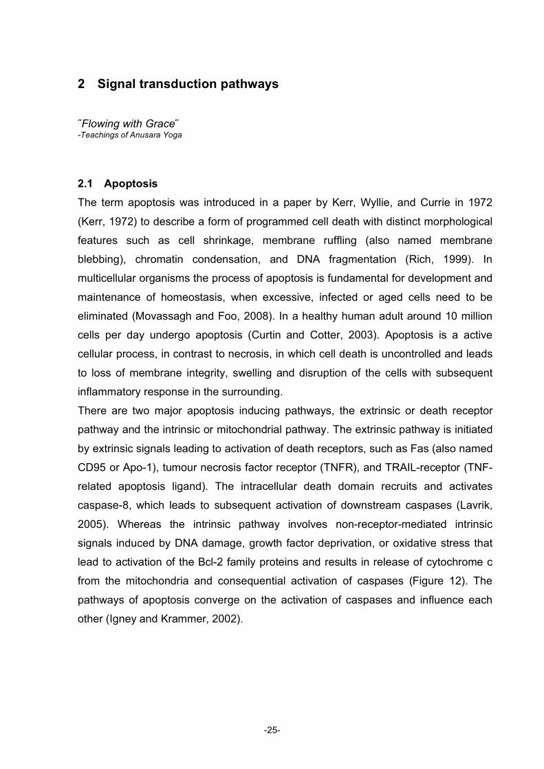
2.1 Apoptosis The term apoptosis was introduced in a paper by Kerr, Wyllie, and Currie in 1972
(Kerr, 1972) to describe a form of programmed cell death with distinct morphological
features such as cell shrinkage, membrane ruffling (also named membrane
blebbing), chromatin condensation, and DNA fragmentation (Rich, 1999). In
multicellular organisms the process of apoptosis is fundamental for development and
maintenance of homeostasis, when excessive, infected or aged cells need to be
eliminated (Movassagh and Foo, 2008). In a healthy human adult around 10 million
cells per day undergo apoptosis (Curtin and Cotter, 2003). Apoptosis is a active
cellular process, in contrast to necrosis, in which cell death is uncontrolled and leads
to loss of membrane integrity, swelling and disruption of the cells with subsequent
inflammatory response in the surrounding.
There are two major apoptosis inducing pathways, the extrinsic or death receptor
pathway and the intrinsic or mitochondrial pathway. The extrinsic pathway is initiated
by extrinsic signals leading to activation of death receptors, such as Fas (also named
CD95 or Apo-1), tumour necrosis factor receptor (TNFR), and TRAIL-receptor (TNF-
related apoptosis ligand). The intracellular death domain recruits and activates
caspase-8, which leads to subsequent activation of downstream caspases (Lavrik,
2005). Whereas the intrinsic pathway involves non-receptor-mediated intrinsic
signals induced by DNA damage, growth factor deprivation, or oxidative stress that
lead to activation of the Bcl-2 family proteins and results in release of cytochrome c
from the mitochondria and consequential activation of caspases (Figure 12). The
pathways of apoptosis converge on the activation of caspases and influence each
other (Igney and Krammer, 2002).
-25-
Figure 12: Simplified diagram of apoptosis pathways (built with templates from
www.cellsignaling.com).
2.1.1 Bcl-2 family
The first protein identified to be involved in apoptosis was Bcl-2, initially characterised
as proto-oncogene in human lymphoma cells (hence the encoding gene was named
B-cell lymphoma-2, bcl-2) (Tsujimoto and Croce, 1986; Vaux, 1988). To date about
20 Bcl-2 family members are identified and can be divided into two functional groups
with either anti- or pro-apoptotic activity. The anti-apoptotic proteins Bcl-2, Bcl-xL,
Bcl-w, and others contain at least four highly conserved bcl-2 homology (BH)
domains. Bcl-2 is exclusively found associated with intracellular membranes,
including the outer mitochondrial membrane, the endoplasmatic reticulum and the
nuclear envelope (Krajewski, 1993). Whereas Bcl-xL is additionally found soluble in
the cytosol, and translocation from the cytosol to the mitochondrial outer membrane
is induced during apoptosis (Hsu, 1997). The pro-apoptotic subfamily can be further
divided into two groups, the multidomain proteins (or BH123), including Bax, Bak,
-26-
and others, and the BH3-only proteins, that contain only one BH3 motif such as Bid,
Bim, Bad, PUMA, Noxa, and some more (Figure 13) (Antonsson, 2004).
Figure 13: Mitochondrial pathway of apoptosis (modified from a model on www.cellsignaling.com).
The BH3-only proteins are pro-apoptotic and function as initial sensors of apoptotic
signals, activating the pro-apoptotic family members. The anti-apoptotic family
members, such as Bcl-2 and Bcl-xL bind to and thereby inhibit the pro-apoptotic
family members Bax and Bak. Activation of the BH3-only proteins results in liberation
of Bax and Bak and thus initiation of apoptosis. The precise mechanism for the
activation of Bax and Bak remains unclear and ‘constitutes the holy grail of apoptosis
research’ (Youle and Strasser, 2008). Liberation of Bax and Bak from binding to their
anti-apoptotic inhibitors Bcl-2 and Bcl-xL leads to several conformational changes
and results in homo-oligomerisation (Chipuk, 2006; Newmeyer and Ferguson-Miller,
2003). These oligomers are believed to build pores in the outer mitochondrial
membrane and induce the mitochondrial outer membrane permeabilisation (MOMP),
-27-
which leads to the release of cytochrome c and other proteins residing in the
mitochondrial intermembranous space, such as apoptosis inducing factor (AIF) and
Smac/DIABLO, an inhibitor of IAPs (inhibitors of apoptosis) (Green and Kroemer,
2004). Progression of apoptosis through activation of caspases, mitochondrial
decomposition and initiation of DNA fragmentation result from these released
molecules (Figure 13).
2.1.2 Cytochrome c
Cytochrome c is a haem-containing protein that participates in the mitochondrial
electron-transport chain, using its heam group to shuttle electrons. However, upon
activation of the intrinsic pathway and MOMP, cytochrome c is released to the cell
plasma where it binds to apoptotic protease-activating factor-1 (Apaf1) that
oligomerises and forms a complex, called apoptosome, which recruits and activates
procaspase-9 (Li, 1997). Active caspase-9 in turn activates downstream effectors,
such as caspase-3 and -7 that lead to execution of apoptosis (Figure 13).
2.1.3 Caspases
Caspases are cysteinyl-aspartic acid proteases that play a key role in apoptotic cell
death and are normally present in healthy cells as inactive precursor zymogens. To
date 11 caspases have been identified in humans (Degterev, 2003). Three groups of
caspases can be formed: the cell death initiators, caspase -8, -9, -2, and -10, the cell
death executors, caspases-3, -6, and -7, and the inflammatory caspases, not
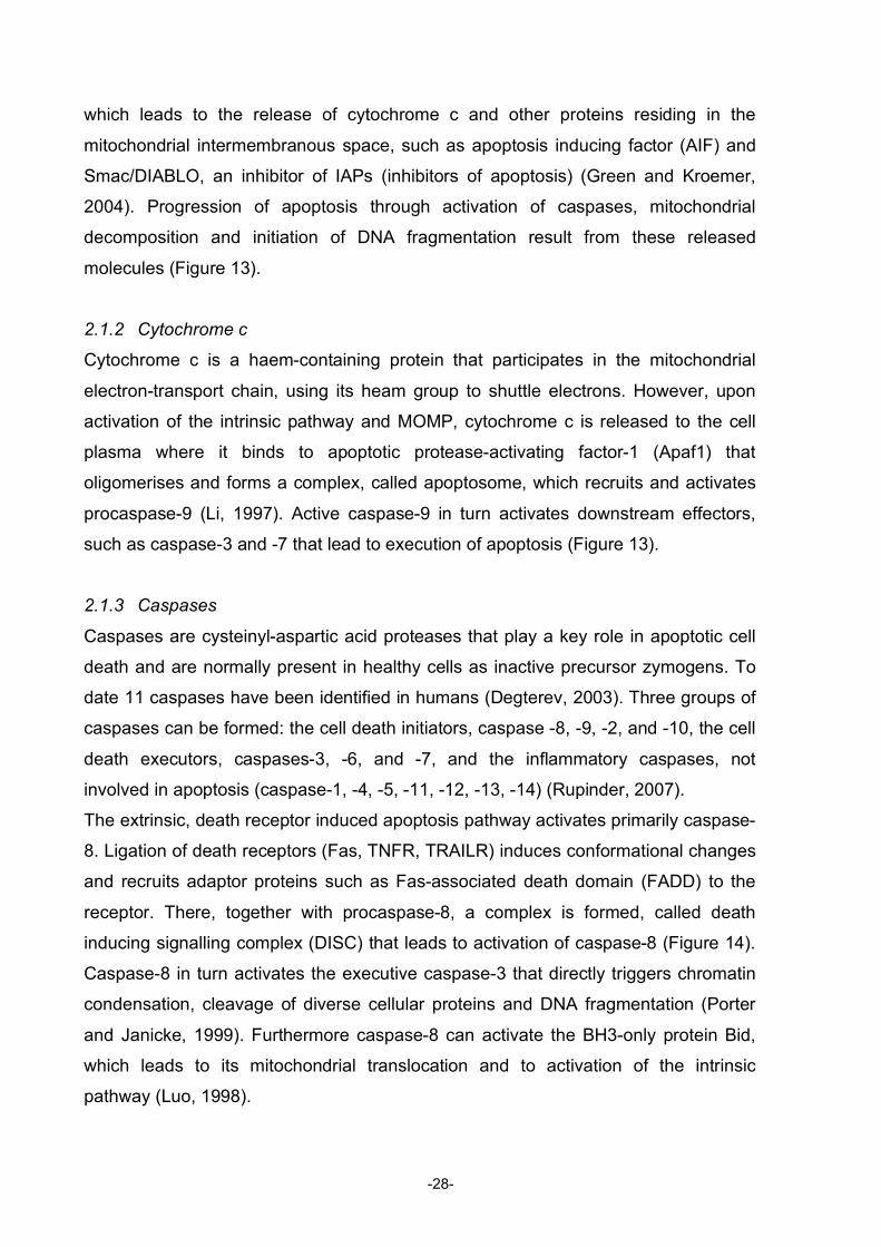
The extrinsic, death receptor induced apoptosis pathway activates primarily caspase-
8. Ligation of death receptors (Fas, TNFR, TRAILR) induces conformational changes
and recruits adaptor proteins such as Fas-associated death domain (FADD) to the
receptor. There, together with procaspase-8, a complex is formed, called death
inducing signalling complex (DISC) that leads to activation of caspase-8 (Figure 14).
Caspase-8 in turn activates the executive caspase-3 that directly triggers chromatin
condensation, cleavage of diverse cellular proteins and DNA fragmentation (Porter
and Janicke, 1999). Furthermore caspase-8 can activate the BH3-only protein Bid,
which leads to its mitochondrial translocation and to activation of the intrinsic
pathway (Luo, 1998).
-28-
Figure 14: Extrinsic pathway and possible points of inhibition (Figure constructed with parts from
www.cellsignaling.com).
2.1.4 IAPs
Although the major way to regulate caspase activity is by proteolytic cleavage, there
are other regulatory pathways. Most importantly the inhibitor-of-apoptosis proteins
(IAPs) that are able to sequester and inactivate pro-caspases and caspases
(LaCasse, 1998). To date eight human IAPs have been identified, including XIAP,
cIAP1, cIAP2, and Survivin. Activity of IAPs, however, is also finely regulated during
apoptosis. The ‘second mitochondrial-derived activator of caspases’ (smac, also
called DIABLO, ‘direct IAP binding protein with low pI’) is released along with
cytochrome c from the mitochondria upon apoptotic stimuli and binds to IAPs in a
manner that liberates caspases from IAP (Figure 14) (Adrain, 2001; Salvesen and
Duckett, 2002).
2.1.5 c-FLIP
Cellular caspase-8 (FLICE)-like inhibitory protein (c-FLIP) is a protein associated with
the DISC, downstream of Fas, TRAIL or TNF receptors (Hyer, 2006; Jin, 2005) and is
-29-
a main regulator of caspase-8 activity (Figure 14). c-FLIP exhibits a dual function of
inhibition or activation of caspase-8 activation downstream of death receptors. In low,
physiological concentrations (1% of caspase-8 content) there is no explicit inhibition
of caspase-8 activation apparent. In higher concentrations, however, c-FLIP inhibits
homodimer formation of caspase-8 and hence prevents activation (Lamkanfi, 2007).
The induced signal leads then to activation of the NFκB pathway and therefore to
pro-survival pathways. Several retroviruses (best characterised in Herpes virus -8)
express FLIP (v-FLIP), which detracts the cell from apoptosis and promotes survival
of virus-infected cells (Thome, 1997).
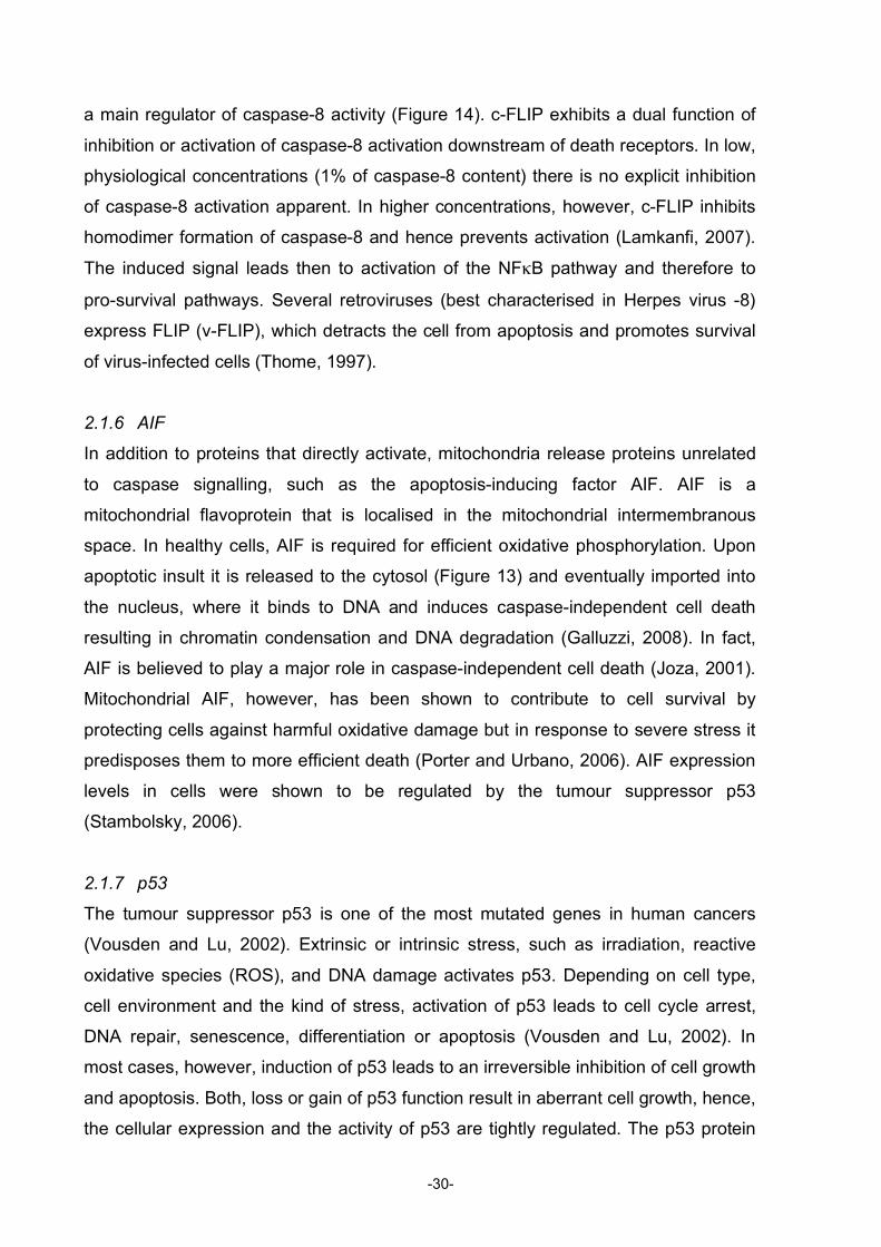
2.1.6 AIF
In addition to proteins that directly activate, mitochondria release proteins unrelated
to caspase signalling, such as the apoptosis-inducing factor AIF. AIF is a
mitochondrial flavoprotein that is localised in the mitochondrial intermembranous
space. In healthy cells, AIF is required for efficient oxidative phosphorylation. Upon
apoptotic insult it is released to the cytosol (Figure 13) and eventually imported into
the nucleus, where it binds to DNA and induces caspase-independent cell death
resulting in chromatin condensation and DNA degradation (Galluzzi, 2008). In fact,
AIF is believed to play a major role in caspase-independent cell death (Joza, 2001).
Mitochondrial AIF, however, has been shown to contribute to cell survival by
protecting cells against harmful oxidative damage but in response to severe stress it
predisposes them to more efficient death (Porter and Urbano, 2006). AIF expression
levels in cells were shown to be regulated by the tumour suppressor p53
(Stambolsky, 2006).
2.1.7 p53
The tumour suppressor p53 is one of the most mutated genes in human cancers
(Vousden and Lu, 2002). Extrinsic or intrinsic stress, such as irradiation, reactive
oxidative species (ROS), and DNA damage activates p53. Depending on cell type,
cell environment and the kind of stress, activation of p53 leads to cell cycle arrest,
DNA repair, senescence, differentiation or apoptosis (Vousden and Lu, 2002). In
most cases, however, induction of p53 leads to an irreversible inhibition of cell growth
and apoptosis. Both, loss or gain of p53 function result in aberrant cell growth, hence,
the cellular expression and the activity of p53 are tightly regulated. The p53 protein
-30-
has a very short half-life and is only present at extremely low levels within the cell
(Pietsch, 2008). Control of the function of p53 is possible at different levels and the
most effective is the control of its plasma levels by Mdm2. Mdm2 is a ubiquitin ligase
that binds to p53 and targets its proteosomal degradation, Mdm2 itself is a
transcriptional target of p53, creating a autoregulatory negative feedback loop
(Harris, 2005). The activation and stabilisation of p53 is generally associated with
inhibition of the function of Mdm2 (Figure 15). Different stress signals allow p53 to
escape Mdm2-mediated protein degradation and to become active. Activation of p53
induces transcription of a myriad of proteins, such as the Bcl-2 proteins Bax, Puma,
and Noxa, the death receptor Fas, caspases-1 and -6, PTEN and many more (Riley,
2008).
Apart from its functions as a transcription factor, p53 directly activates the apoptotic
machinery by translocation to the mitochondria (Caelles, 1994). There, p53 seems to
function analogous to BH3-only members, resulting in oligomerisation of Bak and
Bax and leading to cytochrome c release from the mitochondria (Leu, 2004). This
role of p53, however, has not been studies as extensively as its role as transcription
factor and its relevance it still discussed controversially (Pietsch, 2008).
Figure 15: Regulation of p53
2.1.8 The NFκB pathway
Tumour necrosis factor (TNF) is a multifunctional pro-inflammatory cytokine mainly
produced by macrophages (Wajant, 2003). Besides its function as a death receptor,
TNFR also activates NFκB and c-Jun N-terminal kinase (JNK) pathways. Unlike the
other death receptors, Fas and TRAILR, activation of TNFR does not spontaneously
induce cell death as, the simultaneous NFκB-activation promotes a strong pro-
survival signalling pathway and targets several anti-apoptotic proteins such as Bcl-
xL, IAP, XIAP, and FLIP (Wajant, 2003). Apoptosis is only induced when NFκB
activation is blocked (Wang, 1996).
-31-
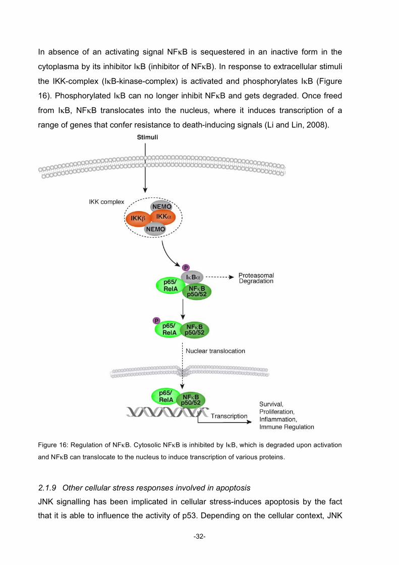
In absence of an activating signal NFκB is sequestered in an inactive form in the
cytoplasma by its inhibitor IκB (inhibitor of NFκB). In response to extracellular stimuli
the IKK-complex (IκB-kinase-complex) is activated and phosphorylates IκB (Figure
16). Phosphorylated IκB can no longer inhibit NFκB and gets degraded. Once freed
from IκB, NFκB translocates into the nucleus, where it induces transcription of a
range of genes that confer resistance to death-inducing signals (Li and Lin, 2008).
Figure 16: Regulation of NFκB. Cytosolic NFκB is inhibited by IκB, which is degraded upon activation
and NFκB can translocate to the nucleus to induce transcription of various proteins.
2.1.9 Other cellular stress responses involved in apoptosis
JNK signalling has been implicated in cellular stress-induces apoptosis by the fact
that it is able to influence the activity of p53. Depending on the cellular context, JNK
-32-
either destabilises p53 by promoting its degradation or stabilises p53 by
phosphorylation (Fuchs, 1998a; Fuchs, 1998b).
Sphingosine and ceramide belong to a group of lipids (sphingolipids) that are
abundant in membranes and are gaining recognition as important signalling
mediators. In healthy cells, sphingomyelin is found predominantly in the outer leaflet
of the plasma membrane. Under stress conditions, however, sphingomyelin turnover
is induced and leads to increased plasma levels of sphingosine and ceramide.
Sphingosine can then be phosphorylated to sphingosine-1-phosphate (S1P) by
sphingosine kinase (SphK). The discovery that S1P regulates cell growth and
suppresses apoptosis triggered the interest in this bioactive lipid mediator. Five
GPCR coupled receptors have been found to mediate the effects of S1P and
stimulating various pathways; JNK, ERK (extracellular signal-regulated kinase), Rac,
Rho, and PI3-kinase/Akt (Takabe, 2008). The first S1P analogue, fingolimod, is in
clinical development and has been described earlier in this introduction.
2.1.10 Disease as a consequence of dysregulated apoptosis
Development of cancer is a multistep process that involves genetic alterations
resulting in unlimited growth of cells. Prominent mechanisms of acquiring resistance
to apoptosis include overactivation of anti-apoptotic proteins or mutation of pro-
apoptotic proteins. Examples of dysregulation of anti-apoptotic proteins include Bcl-2
and Bcl-xL, IAPs, members of the PI3-kinase-Akt pathway and NFκB pathway.
Inactivation of pro-apoptotic signals, on the other hand, is often caused by mutational
changes of p53, Bax, and Apaf-1 and loss of expression of death receptors. The
resistance of cancer cells to apoptosis hinders the success of cancer therapy and
dependant of the kind of mutation therapy needs to be adjusted. Personalised cancer
therapy seems the obvious strategy to pursue, as the type of mutation is fundamental
for successful therapy.
Cancer treatments that specifically target the altered apoptosis pathways are
currently under development. Either by inhibition of the anti-apoptotic members or
activation of pro-apoptotic pathways, cell death is stimulated in cancer cells. For
example the earlier mentioned molecule gossypol, a BH3-mimicking inhibitor of Bcl-2
and Bcl-xL. Inhibition by antisense nucleotide is another strategy, oblimersen, a Bcl-2
antisense oligonucleotide is currently in phase III studies and could reach the market
soon. Several agonist antibodies that target death receptors and induce apoptosis
-33-
are in clinical studies phase I or II, for example mapatumumab and apomab. The
proteasome inhibitor bortezomib (Velcade) blocks proteosomal degradation of
proteins and activates caspase-8 and -9 in vivo, due to inhibition of the NFκB
pathway. Velcade has been approved by the FDA in May 2003 for treatment of
myeloma and has become the first anti-cancer drug on the market to target the
proteasome and thereby, also the first to target NFκB activation (Folmer, 2008).
In neurodegenerative disease the apoptotic pathways are altered in the other
direction and too much apoptosis takes place (Jin, 2005). Neurons naturally possess
very strong anti-apoptotic factors that help them to survive for the lifetime of the
organism. In neurodegenerative diseases such as AD and Parkinson’s disease p53
activation plays a critical role in cell death. The p53 inhibitor pifithrin was shown to
inhibit in vitro amyloid peptide induced neuronal death and protect from neuronal loss
in a Parkinson mouse model (Duan, 2002).
-34-
2.2 PI3K/PIPs/Akt signalling pathway
The serine/threonine kinase Akt, also known as protein kinase B (PKB), plays a
central regulatory role in diverse cellular processes such as homeostasis, survival,
cell cycle progression, and metabolism. Aberrant Akt signalling is observed in cancer
development, insulin resistance and many other pathological conditions. As a general
mechanism, activation of Akt is induced via surface receptor stimulation, where
activated PI3-kinases catalyse the phosphorylation of membrane-bound
phosphoinositides, thus creating docking sites for signalling proteins like Akt (Figure
17). These signalling proteins coordinate complex events that lead to changes in cell
metabolism, cell growth, cell movement and cell survival (Cantley, 2002).
Figure 17: Diagram of the activation of Akt.(modified from www.cellsignaling.com)
2.2.1 Akt pathway
The Akt pathway is activated by receptor tyrosine kinases, B- and T-cell receptors,
cytokine receptors, or G-protein-coupled receptors (GPCRs) that induce production
of phosphatidylinositol-3,4,5-trisphosphates (PtdInsP3) by PI3-kinases
(phosphoinositide 3-kinases). These lipids serve as membrane docking sites for
proteins that harbour pleckstrin-homology (PH) domains, including Akt and its
upstream activator PDK1, thereby recruiting them to the plasma membrane. PDK1
phosphorylates Akt at Thr308, whereas the additional phosphorylation at Ser473 is
-35-
accomplished by the mTOR complex 2 (TORC2) that further increases Akt activity
(Huang and Manning, 2009). Once active, Akt regulates a number of downstream
targets (Figure 18). Three mammalian isoforms of Akt have been identified, Akt1,
Akt2, and Akt3. They share a high degree of structural similarity and more than 80%
sequence homology. Each isoform plays an unique as well as a common role in cells
(Parcellier, 2008).
Figure 18: Effects of Akt activation on different cellular functions (Huang and Manning, 2009).
Akt enhances the survival of cells by blocking the function of proapoptotic proteins
such as inactivation of Bad, a BH3-only protein, by phosphorylation. This prevents
binding of Bad to its target proteins Bcl-2 and Bcl-xL and hence promotes cell
survival. Akt has also been reported to directly phosphorylate capsase-9 and thereby
blocking its activation (Cardone, 1998). Furthermore Akt inhibits the transcription
factor FoxO1 (forkhead box O1) that induces transcription of Bim, another BH3-only
protein stimulating cell death. A further target of Akt that promotes cell survival is
Mdm2, the negative regulator of p53, leading to translocation of Mdm2 into the
nucleus where it inhibits p53 transcriptional activity (Figure 15).
The impact of Akt on cell growth is mainly due to activation of the mTOR complex 1
(TORC1), which is regulated by both nutrient and growth factor signalling. TORC1
regulates protein synthesis by initiation of translation and biogenesis of ribosomes
(Wullschleger, 2006). The enhanced sensitivity of cancer cells exhibiting oncogenic
activation of the Akt-pathway to the mTOR inhibitor rapamycin, illustrates the
importance of TORC1 activation downstream of Akt (Sabatini, 2006).
-36-
Cell proliferation is stimulated by Akt through inhibitiory phosphorylation of the cyclin-
dependent kinase (CDK) inhibitors p21 and p27. Akt thus prevents translocation of
p21 and p27 to the nucleus and attenuates their cell-cycle inhibitory effects (Liang
and Richardson, 2003). Moreover, inhibitory phosphorylation of the glycogen
synthase kinase 3 (GSK3) by Akt leads to cell cycle progression, as inactivation of
GSK3 rescues β-catenin from degradation. β-catenin induces transcription of cyclin
D1, which induces cell cycle progression (Osaki, 2004).
Akt signalling also takes part in the regulation of nutrient uptake and metabolism.
Upon stimulation with insulin, for instance, the glucose transporter Glut4, most likely
through interaction with AS160 (Akt substrate of 160 kDa), translocates to the cellular
membrane and enhances glucose uptake (Sano, 2003). Furthermore, the Akt-
mediated phosphorylation and inhibition of GSK3 stimulates glycogen synthesis.
The Akt pathway interconnects with many other important pathways in the cell.
Several studies have demonstrated that Akt signalling can activate NFκB upon a
variety of stimuli, for instance upon stimulation with platelet-derived growth factor.
Although there are likely to be multiple levels of crosstalk between Akt and NFκB,
one mechanism consists in phosphorylation of IKK by Akt and subsequent activation
of NFκB. Furthermore, activation of Akt influences other pathways such as ERK,
JNK, and MAPK, as their pathways converge upstream.
2.2.2 The phosphoinositide and phosphoinositide-kinases pathways
Hokin and Hokin (1964) have first demonstrated phospholipid turnover in response to
cell stimulation (Hokin and Hokin, 1964). Since then research in this field has yielded
many mechanistic insights of phosphoinositide cell signalling.
Phosphoinositides (PIs) are inositol-containing glycerophospholipids that can be
reversely phosphorylated in several positions on the inositol ring (Figure 19).
Figure 19: Structure of PIs
The family of PIs consists of the mutual non-phosphorylated precursor
phosphatidylinositol (PtdIns) and seven members with different phosphorylation
-37-
patterns on the inositol ring, where the 3-, 4-, and 5- positions can be phosphorylated
by specific kinases (Figure 20) (Wymann and Pirola, 1998). These phosphoinositide-
kinases (PI-kinases) are categorised into three families, depending on the position of
the phosphorylation on the inositol ring: phosphoinositide 3-kinases (PI3-kinases),
phosphoinositide 4-kinases (PI4-kinases or PIP4-kinases), and phosphoinositide 5-
kinases (PI5-kinases or PIP5-kinases).
Figure 20: Interconversions of all the PIs, with indication of the respective enzyme and the disease
resulting when proper function of the enzyme fails (figure from McCrea and De Camilli, 2009).
2.2.3 Phosphoinositide-kinases
The best characterised amongst them are the PI3-kinases that are further grouped
into class I, II, and III, depending on their subunit structure, substrate specificity, and
type of activation.
PI3-kinases class I. The class I PI3-kinases are heterodimeric proteins that catalyse
the formation of phosphatidylinositol-3,4,5-trisphosphate (PtdInsP3) from
phosphatidylinositol-4,5-bisphosphate (PtdIns(4,5)P2). They all consist of a regulatory
-38-
and a catalytic subunit and are subdivided into class IA, that contain the catalytic
subunits p110α, β, or δ, and the class IB that contains the catalytic subunit p110γ
(hence called PI3-kinase γ) (Vanhaesebroeck, 1997). The class IA PI3-kinases are
downstream of receptor tyrosine kinases and can be activated by growth factor or
insulin stimulation, cytokine signalling, or antibody binding, whereas the class IB is
downstream of GPCRs and is activated by Gβγ subunits (Karataeva and Nevinsky,
2007). Although the two classes couple to different surface receptor systems, they
share the small GTPase Ras as an interacting partner (Wymann and Pirola, 1998).
PI3-kinases class II. Class II PI3-kinases are monomers of relatively large molecular
mass (170-220 kDa) and comprise three catalytic isoforms (C2α, C2β, and C2γ).
Unlike class I and III, class II contains no regulatory protein. They mainly catalyse the
production of PtdIns3P from PtdIns and PtdIns(3,4)P2 from PtdIns4P (Falasca and
Maffucci, 2007; McCrea and De Camilli, 2009).
PI3-kinase class III. Class III PI3-kinases have a substrate specificity restricted to
PtdIns. These PI3-kinases are homologous to Vps34 (vacuolar protein sorting 34),
the only PI3-kinase in yeast. Vps34 is closely associated with Vps15, which has been
described as a Vps34 regulatory subunit and was therefore named p150 in
mammals, analogous to the class I regulatory subunits (Backer, 2008;
Vanhaesebroeck, 1997).
PI4-kinases. Two types of PtdIns 4-kinase have been identified. One that only
catalyses PtdIns to PtdIns4P, called PI4-kinase, and one that catalyses PtdInsPs to
PtdIns(3,4)P2 or PtdIns(4,5)P2, respectively, called PIP4-kinase (Jones, 2000; Rusten
and Stenmark, 2006).
PI5-kinases. There are two types of PI5-kinase. A characteristic feature is that they
only phosphorylate PtdInsPs to PtdInsP2. PIKfyve converts PtdIns3P to
PtdIns(3,5)P2, while PIP5-kinase phospohorylates PtdIns4P to PtdIns(4,5)P2 (Krauss
and Haucke, 2007). To date no PI5-kinase is known that phosphorylates PtdIns to
PtdIns5P.
In summary, the PI3-kinases have been well investigated, but little is known about
the other PI-kinases. Studies on PI4- and PI5-kinases are few and their biological
significance not fully understood.
-39-
Figure 21: Localisation of the specific PtdIns at different membranes in the cell (figure from McCrea
and De Camilli, 2009).
2.2.4 Roles of the different phosphoinositides
PtdIns is the most abundant PI and together with PtdIns4P and PtdIns(4,5)P2 they
build the so called canonical pathway. These PIs are kept at constant levels at the
plasma membrane, whereas the other PIs are considered to be low-abundant
signalling molecules that transiently appear upon specific stimulation. Furthermore
PtdIns(4,5)P2 serves as precursor of the intracellular second messengers DAG and
inositol-(1,4,5)-trisphosphate (IP3), that are formed through phospholipase C (PLC)
mediated hydrolysis of PtdIns(4,5)P2. DAG binds and regulates protein kinase C
(PKC) and a variety of other effectors, whereas IP3 triggers calcium release from the
endoplasmic reticulum. PtdIns(4,5)P2 can also be cleaved by phospholipase A2
(PLA2) to generate arachidonic acid, the precursor of inflammatory eicosanoids
(McCrea and De Camilli, 2009).
PtdInsP3. The generation of PtdInsP3 stimulates numerous signalling pathways that
are involved in cell proliferation, metabolism, motility and immune responses.
PtdInsP3 recruits PH domain-containing proteins such as Akt, PDK1, PLC, BKT
(bruton’s tyrosine kinase), and GTPases-activating factors, to the membrane where
they build signalling complexes to produce a specific signal in the cell (Sasaki, 2007).
More than 250 proteins in humans have been found to contain a PH binding domain
and are therefore potential binding partners of PtdInsP3. However, the most
-40-
important effector is Akt. ‘Not all PI3-kinase signalling is mediated via Akt, but clearly
a major part of it’ (Downward, 2004). PtdInsP3 levels resulting after activation of PI3-
kinses class I are tightly regulated by the action of phosphatases such as PTEN
(phosphatase and tensin homologue deleted on chromosome ten) and SHIP (SH2-
containing inositol 5-phosphatase) that dephosphorylate PtdInsP3 to PtdIns(3,4)P2
and PtdIns(4,5)P2, respectively (Figure 20). Deregulated PtdInsP3 signalling is tightly
associated with tumour development, either through mutational changes in receptors
that result in constant activation of PI3-kinases or by mutational changes of PTEN
and hence impaired shut-down of the signal.
PtdInsP2. The bisphosphorylated PIs, PtdIns(3,4)P2, PtdIns(3,5)P2, and PtdIns(4,5)P2
have distinct functions in cell signalling. PtdIns(3,4)P2 is responsible for signal-
extension after PtdInsP3 induction since some of the PtdInsP3 binding proteins also
recognise PtdIns(3,4)P2, namely PDK1 and Akt. The 5-phosphtase SHIP,
responsible for dephosphorylation of PtdInsP3 to PtdIns(3,4)P2 has important roles in
cells of the immune system and in blood platelets (Blero, 2007). SHIP1 deficient mice
display overactive immune responses and platelet clotting. SHIP2 is important for
control of insulin sensitivity (Sasaoka, 2006).
PtdIns(3,5)P2 has a role in membrane and protein trafficking (Figure 21) and is
believed to be only produced via PtdIns3P and subsequent 5-phosphrylation (Michell,
Lemmon, Dove, 2006). Besides its function as a precursor of PtdInsP3, DAG, and
IP3, PtdIns(4,5)P2 has some specific functions in regulation of the actin cytoskeleton,
exo- and endocytoic processes. PtdIns(4,5)P2 is associated with small GTPases and
participates in recruitment and activation of a wide variety of regulatory proteins (Di
Paolo and De Camilli, 2006).
PtdInsP. The phosphoinositide monophosphates were long thought to be mere
intermediate metabolites in the pathway. In yeast the only PI3-kinase Vps34 is
responsible for the production of PtdIns3P and plays a critical role in the sorting of
proteins from the golgi to the vacuole (Pendaries, 2005). In mammals it is involved in
vesicular trafficking (Wurmser, 1999). Besides being an intermediate product on the
way to PtdIns(4,5)P2, PtdIns4P is enriched at the Golgi membrane and targets Golgi-
associated proteins (Figure 21) (Pendaries, 2005). The role of PtdIns5P as signalling
molecule just emerges, as it is the most recently discovered inositol lipid (Rameh,
-41-
1997). It was shown that PtdIns5P levels rise upon thrombin stimulation in platelets
(Morris, 2000) and during osmotic stress in mammalian cells (Sbrissa, 2002).
2.2.5 PI3-kinase/Akt signalling in disease
PI3-kinases were originally discovered because of their association with viral
oncoproteins (Cantley, 1991) and increased PI3-kinase signalling has since then
been found in many cancers. Abberant PI3-kinase/Akt activation occurs in cancer
through a variety of mechanisms. These include mutations or amplification of
upstream regulators such as receptors and PI3-kinases, inactivation of PTEN, or
amplification of Akt. Mutational activation of PI3-kinase IA is found in 15% of all
tumour types, suggesting that this may be the most commonly mutated kinase
(Crabbe, 2007). The PTEN gene is commonly inactivated in cancer cells and is the
second most mutated tumour suppressor gene after p53 (Trotman and Pandolfi,
2003). Another negative regulator of PtdInsP3 is SHIP, and the isoform SHIP2 has
been implicated in development of diabetes. SHIP2 is responsible for the control of
the cellular response to insulin. Elevated levels of SHIP2 expression are found in
cells that show insulin resistance.
A role of PtdIns(4,5)P2 in the early onset of Alzheimer’s Disease in Down Syndrome
has been suggested (Voronov, 2008). Synaptojanin I, a PtdIns(4,5,)P2 phosphatase
plays a critical role in synaptic transmission and is located in the chromosome 21
region that is triplicated in Down Syndrome. Accordingly levels of synaptojanin I are
increased and might add to the early development of AD (McCrea and De Camilli,
2009). Furthermore some hereditary diseases were linked with genetic defects in the
PI-signalling system (Figure 20). Lowe syndrome is a X-linked condition that results
in severe mental retardation, growth defects, and eye defects. It is caused by
mutations in the inositol-5-phosphatase OCRL (occulocerebrorenal syndrome of
Lowe) that dephosphrylates PtdIns(4,5)P2 to PtdIns4P. The cellular causes of the
varied disease phenotypes are not yet understood (Skwarek and Boulianne, 2009).
Another disease cause by impaired PI metabolism is Charcot-Marie-Tooth (CMT)
disease that affects peripheral nerves and is the most common inherited neurological
disorder. Several different subtypes of CMT disease are due to mutations in lipid
phosphatases involved in dephosphorylation of PtdIns(3,5)P2 at either the 3- or 5-
position (Robinson, 2008).
-42-
Targeting of the PI3-kinase pathway is aimed at since the important role of this
pathway has been recognised. Various molecules are in clinical development, the Akt
inhibitor perifosine, PI3-kinase inhibitors, and already in clinical use, the mTOR
inhibitor rapamycin (Yap, 2008). Unfortunately selective inhibition of the PI3-kinases
has not been achieved yet. The non-selective PI3-kinase inhibitor wortmannin and its
role as a model substance in molecular biology were discussed in chapter 1.5.2.
Selective inhibitors of PI3-kinase gamma were once thought to become the ‘aspirine
of the 21st century’ (Ruckle, 2006) but no sufficiently selective inhibitor has been
found yet to fulfil that prophecy.
-43-
2.3 References
Adrain, C., Creagh, E.M., and Martin, S.J. (2001). Apoptosis-associated release of Smac/DIABLO from mitochondria requires active caspases and is blocked by Bcl-2. Embo J 20, 6627-6636.
Antonsson, B. (2004). Mitochondria and the Bcl-2 family proteins in apoptosis signaling pathways. Mol Cell Biochem 256-257, 141-155.
Backer, J.M. (2008). The regulation and function of Class III PI3Ks: novel roles for Vps34. Biochem J 410, 1-17.
Blero, D., Payrastre, B., Schurmans, S., and Erneux, C. (2007). Phosphoinositide phosphatases in a network of signalling reactions. Pflugers Arch 455, 31-44.
Caelles, C., Helmberg, A., and Karin, M. (1994). p53-dependent apoptosis in the absence of transcriptional activation of p53-target genes. Nature 370, 220-223.
Cantley, L.C. (2002). The phosphoinositide 3-kinase pathway. Science 296, 1655-1657.
Cantley, L.C., Auger, K.R., Carpenter, C., Duckworth, B., Graziani, A., Kapeller, R., and Soltoff, S. (1991). Oncogenes and signal transduction. Cell 64, 281-302.
Cardone, M.H., Roy, N., Stennicke, H.R., Salvesen, G.S., Franke, T.F., Stanbridge, E., Frisch, S., and Reed, J.C. (1998). Regulation of cell death protease caspase-9 by phosphorylation. Science 282, 1318-1321.
Chipuk, J.E., Bouchier-Hayes, L., and Green, D.R. (2006). Mitochondrial outer membrane permeabilization during apoptosis: the innocent bystander scenario. Cell Death Differ 13, 1396-1402.
Crabbe, T., Welham, M.J., and Ward, S.G. (2007). The PI3K inhibitor arsenal: choose your weapon! Trends Biochem Sci 32, 450-456.
Degterev, A., Boyce, M., and Yuan, J. (2003). A decade of caspases. Oncogene 22, 8543-8567.
Di Paolo, G., and De Camilli, P. (2006). Phosphoinositides in cell regulation and membrane dynamics. Nature 443, 651-657.
Downward, J. (2004). PI 3-kinase, Akt and cell survival. Semin Cell Dev Biol 15, 177-182.
Duan, W., Zhu, X., Ladenheim, B., Yu, Q.S., Guo, Z., Oyler, J., Cutler, R.G., Cadet, J.L., Greig, N.H., and Mattson, M.P. (2002). p53 inhibitors preserve dopamine neurons and motor function in experimental parkinsonism. Ann Neurol 52, 597-606.
Falasca, M., and Maffucci, T. (2007). Role of class II phosphoinositide 3-kinase in cell signalling. Biochem Soc Trans 35, 211-214.
Folmer, F., Jaspars, M., Dicato, M., and Diederich, M. (2008). Marine natural products as targeted modulators of the transcription factor NF-kappaB. Biochem Pharmacol 75, 603-617.
Fuchs, S.Y., Adler, V., Buschmann, T., Yin, Z., Wu, X., Jones, S.N., and Ronai, Z. (1998a). JNK targets p53 ubiquitination and degradation in nonstressed cells. Genes Dev 12, 2658-2663.
Fuchs, S.Y., Adler, V., Pincus, M.R., and Ronai, Z. (1998b). MEKK1/JNK signaling stabilizes and activates p53. Proc Natl Acad Sci U S A 95, 10541-10546.
-44-
Galluzzi, L., Joza, N., Tasdemir, E., Maiuri, M.C., Hengartner, M., Abrams, J.M., Tavernarakis, N., Penninger, J., Madeo, F., and Kroemer, G. (2008). No death without life: vital functions of apoptotic effectors. Cell Death Differ 15, 1113-1123.
Green, D.R., and Kroemer, G. (2004). The pathophysiology of mitochondrial cell death. Science 305, 626-629.
Harris, L.C. (2005). MDM2 splice variants and their therapeutic implications. Curr Cancer Drug Targets 5, 21-26.
Hokin, L.E., and Hokin, M.R. (1964). The Incorporation of 32p from Triphosphate into Polyphosphoinositides (Gamma-32p)Adenosine and Phosphatidic Acid in Erythrocyte Membranes. Biochim Biophys Acta 84, 563-575.
Hsu, Y.T., Wolter, K.G., and Youle, R.J. (1997). Cytosol-to-membrane redistribution of Bax and Bcl-X(L) during apoptosis. Proc Natl Acad Sci U S A 94, 3668-3672.
Huang, J., and Manning, B.D. (2009). A complex interplay between Akt, TSC2 and the two mTOR complexes. Biochem Soc Trans 37, 217-222.
Hyer, M.L., Samuel, T., and Reed, J.C. (2006). The FLIP-side of Fas signaling. Clin Cancer Res 12, 5929-5931.
Igney, F.H., and Krammer, P.H. (2002). Death and anti-death: tumour resistance to apoptosis. Nat Rev Cancer 2, 277-288.
Jin, F., Liu, X., Zhou, Z., Yue, P., Lotan, R., Khuri, F.R., Chung, L.W., and Sun, S.Y. (2005). Activation of nuclear factor-kappaB contributes to induction of death receptors and apoptosis by the synthetic retinoid CD437 in DU145 human prostate cancer cells. Cancer Res 65, 6354-6363.
Jones, D.H., Morris, J.B., Morgan, C.P., Kondo, H., Irvine, R.F., and Cockcroft, S. (2000). Type I phosphatidylinositol 4-phosphate 5-kinase directly interacts with ADP-ribosylation factor 1 and is responsible for phosphatidylinositol 4,5-bisphosphate synthesis in the golgi compartment. J Biol Chem 275, 13962-13966.
Joza, N., Susin, S.A., Daugas, E., Stanford, W.L., Cho, S.K., Li, C.Y., Sasaki, T., Elia, A.J., Cheng, H.Y., Ravagnan, L., et al. (2001). Essential role of the mitochondrial apoptosis-inducing factor in programmed cell death. Nature 410, 549-554.
Karataeva, N.A., and Nevinsky, G.A. (2007). Enzymes phosphorylating lipids and polysaccharides. Biochemistry (Mosc) 72, 367-379.
Kerr, J.F., Wyllie, A.H., and Currie, A.R. (1972). Apoptosis: a basic biological phenomenon with wide-ranging implications in tissue kinetics. Br J Cancer 26, 239-257.
Krajewski, S., Tanaka, S., Takayama, S., Schibler, M.J., Fenton, W., and Reed, J.C. (1993). Investigation of the subcellular distribution of the bcl-2 oncoprotein: residence in the nuclear envelope, endoplasmic reticulum, and outer mitochondrial membranes. Cancer Res 53, 4701-4714.
Krauss, M., and Haucke, V. (2007). Phosphoinositides: regulators of membrane traffic and protein function. FEBS Lett 581, 2105-2111.
LaCasse, E.C., Baird, S., Korneluk, R.G., and MacKenzie, A.E. (1998). The inhibitors of apoptosis (IAPs) and their emerging role in cancer. Oncogene 17, 3247-3259.
Lamkanfi, M., Festjens, N., Declercq, W., Vanden Berghe, T., and Vandenabeele, P. (2007). Caspases in cell survival, proliferation and differentiation. Cell Death Differ 14, 44-55.
Lavrik, I., Golks, A., and Krammer, P.H. (2005). Death receptor signaling. J Cell Sci 118, 265-267.
-45-
Leu, J.I., Dumont, P., Hafey, M., Murphy, M.E., and George, D.L. (2004). Mitochondrial p53 activates Bak and causes disruption of a Bak-Mcl1 complex. Nat Cell Biol 6, 443-450.
Li, H., and Lin, X. (2008). Positive and negative signaling components involved in TNFalpha-induced NF-kappaB activation. Cytokine 41, 1-8.
Li, P., Nijhawan, D., Budihardjo, I., Srinivasula, S.M., Ahmad, M., Alnemri, E.S., and Wang, X. (1997). Cytochrome c and dATP-dependent formation of Apaf-1/caspase-9 complex initiates an apoptotic protease cascade. Cell 91, 479-489.
Liang, S.X., and Richardson, D.R. (2003). The effect of potent iron chelators on the regulation of p53: examination of the expression, localization and DNA-binding activity of p53 and the transactivation of WAF1. Carcinogenesis 24, 1601-1614.
Luo, X., Budihardjo, I., Zou, H., Slaughter, C., and Wang, X. (1998). Bid, a Bcl2 interacting protein, mediates cytochrome c release from mitochondria in response to activation of cell surface death receptors. Cell 94, 481-490.
McCrea, H.J., and De Camilli, P. (2009). Mutations in phosphoinositide metabolizing enzymes and human disease. Physiology (Bethesda) 24, 8-16.
Morris, J.B., Hinchliffe, K.A., Ciruela, A., Letcher, A.J., and Irvine, R.F. (2000). Thrombin stimulation of platelets causes an increase in phosphatidylinositol 5-phosphate revealed by mass assay. FEBS Lett 475, 57-60.
Movassagh, M., and Foo, R.S. (2008). Simplified apoptotic cascades. Heart Fail Rev 13, 111-119.
Newmeyer, D.D., and Ferguson-Miller, S. (2003). Mitochondria: releasing power for life and unleashing the machineries of death. Cell 112, 481-490.
Osaki, M., Oshimura, M., and Ito, H. (2004). PI3K-Akt pathway: its functions and alterations in human cancer. Apoptosis 9, 667-676.
Parcellier, A., Tintignac, L.A., Zhuravleva, E., and Hemmings, B.A. (2008). PKB and the mitochondria: AKTing on apoptosis. Cell Signal 20, 21-30.
Pendaries, C., Tronchere, H., Racaud-Sultan, C., Gaits-Iacovoni, F., Coronas, S., Manenti, S., Gratacap, M.P., Plantavid, M., and Payrastre, B. (2005). Emerging roles of phosphatidylinositol monophosphates in cellular signaling and trafficking. Adv Enzyme Regul 45, 201-214.
Pietsch, E.C., Sykes, S.M., McMahon, S.B., and Murphy, M.E. (2008). The p53 family and programmed cell death. Oncogene 27, 6507-6521.
Porter, A.G., and Janicke, R.U. (1999). Emerging roles of caspase-3 in apoptosis. Cell Death Differ 6, 99-104.
Porter, A.G., and Urbano, A.G. (2006). Does apoptosis-inducing factor (AIF) have both life and death functions in cells? Bioessays 28, 834-843.
Rameh, L.E., Tolias, K.F., Duckworth, B.C., and Cantley, L.C. (1997). A new pathway for synthesis of phosphatidylinositol-4,5-bisphosphate. Nature 390, 192-196.
Rich, T., Watson, C.J., and Wyllie, A. (1999). Apoptosis: the germs of death. Nat Cell Biol 1, E69-71.
Riley, T., Sontag, E., Chen, P., and Levine, A. (2008). Transcriptional control of human p53-regulated genes. Nat Rev Mol Cell Biol 9, 402-412.
Robinson, F.L., Niesman, I.R., Beiswenger, K.K., and Dixon, J.E. (2008). Loss of the inactive myotubularin-related phosphatase Mtmr13 leads to a Charcot-Marie-Tooth 4B2-like peripheral neuropathy in mice. Proc Natl Acad Sci U S A 105, 4916-4921.
-46-
Ruckle, T., Schwarz, M.K., and Rommel, C. (2006). PI3Kgamma inhibition: towards an 'aspirin of the 21st century'? Nat Rev Drug Discov 5, 903-918.
Rupinder, S.K., Gurpreet, A.K., and Manjeet, S. (2007). Cell suicide and caspases. Vascul Pharmacol 46, 383-393.
Rusten, T.E., and Stenmark, H. (2006). Analyzing phosphoinositides and their interacting proteins. Nat Methods 3, 251-258.
Sabatini, D.M. (2006). mTOR and cancer: insights into a complex relationship. Nat Rev Cancer 6, 729-734.
Salvesen, G.S., and Duckett, C.S. (2002). IAP proteins: blocking the road to death's door. Nat Rev Mol Cell Biol 3, 401-410.
Sano, H., Kane, S., Sano, E., Miinea, C.P., Asara, J.M., Lane, W.S., Garner, C.W., and Lienhard, G.E. (2003). Insulin-stimulated phosphorylation of a Rab GTPase-activating protein regulates GLUT4 translocation. J Biol Chem 278, 14599-14602.
Sasaki, T., Sasaki, J., Sakai, T., Takasuga, S., and Suzuki, A. (2007). The physiology of phosphoinositides. Biol Pharm Bull 30, 1599-1604.
Sasaoka, T., Wada, T., and Tsuneki, H. (2006). Lipid phosphatases as a possible therapeutic target in cases of type 2 diabetes and obesity. Pharmacol Ther 112, 799-809.
Sbrissa, D., Ikonomov, O.C., Deeb, R., and Shisheva, A. (2002). Phosphatidylinositol 5-phosphate biosynthesis is linked to PIKfyve and is involved in osmotic response pathway in mammalian cells. J Biol Chem 277, 47276-47284.
Skwarek, L.C., and Boulianne, G.L. (2009). Great expectations for PIP: phosphoinositides as regulators of signaling during development and disease. Dev Cell 16, 12-20.
Stambolsky, P., Weisz, L., Shats, I., Klein, Y., Goldfinger, N., Oren, M., and Rotter, V. (2006). Regulation of AIF expression by p53. Cell Death Differ 13, 2140-2149.
Takabe, K., Paugh, S.W., Milstien, S., and Spiegel, S. (2008). "Inside-out" signaling of sphingosine-1-phosphate: therapeutic targets. Pharmacol Rev 60, 181-195.
Thome, M., Schneider, P., Hofmann, K., Fickenscher, H., Meinl, E., Neipel, F., Mattmann, C., Burns, K., Bodmer, J.L., Schroter, M., et al. (1997). Viral FLICE-inhibitory proteins (FLIPs) prevent apoptosis induced by death receptors. Nature 386, 517-521.
Trotman, L.C., and Pandolfi, P.P. (2003). PTEN and p53: who will get the upper hand? Cancer Cell 3, 97-99.
Tsujimoto, Y., and Croce, C.M. (1986). Analysis of the structure, transcripts, and protein products of bcl-2, the gene involved in human follicular lymphoma. Proc Natl Acad Sci U S A 83, 5214-5218.
Vanhaesebroeck, B., Leevers, S.J., Panayotou, G., and Waterfield, M.D. (1997). Phosphoinositide 3-kinases: a conserved family of signal transducers. Trends Biochem Sci 22, 267-272.
Vaux, D.L., Cory, S., and Adams, J.M. (1988). Bcl-2 gene promotes haemopoietic cell survival and cooperates with c-myc to immortalize pre-B cells. Nature 335, 440-442.
Voronov, S.V., Frere, S.G., Giovedi, S., Pollina, E.A., Borel, C., Zhang, H., Schmidt, C., Akeson, E.C., Wenk, M.R., Cimasoni, L., et al. (2008). Synaptojanin 1-linked phosphoinositide dyshomeostasis and cognitive deficits in mouse models of Down's syndrome. Proc Natl Acad Sci U S A 105, 9415-9420.
Vousden, K.H., and Lu, X. (2002). Live or let die: the cell's response to p53. Nat Rev Cancer 2, 594-604.
-47-
Wajant, H., Pfizenmaier, K., and Scheurich, P. (2003). Tumor necrosis factor signaling. Cell Death Differ 10, 45-65.
Wang, C.Y., Mayo, M.W., and Baldwin, A.S., Jr. (1996). TNF- and cancer therapy-induced apoptosis: potentiation by inhibition of NF-kappaB. Science 274, 784-787.
Wullschleger, S., Loewith, R., and Hall, M.N. (2006). TOR signaling in growth and metabolism. Cell 124, 471-484.
Wurmser, A.E., Gary, J.D., and Emr, S.D. (1999). Phosphoinositide 3-kinases and their FYVE domain-containing effectors as regulators of vacuolar/lysosomal membrane trafficking pathways. J Biol Chem 274, 9129-9132.
Wymann, M.P., and Pirola, L. (1998). Structure and function of phosphoinositide 3-kinases. Biochim Biophys Acta 1436, 127-150.
Yap, T.A., Garrett, M.D., Walton, M.I., Raynaud, F., de Bono, J.S., and Workman, P. (2008). Targeting the PI3K-AKT-mTOR pathway: progress, pitfalls, and promises. Curr Opin Pharmacol 8, 393-412.
Youle, R.J., and Strasser, A. (2008). The BCL-2 protein family: opposing activities that mediate cell death. Nat Rev Mol Cell Biol 9, 47-59.
-48-
3 Aim of the thesis
The importance of natural products as sources of inspiration for drug discovery and
development and as tools for basic research is undisputed. The combination of
traditional knowledge and modern scientific research has been a successful strategy
to identify new active substances that might serve as lead structures for drug
discovery, and natural products have offered numerous opportunities to study
unknown cellular mechanisms.
The exploitation of natural products is limited by the fact that the modes of action of
numerous compounds are poorly understood. The potential of a given compound can
only be assessed if sufficient information is available regarding their molecular
targets. The natural products analysed during this thesis have both some background
in traditional medicine and have been identified with the aid of functional assays. In
order to assign these observed effects to an interaction on the molecular level, we
studied associated signal transduction pathways in cells, namely:
(i) Phosphorylation of proteins
(ii) Activation of transcription factors
(iii) Concentration of second messengers (i.e. phosphoinositides)
(iv) Localisation of the substance within the cell
In the first part of this thesis the molecular effects of the fungal alkaloid militarinone A
were studied. In an earlier screening for neurotrophic substances, the extract of the
entomogenous fungus Paecilomyces militaris had been shown to induce increased
spike formation in PC12 cells (Schmidt, 2003), and militarinone A had been
subsequent identified as the active metabolite via activity guided fractionation (Figure
22) (Schmidt, 2002).
Figure 22: Structure of militarinone A.
-49-
To uncover the mechanism of this activity we studied the cell signalling pathways
involved in neuronal spike formation and differentiation in two types of neuronal cells
(PC12 and N2a) and the interaction of militarinone A with associated pathways.
In the second part, an alkaloid from the traditional European medicinal plant woad
was analysed. Woad (Isatis tinctoria) contains several known anti-inflammatory
components, namely, tryptanthrin, indirubin, and (E,Z)-3-(3’,5’-dimethoxy-4’-hydroxy-
benzylidene)-2-indolinone (indolinone (Figure 23)). The effects of tryptanthrin as a
potent inhibitor of COX-2 (Danz, 2002), 5-LOX (Oberthur, 2005), and of nitric oxide
production catalyzed by inducible NO synthase (iNOS) (Ishihara, 2000) are well
characterised and account for a good part of the anti-inflammatory activity of the
plant extract. Indirubin is used in Chinese medicine (from isatis indigotica) as an anti-
leukemia treatment and has been shown to inhibit cyclin-dependent-kinase 2 (CDK2)
(Hoessel, 1999) while indolinone was shown to inhibit degranulation of mast cells
(Rüster, 2004).
Figure 23: Structure of indolinone.
The anti-allergic activity of indolinone should be further characterised. Given the
structural similarities with indolin-2-one derived kinase inhibitors, we focussed on
signalling pathways associated with degranulation to determine the mechanism and
point of action of indolinone.
During the studies on indolinone, measurements of intracellular PtdInsP3 (Figure 19)
were initially performed with the current standard methodology that includes
radioactive labelling with 32Pi, ion-exchange HPLC and flow scintillation analysis
(Dove, 1997; Laffargue, 2002). The method proved to be laborious, time-consuming
and potentially hazardous. This led us to develop a non-radioactive method for
-50-
analysis of phosphoinositides based on chromatographic separation and mass
spectrometry detection. This method would allow analysis of all phosphoinositides
without the need for radioactive labelling and should be of interest for many other
laboratories, as it offers new perspectives in detection and relative quantification of
phosphoinositides.
3.1 References Danz, H., Stoyanova, S., Thomet, O.A., Simon, H.U., Dannhardt, G., Ulbrich, H., and Hamburger, M. (2002). Inhibitory activity of tryptanthrin on prostaglandin and leukotriene synthesis. Planta Med 68, 875-880.
Hoessel, R., Leclerc, S., Endicott, J.A., Nobel, M.E., Lawrie, A., Tunnah, P., Leost, M., Damiens, E., Marie, D., Marko, D., et al. (1999). Indirubin, the active constituent of a Chinese antileukaemia medicine, inhibits cyclin-dependent kinases. Nat Cell Biol 1, 60-67.
Ishihara, T., Kohno, K., Ushio, S., Iwaki, K., Ikeda, M., and Kurimoto, M. (2000). Tryptanthrin inhibits nitric oxide and prostaglandin E(2) synthesis by murine macrophages. Eur J Pharmacol 407, 197-204.
Laffargue, M., Calvez, R., Finan, P., Trifilieff, A., Barbier, M., Altruda, F., Hirsch, E., and Wymann, M.P. (2002). Phosphoinositide 3-kinase gamma is an essential amplifier of mast cell function. Immunity 16, 441-451.
Oberthur, C., Jaggi, R., and Hamburger, M. (2005). HPLC based activity profiling for 5-lipoxygenase inhibitory activity in Isatis tinctoria leaf extracts. Fitoterapia 76, 324-332.
Rüster, G.U., Hoffmann, B., and Hamburger, M. (2004). Inhibitory activity of indolin-2-one derivatives on compound 48/80-induced histamine release from mast cells. Pharmazie 59, 236-237.
Schmidt, K., Gunther, W., Stoyanova, S., Schubert, B., Li, Z., and Hamburger, M. (2002). Militarinone A, a neurotrophic pyridone alkaloid from Paecilomyces militaris. Org Lett 4, 197-199.
Schmidt, K., Li, Z., Schubert, B., Huang, B., Stoyanova, S., and Hamburger, M. (2003). Screening of entomopathogenic Deuteromycetes for activities on targets involved in degenerative diseases of the central nervous system. J Ethnopharmacol 89, 251-260.
-51-
-52-
B. PUBLICATIONS
˝It’s a wild dance floor there at the molecular level˝ - Roald Hoffmann in ‘Designing the molecular world’ by Philip Ball (p.83)
1 Promotion of cell death or neurite outgrowth in PC12 and N2a cells by the fungal alkaloid militarinone A depends on basal
expression of p53
-53-
-54-
ORIGINAL PAPER
Promotion of cell death or neurite outgrowth in PC-12 and N2a
cells by the fungal alkaloid militarinone A depends
on basal expression of p53
Peter Kuenzi Æ Sabine Kiefer Æ Anna Koryakina Æ
Matthias Hamburger
Published online: 22 February 2008
Ó Springer Science+Business Media, LLC 2008
Abstract The fungal alkaloid militarinone A (MiliA) was
recently found to stimulate neuronal outgrowth in PC-12
cells by persistant activation of pathways that are also
involved in NGF-mediated differentiation, namely the PI3-
K/PKB and the MEK/ERK pathways. Application of equal
concentrations of MiliA to other cells such as the murine
neuroblastoma cell line N2a resulted in immediate onset of
apoptosis by nuclear translocation of apoptosis inducing
factor (AIF), activation of caspases and c-Jun/AP-1 tran-
scription factor without an intermediate differentiated
phenotype, although minor transient phosphorylation of
PKB and MAPK as well as activation of NF-jB were also
observed. Translocation of AIF was preceded by p53
phosphorylation at Ser15 and blocked by pifithrin a, a
known inhibitor of p53-transcriptional activity. We here
show that both cell types activate the same pathways albeit
in different time scales. This is mainly due to contrasting
basal expression levels of p53, which in turn regulates
expression of AIF. In PC-12 cells, continuous activation of
these pathways after prolonged treatment with 40 lM
MiliA first led to up-regulation of p53, phosphorylation of
p53, release of AIF from mitochondria and its translocation
into the nucleus. Additionally, also activation of the c-Jun/
AP-1 transcription factor was observed, and PC-12 cells
subsequently underwent apoptosis 48–72 h post-treatment.
We report that similar pathways working on different
levels are able to initially shape very divergent cellular
responses.
Keywords Neurite extension � Apoptosis � p53 �
AIF � Natural products
Introduction
Neurotrophic factors are capable of preventing apoptosis in
neuronal cells, and exogenous supplementation with such
factors have been proposed as a new disease-modifying
strategy in the treatment of neurogenerative diseases.
However, clinical trials have been disappointing so far due
to difficulties in delivery of these glycoproteins into the
central nervous system [1]. Small non-peptidic molecules
with neurotrophic properties, and capable of penetrating
the blood–brain barrier, have been proposed as an alter-
native [2]. In the course of a screening for such molecules
we discovered that the fungal alkaloid militarinone A
(MiliA) induced neurite extension in PC-12 cells [3]. Due
to its amphiphilic properties MiliA was rapidly integrated
into the membranous fraction where it triggered PI3-K/
PKB- and ERK1/2-dependent differentiation [4, 5]. PKB
promotes survival of a variety of neuronal cells and has
been shown to be required for neuronal outgrowth [6, 7]. In
addition, phosphorylation and activation of the MAPKs
ERK1/2 [8] have also been connected with neuronal
differentiation [9], albeit its function is not fully clear.
Apoptosis inducing factor (AIF) is a mitochondrial
flavoprotein that is mainly located in the mitochondrial
intermembrane space. In healthy cells, AIF is required for
efficient oxidative phosphorylation. Upon apoptotic insult,
it translocates to the cytosol and subsequently into the
nucleus where it eventually binds to DNA and induces
caspase-independent cell death [10–12]. The concept that
AIF is able to induce apoptotic cell death independent of
caspases is supported by a series of experiments where
P. Kuenzi � S. Kiefer � A. Koryakina � M. Hamburger (&)
Institute of Pharmaceutical Biology, University of Basel,
Kinase assay Appropriate concentrations of PI3-K isoforms p110 α, β, γ GST, γ tr, and δ (fused to
interSH2 domains, a kind gift from Novartis, Basel, Switzerland) were mixed with 10
µg/ml L-α-phosphatidylinositol (Sigma-Aldrich, Buchs, Switzerland) dissolved in 0.3%
octylglucoside kinase buffer (10 mM Tris HCl, 3 mM MgCl2, 50 mM NaCl, 0.8 mM
CHAPS, 1 mM DTT). After 5 min pre-treatment with indolinone or DMSO, 2 µM ATP
(Roche, Basel, Switzerland) dissolved in kinase buffer were added to start the
reaction. Reactions were incubated for 30-90 min, depending on the kinase isoform.
Kinase Glo (Promega, Madison, WI) was added to each well to start the luciferase
reaction. After 15 min incubation at room temperature, plates (white 96 well
luciferase plates (Berthold, Bad Wildbad, Germany) were read out in a luminometer
(Centro LB 960, Berthold) with an integration time of 0.5 s. Samples were run in
duplicate.
Results Inhibition of degranulation in murine bone marrow derived mast cells Indolinone had been previously shown to inhibit compound 48/80-induced histamine
release in rat peritoneal mast cells [12]. We reconfirmed this effect in antigen-
stimulated degranulation of murine bone marrow derived mast cells (BMMC), to
demonstrate that this effect was neither species-dependent nor assay-specific.
-75-
BMMC were sensitised overnight with anti DNP-IgE (100 ng/ml) and indolinone (50
µM) was added 30 min before stimulation where applicable. Degranulation was
simultaneously induced with the antigen DNP-albumin (5 ng/ml) and adenosine (2
µM), as stimulation with antigen DNP-albumin alone only resulted in week responses.
Indolinone pre-treatment reduced the number of degranulated cells from 63.1% ±
8.7% to 23.5% ± 4.6%, only just above Ctrl level (15.0% ± 0.1%) (Fig. 2A & B). Even
though we used a different assay setup, another kind of cells, and changed the type
of degranulation-inducing agents, indolinone clearly inhibited degranulation in
BMMC. Due to interference with indolinone fluorescence occurring at all wavelengths
when excited with 488 or 405 nm, typical hexosaminidase-release measurement had
to be abandoned. Instead, degranulation was measured using Cy5-conjugated
annexin V. Annexin V-Cy5 was exited at 635 nm and emission was measured at
680/30 nm.
Fig. 2 - Degranulation of BMMC. (A) Mast cells were sensitised with IgE antibodies over night and
subsequently challenged with antigen (Ag) and 5 µM adenosine. Indolinone (50 µM) was added to the
cells 30 min before challenge. Cells were stained with annexin V-Cy5 and degranulation was
measured by flow cytometry. Shown is a typical experiment. Region R was determined as approx.
15% degranulation of control cells. (B) Histogram of cells in Region R of three independent
experiments. Shown are the mean ± S.E.: Ctrl: 15.0% ± 0.1%; Ag + Ade: 63.1% ± 8.7%; Ind + Ag +
Ade: 23.5% ± 4.6%.
Reduced phosphorylation of selected signal transduction molecules involved in degranulation Signals that lead to degranulation involve phosphorylation of several signalling
proteins such as PKB, MEK and MAPK. Furthermore, involvement of PKC has been
-76-
suggested [34-36]. Therefore, we checked for phosphorylation of these proteins upon
stimulation with adenosine and analysed for differences between cells pre-treated
with indolinone or untreated controls. Stimulation of BMMC with 5 µM adenosine for
30 s resulted in phosphorylation of PKB (Thr308), MEK1/2 (Ser 217/221), MAPK1/2
(Thr202/Tyr204), and PKC (pan, γ Thr514). Pre-treatment with 50 µM indolinone 30
min prior to stimulation diminished the phosphorylation of PKB, MEK, and MAPK,
whereas phosphorylation of PKC was not affected (Fig. 3A). Pre-treatment with the
PI3-K inhibitor wortmannin also blocked phosphorylation of PKB, MEK, and MAPK.
Quantitative analysis by flow cytometry of adenosine-induced PKB phosphorylation
showed a dose-dependent inhibition after indolinone pre-treatment. Phosphorylation
of PKB was almost completely blocked by 50 µM indolinone in respect to LY pre-
treatment, and partially by 25 µM indolinone (Fig. 3B).
Fig. 3 - Phosphorylation of important signalling molecules involved in degranulation, analyzed by
western blot and flow cytometry. (A) Equal amounts of cellular protein were separated by SDS-PAGE,
transferred to nitrocellulose membrane, and probed using specific antibodies. α-tubulin was used as
an internal loading control. BMMC cells (0.5 x 10⁶) were starved in 2% serum for 4 h, treated with
indolinone or inhibitor (Wort) for 30 min, and then stimulated with adenosine (Ade) for 30 s. Untreated
cells were used as control. Shown are typical results from at least three independent experiments. (B)
Flow cytometry analysis of PKB-phosphorylation. Cells were fixed and stained using specific
antibodies, followed by flow cytometry analysis of 10’000 cells per sample in the FITC channel (FL1).
BMMC cells (0.5 x 10⁶) were starved in 2% serum for 4 h, treated with indolinone (25, 50, and 100 µM)
or LY (50 µM) for 30 min, and then stimulated with adenosine (5 µM) for 30 s. Shown is a typical result
from at least three independent experiments.
-77-
Adenosine-induced rise of PtdIns(3,4,5)P3 levels inhibited by indolinone
Generation of PtdIns(3,4,5)P3 by PI3-K induces translocation of PKB towards the
membrane and its binding to PtdIns(3,4,5)P3 via the pleckstrin homology domain
[37]. Eventually, PDK1 [38] and mTORC2 [39] phosphorylate PKB at Thr308 and
Ser473, respectively.
Fig. 4 - Analysis of PI(3,4,5)P3 levels in BMMC cells and activity of PI3-K class IA and IB isoforms. (A)
BMMC cells were labelled with 32Pi, and phospholipids were extracted, deacylated, and separated by
HPLC. Peaks of interest were integrated and PtdIns(3,4,5)P3 levels expressed in relation to the
integral of PtdIns(4,5)P2. BMMC cells (4 x 10⁶) were starved in 2% serum for 4 h, treated with
indolinone or wortmannin for 30 min, and then stimulated with adenosine for 30 s. Shown are the
mean ± S.E. of three independent experiments. (B) Chromatograms of PtdIns(3,4,5)P3 analysis,
recorded by flow scintillation from a typical experiment. (C) PI3-Ks were co-incubated with ATP and
either indolinone in different concentrations or DMSO as a control. Kinase activity was determined as
consumption of ATP and residual ATP was measured by luciferase reaction. Samples were measured
as duplicates in three independent experiments. Kinase activity of the control was defined as 100%.
We analysed PtdIns(3,4,5)P3 levels in BMMC to determine whether inhibition of PKB-
phosphorylation by indolinone was due to reduced PtdIns(3,4,5)P3 production.
Stimulation of BMMC with adenosine results in elevated PtdIns(3,4,5)P3 levels and
-78-
preliminary experiments showed that PtdIns(3,4,5)P3 levels reached a maximum 30 s
post treatment. PtdIns(3,4,5)P3 levels were quantified relative to PtdIns(4,5)P2 that
was used as internal standard. This ratio increased 15-fold after stimulation with
adenosine. Pre-treatment with indolinone significantly reduced this increase to less
than 5-fold, comparable to pre-treatment with wortmannin (Fig. 4A and 4B).
Inhibition of PI3-Ks class IA and IB by indolinone The activity of all PI3-Ks class IA and IB isoforms (α, β, γ, δ), responsible for the
generation of PtdIns(3,4,5)P3, was tested in an in vitro assay after addition of
different concentrations of indolinone. The γ isoform was tested in two different ways,
as the complete GST tagged enzyme and as a trunctuated form with 144 amino
acids less. We expected reduced kinase activity as the reason for the blocked
increase of PtdIns(3,4,5)P3 levels. Indeed, indolinone inhibited all PI3-K isoforms to
some extent (Fig. 4C). When incubated with 50 µM indolinone the α and the δ
isoform were inhibited to less than 50%, whereas the other isoforms still showed
activities over 60%. Degranulation in BMMC is predominantly induced by PI3-Kγ and
we expected a clear inhibition by indolinone.
Localisation of indolinone in BMMC Due to the fluorescent properties of indolinone itself, we were able to visualise its
localisation in cells directly and monitor its uptake into the cell by flow cytometry.
Although we had expected to see some accumulation at the membrane, analysis of
indolinone-stained cells by confocal microscopy (Fig. 5A) showed equal distribution
throughout the cytoplasm, and no staining of the nucleus. Administration of
indolinone during flow cytometry measurement produced an instant increase in
fluorescence. After less than 3 min no further increase was detected and
fluorescence persisted for the duration of the measurement (15 min). This showed
that indolinone was immediately taken up into the cells and retained in the cytosol
(Fig. 5B and 5C). We further tested if BMMCs would regain their ability to
degranulate after removal of indolinone and incubation in indolinone-free media for
two hours, which was not case (Fig. 5D and 5E).
-79-
Fig. 5 - Appearance and localization of indolinone in BMMCs. Due to the fluorescent properties of
indolinone itself, it was possible to monitor its appearance and localisation in the cell. (A) BMMC were
fixed, stained with indolinone and DRAQ5 and mounted for fluorescence microscopy. The upper panel
illustrates localisation of indolinone in the cell, the lower panel shows DRAQ5 staining of the nucleus.
(B+C) Flow cytometry analysis of BMMCs. (B) Administration of 50 µM indolinone during ongoing
measurement. (C) Fluorescence of BMMC before and after addition of 50 µM indolinone (Ind). (D)
Mast cells were sensitised with IgE antibodies over night and subsequently challenged with antigen
(Ag) and 5 µM adenosine (Ade). 50 µM Indolinone (Ind) was added to the cells for 30 min. Cells were
subsequently washed three times and left for 2 hours or analysed immediately. Cells were stained with
annexin V-Cy5 and degranulation was measured by flow cytometry. Shown is a typical experiment.
Region R was determined as approx. 15% degranulation of control cells. (E) Histogram of cells in
Region R of three independent experiments. Shown are the mean ± S.E.: Ind + Ag + Ade: 23.5% ±
4.6%; Ind + recovery + Ag + Ade: 23.9% ± 3.4%.
-80-
Discussion The only drugs in clinical use that directly inhibit mast cell degranulation are disodium
cromoglycate and its derivative nedocromil [19]. Cromoglycate was developed from
khellin, a furanochromone from the anti-allergic plant Ammi visnaga. It is a mast cell
stabilising agent that has been shown to block ion-channels. However, cromoglycate
likely has additional effects and its mode of action is still not very clear [40]. Various
other agents have been used experimentally to inhibit degranulation [29], but were
not applicable for clinical use. The most recent approaches include Syk kinase
inhibitors [41,42] and anti IgE antibodies. One such anti IgE antibody, Omalizumab
(Xolair), has recently reached the market [43,44].
Extracts of Isatis tinctoria have traditionally been used in the treatment of anti-
inflammatory complaints. Previous studies assigned specific facets of the in vitro
pharmacological spectrum of the extract to specific compounds [7,8] in this complex
multi-component mixture. In the model of compound 48/80 stimulated mast cell
degranulation, indolinone was significantly more potent than disodium cromoglycate
(IC50 of 15 µM (0.0045 µg/ml) vs 1.5 mM). In an earlier study Isatis extract had an
IC50 of 2.3 µg/ml in this assay [3]. Given that indolinone is a minor constituent in the
extract (typical concentrations of 0.04%) [45], it is likely that other components of the
extract contribute to its mast cell stabilising activity.
Marquardt et al. showed suppressed degranulation of BMMCs after addition of the
PI3-K inhibitor wortmannin [46]. Kinase activity of all PI3-K class I isoforms was
inhibited by indolinone. Hence, it can be assumed that degranulation of BMMC was
impaired due to PI3-K inhibition. All PI3-K-subtypes were blocked by indolinone to
some extent, but none of them substantially preferred. PI3-K isoforms important for
mast cell activation are p110γ and δ. Downstream of the high-affinity IgE receptor
FcεRI is the tyrosine kinase-activated p110δ and downstream of the adenosine
receptor the GPCR-activated p110γ [47].
Consequently, phosphorylation of PKB, MEK, and MAPK was blocked due to
inhibition of the upstream PI3-Ks. Adenosine-dependent activation of PKC, however,
was reported to be independent of PI3-K activity and mediated through PLC only
[48,49]. Our findings agree with that model, since adenosine-induced
phosphorylation of PKC persisted after indolinone pre-treatment. Furthermore, the
-81-
intact PKC phosphorylation revealed that indolinone was not an unspecific inhibitor of
phosphorylation but rather blocked all events downstream of the PI3-K.
The calculated distribution coefficient (clogD) of indolinone at pH 7 is 2.28, and polar
surface area (PSA) is 67.8 Å2. These physico-chemical properties explain its rapid
cellular uptake, its distribution in the cytoplasma, and its ability to persist within and
retain its inhibitory role.
Even though the point of action is supposedly localised near or at the cellular
membrane, equal distribution in the cytoplasm may confer some advantage since the
inactive proteins are usually located in the cytoplasm and recruited to the membrane
upon induction.
In this study we have shown that indolinone blocks antigen-induced mast cell
degranulation, decreases adenosine-stimulated production of PtdIns(3,4,5)P3, and
inhibits activity of all PI3-K class I isoforms. The findings corroborate earlier findings
with the lipophilic Isatis extract and thus contribute to a better understanding of
molecular modes of action and synergistic effects of its constituents.
References [1] Danz H. Untersuchungen zur antiinflammatorischen Wirkung und zur Analytik
von Tryptantrin in Isatis tinctoria L. Biologisch-Pharmazeutische Fakultät. Jena: Friedrich-Schiller University, 2000.
[2] Stöger E. Arzneibuch der chineischen Medizin: Wissenschaftliche Verlagsgesellschaft.
[3] Hamburger M. Isatis tinctoria - from the rediscovery of an ancient medicinal plant towards a novel anti.inflammatory phytopharmaceutical. Phytochem Rev 2002;1:333-44.
[4] Heinemann C, Schliemann-Willers S, Oberthur C, Hamburger M, Elsner P. Prevention of experimentally induced irritant contact dermatitis by extracts of Isatis tinctoria compared to pure tryptanthrin and its impact on UVB-induced erythema. Planta Med 2004;70 (5):385-90.
[5] Recio MC, Cerda-Nicolas M, Hamburger M, Rios JL. Anti-arthritic activity of a lipophilic woad (Isatis tinctoria) extract. Planta Med 2006;72 (8):715-20.
[6] Recio MC, Cerda-Nicolas M, Potterat O, Hamburger M, Rios JL. Anti-inflammatory and antiallergic activity in vivo of lipophilic Isatis tinctoria extracts and tryptanthrin. Planta Med 2006;72 (6):539-46.
[7] Danz H, Stoyanova S, Wippich P, Brattstrom A, Hamburger M. Identification and isolation of the cyclooxygenase-2 inhibitory principle in Isatis tinctoria. Planta Med 2001;67 (5):411-6.
[8] Oberthur C, Jaggi R, Hamburger M. HPLC based activity profiling for 5-lipoxygenase inhibitory activity in Isatis tinctoria leaf extracts. Fitoterapia 2005;76 (3-4):324-32.
-82-
[9] Ishihara T, Kohno K, Ushio S, Iwaki K, Ikeda M, Kurimoto M. Tryptanthrin inhibits nitric oxide and prostaglandin E(2) synthesis by murine macrophages. Eur J Pharmacol 2000;407 (1-2):197-204.
[10] Hoessel R, Leclerc S, Endicott JA, Nobel ME, Lawrie A, Tunnah P, Leost M, Damiens E, Marie D, Marko D, Niederberger E, Tang W, Eisenbrand G, Meijer L. Indirubin, the active constituent of a Chinese antileukaemia medicine, inhibits cyclin-dependent kinases. Nat Cell Biol 1999;1 (1):60-7.
[11] Simopoulos AP. Omega-3 fatty acids in inflammation and autoimmune diseases. J Am Coll Nutr 2002;21 (6):495-505.
[12] Ruster GU, Hoffmann B, Hamburger M. Inhibitory activity of indolin-2-one derivatives on compound 48/80-induced histamine release from mast cells. Pharmazie 2004;59 (3):236-7.
[13] Bouerat L, Fensholdt J, Liang X, Havez S, Nielsen SF, Hansen JR, Bolvig S, Andersson C. Indolin-2-ones with high in vivo efficacy in a model for multiple sclerosis. J Med Chem 2005;48 (17):5412-4.
[14] Kaur K, Talele TT. 3D QSAR studies of 1,3,4-benzotriazepine derivatives as CCK(2) receptor antagonists. J Mol Graph Model 2008;27 (4):409-20.
[15] Boechat N, Kover WB, Bongertz V, Bastos MM, Romeiro NC, Azevedo ML, Wollinger W. Design, synthesis and pharmacological evaluation of HIV-1 reverse transcriptase inhibition of new indolin-2-ones. Med Chem 2007;3 (6):533-42.
[16] Bouchikhi F, Rossignol E, Sancelme M, Aboab B, Anizon F, Fabbro D, Prudhomme M, Moreau P. Synthesis and biological evaluation of diversely substituted indolin-2-ones. Eur J Med Chem 2008;43 (11):2316-22.
[17] Atkins M, Jones CA, Kirkpatrick P. Sunitinib maleate. Nat Rev Drug Discov 2006;5 (4):279-80.
[18] Sun L, Tran N, Tang F, App H, Hirth P, McMahon G, Tang C. Synthesis and biological evaluations of 3-substituted indolin-2-ones: a novel class of tyrosine kinase inhibitors that exhibit selectivity toward particular receptor tyrosine kinases. J Med Chem 1998;41 (14):2588-603.
[19] Lüllmann H, Wehling M, Mohr K. Pharmakologie und Toxikologie Arzneimittelwirkungen verstehen - Medikamente gezielt einsetzen. Stuttgart: Georg Thieme, 1999.
[21] Theoharides TC, Kempuraj D, Kourelis T, Manola A. Human mast cells stimulate activated T cells: implications for multiple sclerosis. Ann N Y Acad Sci 2008;1144:74-82.
[23] Marquardt DL, Parker CW, Sullivan TJ. Potentiation of mast cell mediator release by adenosine. J Immunol 1978;120 (3):871-8.
[24] Gilfillan AM, Tkaczyk C. Integrated signalling pathways for mast-cell activation. Nat Rev Immunol 2006;6 (3):218-30.
[25] Ramkumar V, Stiles GL, Beaven MA, Ali H. The A3 adenosine receptor is the unique adenosine receptor which facilitates release of allergic mediators in mast cells. J Biol Chem 1993;268 (23):16887-90.
-83-
[26] Salvatore CA, Tilley SL, Latour AM, Fletcher DS, Koller BH, Jacobson MA. Disruption of the A(3) adenosine receptor gene in mice and its effect on stimulated inflammatory cells. J Biol Chem 2000;275 (6):4429-34.
[27] Schulte G, Fredholm BB. Signalling from adenosine receptors to mitogen-activated protein kinases. Cell Signal 2003;15 (9):813-27.
[28] Laffargue M, Calvez R, Finan P, Trifilieff A, Barbier M, Altruda F, Hirsch E, Wymann MP. Phosphoinositide 3-kinase gamma is an essential amplifier of mast cell function. Immunity 2002;16 (3):441-51.
[29] Ludowyke R, Lagunoff D. Drug inhibition of mast cell secretion. Prog Drug Res 1985;29:277-301.
[30] Demo SD, Masuda E, Rossi AB, Throndset BT, Gerard AL, Chan EH, Armstrong RJ, Fox BP, Lorens JB, Payan DG, Scheller RH, Fisher JM. Quantitative measurement of mast cell degranulation using a novel flow cytometric annexin-V binding assay. Cytometry 1999;36 (4):340-8.
[31] Windmiller DA, Backer JM. Distinct phosphoinositide 3-kinases mediate mast cell degranulation in response to G-protein-coupled versus FcepsilonRI receptors. J Biol Chem 2003;278 (14):11874-8.
[32] Serunian LA, Auger KR, Cantley LC. Identification and quantification of polyphosphoinositides produced in response to platelet-derived growth factor stimulation. Methods Enzymol 1991;198:78-87.
[33] Clarke NG, Dawson RM. Alkaline O leads to N-transacylation. A new method for the quantitative deacylation of phospholipids. Biochem J 1981;195 (1):301-6.
[34] Kalesnikoff J, Galli SJ. New developments in mast cell biology. Nat Immunol 2008;9 (11):1215-23.
[35] Klemm S, Ruland J. Inflammatory signal transduction from the Fc epsilon RI to NF-kappa B. Immunobiology 2006;211 (10):815-20.
[36] Tan SL, Parker PJ. Emerging and diverse roles of protein kinase C in immune cell signalling. Biochem J 2003;376 (Pt 3):545-52.
[37] Brazil DP, Park J, Hemmings BA. PKB binding proteins. Getting in on the Akt. Cell 2002;111 (3):293-303.
[38] Alessi DR, James SR, Downes CP, Holmes AB, Gaffney PR, Reese CB, Cohen P. Characterization of a 3-phosphoinositide-dependent protein kinase which phosphorylates and activates protein kinase Balpha. Curr Biol 1997;7 (4):261-9.
[39] Sarbassov DD, Guertin DA, Ali SM, Sabatini DM. Phosphorylation and regulation of Akt/PKB by the rictor-mTOR complex. Science 2005;307 (5712):1098-101.
[40] Kay AB, Walsh GM, Moqbel R, MacDonald AJ, Nagakura T, Carroll MP, Richerson HB. Disodium cromoglycate inhibits activation of human inflammatory cells in vitro. J Allergy Clin Immunol 1987;80 (1):1-8.
[41] Masuda ES, Schmitz J. Syk inhibitors as treatment for allergic rhinitis. Pulm Pharmacol Ther 2008;21 (3):461-7.
[42] Matsubara S, Li G, Takeda K, Loader JE, Pine P, Masuda ES, Miyahara N, Miyahara S, Lucas JJ, Dakhama A, Gelfand EW. Inhibition of spleen tyrosine kinase prevents mast cell activation and airway hyperresponsiveness. Am J Respir Crit Care Med 2006;173 (1):56-63.
[43] Chang TW, Shiung YY. Anti-IgE as a mast cell-stabilizing therapeutic agent. J Allergy Clin Immunol 2006;117 (6):1203-12; quiz 13.
-84-
[44] Gomez G, Jogie-Brahim S, Shima M, Schwartz LB. Omalizumab reverses the phenotypic and functional effects of IgE-enhanced Fc epsilonRI on human skin mast cells. J Immunol 2007;179 (2):1353-61.
[45] Mohn T, Potterat O, Hamburger M. Quantification of active principles and pigments in leaf extracts of Isatis tinctoria by HPLC/UV/MS. Planta Med 2007;73 (2):151-6.
[46] Marquardt DL, Alongi JL, Walker LL. The phosphatidylinositol 3-kinase inhibitor wortmannin blocks mast cell exocytosis but not IL-6 production. J Immunol 1996;156 (5):1942-5.
[47] Ali K, Camps M, Pearce WP, Ji H, Ruckle T, Kuehn N, Pasquali C, Chabert C, Rommel C, Vanhaesebroeck B. Isoform-specific functions of phosphoinositide 3-kinases: p110 delta but not p110 gamma promotes optimal allergic responses in vivo. J Immunol 2008;180 (4):2538-44.
[48] Kuehn HS, Gilfillan AM. G protein-coupled receptors and the modification of FcepsilonRI-mediated mast cell activation. Immunol Lett 2007;113 (2):59-69.
[49] Vosseller K, Stella G, Yee NS, Besmer P. c-kit receptor signaling through its phosphatidylinositide-3'-kinase-binding site and protein kinase C: role in mast cell enhancement of degranulation, adhesion, and membrane ruffling. Mol Biol Cell 1997;8 (5):909-22.
-85-
-86-
3 Separation and Detection of all Phosphoinositide Isomers
S. Kiefer1, J. Rogger1, A. Koryakina1, A. Melone2, M. Hamburger1, P. Küenzi1*
1Institute of Pharmaceutical Biology, University of Basel, Klingelbergstrasse 50, 4056
Basel, Switzerland
2Institute of Biochemistry and Genetics, Department of Biomedicine, University of
Basel, Mattenstrasse 28, 4058 Basel, Switzerland
*to whom correspondence should be addressed:
Peter Küenzi, Institute of Pharmaceutical Biology, University of Basel
according to the manufacturers instructions. 24 h before transfection, cells were
plated at 106 cells/ 25 cm2 flask, then transfected with 2.6 µg GST-Vps34 and 0.4 µg
Myc-S6K. 30 h after transfection, cells were starved over night and experiments were
performed the following day.
Platelets were isolated from blood of healthy donors. Blood samples were mixed with
acid citrate dextrose ACD (10.1 mM glucose, 30µM citric acid, pH 6.5 in 0.9% NaCl,
all from Sigma-Aldrich) and centrifuged for 5 min at 1000g [29]. Platelet rich plasma
was collected and washed in PBS.
Chemicals used for experiments were: Adenosine (Ade), N-Formyl-Met-Leu-Phe
(fMLP), wortmannin (wort) (all from Sigma-Aldrich).
Phosphoinositide standards used were: Phosphoinositides sodium salt from bovine
brain (Sigma-Aldrich), PtdIns(3,4)P2, PtdIns(3,5)P2, and PtdIns(3,4,5)P2 as 1,2-
dioctanoyl-sn-glycero-3-phosphoinositolphosphates ammonium salt and PtdIns3P
and PtdIns5P as 1-heptadecanoyl-2-(5Z,8Z,11Z,14Z-eicosatetraenoyl)- sn-glycero-3-
phosphoinositolphosphates ammonium salt from Avanti Polar Lipids (Avanti Polar
Lipids, Alabaster, AL).
2.6. Extraction, deacylation, and sample preparation
Extraction of PIs was adapted from Ogiso [24], a modified acidic Bligh-Dyer
extraction [30] with addition of NaCl to the aqueous phase, which helps to reduce
loss of PIs. Briefly, ca. 106 cells were extracted with 2 ml Methanol, 2 ml 1 M HCl,
0.15 ml 2 M NaCl and 2 ml chloroform (Solvents from Scharlau, other reagents from
Sigma-Aldrich). Methanol was supplemented with PhosSTOP (Roche, Basel,
-93-
Switzerland), 1 mM NaF, 3 mM BHT, and 0.5 mM phosphatidic acid (all Sigma-
Aldrich). The two phases were mixed well and centrifuged shortly for separation. The
lower organic phase was removed, evaporated by nitrogen stream and transferred to
deacylation (Fig. 1 B). Dried samples were incubated with methylamine solution in
water/methanol/n-butanol (43: 46: 11) at 53° C for 50 min, all solvent was evaporated
under vacuum, and then extracted with a mixture of n-butanol/petrol ether/ethyl
formiate (20:4:1) and water [31]. The water-phase was dried in vacuum and the
samples were dissolved in 40 µl of solvent A for LC-MS analysis.
-94-
3. Results and discussion 3.1. Separation of phosphoinositides in order of increasing phosphorylation
Separation of anionic or phosphorylated compounds is typically achieved by ion-
exchange chromatography. However, typical ion-pairing reagents are not volatile
and, hence, not compatible with LC-MS. We tested several volatile and MS-
compatible ion-pairing reagents, such as formic acid, ammonium formiate and N,N-
dimethyl-hexylamine (DMHA) and applied them on various columns (Nucleosil C100,
LiChrospher Diol, and Phenomenex Aqua C18). The only acceptable separation of a
phosphoinositide reference mixture was achieved on a short (75 mm) Phenomenex
Aqua column with the addition of DMHA. Subsequently, we tested different gradient
profiles, column temperatures, pH and concentrations of DMHA to optimize
separation. Column temperature had a slight impact, and the best separation was
obtained at 15°C. In contrast, pH of the mobile phase was critical. Best results were
obtained at pH 7, while lower pH values lead to peak tailing and split peaks and
higher pH resulted in shorter retention times. Increase of DMHA concentration from 5
mM to 10 mM and 20 mM did not enhance the quality of the separation. A water-
acetonitrile gradient was applied and the final gradient program was 0.1%-50% ACN
(containing 5 mM DMHA) in 25 minutes, leading to the separation of a PI standard
mixture shown in Fig. 2 A. Peaks shown resulted from 0.1 µg of deacylated
PtdIns(3,4,5)P3 standard mixed with 4 µg of deacylated phosphoinositde extract
(mixture of PtdIns, PtdIns4P and PtdIns(4,5)P2).
Separation of a mixture of PIs standards was also achieved under isocratic
conditions (27% ACN and 73% water (5 mM DMHA)) but separation of biological
samples, however, could not be achieved under these conditions, probably due to
interference with the biological matrix.
To test the applicability of our method to biological samples, we analysed mast cell
extracts. Mast cells are known to produce large amounts of PtdIns(3,4,5)P3 upon
activation that can be provoked in vitro by stimulation with adenosine [28]. Murine
bone marrow derived mast cells (BMMCs) were stimulated with 5 µM adenosine for
30 sec, the lipids were extracted, deacylated and transferred to LC-MS analysis,
where phosphoinositides were separated with method 1. Our method clearly
succeeded in reproducing the increased amounts of PtdIns(3,4,5)P3 upon stimulation
-95-
of mast cells with adenosine, whereas peaks of PtdIns and PtdInsP remained
constant (Fig. 2 B, C).
Fig. 2. A. Extracted ion chromatogram (EIC) of deacylated phosphoinositide standards (PtdIns,
PtdInsP, PtdInsP2, and PtdInsP3) mixture. B. EIC of cell samples from 4 mio BMMC and standards.
Cells were stimulated with Adenosine (Ade) 5 µM for 30 s to induce production of PtdInsP3. C. Peaks
of PtdInsP2 and PtdInsP3 from control cells in relation to stimulated cells, levels of PtdInsP3 increased
after stimulation with Adenosine. Column: Phenomenex Aqua C18 (75 x 2 mm, 3 µm). Solvent A:
H2O/5 mM DMHA, Solvent B: Acetonitrile/5mM DMHA. Gradient: 0.1% B to 50% B in 25 min.
3.2. Regioisomer separation of phosphatidylinositol bisphosphates
To achieve separation of PtdInsP2 regioisomers various columns were tested,
including Nucleosil C100, Atlantis C18, Atlantis T3, and Phenomenex Aqua C18. The
separation was only achieved on a 250 mm long Phenomenex Aqua column with
application of a water-acetonitrile gradient, under addition of 5 mM DMHA as ion-pair
-96-
reagent (method 2). A mixture of standards of all PtdInsP2 isomers was separated in
the elution order of PtdIns(3,4)P2, PtdIns(4,5)P2, and PtdIns(3,5)P2 (Fig. 3 A, B).
Peaks shown resulted from 0.1 µg of deacylated PtdIns(3,5)P2 and PtdIns(3,4)P2
standard mixed with 4 µg of deacylated phosphoinositde extract (mixture of PtdIns,
PtdIns4P and PtdIns(4,5)P2). Several other solvent mixtures and addition of modifiers
were tested. A gradient of water-methanol (method 3) resulted in separation as well,
producing a change in elution order to PtdIns(3,4)P2, PtdIns(3,5)P2, and
PtdIns(4,5)P2 (Fig. 3 C, D).
Fig. 3. A + B. Separation of a standard mixture of PIs containing all PtdInsP2 regioisomers B. Elution
of the isomers of PtdInsP2 in following order: PtdIns(3,4)P2, PtdIns(4,5)P2, and PtdIns(3,5)P2. Column:
Phenomenex Aqua C18 (250 x 4.6 mm, 5 µm). A: H2O, 5 mM DMHA, B: Acetonitrile, 5mM DMHA.
Gradient: 15% B to 35% B in 40 min. C + D. Separation of a standard mixture of PIs containing all
PtdInsP2 regioisomers D. Separation of PtdInsP2 isomers in sequence of PtdIns(3,4)P2, PtdIns(3,5)P2,
and PtdIns(4,5)P2. Column: Phenomenex Aqua C18 (250 x 4.6 mm, 5 µm). A: Methanol/5 mM DMHA,
B: Acetonitrile/5 mM DMHA. Gradient: 15% B to 50% B in 60 min.
As a practical example for the analysis of PtdIns(3,5)P2 in biological samples, we
selected Vps34 transfected HEK 293 cells under hyperosmolar stress [22,25]. The
PI3-kinase Vps34 is known to stimulate osmotic stress related production of
PtdIns(3,5)P2 in yeast [22]. HEK Vps34 were incubated in medium supplemented
with 1M NaCl solution for 10 minutes inducing stimulation of Vps34 and generation of
PtdIns3P that lead to production of PtdIns(3,5)P2. As can be seen in Fig. 4 A + B, the
transfection with Vps34 already induced some production of PtdIns(3,5)P2, which
was then further increased upon NaCl hyperosmotic stimulation.
-97-
Analysis of PtdIns(3,4)P2 in biological samples was performed with human blood
platelets. In comparison to many other cell models, platelets produce PtdIns(3,4)P2 in
relatively large amounts upon activation. PtdIns(3,4,5)P3 is degraded to PtdIns(3,4)P2
by the 5-phosphatase SHIP1 [32]. PtdIns(3,4)P2 is responsible for the persistence of
the signal induced by PtdIns(3,4,5)P3 [8,33]. For detection of PtdIns(3,4)P2, platelets
were stimulated with fMLP for 90 s. This resulted in elevated amounts of
PtdIns(3,4)P2 which were not present in control cells (Fig. 4 C, D).
Fig. 4. Analysis of phosphoinositide cell samples A + B. Extracted ion chromatograms (EIC) of HEK
Vps34 cell samples showed increased amounts of PtdIns(3,5)P2 , that were further increased by
stimulation with 1M NaCl for 10 minutes. C + D. EIC of lipids extracted from platelets, control sample
and stimulated with fMLP for 1 min. Stimulation induced generation of PtdIns(3,4)P2 that was not
present in the control sample. Column: Phenomenex Aqua C18 (250 x 4.6 mm, 5 µm). A: H2O, 5 mM
DMHA, B: Acetonitrile, 5mM DMHA. Gradient: 15% B to 35% B in 40 min.
3.3. Regioisomer separation of phosphatidylinositol monophosphates
Separation of the mono-phosphorylated isomers was only achieved with methanol as
organic modifier (Fig. 5 A, B), while use of acetonitrile resulted in co-elution of
PtdIns3P and PtdIns5P.
We tested the practical applicability of these conditions with Vps34 transfected HEK
cells. Vps34 is stimulated by amino acid addition through a yet unknown mechanism
[34]. Starvation and subsequent amino acid supplementation stimulates Vps34 and
induces the generation of PtdIns3P [35,36]. HEK Vps34 cells were serum and amino
-98-
acid starved for 12 h and 2 h, respectively, stimulated for 30 min by addition of serum
and amino acids, prior to extraction of lipids, deacylation and analysis. The
stimulation with serum and amino acids induced synthesis of PtdIns3P, which, in
contrast, was inhibited by incubation with the pan-PI3-kinase inhibitor wortmannin 15
min prior to and during stimulation with amino acids and serum (Fig. 5 C, D).
Fig. 5. A + B. Extracted ion chromatogram (EIC) and base peak chromatogram (BPC) of mixed
standards of phosphoinositide monophosphates separated according to method 3. Separation of
PtdIns3P, PtdIns4P, and PtdIns5P was achieved. C. BPC and EIC of a HEK Vps34 cell sample. D.
EIC of HEK Vps34 cell samples that were amino acid stimulated and incubated with wortmannin 15
min prior to and during amino acid stimulation. The increase in PtdIns3P following amino acid
starvation and subsequent stimulation was blocked by addition of wortmannin. Column: Phenomenex
Aqua C18 (250 x 4.6 mm, 5 µm). A: Methanol/5 mM DMHA, B: Acetonitrile/5 mM DMHA. Gradient:
15% B to 50% B in 60 min.
3.4. Comparison with radiolabelling method
For comparison with the standard detection method of scintillation analysis, we
applied radiolabelled samples to the newly developed HPLC method combined with
subsequent scintillation analysis. This also gave a reconfirmation of the peaks
measured with MS. The large loop size of 1 ml within the flow scintillation analyser
and a flow rate of only 0.5 ml/min resulted in significant peak broadening and
asymmetry. Nonetheless, peaks of the major PIs could be detected (Fig. 6 A, B). A
-99-
radioactive labelled cell sample, and a labelled standard of PtdIns3P showed all
major PI peaks at the same retention times as when detected with MS (Fig. 6 C).
Furthermore it showed a different retention time than the PtdIns4P peak of the cell
sample.
Fig. 6. A + B. Total ion chromatogram (TIC) and extracted ion chromatogram (EIC) of a HEK Vps34
control cell sample separated according to method 3. C. Chromatogram of parallel online flow
scintillation analysis of the same HEK Vps34 cell sample and a standard of PtdIns3P. Column:
Phenomenex Aqua C18 (250 x 4.6 mm, 5 µm). A: Methanol/5 mM DMHA, B: Acetonitrile/5 mM
DMHA. Gradient: 15% B to 50% B in 60 min.
3.5. Discussion
The separation of PIs differing in degree of phosphorylation was successfully
achieved with method 1 and was successfully applied to analysis of
phosphoinositides in cell samples. The choice of a polar endcapped RP column and
an appropriate volatile ion-pairing reagent was decisive for good separation and
highly symmetrical peak shapes. For analysis of PtdIns(3,4,5)P3 this method offers a
good alternative to currently used HPLC methods using radiolabelling and ion-
-100-
exchange columns. The short analysis time facilitates handling of large sample
numbers.
A satisfactory separation of PtdInsP2 isomers was achieved on the same column
type. However, maximum column length was needed to obtain the required
separation performance. This analytical method offers new perspectives for research
on phosphoinositide-signalling. The low abundant PtdInsP2 isomers can be
separated with method 2 and analysed without the need for radioactive labelling.
Furthermore a complete separation of PtdIns3P, PtdIns4P, and PtdIns5P was
achieved by applying methanol instead of acetonitrile as solvent B (method 3). This
isomer separation has not been possible before. Also the PtdInsP2 isomers were
separated with method 3. However, the changed elution order as compared to
method 2 precludes a complete separation of the highly abundant PtdIns(4,5)P2 from
the minor PtdIns(3,5)P2. Therefore, analysis of PtdIns(3,5)P2 should be performed
with method 2. Depending on the research focus the suitable method can be applied
and produces better separation of isomers than what was possible before.
Separation of PtdInsP2 isomers has been shown before on a cyclodextrin column
[14] but produced co-elution of PtdIns(3,4,5)P3 and PtdIns(3,5)P2. Furthermore
separation of PtdInsP isomers was neither shown nor discussed in that publication.
Compared to a cyclodextrin column the polar endcapped RP-column is more robust,
offers superior separation performance for PIs, and is easier to use with respect to
predictability of chromatographic behaviour.
All methods presented here were tested in relevant biological samples. However, the
extraction procedure of PIs still remains a major concern. As extensively discussed
by Ogiso et al. [24], recovery rates of PIs are generally poor and decrease with
increasing phosphorylation. Due to the amphiphilic properties these lipids are difficult
to extract. The ionic headgroup adsorbs easily to glass surfaces, whereas the lipid
moiety adsorbs to plastic. Glass surfaces adsorption can be prevented by silanized
glassware, plastic surfaces can be pretreated with lipids as adsorption protectants.
We used plastic tubes, lipids as adsorption protectants, phosphatase inhibitors to
prevent artefactual changes in PI patterns, and were able to significantly increase the
recovery of analytes. However, further optimization of the extraction protocol will be
needed in the future to enable a quantitative recovery of deacylated PIs and truly
quantitative analysis.
-101-
Parallel detection with online flow scintillation and mass spectrometry showed the
comparability of the methods and, at the same time, the significantly better peak
shape achieved in LC-MS. There are numerous other drawbacks with radiolabelling,
such as the potential harmfulness of radioisotopes for the user and the environment,
the short half-life of 32Pi, a need for high doses per experiment, high costs, and
limited applicability in long-term experiments due to loss of signal and radioactivity-
related degradation of biomolecules.
The LC-MS methods presented here offer a superior approach for analysis of
intracellular PI-levels including differentiation of PtdInsP and PtdInsP2 isomers.
4. Literature [1] L.C. Skwarek, G.L. Boulianne, Dev Cell 16 (2009) 12. [2] M.P. Wymann, R. Schneiter, Nat Rev Mol Cell Biol 9 (2008) 162. [3] B. Payrastre, K. Missy, S. Giuriato, S. Bodin, M. Plantavid, M. Gratacap, Cell
Signal 13 (2001) 377. [4] I.H. Batty, J. van der Kaay, A. Gray, J.F. Telfer, M.J. Dixon, C.P. Downes,
Biochem J 407 (2007) 255. [5] M.J. Clague, S. Urbe, J. de Lartigue, Exp Cell Res (2008). [6] D. Manna, A. Albanese, W.S. Park, W. Cho, J Biol Chem 282 (2007) 32093. [7] R.H. Michell, V.L. Heath, M.A. Lemmon, S.K. Dove, Trends Biochem Sci 31
(2006) 52. [8] G. Di Paolo, P. De Camilli, Nature 443 (2006) 651. [9] J.B. Morris, K.A. Hinchliffe, A. Ciruela, A.J. Letcher, R.F. Irvine, FEBS Lett 475
(2000) 57. [10] C. Pendaries, H. Tronchere, C. Racaud-Sultan, F. Gaits-Iacovoni, S. Coronas,
S. Manenti, M.P. Gratacap, M. Plantavid, B. Payrastre, Adv Enzyme Regul 45 (2005) 201.
[11] D. Sbrissa, O.C. Ikonomov, R. Deeb, A. Shisheva, J Biol Chem 277 (2002) 47276.
167. [16] H. Hama, J. Torabinejad, G.D. Prestwich, D.B. DeWald, Methods Mol Biol 284
(2004) 243. [17] T.E. Rusten, H. Stenmark, Nat Methods 3 (2006) 251. [18] S.B. Milne, P.T. Ivanova, D. DeCamp, R.C. Hsueh, H.A. Brown, J Lipid Res 46
(2005) 1796. [19] M. Pulfer, R.C. Murphy, Mass Spectrom Rev 22 (2003) 332.
-102-
[20] M.R. Wenk, L. Lucast, G. Di Paolo, A.J. Romanelli, S.F. Suchy, R.L. Nussbaum, G.W. Cline, G.I. Shulman, W. McMurray, P. De Camilli, Nat Biotechnol 21 (2003) 813.
Nature 390 (1997) 187. [23] M. Laffargue, J.M. Ragab-Thomas, A. Ragab, J. Tuech, K. Missy, L.
Monnereau, U. Blank, M. Plantavid, B. Payrastre, P. Raynal, H. Chap, FEBS Lett 443 (1999) 66.
[24] H. Ogiso, R. Taguchi, Anal Chem (2008). [25] C. Nasuhoglu, S. Feng, J. Mao, M. Yamamoto, H.L. Yin, S. Earnest, B. Barylko,
J.P. Albanesi, D.W. Hilgemann, Anal Biochem 301 (2002) 243. [26] T. Gunnarsson, L. Ekblad, A. Karlsson, P. Michelsen, G. Odham, B. Jergil, Anal
Biochem 254 (1997) 293. [27] R. Tuytten, F. Lemiere, W.V. Dongen, E.L. Esmans, H. Slegers, Rapid Commun
Mass Spectrom 16 (2002) 1205. [28] M. Laffargue, R. Calvez, P. Finan, A. Trifilieff, M. Barbier, F. Altruda, E. Hirsch,
M.P. Wymann, Immunity 16 (2002) 441. [29] B. Dewald, M. Baggiolini, Biochim Biophys Acta 888 (1986) 42. [30] E.G. Bligh, W.J. Dyer, Can J Biochem Physiol 37 (1959) 911. [31] N.G. Clarke, R.M. Dawson, Biochem J 195 (1981) 301. [32] M.P. Gratacap, S. Severin, G. Chicanne, M. Plantavid, B. Payrastre, Adv
Enzyme Regul 48 (2008) 240. [33] A. Sorisky, W.G. King, S.E. Rittenhouse, Biochem J 286 ( Pt 2) (1992) 581. [34] P. Gulati, G. Thomas, Biochem Soc Trans 35 (2007) 236. [35] T. Nobukuni, M. Joaquin, M. Roccio, S.G. Dann, S.Y. Kim, P. Gulati, M.P.
Byfield, J.M. Backer, F. Natt, J.L. Bos, F.J. Zwartkruis, G. Thomas, Proc Natl Acad Sci U S A 102 (2005) 14238.
˝When moving forward towards the discovery of the unknown, the scientist is like a traveller who reaches higher and higher summits from which he sees in the distance new countries to explore˝ – Louis Pasteur, Free lance of Science, Chapter III (p.87)
Isolated natural products that had shown diverse biological activity in previously
published tests, namely militarinone A and indolinone were analysed for their direct
effect on molecular signalling processes. Whereas militarinone A had initially shown
neurotrophic activity in PC12 cells, indolinone pronouncedly blocked degranulation of
rat peritoneal mast cells. Degranulation is dependent on phosphoinositide signalling.
This eventually prompted us to have a closer look at phosphoinositide-levels in living
cells and to develop an improved analysis that offers significant advantages over the
current approach using radioactive labelling and ion-exchange HPLC.
Published results of militarinone A reporting it as a neurotrophic substance were
found to describe an intermediate phenomenon restricted to PC12 cells, since other
cell lines underwent apoptosis within 24h. We propose that this difference is due to
varying constitutive levels of p53. Hence, increased neuronal outgrowth could not be
confirmed as a general activity of militarinone A, as this effect was strongly
dependent on the cell type. Although the exact mechanism of action and the direct
target of militarinone A could not be determined, the discovery of militarinone A as an
apoptosis-inducing agent interfering with p53 transcriptional activity may open a new
area for further studies. Regulation of p53 and the plethora of factors influencing its
activity are still not fully understood. Of particular interest are preliminary data
indicating that primary cells (bovine lymph node cells and BMMC) do not undergo
apoptosis even after incubation for one week with militarinone A, whereas all cancer
infected T-cells) died within 72 hours at the latest. This suggests that transformed
cells might be more sensitive to p53-dependent apoptosis, which is supported by the
fact that p53 is mutated in 50% or more of all cancers known.
-105-
The study on militarinone A clearly demonstrates the importance of selecting
appropriate assays for screening and subsequent bioactivity tests. Once an active
extract or substance has been identified its activity should be assessed in various
cells lines of different species to verify the observed effect. Testing of newly identified
natural products on a subset of defined cells would yield a more comprehensive
picture of their bioactivity profile that would allow a better comparison of different
compounds. If all data were fed into a single database, eventually a system biology
or ‘-omics’ –type activity picture would emerge.
In the study on indolinone, we confirmed the initially observed stabilising effect on
mast cells of a different species and in a different assay set-up. We showed that
indolinone efficiently blocked PtdInsP3 production due to inhibition of all class I PI3-
kinases, therefore preventing activation of Akt and subsequent mast cell
degranulation. The concentrations necessary to obtain the observed effect in vitro,
however, were too high to consider in vivo testing.
Since mast cell degranulation depends on phosphoinositide signalling we studied
phosphosinositide levels in living cells upon stimulation. For this purpose, we
developed a method that allows individual analysis of all phosphoinositides, including
all PtdInsP- and PtdInsP2-isomers. This novel method based on ion-pair
chromatographic separation and ESI-MS detection offers substantial perspectives for
application in phosphoinositide-signalling research as it allows relative quantification
of all the different PIs in cells.
Phosphoinositides are upstream members of various important signalling cascades
such as the Akt-, PKC-, and PLC-pathway and, hence, involved in a multiplicity of
cellular processes. While the importance of PtdInsP2 and PtdInsP3 in signalling has
long been recognized, PtdInsP has received little attention only. Our LC-MS method
facilitates PI analysis and will contribute to the further elucidation of their role in
cellular signalling.
In contrast to the industrial drug discovery approach, where chemical libraries are
screened for molecules with activity in a given assay, the strength of natural product
discovery lies in identification of new mechanisms or drug targets. Assignment of an
observed effect to a defined molecular interaction is extremely challenging. In the
-106-
study on militarinone A we could not determine the specific interaction and
mechanism, although we have elucidated parameters involved in its activity.
However, the concept of a step-by-step examination of a signalling pathway
associated with an observed effect (degranulation) and determination of the point of
action was successfully applied in the case of indolinone. This strategy could be
analogously applied for other natural products with preliminary assessed specific
activity but unknown mechanism of action.
-107-
-108-
D. ACKNOWLEDGEMENTS
Zwei Dinge sind zu unserer Arbeit nötig: Unermüdliche Ausdauer und die Bereitschaft, etwas, in das man viel Zeit und Arbeit gesteckt hat, wieder wegzuwerfen -Albert Einstein
First I would like to thank Prof. Hamburger, for giving me the opportunity to do my
PhD thesis in his lab, for setting off into new fields of research and giving me the
freedom to pursue any experiment that seemed appropriate.
My special thanks go to Prof. Jürg Gertsch for having kindly accepted to be the co-
examiner of this work.
I am very grateful to Peter Küenzi, who set up the whole ‘Bioassay’-lab, supervised
my work, introduced me to the fascinating world of cell signalling and discussed all
my scientific and technical problems and questions of meaning. Your never-ending
enthusiasm and good spirit were essential to overcome the various obstacles in the
way. Without you this work would not have been possible.
Many thanks to all the group members of Pharmaceutical Biology; Tobias, for sharing
the office and being so organised and for the never-ending joy for funny swiss-
german expressions. Anna, for sharing the lab, the ups and downs of biological work,
and for showing us that not all Russians like vodka. Maunela, for additional coffee
break chats. Olivier, for support during the practical courses. Mike, for bringing a new
spirit into the group with endless good mood, ‘roter pschütti’ and the paper-party
tradition. Jannine, who spiced-up the work of the group and Inken for her excellent
handicraft skills.
Thanks to Matthias Wymann and his group at the DKBW. Matthias who gave
valuable input, Romina who always helped out with mast cells and mice, Thomas
who performed some of the FACS experiments with me, Ann for the kinase assays
and Anna for the transfected HEK cells.
-109-
Thanks to Verena, Marco, and Emanuel from the FACS facilities, Markus
Dürrenberger for instructions at the confocal microscope and Röbi Wyss for any kind
of workshop tools.
Thanks also to Mrs and Mr Flühmann from the Apotheke Sammet who made it
possible to work part-time in the pharmacy and leave the ivory tower of science from
time to time.
Many thanks go to my friends and family; Kathrin for proof reading and moral as well
as scientific support, Regu for her good friendship, her belief and trust in me, and
Paula for making me laugh whenever possible. To Sigrid and Jörg Sigg for providing
the most beautiful place to live, which has done a lot for my well-being. To my
parents for the good genes, the freedom they always gave me, and their love and
support, with special thanks to my dad for 24h computer hotline and help with
formatting the thesis. And last but not least, to Andreas for the patience and support,
for just being here and being such a wonderful person.