Negative ion photoelectron spectroscopy of the copper-aspartic acid anionand its hydrated complexes
Xiang Li,1 Haopeng Wang,1 Kit H. Bowen,1,a� Ana Martínez,2 Jean-Yves Salpin,3 andJean-Pierre Schermann4
1Department of Chemistry, Johns Hopkins University, Baltimore, Maryland 21218, USA2Departamento de Materia Condensada y Criogenia, Instituto de Investigaciones en Materiales,Universidad Nacional Autónoma de México, Coyoacán 04510, Distrito Federal, Mexico3CNRS–Laboratoire Analyse et Modélisation pour la Biologie et l’Environnement, UMR 8587, Universitéd’Evry Val d’Essonne Bâtiment Maupertuis, , Boulevard François Mitterrand, Evry 91025, France4Department of Biophysics and Biochemical Chemistry, WCU, Seoul National University, Seoul 151-147,South Korea and Laboratoire de Physique des Lasers, Institut Galilée, Université Paris 13,Villetaneuse 93430, France
�Received 10 June 2010; accepted 30 June 2010; published online 24 August 2010�
Interactions of transition metal with proteins play crucialroles in many metabolic processes. Among these, metalcation-amino acid interactions are particularly important. Forthis reason, gas-phase metal-amino acid cationic complexeshave been widely studied as model systems.1–7 Both experi-mental and theoretical studies have been conducted on tran-sition metal cations, e.g., Ni+, Cu+, and Ag+, bound to gly-cine, diglycine, and triglycine, as have such studies on Na+
with aspartic acid, glycine, proline, glutamic acid, aspar-agine, and glutamine. By contrast, both experimental andtheoretical studies on gas-phase metal amino acid anions arescarce.
In the present work, we have generated copper-asparticacid anions and their hydrated species �up to two water mol-ecules� and have recorded their photoelectron spectra. Inter-pretation of these results was conducted by means of densityfunctional theory �DFT� calculations. Using previous compu-tational studies on aspartic acid8,9 and on copper10 as guid-ance, we determined the structure of the experimentally ob-served anionic complex, Cu�Asp�−. Agreement between thecalculated and measured VDE values for this species authen-ticated the computational results. In these complexes, thenegative charge is localized mainly on copper, i.e., we stud-ied the interaction between copper anion and neutral asparticacid counterpart.
II. METHODS
A. Experimental
Negative ion photoelectron spectroscopy is conducted bycrossing a mass-selected beam of negative ions with a fixedfrequency photon beam and energy analyzing the resultantphotodetached electrons. This technique is governed by theenergy-conserving relationship h�=EKE+EBE, where h� isthe photon energy, EKE is the measured electron kinetic en-ergy, and EBE is the electron binding energy. The presentphotoelectron spectra were measured with 3.493 eV photons�third harmonic of a Nd:YAG �yttrium aluminum garnet� la-ser� and calibrated against the spectrum of Cu−. A detaileddescription of the apparatus has been reported elsewhere.11
Parent anions of copper-aspartic acid and its hydratedanalogs were prepared in a laser vaporization source inwhich aspartic acid powder had been pressed into a thin layeron the surface of a copper rod. The second harmonic�532 nm� of a Nd:YAG laser was used to ablate the coatedrod, while the rod was simultaneously rotating and translat-ing. Helium gas at 4 bars was injected from a pulsed valvelocated immediately behind the source for cooling and trans-porting of the ions. For producing the hydrated species, asmall amount of water was placed in the pulsed valve itself;the water vapor seeded the helium gas. The resultant anionsthen entered a linear time-of-flight mass spectrometer formass analysis and selection. Thereafter, a second Nd:YAGlaser was used for photodetachment, and a magnetic bottlewas utilized for electron energy analysis.a�Electronic mail: [email protected].
THE JOURNAL OF CHEMICAL PHYSICS 133, 084303 �2010�
DFT calculations were carried out using the B3LYP12–15
density functional, as implemented in the GAUSSIAN03 set ofprograms.16 Full optimizations without any symmetry con-straints were first carried out with the 6-311G�d� basisset.17–19 The most stable structures were then reoptimized atthe B3LYP /6-31++G�d,p� level of theory. Harmonic vibra-tional frequencies were also estimated at this level to classifythe stationary points as local minima or saddle points, and toestimate the zero-point vibrational energy �ZPE� corrections.Globally, changing the basis set did not lead to substantialstructural differences. In order to compute vertical electrondetachment energies �VDE� of Cu/Asp anionic complexes,further single-point calculations were performed at theB3LYP /6-311++G�3df,2p� level using the geometries ob-tained with B3LYP /6-31++G�d,p�. ZPE corrections wereemployed only for the optimized anion, since VDE calcula-tions do not require the neutral forms to be optimized. VDEwere calculated as the energy difference between the opti-mized anion and its corresponding neutral at the structure ofthe anion. Owing to the fact that a substantial �and adequate�number of isomers were explored during the initial stages ofthis study, we were able to extensively explore the potentialenergy surface in our search for the global minimum. Finally,to give a picture of the distribution of charge within thedifferent species, we have carried out a natural populationanalysis �NPA� at the B3LYP/6-31G���d,p� level by meansof the natural bond orbital �NBO� program.20
III. RESULTS AND DISCUSSION
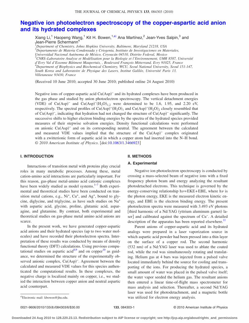
Anion photoelectron spectra of Cu�Asp�−,Cu�Asp�−�H2O�1, and Cu�Asp�−�H2O�2 are presented in Fig.1. The spectrum of Cu�Asp�− shows a broad band starting atEBE=0.9 eV and reaching an intensity maximum at EBE=1.6 eV. This latter value corresponds to the VDE of theCu�Asp�− anion. The VDEs of Cu�Asp�−�H2O�1 andCu�Asp�−�H2O�2 are 1.95 and 2.20 eV, respectively. Thespectral profiles of Cu�Asp�−�H2O�1 and Cu�Asp�−�H2O�2
closely resemble that of Cu�Asp�−, indicating that hydrationhas not changed the structure of Cu�Asp�− significantly;Cu�Asp�− is the chromophore. The successive shifts tohigher electron binding energies by the spectra of the hy-drated species provide measures of their stepwise hydrationenergies. The shift between the VDE of Cu�Asp�− and that ofCu�Asp�−�H2O�1 is 0.35 eV, while the shift between the VDEof Cu�Asp�−�H2O�1 and that of Cu�Asp�−�H2O�2 is 0.25 eV.Thus, 0.35 eV is an estimate of the energy gained whenCu�Asp�− is solvated by a single water molecule, and0.25 eV is an estimate of the energy gained whenCu�Asp�−�H2O�1 is solvated by an additional water mol-ecule.
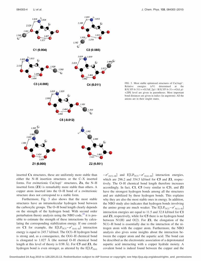
Several initial geometries were investigated in order toobtain a complete set of optimized anionic Cu�Asp�− struc-tures, and these are shown in Fig. 2. The copper atom inter-acts with either canonical or zwitterionic aspartic acid, and insubsequent figures the optimized structures are labeled asCx, and Zx, respectively. For the initial geometries with the
copper atom interacting with both carboxylic groups, the op-timization did not reach completion because the moleculestarted to dissociate during the process. The energies of thosepartially optimized structures are higher than those of theother structures. The results for the most stable structures arereported in Table I and Fig. 3. There are two different fami-lies of structures: those in which the copper atom is insertedin a C–X or X–H bond �X=O or N� and those in where thecopper is not bonded between two atoms. Inserted structuresinclude C1, C2, C3, C4, C5, Z1, and Z2 illustrated in Fig. 3and listed in Table I. Noninserted structures are not shownhere, since they are less stable by more than 1.8 eV, andtherefore, they are not likely to have been generated in thebeam. Table I shows that, at the highest level of calculation,there are six particularly stable structures lying within0.085 eV of one another. In all of these, the copper atom isinserted into a N–H �Z1� or O–H �C1-C5� bond. Insertion ofthe copper into a C–N bond results in a less stable structure�Z2� by 0.501 eV. Note that C3 and C5 are similar in struc-ture. The difference is only a small rotation of the aminogroup, with C5 being slightly more stable than C3. For thisreason, the further discussion will not include C3. Theresults reported in Table I show that the order of the stabilitydepends on the basis set that was used during the optimiza-tion. Z1 is the most stable structure at the B3LYP /6-31++G�d,p� level, while C5 becomes the global minimum atB3LYP /6-311++G�3df,2p� level. However, the energy dif-ference between these structures is very small and is withinthe uncertainties of the calculation.
While it is difficult to determine which structure wouldbe observed experimentally from the total energies resultsalone, the calculated VDE value of each structure is different
FIG. 1. The photoelectron spectra of Cu�Asp�−, Cu�Asp�−�H2O�1, andCu�Asp�−�H2O�2, all recorded at photon energies of 3.493 eV.
084303-2 Li et al. J. Chem. Phys. 133, 084303 �2010�
Downloaded 24 Aug 2010 to 128.220.23.13. Redistribution subject to AIP license or copyright; see http://jcp.aip.org/about/rights_and_permissions
and comparison with the experimentally measured VDEvalue allows us to determine which one was actually ob-served. For structures with the copper atom inserted intoO–H �C1 to C5�, the computed VDE values lie between 1.12to 1.35 eV at the highest level of theory. The calculated VDEvalue of structure, Z1 is 1.74 eV, which is in excellent agree-ment with the experimental result ��1.6 eV�. Thus, we con-clude that a zwitterionic form of aspartic acid with a copper
atom inserted into the N–H bond, i.e., Z1, is the dominantspecies generated in the experiment. As mentioned above,there is evidence that solvating the Cu�Asp�− complex withwater molecules did not change the structure of Cu�Asp�−,i.e., it can be expected to maintain its zwitterionic structureupon hydration.
The theoretical results also provided additional informa-tion about the anionic Cu�Asp�− complexes. Among the O–H
FIG. 2. Initial geometries considered for the Cu�Asp�−
complex
TABLE I. Total �hartree�, ZPE �hartree�, relative, and VDE energies �eV� of the various Cu�Asp�− complexes considered.
Structure Binding of Cu atom
B3LYP /6-31++G�d,p� B3LYP /6-311++G�3df,2p� a
E ZPE�E
�eV�VDE�eV� E
�E�eV�
VDE�eV�
C1 Inserted into O–H bond �2152.820 930 0.118 006 0.027 1.26 �2153.137 741 0.004 1.20C2 Inserted into O–H bond �2152.818 324 0.118 132 0.101 1.16 �2153.134 889 0.085 1.13C3 Inserted into O–H bond �2152.820259 0.117 221 0.024 1.17 �2153.137 293 �0.005 1.16C4 Inserted into O–H bond �2152.819 912 0.117 501 0.041 1.40 �2153.136 995 0.011 1.35C5 Inserted into O–H bond �2152.821 293 0.117 634 0.007 1.20 �2153.137 981 �0.012 1.12Z1 Inserted into N–H bond �2152.822 267 0.118 350 0.000 1.84 �2153.138 242 0.000 1.74Z2 Inserted into C–N bond �2152.805 317 0.118 769 0.473 b �2153.120 257 0.501 b
aObtained with the B3LYP /6-31++G�d,p� geometries.bThese values are not included since this structure is less stable and it is not expected in the experiment.
084303-3 PES of Cu-Asp and its hydrated anions J. Chem. Phys. 133, 084303 �2010�
Downloaded 24 Aug 2010 to 128.220.23.13. Redistribution subject to AIP license or copyright; see http://jcp.aip.org/about/rights_and_permissions
inserted Cx structures, these are uniformly more stable thaneither the N–H insertion structures or the C–X insertedforms. For zwitterionic Cu�Asp�− structures, Zx, the N–Hinserted form �Z1� is remarkably more stable than others. Acopper atom inserted into the O–H bond of a zwitterionicstructure does not correspond to a stable form.
Furthermore, Fig. 3 also shows that the most stablestructures have an intramolecular hydrogen bond betweenthe carboxylic groups. The O–H bond length clearly dependson the strength of the hydrogen bond. With second orderperturbation theory analysis using the NBO code,19 it is pos-sible to estimate the strength of these interactions by calcu-lating the corresponding stabilization energy. If one consid-ers C1 for example, the E�LPO�3�−��
O�4�−H� interaction
energy is equal to 245.7 kJ/mol. The O�3�–H hydrogen bondis strong and, as a consequence, the O�4�–H chemical bondis elongated to 1.027 Å �the normal O–H chemical bondlength at this level of theory is 0.98 Å�. For C5 and Z1, thehydrogen bond is even stronger, as attested by the E�LPO�4�
−��O�3�−H� and E�LPO�3�−��
O�4�−H� interaction energies,which are 296.2 and 354.5 kJ/mol for C5 and Z1, respec-tively. The O–H chemical bond length therefore increasesaccordingly. In fact, C1, C5 �very similar to C3�, and Z1have the strongest hydrogen bonds among all the structuresand are stabilized by these hydrogen bonds. This explainswhy they are also the most stable ones in energy. In addition,the NBO study also indicates that hydrogen bonds involvingthe amino group are much weaker. The E�LPO�2�−��
N�1�−H�interaction energies are equal to 11.5 and 32.8 kJ/mol for C1and Z1, respectively, while for C5 there is no hydrogen bondbetween N1�H� and O�2�. For Z1, the elongation of theN�1�–H bond is essentially due to the interaction of the ni-trogen atom with the copper atom. Furthermore, the NBOanalysis also gives some insights about the interaction be-tween the copper atom and the aspartic acid. The bond canbe described as the electrostatic association of a deprotonatedaspartic acid interacting with a copper hydride moiety. Acovalent bond is indeed found between the copper and the
FIG. 3. Most stable optimized structures of Cu�Asp�−.Relative energies �eV� determined at theB3LYP /6-311++G�3df,2p� / /B3LYP /6-31++G�d,p�+ZPE level are given in parentheses. Most importantbond distances are given in italics �in angstrom�. All theanions are in their singlet states.
084303-4 Li et al. J. Chem. Phys. 133, 084303 �2010�
Downloaded 24 Aug 2010 to 128.220.23.13. Redistribution subject to AIP license or copyright; see http://jcp.aip.org/about/rights_and_permissions
hydrogen atom. Molecular orbitals of the most stable anionicform, Z1, can be found in Fig. 5. Its highest occupied mo-lecular orbital �HOMO� orbital is degenerate and corre-sponds to a nonbonding d orbital of the copper atom. Thisindicates that the extra electron is located on the copperatom. The HOMO-1 orbital of Z1 shows a sigma Cu–Hbonding orbital, corroborating the idea of the covalent bond.The molecular orbitals for other stable anionic structures arefound to be similar.
To give a picture of the charge distribution within thecomplexes, we have carried out a NPA at the B3LYP /6-31++G�d,p� level. For anionic complexes, the natural chargeon the copper atom ranges from 0.38 to 0.45 for all thestructures. Removal of an electron without relaxing the ge-ometry of the systems �vertical transition� significantly modi-fies the natural charges. The most important variation is ob-served for the copper atom, whose natural charge becomes0.99, 1.03, and 0.82 for C1, C5, and Z1, respectively. Cor-respondingly, the negative charge of the hydrogen atombound to the copper atom decreases slightly to �0.54. Thisindicates that some of the anion’s excess electron is delocal-ized over the larger complex, and it further supports the as-
sumption of a copper hydride moiety. One also notices thatfor the anionic Cu�Asp�− complexes, the positively chargedcopper metal atom interacts with two negatively charged at-oms �N and H in the case of Z1 or O and H in the case ofC1-C5�. This contributes to the stabilization of the insertedcompounds.
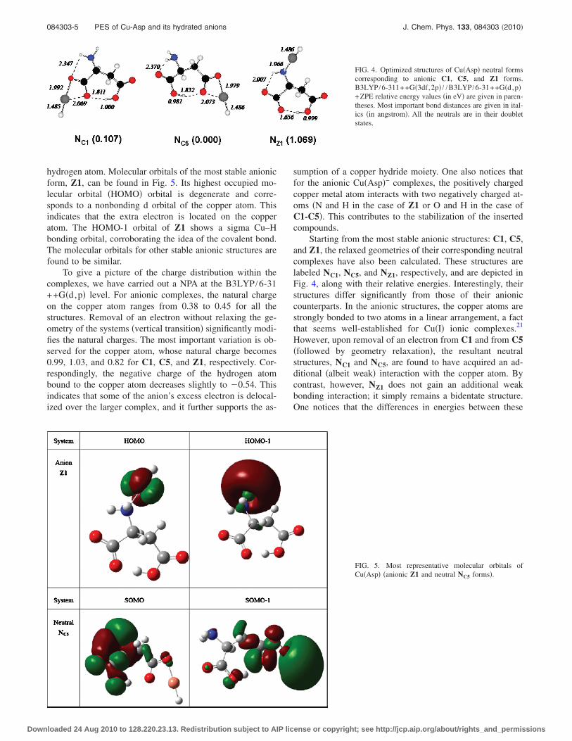
Starting from the most stable anionic structures: C1, C5,and Z1, the relaxed geometries of their corresponding neutralcomplexes have also been calculated. These structures arelabeled NC1, NC5, and NZ1, respectively, and are depicted inFig. 4, along with their relative energies. Interestingly, theirstructures differ significantly from those of their anioniccounterparts. In the anionic structures, the copper atoms arestrongly bonded to two atoms in a linear arrangement, a factthat seems well-established for Cu�I� ionic complexes.21
However, upon removal of an electron from C1 and from C5�followed by geometry relaxation�, the resultant neutralstructures, NC1 and NC5, are found to have acquired an ad-ditional �albeit weak� interaction with the copper atom. Bycontrast, however, NZ1 does not gain an additional weakbonding interaction; it simply remains a bidentate structure.One notices that the differences in energies between these
FIG. 4. Optimized structures of Cu�Asp� neutral formscorresponding to anionic C1, C5, and Z1 forms.B3LYP /6-311++G�3df,2p� / /B3LYP /6-31++G�d,p�+ZPE relative energy values �in eV� are given in paren-theses. Most important bond distances are given in ital-ics �in angstrom�. All the neutrals are in their doubletstates.
FIG. 5. Most representative molecular orbitals ofCu�Asp� �anionic Z1 and neutral NC5 forms�.
084303-5 PES of Cu-Asp and its hydrated anions J. Chem. Phys. 133, 084303 �2010�
Downloaded 24 Aug 2010 to 128.220.23.13. Redistribution subject to AIP license or copyright; see http://jcp.aip.org/about/rights_and_permissions
neutral forms are greater than the differences in energies be-tween their corresponding anionic forms. This is the reasonfor the variation in computed VDE values for the differentstructures. The molecular obitals of the neutral NC5 is shownin Fig. 5. Its singly occupied molecular orbital �SOMO� isdegenerate and located on the aspartic acid molecule, whilethe SOMO-1 is a sigma Cu–H bonding orbital.
ACKNOWLEDGMENTS
The experimental part of this material �K.H.B.� is basedon work supported by the U.S. National Science Foundationunder Grant No. CHE-0809258. The theoretical part of thisstudy �A.M.� was made possible due to funding by DGAPA-PAPIIT �Grant No. IN124602-3�, Consejo Nacional de Cien-cia y Tecnología CONACyT �Grant No. 222506�, and re-sources provided by the Instituto de Investigaciones enMateriales IIM. Some of the calculations were carried outusing a KanBalam supercomputer, provided by DGSCA,UNAM. Part of this work �J.P.S.� was conducted with thesupport of the World Class University Project supported bythe Ministry of Education, Science and Technology of Korea.
1 C. Kapota, J. Lemaire, P. Maitre, and G. Ohanessian, J. Am. Chem. Soc.126, 1836 �2004�.
2 Principles of Mass Spectrometry Applied to Biomolecules �Wiley, New
York, 2006�.3 F. Tureček, Mass Spectrom. Rev. 26, 563 �2007�.4 P. B. Armentrout, A. Gabriel, and R. M. Moision, Int. J. Mass. Spectrom.
283, 56 �2009�.5 L. Rodriguez-Santiago, M. Sodupe, and J. Tortajada, J. Phys. Chem. A
105, 5340 �2001�.6 M. K. Kim and A. E. Martell, J. Am. Chem. Soc. 91, 872 �1969�.7 M. Massaouti and M. Velegrakis, Int. J. Mass. Spectrom. 225, 89 �2003�.8 P. S. Kushwaha and P. C. Mishra, J. Mol. Struct.: THEOCHEM 549, 229�2001�.
9 W. Sang-aroon and V. Ruangpornvisuti, J. Mol. Struct.: THEOCHEM758, 181 �2006�.
10 G. Alagona and C. Ghio, Phys. Chem. Chem. Phys. 11, 776 �2009�.11 M. Gerhards, O. C. Thomas, J. M. Nilles, W.-J. Zheng, and K. H. Bowen,
J. Chem. Phys. 116, 10247 �2002�.12 B. Miehlich, A. Savin, H. Stoll, and H. Preuss, Chem. Phys. Lett. 157,
200 �1989�.13 R. M. Dickson and A. D. Becke, J. Chem. Phys. 99, 3898 �1993�.14 A. D. Becke, J. Chem. Phys. 98, 5648 �1993�.15 A. D. Becke, J. Chem. Phys. 98, 1372 �1993�.16 M. J. Frisch, G. W. Trucks, H. B. Schlegel et al., GAUSSIAN03, Revision
B.05, Gaussian, Inc., Wallingford, CT, 2003.17 A. D. McLean and G. S. Chandler, J. Chem. Phys. 72, 5639 �1980�.18 R. Krishnan, J. S. Binkley, R. Seeger, and J. A. Pople, J. Chem. Phys. 72,
650 �1980�.19 T. Clark, J. Chandrasekhar, G. W. Spitznagel, and P. V. Schleyer, J. Com-
put. Chem. 4, 294 �1983�.20 E.D. Glendening, A.E. Reed, and F. Weinhold, NBO version 3.1.21 A. M. Lamsabhi, M. Yáñez, J.-Y. Salpin, and J. Tortajada, Gas-Phase
Chemistry of Organocopper Compounds �Wiley, New York, 2009�.
084303-6 Li et al. J. Chem. Phys. 133, 084303 �2010�
Downloaded 24 Aug 2010 to 128.220.23.13. Redistribution subject to AIP license or copyright; see http://jcp.aip.org/about/rights_and_permissions